istoatologic inding o strongloides stercoralis in a atient
TRANSCRIPT

© 2021 Asociación Colombiana de Gastroenterología252
Rubén Gustavo Muñoz-Cedeño,1* Linda Patricia Naranjo,2 Gema Rodríguez,3 Virgilio Alvarado-Gallo.4
Histopathologic finding of Strongyloides stercoralis in a patient with Crohn’s disease: Case report
OPEN ACCESS
Citation:Muñoz-Cedeño RG, Naranjo LP, Rodríguez G, Alvarado-Gallo V. Histopathologic finding of Strongyloides stercoralis in a patient with Crohn’s disease: Case report. Rev Colomb Gastroenterol. 2021;36(2):252-256. https://doi.org/10.22516/25007440.501
............................................................................
1 Physician, Gastroenteroly Resident, Hospital de Especialidades Dr. Abel Gilbert Pontón. Guayaquil, Ecuador.
2 Physician, Internal Medicine Resident, Hospital Luis Vernaza. Guayaquil, Ecuador.3 General practitioner, Hospital Universitario de Guayaquil. Guayaquil, Ecuador.4 Gastroenterologist, Hospital General Guasmo Sur. Guayaquil, Ecuador.
*Correspondence: Rubén Gustavo Muñoz-Cedeño. [email protected]
............................................................................
Received: 23/01/20 Accepted: 08/04/20
AbstractClinical case: The following is a rare clinical case in an immunocompro-mised patient with histopathological findings of parasitic infestation. The patient is a middle-aged male who lives in a subtropical area and has a diagnosis of Crohn’s disease treated with corticosteroids and immunomo-dulators. The patient presented with abdominal pain and chronic anemia for 1 year, with negative laboratory tests for parasites and normal acute phase reactants. Gastroscopy and colonoscopy were performed before the consultation (6 months) without relevant findings. Due to the persisten-ce of the symptoms, endoscopic studies were repeated, finding subepithe-lial bleeding with histopathological results of Strongyloides stercoralis. Conclusion: In the context of an immunocompromised patient living in an endemic area and with a torpid evolution, a differential diagnosis should be made always suspecting a parasitic infestation. Although endoscopy is not necessary to diagnose strongyloidiasis, its use may be convenient.
KeywordsNematode infections; Strongyloides stercolaris; Immunocompromised host; Hematemesis; Crohn’s Disease.
Case reportDOI: https://doi.org/10.22516/25007440.501
INTRODUCTION
Strongyloides stercoralis colitis is a serious but treatable disease with a high mortality rate if left untreated. It can last for decades and lead to chronic colitis, similar to that observed in inflammatory bowel disease (IBD). Tropical and subtropical areas provide adequate conditions for its development.
Manifestations of infection can vary from asymptomatic eosinophilia in immunocompetent hosts to disseminated disease with septic shock in immunocompromised hosts, as seen in the case presented, with histopathological fin-dings consistent with Crohn’s disease.
CASE PRESENTATION
This is the case of a 55-year-old male patient, mestizo, businessman, under treatment with corticosteroids and immunomodulators (azathioprine) due to Crohn’s disease. He had a surgical history of appendectomy and cholecys-tectomy performed in 2015. During a routine check-up for abdominal pain and chronic anemia, he was referred to the emergency room after experiencing intermittent melena for a month, which worsened due to hematochezia, mar-ked pallor, asthenia, and arthralgia for a week. Laboratory tests yielded the following results: hemoglobin: 5.80 g/dL, hematocrit: 18.60%, and platelets: 340 000 mm3 (Table 1).
253Histopathologic finding of Strongyloides stercoralis in a patient with Crohn’s disease: Case report
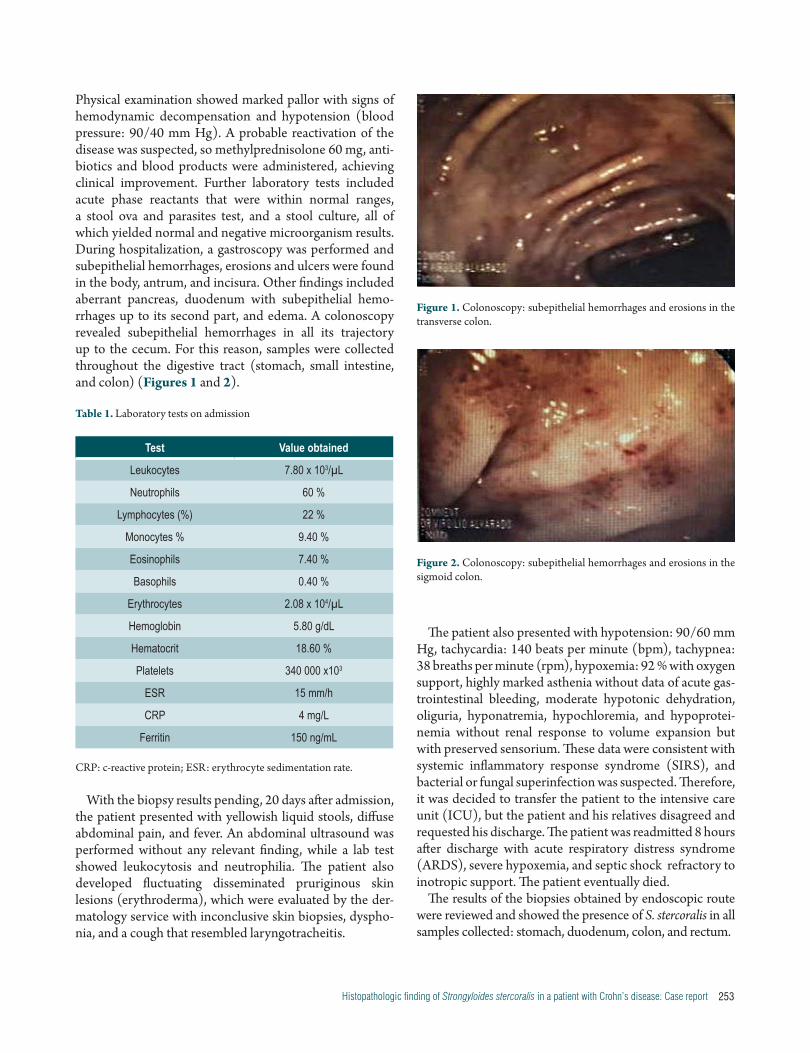
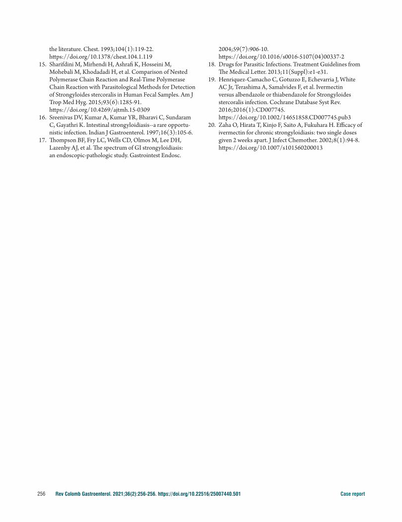
Physical examination showed marked pallor with signs of hemodynamic decompensation and hypotension (blood pressure: 90/40 mm Hg). A probable reactivation of the disease was suspected, so methylprednisolone 60 mg, anti-biotics and blood products were administered, achieving clinical improvement. Further laboratory tests included acute phase reactants that were within normal ranges, a stool ova and parasites test, and a stool culture, all of which yielded normal and negative microorganism results. During hospitalization, a gastroscopy was performed and subepithelial hemorrhages, erosions and ulcers were found in the body, antrum, and incisura. Other findings included aberrant pancreas, duodenum with subepithelial hemo-rrhages up to its second part, and edema. A colonoscopy revealed subepithelial hemorrhages in all its trajectory up to the cecum. For this reason, samples were collected throughout the digestive tract (stomach, small intestine, and colon) (Figures 1 and 2).
Table 1. Laboratory tests on admission
Test Value obtained
Leukocytes 7.80 x 10³/µL
Neutrophils 60 %
Lymphocytes (%) 22 %
Monocytes % 9.40 %
Eosinophils 7.40 %
Basophils 0.40 %
Erythrocytes 2.08 x 10⁴/µL
Hemoglobin 5.80 g/dL
Hematocrit 18.60 %
Platelets 340 000 x103
ESR 15 mm/h
CRP 4 mg/L
Ferritin 150 ng/mL
CRP: c-reactive protein; ESR: erythrocyte sedimentation rate.
With the biopsy results pending, 20 days after admission, the patient presented with yellowish liquid stools, diffuse abdominal pain, and fever. An abdominal ultrasound was performed without any relevant finding, while a lab test showed leukocytosis and neutrophilia. The patient also developed fluctuating disseminated pruriginous skin lesions (erythroderma), which were evaluated by the der-matology service with inconclusive skin biopsies, dyspho-nia, and a cough that resembled laryngotracheitis.
Figure 1. Colonoscopy: subepithelial hemorrhages and erosions in the transverse colon.
Figure 2. Colonoscopy: subepithelial hemorrhages and erosions in the sigmoid colon.
The patient also presented with hypotension: 90/60 mm Hg, tachycardia: 140 beats per minute (bpm), tachypnea: 38 breaths per minute (rpm), hypoxemia: 92 % with oxygen support, highly marked asthenia without data of acute gas-trointestinal bleeding, moderate hypotonic dehydration, oliguria, hyponatremia, hypochloremia, and hypoprotei-nemia without renal response to volume expansion but with preserved sensorium. These data were consistent with systemic inflammatory response syndrome (SIRS), and bacterial or fungal superinfection was suspected. Therefore, it was decided to transfer the patient to the intensive care unit (ICU), but the patient and his relatives disagreed and requested his discharge. The patient was readmitted 8 hours after discharge with acute respiratory distress syndrome (ARDS), severe hypoxemia, and septic shock refractory to inotropic support. The patient eventually died.
The results of the biopsies obtained by endoscopic route were reviewed and showed the presence of S. stercoralis in all samples collected: stomach, duodenum, colon, and rectum.

Rev Colomb Gastroenterol. 2021;36(2):256-256. https://doi.org/10.22516/25007440.501254 Case report
This chronic infection may be clinically inapparent or manifest through skin, gastrointestinal, or pulmonary symptoms, as described in numerous cases of immunosup-pression (HIV, alcoholism, organ transplantation, corticos-teroid use)(6,7) with a high risk of hyperinfection because certain cytotoxic drugs damage cell immunity. The patho-physiology underlying these risk factors, whether disease-related or iatrogenic-induced, is a compromised immune system leading to Th2 helper cell dysfunction; therefore, it is important to detect and eradicate Strongyloides before starting immunosuppressive therapy(8).
About one third of patients with strongyloidiasis are asymptomatic. Serum IgE concentration is high in these settings(9). Gastrointestinal symptoms include diarrhea, abdominal pain, vomiting, and massive gastrointestinal blee-ding secondary to the presence of duodenal ulcers, as well as intestinal occlusion and ileus. Respiratory symptoms include mainly cough and dyspnea. In hyperinfection and dissemi-nation syndromes, pulmonary involvement presents as acute respiratory failure syndrome (ARDS)(10-12).
Eosinophilia is correlated with larva migration and may be present in 83 %-92 % of patients with autoinfection; howe-ver, it is not observed in all patients, but may be the only clue. It may also be absent in cases of hyperinfection or use of immunosuppressive drugs(13,14). For diagnosis, 2 concentra-ted stool samples should be collected to detect the presence of rhabditiform larvae, and serological tests (enzyme immu-noadsorption assay [ELISA] method with immunoglobulin G [IgG] detection), duodeno-jejunal aspiration and stool PCR should be performed(15). The presence of eosinophilia on the blood count is expected, leading to the suspicion of a parasite infection; however, when the parasite has spread, there is no eosinophilia, and the white blood cell count is variable. Upper endoscopy is not needed to diagnose stron-gyloidiasis, although some publications mention that it may give rise to a wide range of endoscopic features(16,17).
The reported patient continued treatment with corticos-teroids and immunomodulators (azathioprine) for approxi-mately 1 year. In view of the suspicion of reactivation, the administration of corticosteroids at induction doses was started with an apparent improvement, which initially dis-pelled the suspicion of a parasitic infestation, mainly due to negative serial stool tests and absence of larvae under endoscopic view. The patient received broad-spectrum antibiotics assuming a possible bacterial or fungal superin-fection. It should be noted that the requested laboratory tests never showed marked eosinophilia, possibly due to his immunosuppression. The presence of this nematode was confirmed through biopsy results in all samples taken from the stomach, duodenum, and colon. Nevertheless, the torpid evolution and progressive deterioration of the
DISCUSSION
Strongyloidiasis is caused by a nematode called Strongy-loides. There are 52 species, but most do not infect humans. The most common and globally distributed human patho-gen is S. stercoralis, which affects 100 million people. The other most common species is Strongyloides fueleborni, which is found sporadically in Africa and New Guinea. S. stercoralis is endemic in tropical and subtropical zones, Southeast Asia, Latin America, sub-Saharan Africa, and the southeastern United States, and may be found sporadically in temperate zones(1).
This parasite develops into its adult stage in the intes-tinal mucosa of the duodenum and jejunum. The adult female produces approximately 12 eggs per day, which hatch and release rhabditoid larvae; thus, this disease is only associated with adult females, rhabditoid larvae, eggs, and, very rarely, filariform larvae in the intestinal mucosa. Histopathological changes caused by this disease in the intestinal mucosa were described in 1962 by De Paola, which include intestinal lesions referred to as catarrhal enteritis, edematous enteritis and ulcerative enteritis, depending on the type of inflammatory infiltrate, villous atrophy, and the presence of ulcers(2,3).
The presentation and evolution of this infection are deter-mined by the interaction between the host and the para-site. Activation of the T helper 2 (Th2) cellular immune response, humoral immune response, or alteration of the mucosal barrier favor the transformation of the rhabditoid larva into filariform, which is considered an autoinfection syndrome that causes a reinfection in the host itself. When crossing the colon mucosa, the filariform larva can carry bacteria and fungi and distribute them throughout the body, causing bacterial or fungal sepsis and Gram-negative meningitis. This capacity for autoinfection can lead to chro-nic long-term disease. The respiratory symptoms are cau-sed by larval infiltration of the vascular and alveolar spaces, resulting in pulmonary edema, bronchopneumonia, and alveolar hemorrhage. The death of the parasites inside the lungs causes intense inflammation and ARDS, and morta-lity can reach up to 80 %(4,5).
Dissemination leads to high mortality (70-90 %). Infection with the nematodes. stercoralis is potentially lethal because of its ability to produce devastating autoinfection (hyperinfection/dissemination), particularly in immuno-suppressed hosts, such as human immunodeficiency virus (HIV) patients who present with elevated levels of interfe-ron gamma (INF-γ) and decreased serum levels of interleu-kin 4 (IL-4) and, consequently, immunoglobulin E (IgE), which allows for a more serious disease and the onset of this syndrome(2,4,5).

255Histopathologic finding of Strongyloides stercoralis in a patient with Crohn’s disease: Case report
patient prevented the search for atypical microorganisms or studies in the alveolar fluid. In this case, it is not possible to know whether the fatal outcome could have been avoi-ded had the patient received treatment with ivermectin or albendazole given the severity of his condition.
In immunocompromised patients who do not respond to conventional treatment (corticosteroids), it is necessary to look for this type of parasite, especially in areas where the risk or prevalence is higher. Since diagnostic tests may be negative, ivermectin could be administered empirically. Treatment with ivermectin is recommended for uncom-plicated strongyloidiasis (grade 1A) and is prescribed as 2 single doses of 200 µg/kg administered over 2 consecutive days. In patients with disseminated disease (hyperinfec-tion), prolonged administration of ivermectin (grade 2C) 200 µg/kg/day for at least 5 to 7 days is suggested, or it may be combined with albendazole 400 mg/orally (VO) 2 times a day for 3 to 7 days. Treatment should be given daily until symptoms resolve and stool tests are negative for at least 2 weeks(18-20).
CONCLUSIONS
In an immunocompromised patient living in an endemic area with a torpid evolution, negative stool tests and nega-tive acute phase reactants, parasitic infestation should be suspected and empirical treatment with anthelmintic ini-tiated. Moreover, if necessary, endoscopic studies should be repeated since the time of infestation and morpholo-gical changes between one study and another cannot be established. Although endoscopy is not necessary for the diagnosis of strongyloidiasis, its use may be convenient, as it was in our case.
Conflicts of interest
None declared by the authors.
Funding sources
None declared by the authors.
REFERENCES
1. Schär F, Trostdorf U, Giardina F, Khieu V, Muth S, Marti H, et al. Strongyloides stercoralis: Global Distribution and Risk Factors. PLoS Negl Trop Dis. 2013;7(7):e2288. https://doi.org/10.1371/journal.pntd.0002288
2. Newberry AM, Williams DN, Stauffer WM, Boulware DR, Hendel-Paterson BR, Walker PF. Strongyloides hyperin-fection presenting as acute respiratory failure and gram-negative sepsis. Chest. 2005;128(5):3681-4. https://doi.org/10.1378/chest.128.5.3681
3. Arévalo Suarez F, Cerrillo Sánchez G. Strongyloides ster-coralis: Hallazgos Histopatológicos en Mucosa Duodenal 1999-2005. Rev Gastroenterol Perú. 2006;26(1):44-8.
4. Gorman SR, Craven DE. Images in clinical medicine. Strongyloides stercoralis hyperinfection. N Engl J Med. 2008;359(11):e12. https://doi.org/10.1056/NEJMicm066791
5. Upadhyay D, Corbridge T, Jain M, Shah R. Pulmonary hyperinfection syndrome with Strongyloides stercoralis. Am J Med. 2001;111(2):167-9. https://doi.org/10.1016/s0002-9343(01)00708-2
6. Woodring JH, Halfhill H 2nd, Reed JC. Pulmonary strongyloidiasis: clinical and imaging features. AJR Am J Roentgenol. 1994;162(3):537-42. https://doi.org/10.2214/ajr.162.3.8109492
7. Kim JH, Kim DS, Yoon YK, Sohn JW, Kim MJ. Donor-Derived Strongyloidiasis Infection in Solid Organ Transplant Recipients: A Review and Pooled Analysis.
Transplant Proc. 2016;48(7):2442-2449. https://doi.org/10.1016/j.transproceed.2015.11.045
8. Fardet L, Généreau T, Cabane J, Kettaneh A. Severe strongyloidiasis in corticosteroid-treated patients. Clin Microbiol Infect. 2006;12(10):945-7. https://doi.org/10.1111/j.1469-0691.2006.01443.x
9. Concha R, Harrington W Jr, Rogers AI. Intestinal stron-gyloidiasis: recognition, management, and determinants of outcome. J Clin Gastroenterol. 2005;39(3):203-11. https://doi.org/10.1097/01.mcg.0000152779.68900.33
10. Robinson RD, Lindo JF, Neva FA, Gam AA, Vogel P, Terry SI, et al. Immunoepidemiologic studies of Strongyloides stercoralis and human T lymphotropic virus type I infec-tions in Jamaica. J Infect Dis. 1994;169(3):692-6. https://doi.org/10.1093/infdis/169.3.692
11. Scowden EB, Schaffner W, Stone WJ. Overwhelming strongyloidiasis: an unappreciated opportunistic infection. Medicine (Baltimore). 1978;57(6):527-44.
12. Strazzella WD, Safirstein BH. Asthma due to parasitic infes-tation. N J Med. 1989;86(12):947-9.
13. Woodring JH, Halfhill H 2nd, Berger R, Reed JC, Moser N. Clinical and imaging features of pulmonary strongyloidia-sis. South Med J. 1996;89(1):10-9. https://doi.org/10.1097/00007611-199601000-00002
14. Lessnau KD, Can S, Talavera W. Disseminated Strongyloides stercoralis in human immunodeficiency virus-infected patients. Treatment failure and a review of
Rev Colomb Gastroenterol. 2021;36(2):256-256. https://doi.org/10.22516/25007440.501256 Case report
2004;59(7):906-10. https://doi.org/10.1016/s0016-5107(04)00337-2
18. Drugs for Parasitic Infections. Treatment Guidelines from The Medical Letter. 2013;11(Suppl):e1-e31.
19. Henriquez-Camacho C, Gotuzzo E, Echevarria J, White AC Jr, Terashima A, Samalvides F, et al. Ivermectin versus albendazole or thiabendazole for Strongyloides stercoralis infection. Cochrane Database Syst Rev. 2016;2016(1):CD007745. https://doi.org/10.1002/14651858.CD007745.pub3
20. Zaha O, Hirata T, Kinjo F, Saito A, Fukuhara H. Efficacy of ivermectin for chronic strongyloidiasis: two single doses given 2 weeks apart. J Infect Chemother. 2002;8(1):94-8. https://doi.org/10.1007/s101560200013
the literature. Chest. 1993;104(1):119-22. https://doi.org/10.1378/chest.104.1.119
15. Sharifdini M, Mirhendi H, Ashrafi K, Hosseini M, Mohebali M, Khodadadi H, et al. Comparison of Nested Polymerase Chain Reaction and Real-Time Polymerase Chain Reaction with Parasitological Methods for Detection of Strongyloides stercoralis in Human Fecal Samples. Am J Trop Med Hyg. 2015;93(6):1285-91. https://doi.org/10.4269/ajtmh.15-0309
16. Sreenivas DV, Kumar A, Kumar YR, Bharavi C, Sundaram C, Gayathri K. Intestinal strongyloidiasis--a rare opportu-nistic infection. Indian J Gastroenterol. 1997;16(3):105-6.
17. Thompson BF, Fry LC, Wells CD, Olmos M, Lee DH, Lazenby AJ, et al. The spectrum of GI strongyloidiasis: an endoscopic-pathologic study. Gastrointest Endosc.