isolation pseudomonassolanacearum-specific dna …aem.asm.org/content/58/11/3751.full.pdf · appl....
TRANSCRIPT

Vol. 58, No. 11
Isolation of a Pseudomonas solanacearum-Specific DNAProbe by Subtraction Hybridization and Construction ofSpecies-Specific Oligonucleotide Primers for Sensitive
Detection by the Polymerase Chain ReactionSUSAN E. SEAL,t* LUKE A. JACKSON, AND MICHAEL J. DANIELS
The Sainsbury Laboratory, John Innes Centre, Colney Lane, Norwich, Norfolk NR4 7UH, United Kingdom
Received 24 March 1992/Accepted 6 September 1992
A subtraction hybridization technique was employed to make a library enriched for Pseudomonassolanacearum-specific sequences. One cloned fragment, PS2096, hybridized under stringent conditions to DNAof 82 P. solanacearum strains representing all subgroups of the species. Other plant-associated bacteria,including closely related species such as Pseudomonas cepacia, Pseudomonas pickettii, or Pseudomonas syzygii,did not hybridize to PS2096. A minimum number of between 4 x 10i and 4 x 10' P. solanacearum cells couldroutinely be detected with PS2096 labelled either with [32P]dCTP or with digoxigenin-11-dUTP. To improvethe sensitivity of detection, PS2096 was sequenced to allow the construction of specific oligonucleotide primersto be used for polymerase chain reaction (PCR) amplification. After 50 cycles of amplification, 5 to 116 cells,depending on the strain, could reproducibly be detected by visualization of a 148-bp PCR product on an
agarose gel. A preliminary field trial in Burndi with the probe and PCR primers has confirmed that they are
sensitive tools for specifically detecting low-level infections of P. solanacearum in potato tubers.
Pseudomonas solanacearum E. F. Smith causes bacterialwilt, which is one of the most important and widely spreadbacterial diseases of crops in the tropics, subtropics, andwarm temperate regions of the world. The disease has beenrecorded on several hundred species representing 44 familiesof plants (23). Strains of P. solanacearum compose a com-plex taxonomic group, with subspecific groupings consistingof either races, based on host range (10), or biovars, basedon the catabolism of certain sugars and sugar alcohols (22).There are many reports of bacterial wilt on potatoes (e.g.,
9, 11, 31, 32) and bananas (e.g., 1, 8, 27) having been spreadwithin and between countries in latently infected plantingmaterial. Other crops such as tomatoes have also beenshown to be capable of carrying latent infections of P.solanacearum (35). These examples emphasize the need foreffective quarantine measures against P. solanacearum, notonly for tropical and subtropical countries but also fortemperate-zone countries where race 3 could pose a threat topotato crops, illustrated by the experience in Sweden (32).The success of quarantine procedures relies on the use ofsimple yet sensitive detection techniques. Current identifi-cation methods rely on a series of biochemical tests on
purified single colonies which requires 1 to 2 weeks before a
species identification is possible. More recently, methodssuch as metabolic profiling (5) or computer-assisted fattyacid profiling (43), although speeding up the process, stillrequire purification of a single P. solanacearum colony,which may be obscured by the more-rapid growth of otherplant-associated microorganisms. Specific monoclonal or
polyclonal antisera to P. solanacearum that do not reactwith closely related species, such as Pseudomonas pickettiior Pseudomonas syzygii, have not become available to date.DNA probes have been developed and applied success-
* Corresponding author.t Present address: Natural Resources Institute, Central Avenue,
Chatham Maritime, Kent ME4 4TB, United Kingdom.
fully in the detection and identification of human pathogens(18, 21, 25, 46) and plant-pathogenic bacteria (20, 29, 37, 40,45). Development of a DNA probe specific for P. solan-acearum is complicated by the genetic diversity of thisspecies (12, 42) as well as a high degree of DNA homology toP. pickettii (36) and the clove disease bacterium P. syzygii (4,38). P. solanacearum, P. syzygii, and P. pickettii are foundwithin the same DNA-DNA homology group of the Pseudo-monas rRNA homology group II (33, 38). In addition, a
closely related pseudomonad "Pseudomonas celebense"(19), which causes blood disease of members of the familyMusaceae in Indonesia, reacts with some P. solanacearumDNA probes (13, 14, 42) but shows cultural properties (17)different from those of P. solanacearum strains.
In order to avoid the laborious screening of randomlycloned DNA fragments for their specificity for P. solan-acearum, we utilized a simple subtraction hybridizationmethod to enrich for such sequences. A P. solanacearum-specific DNA fragment was isolated and tested for itssuitability as a nonradioactive diagnostic probe which couldbe used as a substitute for current detection and identifica-tion procedures. The P. solanacearum-specific DNA was
sequenced, allowing the construction of specific oligonucle-otide primers for detection of lower numbers of bacteria bypolymerase chain reaction (PCR) amplification.
MATERIALS AND METHODS
Bacterial strains, culture media, and growth conditions.The characteristics and sources of the strains tested in thisstudy are shown in Table 1. P. solanacearum was routinelycultured at 28°C in CPG broth (24) or on CPG platescontaining 15 g of agar per liter and 0.1 g of 2,3,5,-triphe-nyltetrazolium chloride per liter to ensure that the fluidal(virulent) morphology was selected during subculturing (26).Escherichia coli strains were grown at 37°C in LB medium(30). All other bacterial species used were grown in NYG
3751
APPLIED AND ENVIRONMENTAL MICROBIOLOGY, Nov. 1992, p. 3751-37580099-2240/92/113751-08$02.00/0
on May 31, 2018 by guest
http://aem.asm
.org/D
ownloaded from

APPL. ENVIRON. MICROBIOL.
TABLE 1. Bacterial strains
Organism and strain Race Biovar
Pseudomonas solanacearumGM18131UW25UW26UW30UW90UW256UW275UW278R563R142R330R361R568R573R578R583R132R306R314R374S9S729S825T456T494UW8UW119UW130UW147UW255UW380GMI1000GMI1336PD1682R27TomMUW27UW74UW151UW359UW360UW369UW378UW361UW373UW160UW167GM18133JEBUG1JEBUG2JEBUG4P13P23R150R152Rl55aR161R203R367R372R481R484R497R368R570R579R589R598
1 1
1 1
1 1
1 1
1C 1
1 1
1 1
1 1
1 1
2N2dN2N2N2N2N2
1 31 31 31 31 31 31 31 31 31 31c 31 31 31 31 31 41c 41 41 41 41 41 41 41 41 41 41 41 51c 5
2 12 12 12Ad 12A 12A 12A 12A 1
2 1
2 1
2 1
2 1
2 1
2 1
2 1
2A 1
2A 12A 12 12 1
2 1
2 1
2 1
Tobacco, Colombia, Granada S247Tomato, United States, Kelman K60Tomato, United States, Kelman K74Tomato, Trinidad, Dudman K136Tobacco, Brazil, Robbs ENA521Potato, Costa Rica, Gonzalez G-7Melampodium perfoliatum, Costa Rica, SequeiraTobacco, Mexico, FucikovskyPotato, Peru, Pinedo CIP120Clove, Indonesia, Eden-Green S710Potato, Brazil, Reifschneider 68Potato, Peru, Turkensteen CIP61Potato, Brazil, Neto 172, CIP226Soil, Peru, Martin CIP162Eggplant, Peru, Aley SR130Potato, Peru, Martin CIP172Potato, Fiji, Hayward B2122Potato, Brazil, Reifschneider 52Potato, Cameroun, Pirkko Hay CIP292Heliconia sp., Costa Rica, Sequeira S118, UW6Peanut, Malaysia, SealClove, Indonesia, Eden-GreenClove, Indonesia, Eden-GreenCastor bean, Indonesia, SupriadiPeanut, Indonesia, Subandiah 1105BEupatorium odoratum, Costa Rica, Sequeira K201Potato, Costa Rica, Gonzalez S213Tomato, Peru, Sequeira S225Tobacco, Australia, Hayward S240Pepper, Costa Rica, Gonzalez G-1Olive, China, He OPS2Tomato, Guyana (3)hrp mutant of GMI1000Ginger, Thailand, SardsudPotato, Indonesia, Hayward B050Tomato, Malaysia, SealTobacco, United States, Kelman K105Potato, Sri Lanka, CMI no. B2861Ginger, Australia, Hayward S244Ginger, The People's Republic of China, He ZPS1Mulberry, The People's Republic of China, He MPS5Peanut, The People's Republic of China, He PPS14Olive, The People's Republic of China, He OPS1Mulberry, The People's Republic of China, He MPS4Mulberry, The People's Republic of China, He MPS2Plantain, Peru, Sequeira S253Banana, Costa Rica, Sequeira K160Plantain, Colombia, Thurston S210Banana, The Philippines, ElphinstoneBanana, The Philippines, ElphinstoneBanana, The Philippines, ElphinstoneBanana, The Philippines, SealBanana, The Philippines, SealBanana, Costa Rica, Buddenhagen BUD100Heliconia sp., Colombia, Buddenhagen H249Banana, Grenada, Eden-Green M107aBanana, Grenada, Eden-Green GlPlantain, Guyana, McDonald, CMI no. B8365Plantain, Colombia, Kelman, UW181Plantain, Costa Rica, Sequeira S232, UW139Banana, The Philippines, Eden-Green P10Banana, The Philippines, Eden-Green P14Banana, The Philippines, Eden-Green P12Plantain, Colombia, Kelman K254Plantain, Costa Rica, CIP19, UW155Plantain, Costa Rica, Sequeira S233, UW140Plantain, Colombia, Granada G28Banana, Grenada, Hunt SlOlci
Continued
Aw
w
w
w
w
w
w
w
RRRRRRRRRRRDRRBBw
w
w
w
w
w
AAJ
RDw
w
w
w
w
w
w
w
w
w
w
ARRRDDRRRRRRRRRRRRRRR
I on followingpage
Origin' Sourceb
3752 SEAL ET AL.
on May 31, 2018 by guest
http://aem.asm
.org/D
ownloaded from

P. SOLANACEARUM-SPECIFIC PROBE AND PRIMERS FOR PCR 3753
TABLE 1-Continued
Organism and strain Race Biovar Origina Sourceb
SBBUR1BUR2BUR3BUR4E/225aE/301E/310E/N35-89E/1677AGM18141K46P2R39UW19UW23UW73
Pseudomonas celebense (blooddisease bacterium)
P02R229R230R234R604T340
Pseudomonas pickettiiE1625NC11149
Pseudononas syzygiiR001R004
Pseudomonas cepacia GMI8101Pseudomonas synngae pv. tabaciAlcaligenes eutrophus GMI8105Alcaligenesfaecalis E/1643Envinia carotovora E/412Erwinia chrysanthemi (NCPPB 2030)Erwinia herbicola (NCPPB 2971)Envinia sp. E/422bEscherichia coli ED8767Kiebsiella pneumoniae E/316Kiebsiella sp. E/1626aXanthomonas campestris pv.
campestris 8004Xanthomonas campestris pv.
campestris 45Xanthomonas campestnis pv. graminisXanthomonas campestris pv.
holcicolaXanthomonas campestris pv.
malvacearumXanthomonas campestris pv.
vesicatoria NCPPB 2595
2A 1 Banana, The Philippines, Soguilon SB12 Potato, Burundi, Seal2 Potato, Burundi, Seal2 Potato, Burundi, Seal2 Potato, Burundi, Seal2 Potato, Ethiopia, Wondimagegne
3 2 Potato, Ethiopia, Wondimagegne2 Potato, Ethiopia, Wondimagegne
3 2 Potato, Ethiopia, Wondimagegne3 2 Potato, Ethiopia, Wondimagegne3 2 Potato, Colombia, Thurston S2063 2 Potato, Kenya, Forde3 2 Potato, The Philippines, Seal3 2 Potato, Egypt, Lelliott NCP9093 2 Potato, Colombia, Thurston S2053 2 Potato, Israel, Volcani K563 2 Potato, Sri Lanka, CMI no. B2768
Plantain, Indonesia, BaharuddinBanana, Indonesia, Eden-Green T389Banana, Indonesia, Eden-Green T334Banana, Indonesia, Eden-Green T391Banana, Indonesia, Hartati t509Banana, Indonesia, Eden-Green
Soil/potato, Ethiopia, Wondimagegne
Clove, Indonesia, Eden-GreenHindola fulva, Indonesia, Eden-Green
a Host, country, isolator, and alternative strain designations (where applicable) are given.b Strains were contributed by the sources designated as follows: A, M. Arlat and P. Barberis, CNRS-INRA, Auzeville, Castanet-Tolosan Cedex, France; B,
Supriadi, Balai Penelitian Tanaman Rempah dan Obat, J1. Cimanggu 3, Bogor, Indonesia; D, M. Daniels and S. Seal, The Sainsbury Laboratory, Norwich, UnitedKingdom; E, E. Wondimagegne and J. Turner, University of East Anglia, Norwich, United Kingdom; G, B. Baharuddin and K. Rudolph, Institut furPflanzenpathologie oder Pflanzenschutz der Universitat, Gottingen Universitat, D-3400 G6ttingen, Germany; J, J. Janse, Plant Protection Service, BacteriologyDepartment, 6700 HC Wageningen, The Netherlands; N, National Collection ofType Cultures, London, United Kingdom; P, C. Soguilon and A. Quimio, UniversityofThe Phillipines at Los Banos, College, Laguna, The Philippines; R, S. Eden-Green, J. Elphinstone, and S. Forde, Rothamsted Experimental Station, Harpenden,Hertfordshire AL5 2JQ, United Kingdom; W, D. Cook and L. Sequeira, Department of Plant Pathology University of Wisconsin-Madison, Madison, Wis.
C Nonpathogenic strains.d N2, new biovar 2 (lowland strains); 2A, banana "bugtok" strains.
medium (15). Whole P. solanacearum cells to be used forproviding template for PCR amplifications were grown inMM minimal medium (6) to ensure adequate lysis of thecells. Lysis was performed by boiling 100 ,ul of an overnight
MM culture (or a loopful of bacteria from a colony resus-pended in 100 plA of sterile distilled water) for 5 min. Afterbeing cooled to room temperature, 1 to 5 pul of the boiledculture was used per reaction without further treatment.
pDDDDEEEEEARDRwww
GRRRRR
EN
RRAEAEEEEEDEED
D
DD
D
D
VOL. 58, 1992
on May 31, 2018 by guest
http://aem.asm
.org/D
ownloaded from

APPL. ENVIRON. MICROBIOL.
DNA manipulations. Bacterial DNA was isolated by themethod of Boucher et al. (7). The procedures for agarose gelelectrophoresis, Southern blotting, preparation of competentcells, ligation, and transformation were done as described byManiatis et al. (28). Restriction enzyme digestions werecarried out according to the conditions defined by the sup-plier, using S U of enzyme per pug of DNA. Hybridizationswere performed at 65°C with probe DNA labelled with either[32P]dCTP or digoxigenin-11-dUTP, which was detected bychemiluminescence, according to the manufacturer's instruc-tions (Boehringer Mannheim). Dot blots were prepared on
Boehringer positively charged nylon membranes with the aidof a Hybridot manifold (Bethesda Research Laboratories,Life Technologies, Inc.). Cells were grown in minimal me-dium and adjusted to an optical density of 0.3 at 600 nm
(approximately 4 x 10⁸ CFU/ml). Serial 10-fold dilutions weremade with sterile distilled water, and 100-pA aliquots of eachdilution were added to the membranes. Cell dots were lysedby placing the membrane on Whatman 3 MM paper soaked in1.5 M NaCl-0.5 M NaOH for 7 min, neutralized for 5 min in1.5 M NaCl-0.5 M Tris-Cl (pH 7.2)-i mM EDTA, and fixedby being boiled for 1 min in sterile distilled water.
Subtraction hybridization. Sequences present in one strain(the target strain) not present in another strain (the driverstrain) were enriched by the removal of common DNA. Thetechnique employed was based on increasing the rate ofreassociation of DNA molecules by the presence of a highconcentration of inorganic phosphate (3). Xanthomonascampestnrs pv. vesicatoria was used as the driver strain, and250 pug of DNA was sheared by ultrasonication (modelSoniprep 150; Medical Scientific Equipment) to a size rangeof 1 to 3 kb and then mixed with 1 ,ug of MboI-digested P.solanacearum UW25 DNA. The mixture was denatured at100°C for 5 min and then allowed to reassociate for 18 h at86°C in 2.4 M phosphate buffer (pH 6.8). The reassociatedDNA mixture was dialyzed extensively against 10 mMTris-Cl-1 mM EDTA (pH 8.0), precipitated with ethanol,and redissolved in 250 of sterile distilled water. Ligationswere carried out overnight at 12°C, each reaction mixturecontaining S ptl of subtracted mixture and 0.15 p,g of phos-phatase-treated BamHI-digested pBR322 DNA. Aliquots ofthe ligation mixture were transformed into competent E. coliED8767 cells, and transformants were selected on LB platessupplemented with 100 pg of ampicillin per ml. Transfor-mants containing recombinant plasmids were tetracyclinesensitive. Preparation of the insert DNA from the clones wascarried out by PCR amplification using oligonucleotide prim-ers corresponding to sequences flanking the BamHI site ofpBR322. Sequencing was performed with a Sequenase kit(United States Biochemical Corp., Cleveland, Ohio).PCR amplifications. PCR amplifications were performed
with DNA thermal cyclers (Perkin-Elmer Cetus). Reactionvolumes (25 to 50 ,ul) contained lx PCR buffer (10 mMTris-Cl [pH 8.3] at 25°C, 50 mM KCl, 1.5 mM MgCl2, 0.001%[wt/vol] gelatin [G-2500; Sigma], 0.05% [vol/vol] NonidetP-40 [N-3516; Sigma], 0.05% [vol/vol] Tween 20 [P-1379;Sigma]), 0.2 mM (each) dNTP, 1.25 U of AmpliTaq poly-merase (Perkin-Elmer Cetus) per 50 of reaction volume, 1p,M primers, and template DNA. The primers were synthe-sized with an Applied Biosystems 391 DNA synthesizer.Each reaction mixture was overlaid with 2 drops of lightmineral oil (M-3516; Sigma), heated at 96°C for 2 min toensure complete denaturation of the template DNA, andthen cycled through a temperature profile. For amplificationof the insert DNA from subtraction hybridization pBR322clones, the primers constructed were BF (5'ATGCGTCCG
GCGTAGA3') and BR (5'CACTATCGACTACGCGATCA3'). Bacteria were inoculated into LB containing ampicillinand allowed to grow at 37°C for 1 h or until growth was justvisible. Reaction mixtures (25 ,ul) were set up by using 1 RIof culture as template. After 2 min at 96°C, the reactionmixtures were cycled 30 times through phases of denatur-ation (94°C for 30 s), annealing (48°C for 30 s), and extension(72°C for 1 min), with a final extension period of 10 min at72°C to allow all extension products to be completed.Samples (5 ,ul) of reaction mixtures were electrophoresed in2% agarose gels (A-6013; Sigma) in lx TBE (8.9 mM Tris,0.25 mM Na2EDTA, 0.89 mM boric acid [pH 8.3]). Allproducts larger than the product from control E. coli cellsharboring pBR322, i.e., 64 bp, have inserts.For amplification of the P. solanacearum-specific product,
primers PS96-H (5'TCACCGAAGCCGAATCCGCGTCCATCAC3') and PS96-I (5'AAGGTGTCGTCCAGCTCGAACCCGCC3') were used. After 2 min of denaturation at 96°C,50-pI reaction volumes were cycled 35 to 50 times throughphases of denaturation (94°C for 10 s), annealing (64°C for 20s), and extension (74°C for 20 s), with a final extension for 10min. The PCR products were resolved by running 15 pA on2.0% agarose gels and staining with ethidium bromide.Axenic tomato plants. Axenic tomato plants were grown
essentially as described by Boucher et al. (6), with theexception that seedlings were grown in sterilized soil insterile 100-ml plastic screw-capped containers (Richardson'sof Leicester, Leicester, England). Seedlings were inoculatedat the two-leaf stage by stabbing _106 P. solanacearum cellsinto the stems with a sharpened tungsten wire. After 2 daysat 28°C, the bacteria were reisolated by cutting the stembelow the inoculation point and placing the stem in 500 RIu ofsterile distilled water for 5 min. The stem was subsequentlyremoved, the water was boiled for 5 min, and 10 pI of thereaction mixture was used as a template in PCR procedures.
RESULTS
One hundred twenty-one clones containing subtracted-P.solanacearum DNA obtained were obtained, of which 44had inserts that were large enough (>100 bp) to be labelledadequately with digoxigenin-11-dUTP. Fifteen of these in-serts did not hybridize to all P. solanacearum strains, and 16of the inserts hybridized under stringent conditions to someof the closely related species. Twelve of the 44 clonescontained moderately to highly repetitive sequences. Al-though a highly repeated sequence would allow greatersensitivity of detection, the copy number of these sequenceswas generally extremely variable between P. solanacearumstrains, and hence, sensitivity of detection would also varygreatly. The insert PS2096 from one plasmid was selected forfurther study as it showed less variation in copy numberbetween strains and lay within an apparently well-conserved-0.9-kb EcoRI fragment. Probe PS2096 hybridized to 82 of85 tested P. solanacearum strains in Southern (Fig. 1) andcell (Fig. 2) blots. Three strains (UW119, UW373, andGM11336) did not hybridize to PS2096 under stringent con-ditions, nor did 27 strains from other bacterial species (Table1). PS2096 was tested on membranes containing squashes ofpotato stems and tubers collected at field stations in Bu-rundi. PS2096 hybridized to five of eight samples from wiltedplants, but not to the three healthy plant controls for whichthe results are shown in Fig. 3.To determine the sensitivity of this probe, dilutions of
exponentially growing cultures were dotted onto nylon mem-branes and probed with PS2096 labelled either with
3754 SEAL ET AL.
on May 31, 2018 by guest
http://aem.asm
.org/D
ownloaded from
P. SOLANACEARUM-SPECIFIC PROBE AND PRIMERS FOR PCR
I~ 0
U)~~ u-V) V
101
T-101r-(oC4Co)
0a to X @
:==m =Xm MC .m OM
2 kb,
0.9 kb
g. .. . .
0. .
... .J @1
'1
0
I
_. _a go
u _ACvQb. .
'6
NcN
CC co xecoX0 co t) to0 00 00m
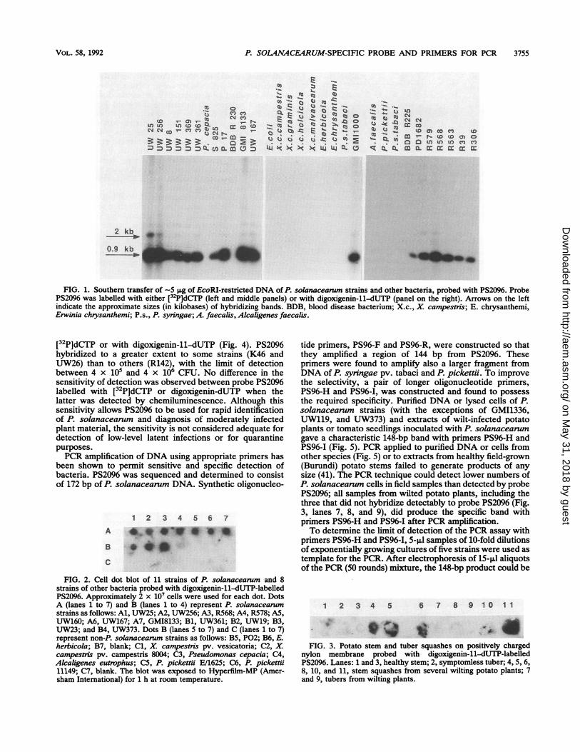
FIG. 1. Southern transfer of -5 SLg of EcoRI-restricted DNA of P. solanacearun strains and other bacteria, probed with PS2096. ProbePS2096 was labelled with either [32P]dCT7P (left and middle panels) or with digoxigenin-11-dUTP (panel on the right). Arrows on the leftindicate the approximate sizes (in kilobases) of hybridizing bands. BDB, blood disease bacterium; X.c., X. campestris; E. chrysanthemi,Erwinia chrysanthemi; P.s., P. syringae; A. faecalis, Alcaligenes faecalis.
[32P]dCTP or with digoxigenin-11-dUTP (Fig. 4). PS2096hybridized to a greater extent to some strains (K46 andUW26) than to others (R142), with the limit of detectionbetween 4 x 10⁵ and 4 x 10⁶ CFU. No difference in thesensitivity of detection was observed between probe PS2096labelled with [32P]dCTP or digoxigenin-dUTP when thelatter was detected by chemiluminescence. Although thissensitivity allows PS2096 to be used for rapid identificationof P. solanacearum and diagnosis of moderately infectedplant material, the sensitivity is not considered adequate fordetection of low-level latent infections or for quarantinepurposes.PCR amplification of DNA using appropriate primers has
been shown to permit sensitive and specific detection ofbacteria. PS2096 was sequenced and determined to consistof 172 bp of P. solanacearum DNA. Synthetic oligonucleo-
1 2 3 4 5 6 7A
B
FIG. 2. Cell dot blot of 11 strains of P. solanacearum and 8strains of other bacteria probed with digoxigenin-11-dUTP-labelledPS2096. Approximately 2 x 10⁷ cells were used for each dot. DotsA (lanes 1 to 7) and B (lanes 1 to 4) represent P. solanaceanumstrains as follows: Al, UW25; A2, UW256; A3, R568; A4, R578; A5,UW160; A6, UW167; A7, GMI8133; Bi, UW361; B2, UW19; B3,UW23; and B4, UW373. Dots B (lanes 5 to 7) and C (lanes 1 to 7)represent non-P. solanacearum strains as follows: B5, P02; B6, E.herbicola; B7, blank; Cl, X. campestris pv. vesicatoria; C2, X.campestris pv. campestris 8004; C3, Pseudomonas cepacia; C4,Alcaligenes eutrophus; C5, P. pickettii E/1625; C6, P. pickettii11149; C7, blank. The blot was exposed to Hyperfilm-MP (Amer-sham International) for 1 h at room temperature.
tide primers, PS96-F and PS96-R, were constructed so thatthey amplified a region of 144 bp from PS2096. Theseprimers were found to amplify also a larger fragment fromDNA of P. syningae pv. tabaci and P. pickettii. To improvethe selectivity, a pair of longer oligonucleotide primers,PS96-H and PS96-I, was constructed and found to possessthe required specificity. Purified DNA or lysed cells of P.solanacearum strains (with the exceptions of GMI1336,UW119, and UW373) and extracts of wilt-infected potatoplants or tomato seedlings inoculated with P. solanacearumgave a characteristic 148-bp band with primers PS96-H andPS96-I (Fig. 5). PCR applied to purified DNA or cells fromother species (Fig. 5) or to extracts from healthy field-grown(Burundi) potato stems failed to generate products of anysize (41). The PCR technique could detect lower numbers ofP. solanacearun cells in field samples than detected by probePS2096; all samples from wilted potato plants, including thethree that did not hybridize detectably to probe PS2096 (Fig.3, lanes 7, 8, and 9), did produce the specific band withprimers PS96-H and PS96-I after PCR amplification.To determine the limit of detection of the PCR assay with
primers PS96-H and PS96-I, 5-,ul samples of 10-fold dilutionsof exponentially growing cultures of five strains were used astemplate for the PCR. After electrophoresis of 15-,ul aliquotsof the PCR (50 rounds) mixture, the 148-bp product could be
1 2 3 4 5 6 7 8 9 10 1 1
FIG. 3. Potato stem and tuber squashes on positively chargednylon membrane probed with digoxigenin-11-dUTP-labelledPS2096. Lanes: 1 and 3, healthy stem; 2, symptomless tuber; 4, 5, 6,8, 10, and 11, stem squashes from several wilting potato plants; 7and 9, tubers from wilting plants.
VOL. 58, 1992 3755
on May 31, 2018 by guest
http://aem.asm
.org/D
ownloaded from

APPL. ENVIRON. MICROBIOL.
A
1 2 3 4 5 6 7 8
i
4X10⁷
4X10⁶
4X10⁵
FIG. 4. Comparison of sensitivity of probe PS2096 labelled with[32P]dCTP (A) and with digoxigenin-11-dlUTP (B). Detection was
done by autoradiography on Kodak XAR5 film for 1 day at -70°C(A) or by luminography on Hyperfilm-MP (Amersham) for 1 h atroom temperature (B). Lanes: 1, E. herbicola; 2, P. solanacearumUW26 mixed with 107 E. herbicola cells; lanes 3 to 10, P. solan-acearum UW26, R361, R289, R330, R142, K46, K46, and UW8,respectively.
seen from samples of strains UW25, UW19, S825, UW167,and R142, containing 5, 6, 14, 22, and 116 cells, respectively.To determine the sensitivity of the PCR assay in the pres-
ence of large numbers of other bacteria, cells of P. solan-acearum UW19 and UW25 were mixed with 20-, 200-,2,000-, and 20,000-fold excesses of either Erwinia herbicolaor X. campestis pv. campestris cells. The sensitivity of thePCR method remained the same when there was a 20- or
200-fold excess of E. herbicola or X. campestris pv. campes-
tris cells. However, with 2,000- and 20,000-fold excesses,
the level of reproducible detection was decreased 10-fold.
DISCUSSION
A 172-bp P. solanacearum-specific probe, PS2096, was
isolated by screening 44 subtraction hybridization libraryclones against 85 P. solanacearum strains and 27 strainsfrom other bacterial species. The 85 P. solanacearum strainsoriginated from 49 different host-country combinations andwere therefore considered to be a representative sample ofthe species. Only three P. solanacearum strains, GMI1336,UW119, and UW373, did not hybridize to PS2096 under
A _fi 7 .1 1Q 11 12U 1 1 17Z 1a 12 2Q
FIG. 5. Electrophoretic analysis of PCR-amplified DNA from P.solanacearum and other bacteria by using the P. solanacearum-specific primer pair PS96-H and PS96-I. The arrow on the left showsthe size of the specific PCR product. Lanes: 1 to 14, P. solan-acearum UW278, UW73, UW360, R361, R563, UW361, UW160,GMI8133, R142, R289, UW373, UW74, UW378, and PD1682,respectively; 15, E. herbicola; 16, P. cepacia; 17, A. eutrophus;18, P. syingae pv. tabaci; 19, P. celebense T340; 20, P. syzygiiR001.
stringent conditions. These three strains have previouslybeen reported not to hybridize to certain clones containinggenes required for pathogenicity and production of a hyper-sensitive response on an incompatible plant (hrp genes [7])(12). Failure of PS2096 to hybridize to these three nonpath-ogenic strains is therefore not a limitation to the diagnostictest in practice. No significant relatedness was found be-tween the PS2096 sequence and sequences present in theEMBL/GenBank Data Library or with the hrp clone pVir2(2). However, many naturally occurring avirulent strainscarry large deletions of hundreds of kilobases including thehrp genes (7), so it is possible that PS2096 is derived from aregion close to the hrp genes.MboI-digested DNA (fragments of <600 bp) was used as
the target DNA for subtraction hybridization to reduce theprobability of generating DNA fragments that contain bothP. solanacearum-specific and linked sequences not specificto P. solanacearum. Subtraction could perhaps be improvedby using driver DNA from a species more closely related toP. solanaceamm, such as P. pickettii or P. syzygii, than X.campestis pv. vesicatoria and by using multiple rounds ofsubtraction (44). The efficiency of the subtraction was notstudied in detail. Six of 25 probes tested did hybridize to X.campestris pv. vesicatoria, but the proportion of clones froman unsubtracted library that would cross-hybridize is un-known.The DNA probe PS2096 offers a rapid and precise identi-
fication method but cannot, however, detect less than -105to 106 bacteria and hence is insufficiently sensitive to detectlow-level infections. This problem could be overcome byamplification by selective growth of the bacteria on a mem-brane, followed by lysis and hybridization with PS2096. Thealternative approach used was to sequence the DNA probeto allow construction of specific oligonucleotide primers formore-sensitive detection through the application of PCRtechnology. Between 5 and 116 CFU of P. solanacearumcould be detected with primers PS96-H and PS96-I and 50rounds of amplification. As the PCR technique does notdistinguish between viable and nonviable organisms, thismight be an overestimate of the sensitivity. The sensitivity iscomparable, however, to that reported for Listeria monocy-togenes (16), for which a minimum number of 542 cells(viable and nonviable) could reliably be detected after only35 PCR amplification rounds. The sensitivity could be im-proved further by hybridization of the PCR product with aninternal part of the amplified fragment (37) or by a secondround of amplification (16). Either of these methods shouldbring the sensitivity down to the single-cell stage. It may alsobe necessary to use these two-step procedures for fieldsamples that contain a large excess (22,000-fold) of non-P.solanacearum cells, as this was found to reduce the repro-ducible level of sensitivity by an order of magnitude.
This work is the first report describing a DNA probespecific for the species P. solanacearum. Recently, Cookand Sequeira (14) utilized a subtraction hybridization proce-dure to enrich for P. solanacearum race 3-specific sequencesand obtained a probe that reacted with all race 3 strains butwith only 5 of 90 non-race 3 strains tested. They did notreport whether their probe hybridizes to closely relatedpathogens such as P. syzygii, P. cepacia, or P. pickettii.Although P. pickettii is usually recorded from human infec-tions arising from contaminated water supplies, one isolate,E1625, was supplied to our laboratory as originating from awilt-infected potato field in Ethiopia. Should P. pickettiistrains be commonly found in soil, it is clearly important thatprobes should not cross-react with this species. Eight of 15
3756 SEAIL ET AL.
148 bpNo
on May 31, 2018 by guest
http://aem.asm
.org/D
ownloaded from

P. SOLANACEARUM-SPECIFIC PROBE AND PRIMERS FOR PCR 3757
subtraction hybridization probes tested did hybridize to P.pickettii strains under stringent conditions, confirming thehigh genetic relatedness previously reported for these twospecies (36).The PCR can be used specifically to detect very low
numbers of P, solanacearum cells, without the need for priorenrichment or cultivation. In many instances, it is quickerand advantageous to use methods that do not require cultur-ing of the organism. Sele-ti_e media have been shown toreduce the efficiency with whi_cEWicroorganisms are recov-ered from the environment (39). Moreover, bacteria canenter a nonculturable but apparently viable state (34) and notbe detected by traditional isolation procedures.Many questions regarding the epidemiology of bacterial
wilt have remained unanswered to date partly because of thelack of a simple, rapid, and sensitive identification proce-dure. Seed has been suggested to be a vehicle for the spreadof bacterial wilt, but no conclusive data have been reported(23). Although many weeds have been shown to be symp-tomless carriers ofP. solanacearum (23), there are probablyalso many unknown hosts that maintain high levels ofinoculum between successive crops. The application ofprobe PS2096 and PCR primers PS96-H and PS96-I shouldenable such aspects of the epidemiology to be investigated.
ACKNOWLEDGMENTS
We thank all who kindly donated strains to our collection, J.Elphinstone for providing information on the pathogenicity, and D.Berrios, S. Eden-Green, J. Elphinstone, K. Y. Lum, and L.Skoglund for their help with field testing. We are also grateful to D.Love for providing useful information on the subtraction hybridiza-tion protocol.
This work was commissioned by the Natural Resources Institute,Chatham, United Kingdom (project X0082), and was carried outunder MAFF license no. PHF 1185A/99(81), PHF 1185B/10(111),and PHF 1185A/62(11), issued under the Plant Health (Great Britain)Order, 1987. The Sainsbury Laboratory is supported by the GatsbyCharitable Foundation.
REFERENCES1. Akiew, E. B., K. Hyde, L. Diatloff, and R. Peterson. 1990.
Bacterial wilt of Heliconia plants from Oahu, Hawaii. ACIARBacterial Wilt Newsl. 6:5.
2. Arlat, M., S. Genin, and C. Gough. Personal communication.3. Avery, R J., J. D. Norton, J. S. Jones, D. C. Burke, and A. G.
Morris. 1980. Interferon inhibits transformation by murine sar-coma viruses before integration of provirus. Nature (London)288:93-95.
4. Bennett, C. P. A., P. Hunt, and A. Asman. 1985. Association ofa xylem-limited bacterium with Sumatra disease of cloves inIndonesia. Plant Pathol. 34:487-494.
5. Bochner, B. R., and M. A. Savageau. 1977. Generalized indica-tor plates for genetic, metabolic, and taxonomic studies withmicroorganisms. Appl. Environ. Microbiol. 33:434 444.
6. Boucher, C. A., P. A. Barberis, A. P. Trigalet, and D. A.Demery. 1985. Transposon mutagenesis of Pseudomonas solan-acearum: isolation of TnS-induced mutants. J. Gen. Microbiol.131:2449-2457.
7. Boucher, C. A., F. Van Gijsegem, P. A. Barberis, M. Arlat, andC. ZischeL 1987. Pseudononas solanacearum genes controllingboth pathogenicity on tomato and hypersensitivity on tobaccoare clustered. J. Bacteriol. 169:5626-5632.
8. Buddenhagen, I. W. 1961. Bacterial wilt of bananas: history andknown distribution. Trop. Agric. Trinidad 38:107-121.
9. Buddenhagen, I. W. 1986. Bacterial wilt revisited, p. 126-143. InG. J. Persley (ed.), Bacterial wilt disease in Asia & the SouthPacific. Proc. Int. Workshop, PCARRD, Los Bafios, The Phill-ipines, 8-10 Oct. 1985. ACIAR Proc. 13, Canberra, Australia.
10. Buddenhagen, I. W., L. Sequeira, and A. Kelman. 1962. Desig-
nation of races in Pseudomonas solanacearum. Phytopathology52:726.
11. Ciampi, L., L. Sequeira, and E. R. French. 1980. Latentinfection of potato tubers by Pseudomonas solanacearum. Am.Potato J. 57:377-386.
12. Cook, D., E. Barlow, and L. Sequeira. 1989. Genetic diversity ofPseudononas solanacearum: detection of restriction fragmentlength polymorphisms that specify virulence and the hypersen-sitive response. Mol. Plant-Microbe Interact. 2:113-121.
13. Cook, D., E. Barlow, and L. Sequeira. 1991. DNA probes astools for the study of host-pathogen evolution: the example ofPseudomonas solanacearum, p. 103-108. In H. Hennecke andD. P. Verma (ed.), Advances in molecular genetics of plant-microbe interactions, vol. 1. Kluwer, Dordrecht, The Nether-lands.
14. Cook, D., and L. Sequeira. 1991. The use of subtractive hybrid-ization to obtain a DNA probe specific for Pseudomonassolanacearum race 3. Mol. Gen. Genet. 227:401-410.
15. Daniels, M. J., C. E. Barber, P. C. Turner, W. G. Cleary, andM. Sawczyc. 1984. Isolation of mutants of Xanthononascampestris pv. campestris showing altered pathogenicity. J.Gen. Microbiol. 130:2447-2455.
16. Deneer, H. G., and I. BoychukL 1991. Species-specific detectionof Listeria monocytogenes by DNA amplification. Appl. Envi-ron. Microbiol. 57:606-609.
17. Eden-Green, S. J., and H. Sastraatmadja. 1990. Blood diseasepresent in Java. FAO Plant Prot. Bull. 38:49-50.
18. Fitts, R., M. Diamond, C. Hamilton, and M. Neri. 1983. DNA-DNA hybridization assay for detection of Salmonella spp. infoods. Appl. Environ. Microbiol. 46:1146-1151.
19. Gaumann, E. 1923. Onderzoekeningen over de bloedziekte derbananen op Celebes II. Mededeelingen Instit. Plantenziekten59:1-47.
20. Gilbertson, R. L., D. P. Maxwell, D. J. Hagedorn, and S. A.Leong. 1989. Development and application of a plasmid DNAprobe for detection of bacteria causing common bacterial blightof bean. Phytopathology 79:518-525.
21. Gridmont, P. A. D., F. Gridmont, N. Desplaces, and P. Tschen.1985. DNA probe specific for Legionella pneumophila. J. Clin.Microbiol. 21:431-437.
22. Hayward, A. C. 1964. Characteristics of Pseudomonas solan-acearum. J. Appl. Bacteriol. 27:265-277.
23. Hayward, A. C. 1991. Biology and epidemiology of bacterial wiltcaused by Pseudomonas solanacearum. Annu. Rev. Phyto-pathol. 29:65-87.
24. Hendrick, C. A., and L. Sequeira. 1984. Lipopolysaccharide-defective mutants of the wilt pathogen Pseudononas solan-acearum. Appl. Environ. Microbiol. 48:389-395.
25. Hill, W. E., W. L. Payne, and C. C. G. Aulisio. 1983. Detectionand enumeration of virulent Yersinia enterocolitica in food byDNA colony hybridization. Appl. Environ. Microbiol. 46:636-641.
26. Kelman, A. 1954. The relationship of pathogenicity of Pseudo-monas solanacearum to colony appearance on a tetrazoliummedium. Phytopathology 44:693-695.
27. Lehman-Danziger, H. 1987. The distribution of Moko disease inCentral and South America and its control on plantains andbananas, p. 130-155. In Seminar proceedings, improving citrusand banana production in the Caribbean through phytosanita-tion, St. Lucia, West-Indies, 2-5 September 1986. CTA/CARDI, Wageningen, The Netherlands.
28. Maniatis, T., E. F. Fritsch, and J. Sambrook 1982. Molecularcloning: a laboratory manual. Cold Spring Harbor Laboratory,Cold Spring Harbor, N.Y.
29. Manulis, S., Y. Gafni, E. Clark, D. Zutra, Y. Ophir, and I.Barash. 1991. Identification of a plasmid DNA probe for detec-tion of strains of Envinia herbicola pathogenic on Gypsophilapaniculata. Phytopathology 81:54-57.
30. Miller, J. H. 1972. Experiments in molecular genetics. ColdSpring Harbor Laboratory Press, Cold Spring Harbor, N.Y.
31. Nyangeri, J. B., E. M. Gathuru, and D. M. Mukunya. 1984.Effect of latent infection on the spread of bacterial wilt ofpotatoes in Kenya. Trop. Pest Manage. 30:163-165.
VOL. 58, 1992
on May 31, 2018 by guest
http://aem.asm
.org/D
ownloaded from

APPL. ENVIRON. MICROBIOL.
32. Olsson, K. 1976. Experience of brown rot caused by Pseudo-monas solanacearum (Smith) Smith in Sweden. EPPO (Eur.Mediterr. Plant Prot. Organ.) Bull. 6:199-207.
33. Palleroni, N. J. 1984. Family I. Pseudomonadeae Winslow,Broadhurst, Buchanan Krumwiede, Rogers and Smith 1917,555AL*, p. 141-219. In N. R. Krieg and J. G. Holt (ed.),Bergey's manual of systematic bacteriology, vol. I. Williams &Wilkins, Baltimore.
34. Pickup, R. W. 1991. Development of molecular methods for thedetection of specific bacteria in the environment. J. Gen.Microbiol. 137:1009-1019.
35. Prior, P., M. Beramis, M. Chillet, and J. Schmit. 1990. Prelim-inary studies for tomato bacterial wilt (Pseudomonas solan-acearum E. F. Sm.) resistance mechanisms. Symbiosis 9:393-400.
36. Ralston, E., N. J. Palleroni, and M. Doudoroff. 1973. Pseudo-monas pickettii, a new species of clinical origin related toPseudomonas solanacearum. Int. J. Syst. Bacteriol. 23:15-19.
37. Rasmussen, 0. F., apd B. S. Wulff. 1990. Identification and useofDNA probes for plant pathogenic bacteria. In Fifth EuropeanCongress on Biotechnology, Copenhagen, July 8-13, 1990.Proceedings, vol. 2.
38. Roberts, S. J., S. J. Eden-Green, P. Jones, and D. J. Ambler.1990. Pseudomonas syzygii sp. nov., the cause of Sumatradisease of cloves. Syst. Appl. Microbiol. 13:34-43.
39. Roszak, D. B., and R. R. Colwell. 1987. Survival strategies ofbacteria in the natural environment. Microbiol. Rev. 51:365-379.
40. Schaad, N. W., H. Azad, R. C. Peet, and p{. J. Panopolus. 1989.Identification of Pseudomonas syrngae pv., phaseolicola by aDNA hybridization probe. Phytopathology 79:903-907.
41. Seal, S., J. Elphinstone, L. Skoglund, and D. Berrios. 1992.Detection of Pseudomonas solanacearum latent infections inseed potatoes during their multiplication irq Burundi. ACIARBacterial Wilt Newsl. 8:2-3.
42. Seal, S., L. Jackson, and M. Daniels. Unpublished data.43. Stead, D. E. 1988. Identification of bacteria by computer-
assisted fatty acid profiling. Acta Hortic. (The Hague) 225:39-46.
44. Straus, D., and F. M. Ausubel. 1990. Genomic subtraction forcloning DNA corresponding to deletion mutations. Proc. Natl.Acad. Sci. USA 87:1889-1893.
45. Thompson, E., J. V. Leary, and W. W. C. Chun. 1989. Specificdetection of Clavibacter michiganense subsp. michiganense bya homologous DNA probe. Phytopathology 79:311-314.
46. Wood, P. K., J. G. Morrie, P. L. C. Small, 0. Sethabutr, M.Regina, L. Trabulsi, and J. B. Kaper. 1986. Comparison of DNAprobes and the Sereny test for identification of invasive Shigellaand Escherichia coli strains. J. Clin. Microbiol. 24:498-500.
3758 SEAL ET AL.
on May 31, 2018 by guest
http://aem.asm
.org/D
ownloaded from