isolation, detection and genomic differentiation … · yang tinggi. dendrogram yang dihasilkan...
TRANSCRIPT

ISOLATION, DETECTION AND GENOMIC DIFFERENTIATION OF Escherichia
coli FROM AQUATIC ENVIRONMENTS IN KELANTAN, MALAYSIA.
RATHI DEVI A/P ATHIMURTHY
SGF 070016
Thesis submitted in partial fulfillment of the requirements for the degree of Master of
Biotechnology
MASTER OF BIOTECHNOLOGY
FACULTY OF SCIENCE
UNIVERSITY MALAYA
KUALA LUMPUR, MALAYSIA
2009

UNIVERSITI MALAYA
ORIGINAL LITERARY WORK DECLARATION
Name of Candidate: Rathi Devi a/p Athimurthy I.C/Passport No: 831030-01-6538
Registration/Matric No: SGF 070016
Name of Degree: Masters of Biotechnology (M.Biotech)
Title of Project Paper/Research Report/Dissertation/Thesis (“this Work”):
Isolation, detection and genomic differentiation of Escherichia coli from aquatic environments
in Kelantan.
Field of Study: Biotechnology
I do solemnly and sincerely declare that:
(1) I am the sole author/writer of this Work;(2) This Work is original;(3) Any use of any work in which copyright exists was done by way of fair dealing and forpermitted purposes and any excerpt or extract from, or reference to or reproduction ofany copyright work has been disclosed expressly and sufficiently and the title of theWork and its authorship have been acknowledged in this Work;(4) I do not have any actual knowledge nor do I ought reasonably to know that the makingof this work constitutes an infringement of any copyright work;(5) I hereby assign all and every rights in the copyright to this Work to the University ofMalaya (“UM”), who henceforth shall be owner of the copyright in this Work and that anyreproduction or use in any form or by any means whatsoever is prohibited without thewritten consent of UM having been first had and obtained;(6) I am fully aware that if in the course of making this Work I have infringed any copyrightwhether intentionally or otherwise, I may be subject to legal action or any other actionas may be determined by UM.
Candidate’s Signature Date
Subscribed and solemnly declared before,
Witness’s Signature Date
Name: Prof Dr Thong Kwai Lin
Designation: Supervisor

ABSTRACT
Diarrhea caused by Escherichia coli is one of the main diseases associated with
water supply and sanitation. The main aims of this study were to isolate and confirm the
presence of E. coli in selected aquatic environments in Bachok, Kelantan, as well as to
determine the incidence of their virulence genes and the genomic diversity among the
isolates. Fifty water samples from various aquatic environments of Bachok, Kelantan
were examined to determine their microbiological quality by applying both phenotypic
and genotypic methods for detection of total coliform and E. coli. The presence of total
coliform was significantly correlated to E. coli (p<0.05). Based on biochemical tests,
78% of the samples had E. coli with an average density of 1 x 10⁶ cfu/100mL. Among
the 39 isolates recovered, 74% (29 isolates) of E. coli were positive for the phoA gene,
which is the housekeeping gene for E. coli. A hexaplex PCR was performed to detect six
virulence genes in pathogenic E. coli using 5 sets of primers (ST1, LT1, LT2, VT and
AE). E. coli from only one sample (EC15) was positive for the LT1 gene, which codes
for LT-heat labile toxin. Antimicrobial susceptibility tests (AST) showed that only
ETEC isolate was resistant to ampicillin, chloramphenicol and trimethoprim-
sulfamethoxazole. The rest of E. coli isolates were susceptible to the tested antibiotics.
The analysis of genomic diversity of E. coli isolates by Repetitive Extragenic
Palindromic (REP)-PCR generated 27 patterns (F=0.26-1.0). The REP-PCR profiles
were reproducible and the multiple DNA fingerprints showed that the E. coli isolates
were genetically diverse. A dendrogram generated by the UPGMA algorithm showed 4
clusters of E. coli isolates based on 80% similarity. Overall, REP-PCR generated high
genetic variability within the E. coli isolates. The finding in the study indicates that

REP-PCR is a promising molecular method for determining the genomic diversity of
environmental E. coli strains.
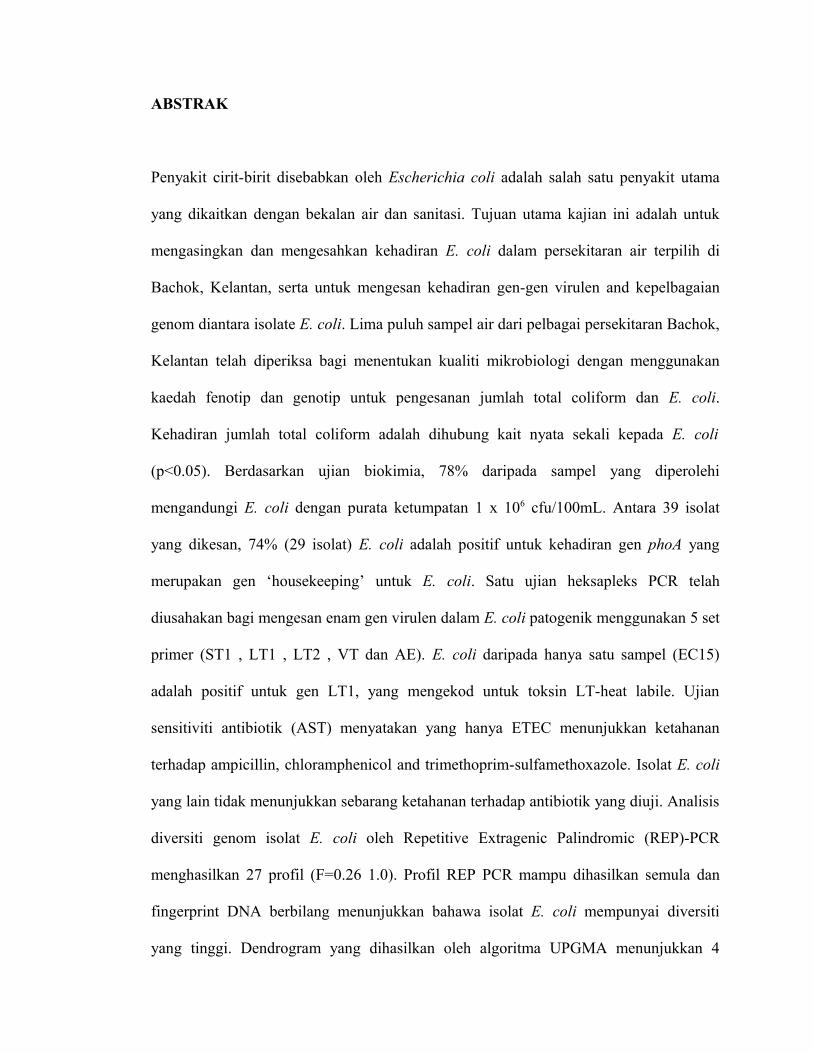
ABSTRAK
Penyakit cirit-birit disebabkan oleh Escherichia coli adalah salah satu penyakit utama
yang dikaitkan dengan bekalan air dan sanitasi. Tujuan utama kajian ini adalah untuk
mengasingkan dan mengesahkan kehadiran E. coli dalam persekitaran air terpilih di
Bachok, Kelantan, serta untuk mengesan kehadiran gen-gen virulen and kepelbagaian
genom diantara isolate E. coli. Lima puluh sampel air dari pelbagai persekitaran Bachok,
Kelantan telah diperiksa bagi menentukan kualiti mikrobiologi dengan menggunakan
kaedah fenotip dan genotip untuk pengesanan jumlah total coliform dan E. coli.
Kehadiran jumlah total coliform adalah dihubung kait nyata sekali kepada E. coli
(p<0.05). Berdasarkan ujian biokimia, 78% daripada sampel yang diperolehi
mengandungi E. coli dengan purata ketumpatan 1 x 10⁶ cfu/100mL. Antara 39 isolat
yang dikesan, 74% (29 isolat) E. coli adalah positif untuk kehadiran gen phoA yang
merupakan gen ‘housekeeping’ untuk E. coli. Satu ujian heksapleks PCR telah
diusahakan bagi mengesan enam gen virulen dalam E. coli patogenik menggunakan 5 set
primer (ST1 , LT1 , LT2 , VT dan AE). E. coli daripada hanya satu sampel (EC15)
adalah positif untuk gen LT1, yang mengekod untuk toksin LT-heat labile. Ujian
sensitiviti antibiotik (AST) menyatakan yang hanya ETEC menunjukkan ketahanan
terhadap ampicillin, chloramphenicol and trimethoprim-sulfamethoxazole. Isolat E. coli
yang lain tidak menunjukkan sebarang ketahanan terhadap antibiotik yang diuji. Analisis
diversiti genom isolat E. coli oleh Repetitive Extragenic Palindromic (REP)-PCR
menghasilkan 27 profil (F=0.26 1.0). Profil REP PCR mampu dihasilkan semula dan
fingerprint DNA berbilang menunjukkan bahawa isolat E. coli mempunyai diversiti
yang tinggi. Dendrogram yang dihasilkan oleh algoritma UPGMA menunjukkan 4

cluster isolat E. coli berdasarkan 80% persamaan. Keseluruhannya, REP PCR
menghasilkan variasi genetik yang tinggi dalam isolat E. coli. Penemuan dalam kajian
itu menunjukkan yang REP-PCR adalah satu kaedah molekular yang baik untuk
penentuan diversiti genom E. coli dari alam sekitar.

ACKNOWLEDGEMENTS
I take this opportunity to extend my deep sense of gratitude and words of appreciation
towards those who helped me during the pursuit of my present study. Firstly, I would
like to express my deepest gratitude to my supervisor, Prof. Dr. Thong Kwai Lin for
guidance and strength that she bestowed during the entire project. I deeply appreciate
her patient advice throughout the project.
I also have great appreciation for the Bachok team whose combined efforts helped me a
lot in the success of my work. I would like to extent thanks to all the staff members of
Genetic and Molecular Biology Department for the guidance and technical assistants.
Special thanks go to Mr. Kudus and Cik Hazalina. I also extend gratefulness to the IOES
and IPPP of University Malaya for providing research funding to aid in the cost of the
project. Besides that, I am grateful to my fellow lab mates for the assistance, care and
friendship. Special thanks to Vimala for her kind assistance and thought sharing.
I would like to express my heartfelt gratitude to my mum, sister and loved one for the
unconditional support and love. Not forgetting my dad who is my inspiration in life. Last
but not the least, I would like to thank all those who helped directly or indirectly in
completion of this study. My most sincere thanks to the Almighty God who made
everything possible.

PUBLICATION AND PRESENTATION
Publication:
Rathi, A., Thong, K. L. and Chong, V. C. Isolation, detection and genomic
differentiation of Escherichia coli from aquatic environments in Kelantan, Malaysia.
- Paper submitted to Malaysian Journal of Science (MJS) on April 2009.
Presentations:
Thong, K. L., Vimala, B., Rathi, A., Lim, S. Y. and Tim, S. C. Microbial source
tracking in an aquatic ecosystem in Bachok, Kelantan, Malaysia.
- Poster presented at the International Microbial Biotechnology Conference
(IMBC) and Workshop on Metagenome, Jakarta, Indonesia on 11th -13th Dec
2008.
Thong, K. L., Vimala, B., Rathi, A., Lim, S. Y., Tim, S. C. and Chong, V. C. Microbial
source tracking in an aquatic ecosystem in Bachok, Kelantan, Malaysia.
- Poster presented at the South China Sea Conference (SCS), Kuantan Pahang on
25th -29th Nov 2008.

CONTENTS
Abstract iii
Abstrak v
Acknowledgments vii
Publication and Presentation viii
Contents ix
List of Figures xiv
List of Tables xvi
Chapter 1: Introduction
1.1 Background of the study 1
1.2 Objectives of the study 6
1.3 Scope of the study 6
Chapter 2: Literature review
2.1 Water Quality and Sanitation 7
2.2 Microbial Contamination 7
2.3 Water quality indicator 8
2.4 Escherichia coli 9
2.5 Pathogenic E. coli 10
2.6 Molecular Approach 12
2.7 Detection of pathogenic strains 13
2.7.1 Phenotypic Assays

2.7.2 Molecular Assays
2.8 Microbial source tracking 15
2.8.1 Phenotypic source tracking
2.8.1.1 Antibiotic resistance analysis (ARA)
2.8.2 DNA based source tracking
2.9 Rep-PCR 18
2.9.1 Advantages of rep-PCR Analysis
Chapter 3: Materials and methods
3.1 Materials 21
3.1.1 Sampling 21
3.1.2 E. coli isolates 21
3.1.3 Media and reagents 22
3.1.3.1 Media for bacterial growth
3.1.3.2 Materials for Biochemical Tests
3.1.3.3 Other Solutions
3.1.4 PCR Materials and Equipments 26
3.1.4.1 Materials and Equipments for PCR assays
3.1.4.2 Materials and Equipments for Agarose Gel Electrophoresis
3.2 Methods 31
3.2.1 Collection of water samples 31
3.2.2 Media preparation 31
3.2.3 Filtration of water samples 31
3.2.4 Isolation of E. coli and other coliforms 32
3.2.5 Biochemical tests 32
3.2.5.1 Indole test
3.2.5.2 Methyl red test
3.2.5.3 Voges Proskauer test
3.2.5.4 Citrate utilization test
3.2.5.5 Triple Sugar Iron (TSI) test
3.2.6 API 20E 35
3.2.7 Total DNA extraction from bacterial culture 35
3.2.8 Polymerase Chain Reaction (PCR) assay 36
3.2.8.1 PCR material and master mixture preparations
3.2.8.2 Confirmation of E. coli using Monoplex PCR assay
3.2.8.3 Toxin genes detection using Multiplex PCR assay
3.2.8.4 PCR conditions
3.2.8.5 Running Agarose Gel Electrophoresis
3.2.9 Antimicrobial agent susceptibility testing (ARA) 42
3.2.9.1 Preparation of inoculum
3.2.9.2 Inoculation of Mueller-Hinton agar
3.2.10 Repetitive extragenic palindromes (REP)-PCR amplification
3.2.11 Interpretation of fingerprints 45
Chapter 4: Results
4.1 Sampling and isolation of total coliform and E. coli 46
4.2 Correlation between total coliform and E. coli 50

4.2.1 Comparison of total coliform and E. coli colony counts isolated 52
at different holding time
4.3 Identification of E. coli isolates 54
4.3.1 Confirmation by using biochemical tests 54
4.3.2 Confirmation by using API 20E 56
4.3.3 Optimization of monoplex PCR using different primer concentration 57
4.3.3.1 Confirmation by using monoplex PCR assay 58
4.4 Virulence gene detection 61
4.4.1 Detection of E. coli 0157:H7 by selective plating 62
4.5 Antimicrobial susceptibility test of E. coli strains 63
4.6 Genetic diversity of E. coli using PCR based subtyping 64
4.6.1 Optimization of REP-PCR using different primers 64
4.6.2 REP-PCR of isolates 65
4.6.3 Cluster analysis of E. coli isolates 66
4.6.3 Comparison of REP-PCR of E. coli isolates from clinical 69
and food samples
4.6.4 Cluster analysis of E. coli from different sources 73
Chapter 5: Discussion
5.1 Isolation and identification of E. coli 74
5.2 Survival of E. coli and total coliform at different holding time 76
5.3 PCR confirmation of E. coli isolates 78
5.4 Prevalence of virulence genes 79
5.5 Antimicrobial Susceptibility test 80

5.6 Genomic diversity among isolates based on REP-PCR 81
Chapter 6: Conclusion
6.1 Conclusion 83
References 84
Appendixes 102

LIST OF FIGURES
Fig. 1.1a Sampling sites around Bachok, Kelantan. 3
Fig.4.1a E. coli produce dark blue to violet colonies on CCA 47
plates (arrow). Total coliform produce salmon to red colonies.
Fig. 4.1b E. coli produce greenish metallic sheen colonies on EMB 47
agar plates.
Fig. 4.2a Correlation of total coliform and E. coli (p<0.05) 51
Fig. 4.2b Comparison of colony count of total coliform and E. coli 53
isolated at different holding time.
Fig. 4.3a E. coli negative for citrate utilization. 55
Fig. 4.3b E. coli in TSI tubes produce an acid butt, an acid slant and gas. 55
Fig. 4.3c E. coli was positive for indole production. 55
Fig. 4.3d E. coli was positive for methyl red test. 55
Fig. 4.3e API 20E kits shows the identification of E. coli 57
Fig. 4.3f Representative gel of monoplex PCR for phoA gene detection 57
of 11 presumptive E. coli isolates.
Fig. 4.3g Representative gel of monoplex PCR for phoA gene detection 58
of 10 presumptive E. coli isolates (primer- 0.1µM).
Fig. 4.4a Representative gel showing multiplex PCR using 62
5 sets of primers.
Fig. 4.5a Antimicrobial susceptibility test plates of sample 15 which was 63
resistant to ampicillin, chloramphenicol and trimethoprim-
sulfamethoxazole.

Fig. 4.6a Representative gel showing REP-PCR for three isolates 64
(EC6, EC7, EC44) using four different types of primers.
Fig. 4.6b Representative DNA fingerprints of 11 E. coli isolates generated 66
by REP1R(b) primer.
Fig. 4.6c Dendrogram showing the result of cluster analysis of the 68
REP-PCR patterns from 29 isolates of E. coli generated with
GelCompar software by the UPGMA method. The different
fingerprint patterns and location of samples are indicated.
Fig. 4.6d Representative DNA fingerprint of clinical and food E. coli 70
isolates generated by REP 1R(b) primer.
Fig. 4.6e Dendrogram showing the result of cluster analysis of the 72
REP-PCR patterns from 49 isolates of E. coli generated with
GelCompar software by the UPGMA method. The different
fingerprint patterns and location of samples are indicated.
LIST OF TABLES

Table 3.1a The bacterial control strains and their respective genes. 22
Table 3.1b Primers used for detection of housekeeping gene (monoplex) 28
and virulence genes (multiplex) of E. coli.
Table 3.1c Primers used for REP-PCR assays. 29
Table 3.2a Monoplex PCR master mixture. 38
Table 3.2b Multiplex PCR master mixture. 38
Table 3.2c PCR amplification condition for monoplex and multiplex PCR 39
assays.
Table 3.2d PCR amplification condition for REP- PCR assay. 44
Table 3.2e REP-PCR Master mixture. 44
Table 4.1c Colony count of total coliform and E. coli for fifty samples at 48
two different holding time.
Table 4.2a Pearson correlation of E. coli and total coliform. 50
Table 4.3a Results for API 20E test. 56
Table 4.3b Summary of E. coli recovery from different confirmation stages. 59
Table 4.6a Sources and origin of clinical and food sample used. 69
Table 4.1a Global positioning system (GPS) of fifty samples 101
collected around Bachok, Kelantan.
Table 4.5a Antimicrobial susceptibility test result of 29 E. coli 103
strains.
Table 4.1b Salinity and other detailed particulars of 15 samples 104
CHAPTER 1: INTRODUCTION