ionizing radiation in medical imaging and efforts in dose optimization
TRANSCRIPT

17
Ionizing Radiation in Medical Imaging and Efforts in
Dose Optimization
Varut Vardhanabhuti and Carl A. Roobottom Derriford Hospital and
Peninsula College of Medicine and Dentistry, Plymouth,
United Kingdom
1. Introduction
Medical-related radiation is the largest source of controllable radiation exposure to humans
and it accounts for more than 95% of radiation exposure from man-made sources. Its direct
benefits in modern day medical practices are beyond doubt but risks-benefits ratios need to
be constantly monitored as the use of ionizing radiation is increasing rapidly. From 1980 to
2006, the per-capita effective dose from diagnostic and interventional medical procedures in
the United States increased almost six fold, from 0.5 to 3.0mSv, while contributions from
other sources remained static (NCRP report no 160, 2009).
This chapter will review radiation exposure from medical imaging initially starting from a historical viewpoint as well as discussing innovative technologies on the horizon. The challenges for the medical community in addressing the increasing trend of radiation usage will be discussed as well as the latest research in dose justification and optimization.
2. Sources and trends
Medical radiation is by far the largest artificial source of population exposure to ionising
radiation, accounting for 90% of all doses from artificial sources (United Nations, 2010). This
radiation burden is increasing. In the US, this increase has been primarily due to the
increasing use of computed tomography (CT). Despite the fact that CT only accounts for
11% of the examinations it contributes 68% of collective dose. In comparison, conventional
radiography constitutes 90% of the examinations but only 19% of collective dose (Mettler et
al., 2009). From 1980s to 2006, there is an increase of approximately 6-fold in cumulative
effective dose per individual in the US from 0.5 to 3mSV (NCRP report no 160., 2009).
As an overall exposure to all kinds of radiation to humans, medical-related radiation
exposure now accounts for 48% (an increase from 15%) with background radiation
remaining relative static at 50%. The remaining 2% of total radiation exposure came from
consumer-related products and activities. These include cigarette smoking, building
materials, commercial air travel, mining and agriculture.
www.intechopen.com

Current Topics in Ionizing Radiation Research 364
Table 1. Changes in collective effective dose and effective dose per individual in the US population between early 1980s and 2006.
Fig. 1. Exposure of the US population in early 1980s and in 2006 according to NCRP report no 93 and 160.
From data in 2006, in radiology and nuclear medicine procedures, the biggest contribution
as previously mentioned is from computed tomography (49%), followed by nuclear
medicine studies (26%), then interventional procedures (14%). The remaining 11% came
from radiographic and fluoroscopic studies despite accounting for more than 74% of total
number of procedures. Not surprisingly, the per capita dose for CT is highest at 1.47 mSv.
Data is summarised in the following table.
www.intechopen.com

Ionizing Radiation in Medical Imaging and Efforts in Dose Optimization 365
Data taken from NRCP report no 160
Table 2. Estimated number and collective effective doses from radiologic and nuclear medicine procedures in the US for 2006.
The overall trend is similar in the UK. A recent Health Protection Agency publication (Hart et al., 2010) showed a 28% increase in 2008 compared to 1997/8 in the United Kingdom. This increase is again mainly due to a doubling of the computed tomography (CT) examinations performed over this same period. The increase in radiation dose in the UK is thought to be modest and this is partly due to the number of examinations performed. A comparison with other European countries show that the 19 out of 20 most commonly performed examinations, UK shows a lower than European average in frequency (Aroua et al., 2010). Only barium enemas were shown to be more frequent than average compared to the European counterparts. In terms of dose, 17 out of 20 examinations in the UK are less on average dose compared to their European counterparts. This illustrates that even in a country like United Kingdom, the trend for increasing use of medically related ionising radiation continues apace despite the fact that there has been more traditional emphasis on
Table 3. Collective dose per caput in 2008 for the “top 20” examinations in Europe (from Aroua et al, 2010).
www.intechopen.com

Current Topics in Ionizing Radiation Research 366
dose awareness. Table below shows annual collective dose per capita of European countries. The UK collective dose per caput from all medical and dental X-ray examinations stands at 0.3 mSv. This number can be compared with corresponding values of 2.2 mSv assessed for medical and dental X-rays in the USA in 2006 (NCRP, 2009) and the similar figure of 1.9 mSv (UNSCEAR., 2010) as the average for people living in Healthcare Level 1 (HCL1) countries. The UK collective dose per caput is clearly very low for a HCL1 country. For CT, the UK annual per caput dose stands at 0.27 mSv. This number can be compared with corresponding levels of 1.5 mSv from CT in the USA in 2006 (NCRP., 2009) and 0.74 mSv from CT in Canada in 2006 (Chen and Moir., 2010). The UK per caput dose from CT is relatively low compared to North American countries. At the time of writing, there is likely to be an even further increase in the use of CT compared to the level quoted in 2006. This is mainly due to rapid rise in the use of CT in emergency setting (White and Kuo, 2007; Street et al, 2009) and also the increasing use of PET/CT (Elliot A, 2009; Chawla et al, 2010; Devine et al, 2010). However, the dose increase may not be as high as recent concentrated efforts in the medical community have focused on strategies in dose reduction. Recent effective novel innovations have helped to reduce some of the dose burden (see later).
3. Risks
Ionizing radiation has long been known to increase the risk of cancer and is officially classified as a carcinogen by the International Agency for Research on Cancer (IARC). It is now on the official list of carcinogen by the World Health Organization (IARC list of carcinogen, 2011). The exact relationship between dose exposure and cancer induction, however, is complex and several issues merit further discussion. First, perceived medical-related radiation has traditionally been quantified in comparison with radiation we all receive from background levels. It is worth noting that medical-related radiation exposure is not the same as background radiation. The exposure from background radiation is generally of mixed-energy particles (high and low LET-radiation) while the exposure from diagnostic medical procedures is generally of low-energy x-rays. Low-LET radiation deposits less energy in the cell along the radiation path and is considered less destructive per radiation track than high-LET radiation. Examples of low-LET radiation include X-rays and γ-rays (gamma rays), which are used in medical imaging. Low-LET radiation produces ionizations sparsely throughout a cell; in contrast, high-LET radiation transfers more energy per unit length as it traverses the cell and is more destructive per unit length. Background radiation are naturally occurring from sources in soil, rocks, bricks and building material, and from radon gas which seeps out to homes. Radon is a colourless, odourless gas that emanates from the earth and as it decays also emits LET radiation. Average annual exposures worldwide to natural radiation sources (both high and low LET) would generally be expected to be in the range of 1–10 mSv, with 2.4 mSv being the present estimate of the central value (UNSCEAR, 2000). Of this amount, about one-half (1.2 mSv per year) comes from radon and its decay products. Some areas will have more radon gas background than others (e.g. Cornwall in the UK generally has twice the amount of background radiation than the rest of the UK). After radon, the next highest percentage of natural ionizing radiation exposure comes from cosmic rays. This is followed by terrestrial sources, and “internal” emissions. Cosmic rays
www.intechopen.com

Ionizing Radiation in Medical Imaging and Efforts in Dose Optimization 367
are particles that travel through the universe and are filtered by the earth atmosphere (therefore this varies from sea level to higher altitudes where less atmospheric filtration occurs). The Sun is a source of some of these particles. Other particles come from exploding stars called supernovas. The amount of terrestrial radiation from rocks and soils varies in different parts of the world. Much of this variation is due to differences in background radon levels. “Internal” emissions come from radioactive isotopes in food and water with uranium and thorium series of radioisotopes present in food and drinking water (UNSCEAR, 2000). To quantify medical exposures (which are of predominantly low-LET radiation) to background radiation (which are of mixed-LET radiation) is uncertain due to this respect but it is now generally accepted that of the 2.4mSv total average background radiation of mixed-LET, the total average annual population exposure worldwide due to low-LET radiation would generally be expected to be in the range of 0.2–1.0 mSv, with 0.9 mSv being the present estimate of the central value. The pie chart below illustrates proportions of high and low-LET radiation of the background radiation worldwide (adapted from BIER VII, 2006).
Fig. 2. Pie chart proportions of high and low-LET radiation of the background radiation worldwide (adapted from BIER VII, 2006).
Ubiquitous background radiation represents exposure to whole population (all ages, gender, health status) whereas medical radiation distributions are often skewed towards higher age groups and also sicker individuals. There may also be skewed in gender distribution (there is rightly a reluctance to image patients who are pregnant, for example). Therefore, one must acknowledge that the background radiation level is an approximation and will vary from individual to individual living at different locality having varying exposures.
www.intechopen.com

Current Topics in Ionizing Radiation Research 368
Current radiation protection standards and practices are based on the premise that any
radiation dose, no matter how small, can result in detrimental health effects, such as cancer
and genetic damage. Further, it is assumed that these effects are produced in direct
proportion to the dose received, i.e., doubling the radiation dose results in a doubling of the
effect. These two assumptions lead to a dose-response relationship, often referred to as the
linear no-threshold model, for estimating health effects at doses of interest. Although, this is
of much benefit in practical terms in risk estimation in the context of radiation protection
quantification, there is, however, substantial scientific evidence that this model is an
oversimplification of the dose-response relationship. In particular, it results in an
overestimation of health risks in the low dose range. Biological mechanisms including
cellular repair of radiation injury, which are not accounted for by the linear, no-threshold
model, reduce the likelihood of cancers and genetic effects. Therefore, it is now generally
accepted that quantification of estimated health risk in the dose range similar to that of
background radiation should be strictly qualitative and encompass a range of hypothetical
health outcomes, including the possibility of no adverse health effects at such low levels
(Burk Jr RJ 1996, updated in 2004).
Biological Effects of Ionizing Radiation (BEIR) VII committee defines low doses of ionizing
radiation as less than 100mSv and agrees that at doses of 100 mSv or less, statistical
limitations make it difficult to evaluate cancer risk. The current best estimation model for
risks associated with low dose radiation exposure is that approximately one individual in
100 persons would be expected to develop cancer (solid cancer or leukemia) from a dose of
100 mSv while approximately 42 of the 100 individuals would be expected to develop solid
cancer or leukemia from other causes (BEIR VII, 2006).
It is now generally accepted that at effective doses above 50 mSv the risk of cancer induction
increases linearly with dose, this dose-response relation has not been demonstrated at doses
below 50 mSv. Below 50 mSv no convincing epidemiological evidence exists currently for
cancer risk induction.
There is, however, more convincing evidence to support risk of cancer induction using high
dose radiation. A recent article from the USA estimated that CT scans performed in 2007
could result in 29,000 future cancers based on current risk estimations (Berrington de
Gonzales et al., 2009). The risk to an individual patient is also high. For example a cardiac
scan in a 20-year-old female can produce a lifetime cancer risk from that scan of
approaching 1% (Einstein et al., 2007). Specific effects of radiation are thought to be either deterministic and/or stochastic although the exact relationship is difficult to quantify with certainty. There is reasonable epidemiological evidence (though not definitive) from 30,000 A-bomb survivors that organ doses from 5 to 125 mSv result in a very small but statistically significant increase in cancer risk (Preston et al., 2007). Other low dose epidemiological studies from the occupational exposure of radiation workers are also generally in keeping with this trend with increasing cancer risk with increasing radiation dose (Muirhead et al., 2009 and Cardis et al., 2007). Of note, the third analysis of the National Registry for Radiation Workers in the UK (NRRW-3 from Muirhead et al., 2009) provides the most up-to-date and precise information on the risks of occupational radiation exposure based on cancer registrations as well as mortality from an enlarged cohort of 174,541 workers dating back to 1955. Data were available for 26,731 deaths during 3.9 million person-years of total follow-up period. It clearly shows a statistically significant increasing trend with dose in both mortality and incidence for all
www.intechopen.com

Ionizing Radiation in Medical Imaging and Efforts in Dose Optimization 369
malignant neoplasms (leukaemia and several solid organs tumours). Table below summarises the findings from NRRW-3. Despite this, one must bear in mind that radiogenic excess cancer risk associated with medical-related radiation is in orders of magnitude smaller than the spontaneous cancer risk (Burk Jr RJ 1996, updated in 2004 and BEIR VII (2006).
Table 4. Comparison of estimates of ERR per Sv (and 90% CI) for cancer in NRRW, the 15-country nuclear worker and the Japanese A-bomb survivors.
In addition, there appears to be other determinants that also affect the risk of developing
cancers. Among these, genetic considerations, age at exposure, sex and fractionation and
protraction of exposure appear to play important roles.
Genetic considerations
There are 2 epidemiological studies that suggest that there is a subgroup of population who
are more likely to develop cancer when exposed to radiation although in neither case has
the genes responsible for the increased radiosensitivity been identified. Ronckers et al (2008)
performed a retrospective study looking at 3,010 young women with scoliosis who regularly
underwent radiographic follow-up to monitor disease progression between 1912 and 1965.
They had found that there was a borderline but significant dose response in a subset of
women with a family history of breast cancer in first- or second-degree relatives. Flint-
Richter et al (2007) performed a case-control study looking at children who were epilated
with x-rays for the treatment of tinea capitis and found that 1% of children developed
meningioma with marked clustering in certain families suggestive of genetic susceptibility
to the development of tumours after exposure to radiation. A meta-analysis by Jansen-van
der Weide (2010) also supports increase risk of radiation in genetically susceptible group.
They performed a meta-analysis from seven studies evaluating the effects of low-dose
radiation exposure, such as mammography, on cancer risk in women with a familial or
genetic predisposition. They had found that there is an increased risk with exposure to
radiation that results in 1.3 times increased breast cancer risk. The risk is also higher in
women who were exposed before the age of 20 or who were frequently exposed to
radiation.
www.intechopen.com

Current Topics in Ionizing Radiation Research 370
Age at exposure
Following on from the last point, there is convincing evidence to support a relationship
between life-time attributable risk of cancer incidence and age at exposure. Graph below
show analysis of lifetime attributable risk of radiation-induced cancer incidence derived
from BEIR VI committee based on data of A-bomb survivors. This, in general, supports that
children are more radiosensitive than adults. However, it is also true that social and
environmental factors play a role as for some solid cancers, these risks do not decline with
age. When looking at effective risks ratio (ERR), there is little difference in risk between 10
and 40 years of age, while for some cancers such as lung and bladder, there appears to be a
significant increase in risk with increasing age of exposure.
Fig. 3. Lifetime attributable risk of radiation-induced cancer incidence, as a function of age at exposure for males and females (data based on BEIR VII, 2006). (with permission from Hricak et al, 2011)
Sex
BEIR VI report supports the notion that there is substantially higher lifetime attributable risk
of cancer incidence in females compared with males. Figure 5 shows breast cancers risks are
higher, but what is more notable is the risk for lung and bladder cancer in women is much
higher than in men. This is, in spite of the fact that in 1945 Japan, men were heavy smokers
while smoking was deemed as very uncommon in women.
Fractionation and protraction of exposures
It was previously thought that radiation risks per unit dose at low levels and at low dose rates were smaller than that of higher dose and dose rates. This is due to the perceived influence of DNA repair. A suggested dose and dose rate effectiveness factor (this is a multiplication factor use for low dose rates compared to high dose rates) by the ICRP was two, while BEIR VII suggested a value of 1.5. There are a couple of studies that have already been mentioned but are again worth mentioning in this regard. First the International Agency for Research on Cancer 15-country study looked at around 600,000 nuclear workers who were exposed to an average cumulative dose of 19mSv. The estimated ERR for this cohort for developing solid cancers was almost four times larger than that for the A-bomb
www.intechopen.com

Ionizing Radiation in Medical Imaging and Efforts in Dose Optimization 371
Fig. 4. Comparison of site-specific gender (top), age-at-exposure (middle) and attained-age (bottom) effects on standardised ERR1Gy estimates for selected sites and all solid cancers. (with permission from Preston et al, 2007)
www.intechopen.com

Current Topics in Ionizing Radiation Research 372
survivors. However, the study noted that there are likely to be important confounders. First, the results are likely to be skewed by the Canadian workers who were relatively few in number with high number of death rates. Second, the predominance of lung cancer suggests possible confounding effect of smoking. More recently, the update from the National Registry for Radiation Workers in the UK (NRRW-3 from Muirhead et al., 2009) followed cohorts with cumulative dose of 25mSv. Cancer risks do increase with dose and the estimated ERR per Sievert was very similar to that for the A-bomb survivor suggestive of a rather small reduction in cancer risk induction with dose protraction which is somewhat surprising. These 2 large studies in themselves suggest that it is not at all clear what the relationship is between dose fractionation and protraction with the risk of cancer induction. Currently, there are several epidemiological studies following up patients who were exposed to CT at a young age in UK, Australia, Canada, France, Israel and Sweden. Results of such studies will shed more light into the precise influence of radiation and add to the existing body of evidence. This is likely to take time, however, due to the inherent nature of these studies.
4. Strategies in dose optimisations in Computed Tomography (CT)
The fact that CT accounts for most of the ionising radiation used in medical imaging is the
reason for its focus on this chapter. Other imaging modalities will also be discussed but in
lesser detail.
Dose elimination
Whilst, undoubtedly medical imaging using ionising radiation has several advantages, one must bear in mind that the best way to reduce radiation is to not perform the investigation at all. There is evidence that increasingly CT has been over utilised in various clinical settings meaning that unnecessary scans are being performed, or incorrect examinations are being performed without appropriate justification. The usual practice is to refer to clinical guidelines or appropriateness criteria to direct or justify an examination according to a clinical scenario. The American College of Radiology (ACR), the Royal College of Radiologists in the UK (RCR), and the European Commission all have published decision guidelines for the appropriate use of CT in different clinical scenarios. A retrospective study was performed in a level I trauma centre looking at appropriateness of scans (Hadley et al, 2006). It was found that 44% of the studies ordered would not have been indicated had the guidelines been rigorously followed. One recent innovative approach addressing this has been to incorporate these guidelines into computerised imaging order entry system. Pre-authorisation of CT examinations according to the ACR and RCR guidelines were utilised in an institution which showed significant deferral rate and substantial decrease in the use of CT and MRI. After reauthorisation was implemented, CT annual performance rates decreased from 25.9 examinations per 1,000 in 2000 to 17.3 per 1,000 in 2003 (Blachar et al, 2006). Despite being evidence-based and recommended for routine clinical usage, these guidelines still show poor uptake in general usage (Bautista et al, 2009). When a survey was performed looking at how physicians decide what the best imaging test to use for their patients, the use of ACR Appropriate criteria showed very low uptake in one institution (2.4%) compared with other available resources (e.g. Radiologist consult, specialty journal, UpToDate, Google, Pubmed, etc).
www.intechopen.com

Ionizing Radiation in Medical Imaging and Efforts in Dose Optimization 373
Dose reduction
CT dose reduction can broadly be divided into ways to reduce the total radiation emitted by the X-ray tube and ways to reduce scanning time. Ways to reduce scanning time include ECG gating in cardiac studies, or increasing the pitch of the scanner, for example. Traditionally, the ways to reduce total dose have included X-ray beam filtration, X-ray beam collimation, X-ray tube current modulation and adaptation for patient body habitus (automatic exposure control), peak kilovoltage optimisation, improved detection system efficiency, low dose protocols for specific indications (e.g. CT KUB for renal stones). Since the 1980s, a number of technical innovations have been responsible for dose reduction in CTs including the use of solid state scintillating detectors, electronic circuitry, multi-detector arrays, more powerful x-ray tubes and beam-shaping filters. More recently, a number of newer dose reduction techniques have gained widespread acceptance and these are as follows:
Automatic exposure control
Automatic exposure control (AEC) is one of the most important techniques in clinical practice to reduce radiation dose without compromising image quality. AEC is a broad term that encompasses not only tube current modulation (to adapt to changes in patient attenuation), but also determining and delivering the “right” dose for any patient (infant to obese) in order to achieve the diagnostic quality images. It is technologically possible for CT systems to adjust the x-ray tube current in real-time in response to variations in x-ray intensity at the detector (McCollough et al, 2005), much as fluoroscopic x-ray systems adjust exposure automatically. The modulation may be fully pre-programmed, occur in near-real time by using a feedback mechanism, or incorporate pre-programming and a feedback loop.
Tube current modulation
This is done by maintaining a constant image noise level through longitudinal and/or angular modulation of x-ray tube current according to patient size, shape and the resultant attenuation. This means that the tube current varies across different scan length. This is in contrast to fixed tube current methods where it is constant throughout the scan length meaning in effect that in certain areas, there are wasted radiation as one will not yield increased diagnostic capability.
Longitudinal (z) mA modulation
In the longitudinal (z-axis) modulation (AutomA) technique, the basic strategy is to provide predictable image quality to achieve a reliable diagnosis with the lowest necessary radiation dose depending on patient size and attenuation. This is done along the patient’s longitudinal axis (i.e. shoulders to pelvis). For a specific patient anatomy and diagnostic task, a specific parameter (defined as noise index - NI) is prescribed by the user to specify the targeted image quality that represents the average noise in the centre of an image of a uniform water phantom. A 5% decrease in NI demands approximately a 10% radiation increase, whereas a 5% increase in NI decreases radiation dose by approximately 10% (Karla et al, 2004a). Therefore, an appropriate NI selection is imperative to control the balance between radiation dose and image quality. This is often recommended by a combination of manufacturer recommendation and clinical experience. This also varies depending on the type of scan performed. With a given NI, the AutomA automatically adjusts x-ray tube current in the scan to maintain the same noise level in all images regardless of patient size and attenuation. Previous studies have shown that in abdominal CT studies a NI of 10.5 to
www.intechopen.com

Current Topics in Ionizing Radiation Research 374
15 leads to a reduction in radiation dose by 16.6-53.3% in comparison to that using a constant x-ray tube current (Karla et al, 2004b).
Angular (x,y) mA modulation
Angular (x,y) mA modulation addresses the variation in x-ray attenuation around the patient by varying the mA as the x-ray tube rotates about the patient (e.g. in the A.P. versus lateral direction). The operator chooses the initial mA value, and the mA is modulated upward or downward from the initial value within a period of one gantry rotation. As the x-ray tube rotates between the AP and lateral positions, the mA can be varied according to the attenuation information from the CT radiograph (i.e. Scout image), or in near real-time according to the measured attenuation from the 180° previous projection. Using both angular and longitudinal mA modulation, significant dose reduction can be achieved. Although an approximately 50% reduction in dose has been found with automatic exposure control, the system is not foolproof. It seems that at the extremes (i.e. smaller and larger patient sizes), there needs to be adjustment of the noise level such that a higher noise level was recommended for a large sized patient to avoid a higher radiation exposure (Karla, 2004a), and a lower noise level for smaller patients was also suggested by a previous investigation (Karla, 2004b). Some of these studies have shown that there is an influence of patient weight on image quality and dose when a constant noise level is chosen for all patient sizes. Kalra et al (2004b) showed that smaller patients (in their study, defined as having weight less than 68 kg and corresponding to smaller transverse and anteroposterior diameters) had subjective image quality scores lower than larger patients (weights greater than 68 kg) despite using a fixed noise index parameter. Adjusting noise level based on weight alone is also fraught with difficulties. Several studies have shown that weight is not the ideal factor for required dose calculations. This is because two patients with the same weight can have different regional dimensions and tissue attenuation properties on CT scanning, which can affect the image quality significantly (Karla et al, 2003). Schindera et al (2008) found that a phantom with increased anthropomorphic size received significantly increased skin and deep organ dose than a smaller sized phantom for fixed noise level. Some people have proposed a correction factor for patient size to find the optimal noise index using a combination of patient’s weight, BMI or information body diameter from CT scout images (Li et al, 2011).
Tube Angle Start Position and Pitch
Various investigators have demonstrated that there are significant dose variations with a sinusoidal pattern on the peripheral of a CTDI 32 cm phantom or on the surface of an anthropomorphic phantom when helical CT scanning is performed, resulting in the creation of “hot” spots or “cold” spots (Svandi et al, 2009). Exploiting this in conjunction with adjustment of pitch can result in dose saving (Zhang et al, 2009). For example, at a pitch of 1.5 scans, the dose is usually lowest when the tube start angle is such that the x-ray tube is posterior to the patient when it passes the longitudinal location of the organ. For pitch 0.75 scans, the dose is lowest when the tube start angle is such that the x-ray tube is anterior to the patient when it passes the longitudinal location of the organ. For organs that have a relatively small longitudinal extent, dose can vary considerably with different start angles. While current MDCT systems do not provide the user with the ability to control the tube start angle, these results indicate that in these specific situations pitch 1.5 or pitch 0.75, small organs and especially small patients, there could be significant dose savings to organs if that functionality adjustment was available.
www.intechopen.com

Ionizing Radiation in Medical Imaging and Efforts in Dose Optimization 375
Fig. 5. Diagrammatic illustration of differences between fixed mA (a), z-modulation (b) and combined z and angular modulation (c). Note that the use of fixed mAs means that mA is too high for most parts of the scan. Alternatively, solely relying on z-axis modulation can also mean that tube currents are higher than fixed mA at the edges of the scan where the tube current is increased to account for shoulder girdles and bony pelvis (which inherently have higher attenuation than body scan). (images are authors’ own reproduction, modified from Hricak et al, 2011 with permission)
www.intechopen.com

Current Topics in Ionizing Radiation Research 376
Tube voltage
There have been various studies looking at the adjustment of tube potential. The most
reliable determinant when adjusting the tube voltage at present is the patient’s BMI but
factors such as fat distribution (e.g. around the thorax) should also be considered. The
traditional setting for voltage has been at 120kV. Diagnostic images have been shown to be
possible at 100 or even at 80kV (Hausleiter et al, 2010). This can be done according to the
patient’s size with published reports demonstrating up to 70% reduction in chest scans and
40% reduction in abdominal scans (Yu et al, 2010) by lowering the kV in selected groups to
100kV from 120kV. Lowering the voltage decreases penetration (therefore less useful in
patients with high BMI) but also results in increased subject contrast as it approximates
towards the k-edge of iodine (33 keV). This means that there is superior enhancement of
iodine at lower potentials. As a result, there is improved conspicuity of hypervascular or
hypovascular pathologies in contrast-enhanced studies as there is improved contrast-to-
noise ratio. There is a trade-off in that there is increase image noise mainly due to higher
absorption of low-energy photons by the patient. Therefore, this only works best in patients
of smaller sizes and the improved contrast-to-noise is negated in larger patients. Dose is
proportional to the current but to the square of tube voltage and so it remains more
advantageous to lower the kV than the mAs.
Cardiac CT
Various innovations related to cardiac CT have markedly reduced the dose of this
traditionally high dose examination and are worth discussing in more detail (Roobottom et
al, 2010). The 2 main methods of data acquisitions in cardiac CT angiographic studies are
inherently linked to the cardiac cycle (ECG-gating). These are retrospective and prospective
gating. The former tends to be utilised when the heart rate is greater than 65 beats per
minute (bpm) or irregular heart rate and the latter with regular heart rate below 65 bpm.
Radiation reduction strategies for these are as follows.
ECG-linked tube current modulation
Traditionally, tube current is applied throughout the cardiac cycle in retrospective
acquisition. But since the coronary arteries are best image at end diastole where they are
most still (usually 75% of the RR interval), the tube current only needs to be maximum at
this point. At all other points, the tube currents can be reduced (see Fig. 6 below). Phase data
is still available for the rest of the cardiac cycle (although image will be affected by quantum
mottle) and functional data can still be obtained. The traditional ECG-gated retrospective
spiral acquisition, though results in good image quality, the dose still poses concerns. Thus
sequential scanning as an alternative has been developed (Schoenhagen P, 2008).
Prospective axial gating
Axial acquisition (rather than spirally) on a pre-defined block (e.g. 4cm) can be used – so
called ‘step and shoot’ technique (Stolzmann et al, 2008) – is a more radiation-efficient
technique in that the tube current is only applied in that block and is zero outside that pre-
determined window (see Fig 7, below). This results in a dramatic reduction in dose to a level
where 5mSv and below can be regularly achieved. The current reconstruction technique
limitation, however, determines that the heart rate needs to be below 65bpm. Therefore
aggressive beta blockade should be utilised where possible to achieve this (unless there are
www.intechopen.com

Ionizing Radiation in Medical Imaging and Efforts in Dose Optimization 377
contraindications). This technique is also prone to step artefacts. The heart rate also needs to
be regular but the use of more ‘padding’ with centering of the acquisition to include end-
systolic (35%) to end-diastolic (85%) phases can combat this to a certain extent. This also
allows multi-phase analysis. Expanding phase data, however, increases dose penalty but
still remains advantageous as this is still less than the use of retrospective spiral scanning,
even with aggressive dose modulation.
Fig. 6. ECG-linked dose modulation with maximum tube current during diastolic phase and minimum for the rest of the cardiac cycle. (images are authors’ own reproduction)
www.intechopen.com

Current Topics in Ionizing Radiation Research 378
Fig. 7. The “step and shoot” technique with sequential axial scans covering the entire heart. (images are authors’ own reproduction)
Scan length
Scan length should be kept at a minimum to lower the dose. In general CT, this is done by
the operator from the scout view obtained on initial scanning. In certain studies, increase
coverage is required but this is difficult to ascertain on the scanned projection radiograph
(i.e. scanogram, scoutview or topogram). For example, increase coverage is needed in
patients with vein grafts (e.g. internal mammary artery grafts require coverage from lung
apex). At our institution, we performed the initial low dose unenhanced scan to look for
coronary calcifications which also allows accurate delineation of the coronary arteries. The
added information means that we do not have to perform overcoverage (and therefore
eliminate unnecessary radiation) and we now routinely perform this at our centre
(Roobottom et al, 2010).
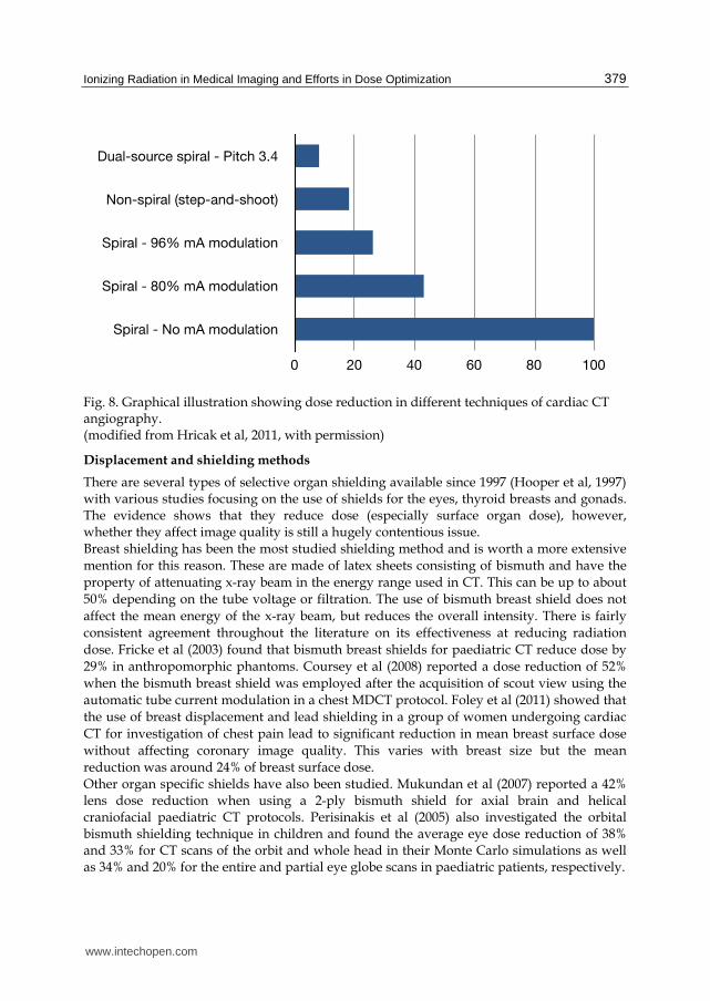
High pitch spiral acquisition
High pitch ECG-gated acquisition using dual source CT scanner is sufficiently fast enough
so that in a patient that has a slow heart rate, images of the whole heart can be acquired
within a single heart beat. In particular, cardiac imaging have shown great efforts in dose
reduction where a dose reduction of up to 90% has been achieved using novel acquisition
techniques (Flohr et al, 2009 and McCollough et al, 2009).
www.intechopen.com
Ionizing Radiation in Medical Imaging and Efforts in Dose Optimization 379
Fig. 8. Graphical illustration showing dose reduction in different techniques of cardiac CT angiography. (modified from Hricak et al, 2011, with permission)
Displacement and shielding methods
There are several types of selective organ shielding available since 1997 (Hooper et al, 1997) with various studies focusing on the use of shields for the eyes, thyroid breasts and gonads. The evidence shows that they reduce dose (especially surface organ dose), however, whether they affect image quality is still a hugely contentious issue. Breast shielding has been the most studied shielding method and is worth a more extensive
mention for this reason. These are made of latex sheets consisting of bismuth and have the
property of attenuating x-ray beam in the energy range used in CT. This can be up to about
50% depending on the tube voltage or filtration. The use of bismuth breast shield does not
affect the mean energy of the x-ray beam, but reduces the overall intensity. There is fairly
consistent agreement throughout the literature on its effectiveness at reducing radiation
dose. Fricke et al (2003) found that bismuth breast shields for paediatric CT reduce dose by
29% in anthropomorphic phantoms. Coursey et al (2008) reported a dose reduction of 52%
when the bismuth breast shield was employed after the acquisition of scout view using the
automatic tube current modulation in a chest MDCT protocol. Foley et al (2011) showed that
the use of breast displacement and lead shielding in a group of women undergoing cardiac
CT for investigation of chest pain lead to significant reduction in mean breast surface dose
without affecting coronary image quality. This varies with breast size but the mean
reduction was around 24% of breast surface dose. Other organ specific shields have also been studied. Mukundan et al (2007) reported a 42% lens dose reduction when using a 2-ply bismuth shield for axial brain and helical craniofacial paediatric CT protocols. Perisinakis et al (2005) also investigated the orbital bismuth shielding technique in children and found the average eye dose reduction of 38% and 33% for CT scans of the orbit and whole head in their Monte Carlo simulations as well as 34% and 20% for the entire and partial eye globe scans in paediatric patients, respectively.
www.intechopen.com

Current Topics in Ionizing Radiation Research 380
However, despite some evidence supporting the use of shielding, routine use of these
shields has been called into question. Geleijns et al (2006) and Vollmar et al (2008) reported
that although dose reduction can be achieved with the use of shields, their use is also
associated with significant artefacts and increase in image noise. Their findings were
supported by Kalra et al (2009) indicating that as well as increasing noise, there is
artifactually increased in attenuation values in the region immediately behind the shields.
More recently, Lee et al (2011) showed that the use of thyroid shielding with cotton wool
spacer reduces the dose to thyroid by up to 27% without affecting image noise although it
was noted that there was noticeably increase in the attenuation values of the superficial neck
structures such as the neck muscles. It was thought that this was likely related to a metal
artefact caused by bismuth implanted within the shield itself.
Some of the streak artefacts in earlier studies can partly be explained by close apposition of
the shields to the skin surface. This was the case for both Geleijns et al (2006) and Vollmar et
al (2008). When a spacer or plastic shields are used the streak artefacts are noticeably
eliminated (Hohl et al 2006, Karla et al, 2009 and Lee et al, 2011). However, the increase in
attenuation appears to be a real issue (Karla et al, 2009, Lee et al, 2011). This could have
implications when looking at coronary artery calcifications, renal cyst and adrenal mass
characterisation, for example, where increase in attenuation is used specifically to
characterise disease entities.
Another contentious issue concerns image quality. Although some of the studies have
examined the effect on image quality, the robustness of these assessments has also been
called into question. In particular, the image quality was not quantified objectively but
rather only in qualitative terms with statements such as “we did not see any differences in
quality between the shielded and unshielded lung” (Yilmaz et al, 2007). Some investigators
have argued that when noise and image quality were analysed objectively and in a robust
manner, studies appear to suggest that there is increase in image noise and deterioration of
image quality (Geleijns et al, 2006, Vollmarr et al, 2008, Karla et al, 2009). Although, it must
be noted that a more recent study by Lee et al (2011) looking at bismuth thyroid shield did
not show significant differences in mean noise values. In general, there needs to be more
evidence that robustly assess image quality prior to the routine use of shielding to be
universally accepted.
Another issue concerns the effect on image reconstruction and wasted radiation. Some
investigators argue that bismuth shielding has very different effects on patient dose for the
frontal (AP) projection compared to the dorsal (PA) projection (Geleijns et al, 2010). The
attenuation of the incoming and exiting beam ultimately has an effect of image quality and
dose efficiency. It is argued that if the x-ray tube was in the dorsal position, the exiting x-ray
beam is attenuated prior to reaching the detector and therefore this wastes unnecessary
radiation, which may have been useful for image formation process. They argue that if the
aim of bismuth shield is to reduce low energy photons that mainly deposit their energies on
the surface of the patients, then this can be similarly achieved with lowering the tube
current without the added artefacts. The use of bismuth shielding in combination with automated tube current modulation has also been investigated. This is also fraught with danger as there is the potential for either a dose increase to the patient or unequal noise levels within/between images. The reason for this is that the AEC system may attempt to increase the dose to account for the extra patient attenuation. Some of this can be eliminated by placing the shields after the scanned
www.intechopen.com

Ionizing Radiation in Medical Imaging and Efforts in Dose Optimization 381
projection radiograph (i.e. scanogram, scoutview or topogram), however, some manufacturer system performs continual monitoring of patient attenuation and adapts tube current in real time (e.g. when angular modulation is used - see earlier section). If this is the case, then the use of shielding can be detrimental and would actually increase the dose to the patient.
Fig. 9. Comparison of 120 kVp CT beam x-ray spectra with no shield, reduced mAs or 2-ply Bi shield. Note that the reduction of the low-energy photons is substantial in the 2-ply bismuth shield compared to the reduced tube current. (with permission from Kim et al, 2010)
Patient factors
Increasingly, we are moving towards tailoring our examination according to individual patient to optimise image quality and minimising dose. Previous discussion regarding tube current modulation have already shed light on the benefits of this. In general, factors such as age, chest circumference, body mass index, and specific individual factors such as presence of stents and coronary calcification may influence the type of study being performed as well as dose limitation.
Patient scan length
Patient scan length is also worthy of consideration. Larger length scans produce larger dose. In the PROTECTION I trial, the average dose for a cardiac CT angiography was 12mSv with the average scan volume of 12cm (Hausleiter et al, 2006)). For cardiac scanning (at least for retrospective scanning), an increase in 1cm results in an increase of approximately 1mSv of added radiation. The volume of acquisition must therefore be tailored to suit each individual patient. Both radiologists and radiographers will need to have active role in selecting this.
www.intechopen.com

Current Topics in Ionizing Radiation Research 382
Special patient groups
Paediatric patients
Usual dose reduction strategies such as tube current modulation, ECG-gating, etc as is used
in adults can be similarly applied to children with further dose reduction. Adjustment of kV
to 80kv is not an unreasonable approach as well as changing mAs values by employing
weight-based specific protocols (Ben Saad et al, 2009, Lee et al, 2006, Young et al 2011).
Women’s imaging and pregnancy
Efforts in dose reduction in pregnant patients owing to dose concerns to the foetus have
previously been to not perform the study unless it is absolutely paramount. If this was
deemed necessary, then various approaches have been adopted but mostly revolve around
the use of shielding (see above). Some specific adjustment of protocols have been tried for
certain clinical scenario such as for investigation of pulmonary embolism. Litmanovich et al
(2009) compared reduced-dose pulmonary CT angiography (200 mA and 100 kV) with
matched control group standard protocol (400 mA, 120 kv). The CT dose index, dose-length
product, effective dose, image quality, and signal-to-noise ratio were assessed. There was a
significant dose reduction of more than 65% using low dose protocols while maintaining
diagnostic imaging quality. Though the dose to the chest has been substantially reduced, the
dose to the foetus due to scatter radiation still poses concerns. Others have adopted a more
simple approach to tackle this. Danova et al (2010) used lead aprons as shielding to the
uterus when scanning thoracic CT achieving up to 34% reduction using the wrap around
apron to cover for scatter radiation demonstrating that protective aprons are an effective
dose reduction technique without additional costs and little effect on patient examination
time.
Iterative reconstruction
Out of all the dose reduction strategies discussed, iterative reconstruction shows the most
promise. CT workstations have used filtered back projection as the preferred method for
producing images from the raw data acquired by the receptors. Filtered Back-Projection
(FBP) algorithm has been used as the foundation of commercially available CT
reconstruction techniques since the 1970’s. Comparatively, it is robust and relatively
undemanding on computer processing. It is still widely used today and considered
acceptable for clinical diagnosis, but it does not provide optimal results for depiction of the
patient as it makes many incorrect assumptions about the data. This is apparent in the
inherent level of artefacts and noise in FBP images. To compensate for such noise, larger
patient doses are required to overcome this.
Iterative reconstruction algorithms have been put forward as promising recent advances in
CT technology but were in fact initially proposed by Shepp and Vardi back in 1982. Only
recently, this has been shown to be superior to filtered back projection algorithms for noise
reduction (Liu et al, 2007). Even though iterative reconstruction algorithms exhibit great
advantages in situations of low signal to noise ratio, their use in real-time CT was previously
limited by the time and computing power required to perform the iterations. Recently,
however, due to improving computing technology, it is now possible to utilise various
facets of iterative reconstruction to reduce the noise and thus achieving significant dose
reduction.
www.intechopen.com

Ionizing Radiation in Medical Imaging and Efforts in Dose Optimization 383
- Adaptive Statistical Iterative Reconstruction (ASIR) Adaptive statistical iterative reconstruction (ASIR) is a post-processing method marketed by
GE (General Electric Medical systems, WI, USA) where images are obtained by applying
adaptive statistical iterative reconstruction to filtered back projection images. The images are
obtained in a low dose mode and the noise in the image is then reduced by applying ASIR.
The amount of noise reduction applied can be varied from 1% to 100%. This technique
allows modest (up to 40%) dose reduction with similar recorded levels of noise in the image.
Several manufacturers have subsequently released similar technology and now all
manufacturers are offering iterative reconstruction methods and a means for improved
image quality and dose reduction.
There is increasing amount of research to suggest that utilising iterative reconstruction
improves image quality, and thereby allows for lowering of kV and mAs and thereby
reducing the dose (Prakash et al, 2010). Marin et al (2010) utilized ASIR at low kV and high
mAs setting comparing with standard of care FBP technique scanning hepatic organs and
found that there is improvement in both image quality and reduction in dose. Similarly,
Singh et al (2010) found that abdominal scanning using ASIR compared with FBP yields
significant benefit in terms of improved image quality. Due to improved contrast-to-noise
ratio, lower radiation parameters can be utilised thereby achieving significant dose
reduction. Similar results have been shown using chest CT in comparing ASIR with
standard FBP. Leipsic et al (2011) showed that compared with FBP images, ASIR images had
significantly higher subjective image quality, less image noise, and less radiation dose with
around 30% reduction on average. Studies that have high dose burden such as CT
colonography also shows much promise with the use of new ASIR technique. Flicek et al
(2010) utilised ASIR in a pilot study using 18 patients undergoing CT colonography using an
altered protocol of standard scan 50mAs (supine scan) and 25mAs (prone scan with 40%
ASIR). The results show that the radiation dose can be reduced 50% below currently
accepted low-dose techniques without significantly affecting image quality when ASIR is
used. More recent investigations appear to confirm that using ASIR yields benefits in
achieving dose reduction as well as improved image quality. Pontana et al (2011a) studied
the utility of iterative reconstruction algorithm in comparison to FBP on 80 patients and
found that iterative reconstructions provided similar image quality compared with the
conventionally used FBP reconstruction at 35% less dose and also suggested that even
higher dose reductions than 35% may be feasible by using higher levels of iterations.
Significant noise reduction can also be achieved using the same dose/raw data (Pontana et
al 2011b). More and more evidence are appearing in the literature for specific uses of ASIR
in specific clinical setting (e.g. CT enterography in Crohn’s patients - Kambadakone et al
2011; coronary CT angiography - Leipsic et al 2011) further emphasising its increasing
usefulness in quest for improved image quality and dose reduction.
- Model-Based Iterative Reconstruction (MBIR) Compared with ASIR, newer method so called ‘Model-Based Iterative Reconstruction
(MBIR)’ has now been developed and instead of relying on a single model (as is used in
ASIR), performs multiple iterations from multiple models. These models account for a
complete three-dimensional assumptions that comprises of focal spot, beam shape, voxel
size, and size of detector. In addition, MBIR also accounts for noise from photon flux as well
as system noise from the scanner itself.
www.intechopen.com

Current Topics in Ionizing Radiation Research 384
There is currently no literature research on the practical applications on MBIR as this has only recently been commercialised in December 2010. Preliminary work by the authors on phantoms (unpublished data) have revealed that MBIR shows the most reduction in terms of noise and is superior in terms of objective and subjective image quality and diagnostic confidence compared with ASIR and FBP technique. Dose can be further reduced by up to 80%. Studies are underway to see how image quality compares with traditional methods of FBP and ASIR. From the preliminary work, there is a distinct possibility of achieving body scanning at under 1mSv thus paving a way for significant dose reduction of up to 80% of current levels.
Fig. 10. Side-by-side comparison of scanned torso phantom showing different image quality between traditional filtered back projection, ASIR and MBIR. Note that despite MBIR being acquired at very low dose, this still shows remarkable superior image quality. (images are authors’ own work)
www.intechopen.com

Ionizing Radiation in Medical Imaging and Efforts in Dose Optimization 385
Garnet technology
Recently, Garnet-based detectors have been developed which are 100 times faster and have 25% less afterglow compared to the traditional GOS (Gd2O2S)-based detectors. Reduced inherent noise as well as greater contrast resolution should allow for lower voltage/current techniques even for patients with high BMI. Moreover, due to greater contrast resolution, it is hoped that this might aid more accurate detection of in-stent restenosis in cardiac CT angiography – something that is difficult to assess accurately previously due to streak artefact (Cademartiri et al, 2007; Haraldsdottir et al 2010).
320-Row detector
With new 320-row detectors now available, axial volumetric scanning of a 16cm segment range in a single 0.35s rotation with an acquisition configuration of 320x0.5mm is now possible. This has the advantage of preventing overranging, and offers high spatial and temporal resolution. This has the potential to reduce the dose considerably. It has been determined that axial volumetric scanning constitutes a dose saving of up to 55% (Kroft et al, 2010; Al-Mousily et al 2011).
5. Dose reduction in fluoroscopy
At the turn of the century, there is little awareness of the risk of associated with fluoroscopic procedures, and in particular fluoroscopically-guided intervention. This has in some parts been due to the lack of adequate measuring equipment, complex dose relationships, and false sense of perceived lack of risk. The mood has gradually shifted since 2 key publications in 2004 calling for better dose management for fluoroscopically-guided interventions (Hirshfeld Jr et al 2004; Miller et al, 2004). Since then the FDA in the US have included some of the recommended requirements into new safety-related regulations for manufacturers, and these include features such as last image hold, display of cumulative exposure time, cumulative air kerma, and real-time display of the air kerma dose rate. Dose delivery in fluoroscopic procedure is complex interactions of numerous factors. There are some excellent review articles (Hirshfeld Jr et al 2004; Steckler et al, 2009) but in-depth discussion is beyond the scope of this chapter. The issue of adequate dose monitoring and effective dose estimations have previously been called to question. The latest dose management technology, which is a feature that is made available on all angiographic equipment in the US, is ‘cumulative dose at reference point’. This actually refers to the cumulative ‘free-in-air’ air kerma and quantify total amount of radiation delivered to a specific point located a fixed distance from the x-ray source. Although, actual dose to patient is a complex, procedure-specific, and varies between operators and is dynamic process (patient’s movement during procedure will affect dose received, for example), the cumulative dose at reference point has been shown to correlate reasonably well with the absorbed dose at a specific skin site (Miller et al, 2003).
6. Dose reduction efforts in nuclear medicine
In the US, between 1972 and 2005, diagnostic nuclear medicine procedures increased 5- to 6-fold whereas the U.S. population increased by approximately 50%. Between 1996-2005, there was 5% annual growth in the number of nuclear medicine procedures while the growth of the U.S. population has been less than 1% annually. Between 1982 and 2005, the estimated
www.intechopen.com

Current Topics in Ionizing Radiation Research 386
per capita effective dose from in vivo diagnostic nuclear medicine increased by 550% and the collective effective dose increased by 720% (Mettler et al, 2008). In fact, the estimated 2005 per capita effective dose from diagnostic nuclear medicine (0.75 mSv) is greater than the total per capita dose from both diagnostic radiology and nuclear medicine examinations combined in 1982 (0.14 and 0.40 mSv, respectively). As might also be suspected, the estimated 2005 collective effective dose from diagnostic nuclear medicine (220,000 person Sv) is greater than the total per capita dose from both diagnostic radiology and nuclear medicine examinations in 1982 (32,600 and 92,000 person Sv, respectively).
Table 5. Number of Nuclear Medicine Examinations Performed in the U.S. between 1972-2005. (with permission from Mettler et al, 2008)
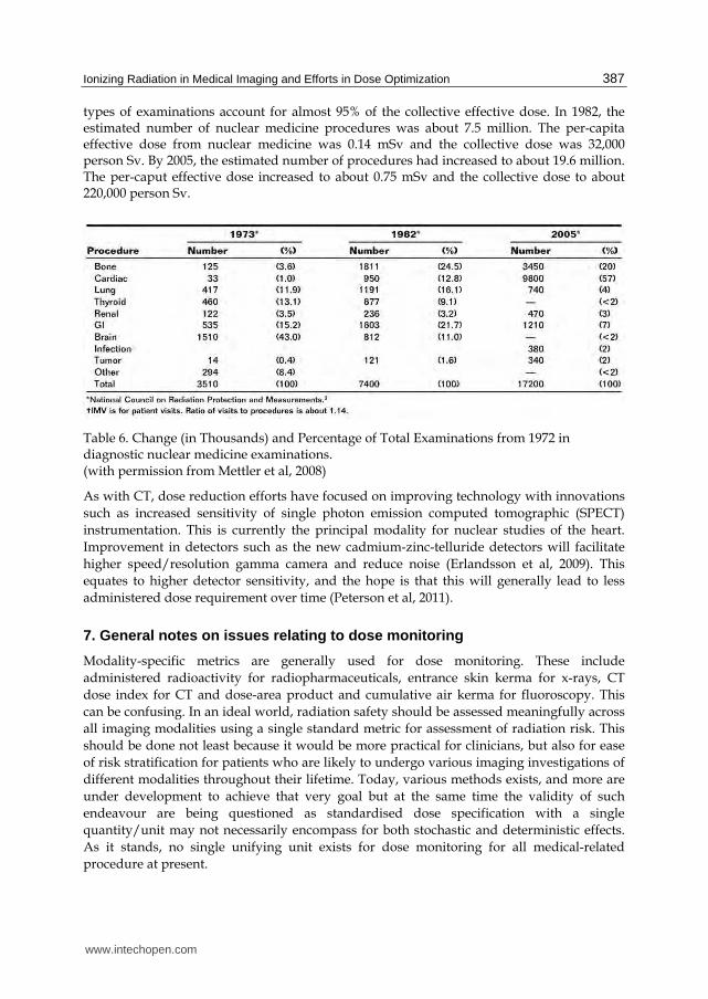
There has been a marked shift in the type of procedures with the studies of the brain and thyroid decreasing from a combined percentage of more than 56% of all procedures in 1973 to less than 4% in 2005. The most dramatic increase occurred in cardiac procedures increasing from 1% in 1973 to 57% in 2005. Cardiac studies are relatively high dose procedures and account for more than 85% of the effective dose to the patient population. Currently, more than 75% of all studies fall into 2 categories- cardiac and bone and these 2
www.intechopen.com
Ionizing Radiation in Medical Imaging and Efforts in Dose Optimization 387
types of examinations account for almost 95% of the collective effective dose. In 1982, the estimated number of nuclear medicine procedures was about 7.5 million. The per-capita effective dose from nuclear medicine was 0.14 mSv and the collective dose was 32,000 person Sv. By 2005, the estimated number of procedures had increased to about 19.6 million. The per-caput effective dose increased to about 0.75 mSv and the collective dose to about 220,000 person Sv.
Table 6. Change (in Thousands) and Percentage of Total Examinations from 1972 in diagnostic nuclear medicine examinations. (with permission from Mettler et al, 2008)
As with CT, dose reduction efforts have focused on improving technology with innovations
such as increased sensitivity of single photon emission computed tomographic (SPECT)
instrumentation. This is currently the principal modality for nuclear studies of the heart.
Improvement in detectors such as the new cadmium-zinc-telluride detectors will facilitate
higher speed/resolution gamma camera and reduce noise (Erlandsson et al, 2009). This
equates to higher detector sensitivity, and the hope is that this will generally lead to less
administered dose requirement over time (Peterson et al, 2011).
7. General notes on issues relating to dose monitoring
Modality-specific metrics are generally used for dose monitoring. These include
administered radioactivity for radiopharmaceuticals, entrance skin kerma for x-rays, CT
dose index for CT and dose-area product and cumulative air kerma for fluoroscopy. This
can be confusing. In an ideal world, radiation safety should be assessed meaningfully across
all imaging modalities using a single standard metric for assessment of radiation risk. This
should be done not least because it would be more practical for clinicians, but also for ease
of risk stratification for patients who are likely to undergo various imaging investigations of
different modalities throughout their lifetime. Today, various methods exists, and more are
under development to achieve that very goal but at the same time the validity of such
endeavour are being questioned as standardised dose specification with a single
quantity/unit may not necessarily encompass for both stochastic and deterministic effects.
As it stands, no single unifying unit exists for dose monitoring for all medical-related
procedure at present.
www.intechopen.com
Current Topics in Ionizing Radiation Research 388
8. Conclusion
In summary, recent efforts in dose reduction in the medical community has arisen due to increasing awareness of radiation-related risks. As we perform more imaging, the risk to the population becomes more concerning. Trends to this effect show that this is increasing at an almost exponential rate. Fortunately, the medical community has made great strides both in utilising new technologies but also aided by closer examination of current practices. Some of the improvements can be implemented right away without much efforts and costs but rely on adherence to already available guidelines and decision making systems. Others rely on the use of new technologies that should make it easier for clinicians to both monitor accurately the dose delivered to the patient but also aid in dose reduction/optimization. In the future, medical imaging is likely to become intertwined with individualised medicine to such an extent that each patient will receive an appropriate test, at an appropriate dose level and according to his or her specific characteristics. Tailoring an investigation to the individual patient should become the norm rather than a one-size-fits-all solution. Several advances are on the horizon and further progress in computational power, new tube design, novel detectors and advances in medical physics and engineering will likely overcome current limitations which can only be beneficial.
9. References
Al-Mousily F, Shifrin RY, Fricker FJ, et al. (2011). Use of 320-detector computed tomographic angiography for infants and young children with congenital heart disease. Pediatr Cardiol, 32:426-32.
Aroua A, Olerud HM et al (2010). Collective doses from medical exposures: an inter-comparison of the “Top 20” radiological examinations based on the EC guidelines RP 154. Proceedings of the Third European IRPA Congress, June 2010, Helsinki, Finland.
Bautista, A.B., Burgos, A., Nickel, B.J., Yoon, J.J., Tilara, A.A. & Amorosa, J.K., (2009). Do clinicians use the American College of Radiology Appropriateness criteria in the management of their patients? American Journal of Roentgenology, 192(6), p. 1581.
BEIR VII report, (2006). Health Risks from Exposure to Low Levels of Ionizing Radiation: BEIR VII Phase 2, U.S. Nuclear Regulatory Commission. Washington, DC: National Academies Press.
Ben Saad M, Rohnean A, Sigal-Cinqualbre A et al (2009) Evaluation of image quality and radiation dose of thoracic and coronary dual-source CT in 110 infants with congenital heart disease. Pediatr Radiol 39:668-676
Berrington de Gonzalez A, Mahesh M, Kim K et al. (2009). Projected Cancer Risks From Computed Tomographic Scans Performed in the United States in 2007. Arch Intern Med, 169:2071-2077.
Blachar A, Tal S, Mandel A, Novikov I, Polliack G, Sosna J, Freedman Y, Copel L, Shemer J. (2006). Preauthorization of CT and MRI examinations: assessment of a managed care preauthorization program based on the ACR Appropriateness Criteria and the Royal College of Radiology guidelines. J Am Coll Radiol. Nov;3(11):851-9.
Burk Jr, R.J. (1996). Radiation risk in perspective: position statement of the Health Physics Society, Health Physics Society Website. www. hps. org/documents/risk\_ps010-1. pdf. Published January. Updated in 2004.
www.intechopen.com

Ionizing Radiation in Medical Imaging and Efforts in Dose Optimization 389
Cardis, E., Vrijheid, M., Blettner, M., Gilbert, E., Hakama, M., Hill, C., Howe, G., Kaldor, J., Muirhead, C.R. & Schubauer-Berigan, M. (2007). The 15-Country Collaborative Study of Cancer Risk among Radiation Workers in the Nuclear Industry: estimates of radiation-related cancer risks, Radiation research, 167(4), pp. 396-416.
Cademartiri F, Schuijf JD, Pugliese , et al. (2007). Usefulness of 64-slice multislice com- puted tomography coronary angiography to assess in-stent restenosis. J Am Coll Cardiol, 49:2204–10.
Chawla SC, Federman N, Zhang D, et al. (2010). Estimated cumulative radiation dose from PET/CT in children with malignancies: a 5-year retrospective review. Pediatr Radiol. 40:681-686.
Chen J and Moir D (2010). An estimation of the annual effective dose to the Canadian population from medical CT examinations. J Radiol Prot, 30 (2), 131.
Coursey C, Frush DP, Yoshizumi T et al (2008) Pediatric chest MDCT using tube current modulation: effect on radiation dose with breast shielding. AJR 190:W54-W61
Danova D, Keil B, Kästner B, Wulff J, Fiebich M, Zink K, Klose KJ, Heverhagen JT. (2010). Reduction of uterus dose in clinical thoracic computed tomography. Rofo. Dec;182(12):1091-6.
Devine CE, Mawlawi O. (2010). Radiation safety with positron emission tomography and computed tomography. Semin Ultrasound CT MR. Feb;31(1):39-45. Review.
Einstein AJ, Henzlova MJ, Rajagoplana S. (2007). Estimating Risk of Cancer Associated With Radiation Exposure From 64-Slice Computed Tomography Coronary Angiography. JAMA. 298(3):317-323
Elliott A. (2009). Issues in medical exposures. J Radiol Prot. Jun;29(2A):A107-21. Erlandsson, K., Kacperski, K., van Gramberg, D. & Hutton, B.F. (2009). Performance
evaluation of D-SPECT: a novel SPECT system for nuclear cardiology, Physics in medicine and biology, 54, p. 2635.
Flicek, K.T., Hara, A.K., Silva, A.C., Wu, Q., Peter, M.B. & Johnson, C.D. (2010). Reducing the radiation dose for CT colonography using adaptive statistical iterative reconstruction: A pilot study, AJR. 195(1), pp. 126-31.
Flint-Richter, P. & Sadetzki, S. (2007). Genetic predisposition for the development of radiation-associated meningioma: an epidemiological study, The Lancet Oncology, 8(5), pp. 403-10.
Flohr TG , L eng S, Yu L, et al. (2009). Dual-source spiral CT with pitch up to 3.2 and 75 ms temporal resolution: image reconstruction and assessment of image quality. Med Phys, 3 6 ( 12 ): 5641 -5 653 .
Foley, S.J., McEntee, M.F., Achenbach, S., Brennan, P.C., Rainford, L.S. & Dodd, J.D. (2011). Breast Surface Radiation Dose During Coronary CT Angiography: Reduction by Breast Displacement and Lead Shielding, American Journal of Roentgenology, 197(2), pp. 367-73.
Fricke BL, Donnelly LF, Frush DP et al. (2003). In-plane bismuth breast shields for pediatric CT: effects on radiation dose and image quality using experimental and clinical data. AJR 180:407-411
Geleijns, J., Wang, J. & McCollough, C. (2010). The use of breast shielding for dose reduction in pediatric CT: arguments against the proposition, Pediatric radiology, pp. 1-4.
www.intechopen.com

Current Topics in Ionizing Radiation Research 390
Geleijns J, Salvado AM, Veldkamp WJ et al. (2006). Quantitative assessment of selective in-plane shielding of tissues in computed tomography through evaluation of absorbed dose and image quality. Eur Radiol 16:2334-2340
Hadley, J.L., Agola, J. & Wong, P. (2006). Potential impact of the American College of Radiology appropriateness criteria on CT for trauma, American Journal of Roentgenology, 186(4), p. 937.
Hart, Wall, Hillier, Shrimpton. (2010). HPA-CRCE-012 - frequency and collective dose for medical and dental x-ray examinations in the UK, 2008. Health protection agency December 2010: Available from:
http://www.hpa.org.uk/Publications/Radiation/CRCEScientificAndTechnicalReportSeries/HPACRCE012/. Accessed 5 May 2011.
Hausleiter et al. (2010) Image Quality and Radiation Exposure With a Low Tube Voltage Protocol for Coronary CT Angiography Results of the PROTECTION II Trial. JACC Cardiovasc Imagingvol. 3 (11) pp. 1113-23
Haraldsdottir et al. (2010) Diagnostic accuracy of 64-slice multidetector CT for detection of in-stent restenosis in an unselected, consecutive patient population. European Journal of Radiology vol. 76 (2) pp. 188-94
Hirshfeld JW Jr, Balter S, Brinker JA, Kern MJ, Klein LW, Lindsay BD, Tommaso CL, Tracy CM, Wagner LK, Creager MA, Elnicki M, Hirshfeld JW Jr, Lorell BH, Rodgers GP, Tracy CM, Weitz HH. (2004). American College of Cardiology Foundation; American Heart Association; American College of Physicians. ACCF/AHA/HRS/SCAI clinical competence statement on physician knowledge to optimize patient safety and image quality in fluoroscopically guided invasive cardiovascular procedures. A report of the American College of Cardiology Foundation/American Heart Association/American College of Physicians Task Force on Clinical Competence and Training. J Am Coll Cardiol. Dec 7;44(11):2259-82.
Hopper KD, King SH, Lobell ME et al. (1997). The breast: in-plane x-ray protection during diagnostic thoracic CT - shielding with bismuth radioprotective garments. Radiology 205:853-858
Hohl C, Wildberger JE, Suss C, et al. (2006). Radiation dose reduction to breast and thyroid during MDCT: effectiveness of an in-plane bismuth shield. Acta Radiol; 47:562-567
Hausleiter J, Meyer T, Hadamitzdy et al. (2009). Estimated radiation dose associated with cardiac CT angiography. JAMA ;301:500-7.
International Agency for Research on Cancer (IARC) Web site: www.iarc.fr IARC Carcinogen Monographs: http://monographs.iarc.fr. Accessed 6 Aug 2011.
Israel, G.M., Cicchiello, L., Brink, J. & Huda, W. (2010). Patient size and radiation exposure in thoracic, pelvic, and abdominal CT examinations performed with automatic exposure control, American Journal of Roentgenology, 195(6), p. 1342.
Jansen-van der Weide, M.C., Greuter, M.J.W., Jansen, L., Oosterwijk, J.C., Pijnappel, R.M. & de Bock, G.H. (2010). Exposure to low-dose radiation and the risk of breast cancer among women with a familial or genetic predisposition: a meta-analysis, European radiology, pp. 1-10.
Kambadakone, A.R., Chaudhary, N.A., Desai, G.S., Nguyen, D.D., Kulkarni, N.M. & Sahani, D.V. (2011). Low-Dose MDCT and CT Enterography of Patients With Crohn
www.intechopen.com

Ionizing Radiation in Medical Imaging and Efforts in Dose Optimization 391
Disease: Feasibility of Adaptive Statistical Iterative Reconstruction, American Journal of Roentgenology, 196(6), p. W743.
Kalra, M.K., Maher, M.M., Toth, T.L., Hamberg, L.M., Blake, M.A., Shepard, J.A. & Saini, S. (2004). Strategies for CT Radiation Dose Optimization1, Radiology, 230(3), p. 619. – 2004a
Kalra MK, Maher MM, Kamath RS, et al. (2004). Sixteen-detector row CT of abdomen and pelvis: study for optimization of Z-axis modulation tech-nique performed in 153 patients. Radiology; 233:241-249. – 2004b
Kalra MK, Maher MM, Prasad SR, et al. (2003). Correlation of patient weight and cross-sectional dimensions with subjective image quality at standard dose abdominal CT. Korean J Radiol; 4:234-238.
Kalra MK, Dang P, Singh S et al. (2009). In-plane shielding for CT: effect of off-centering, automatic exposure control and shield-to-surface distance. Korean J Radiol 10: 156-163
Kim, S., Frush, D.P. & Yoshizumi, T.T. (2010). Bismuth shielding in CT: support for use in children, Pediatric radiology, pp. 1-5.
Kroft LJ, Roelofs JJ, Geleijins J. (2010). Scan time and patient dose for thoracic imaging in neonates and small children using axial volumetric 320-detector row CT compared to helical 64-, 32-, and 16- detector row CT acquisitions. Pediatr Radiol ;40:294-300.
Lee, Y.H., Park, E., Cho, P.K., Seo, H.S., Je, B.K., Suh, S. & Yang, K.S. (2011). Comparative Analysis of Radiation Dose and Image Quality Between Thyroid Shielding and Unshielding During CT Examination of the Neck, American Journal of Roentgenology, 196(3), p. 611.
Lee T, Tsai IC, Fu YC. (2006). Using multi-detector row CT in neonates with complex congenital heart disease to replace diagnostic cardiac catheterization for anatomical investigation—initial experiences in technical and clinical feasibility. Pediatr Radiol 36:1273-1282
Leipsic, J., Nguyen, G., Brown, J., Sin, D. & Mayo, J.R. (2010). A prospective evaluation of dose reduction and image quality in chest CT using adaptive statistical iterative reconstruction, AJR. American journal of roentgenology, 195(5), pp. 1095-9.
Leipsic, J., Heilbron, B.G., and Hague, C. (2011). Iterative reconstruction for coronary CT angiography: finding its way. Int J Cardiovasc Imaging, Feb 27. (Epub ahead of print].
Leschka et al. (2008). Low kilovoltage cardiac dual-source CT: attenuation, noise and radiation dose. Eur Radiol ;18(9):1809-1817.
Li, J., Udayasankar, U.K., Tang, X., Toth, T.L., Small, W.C. & Kalra, M.K. (2011). Patient Size Compensated Automatic Tube Current Modulation in Multi-detector Row CT of the Abdomen and Pelvis, Academic radiology, 18(2), pp. 205-11.
Litmanovich D, Boiselle PM, Bankier AA, Kataoka ML, Pianykh O, Raptopoulos V. (2009). Dose reduction in computed tomographic angiography of pregnant patients with suspected acute pulmonary embolism. J Comput Assist Tomogr. Nov-Dec;33(6): 961-6.
Liu YJ, Zhu PP, Chen B, et al. (2007). A new iterative algorithm to reconstruct the refractive index. Phys Med Biol, 52:L5 – L13
Marin, D., Nelson, R.C., Schindera, S.T., Richard, S., Youngblood, R.S., Yoshizumi, T.T. & Samei, E. (2010). Low-tube-voltage, high-tube-current multidetector abdominal CT:
www.intechopen.com
Current Topics in Ionizing Radiation Research 392
improved image quality and decreased radiation dose with adaptive statistical iterative reconstruction algorithm--initial clinical experience, Radiology, 254(1), pp. 145-53.
McCollough CH, Bruesewitz MR, Kofler JM. (2006). CT Dose Reduction and Dose Management Tools: Overview of Available Options; Radiographics March – April, 26:2: 503 – 512.
McCollough CH. (2005). Automatic exposure control in CT: are we done yet? Radiology Dec; 237(3): 755-756.
McCollough C H, Leng S, Schmidt B, Allmendinger T, Eusemann C, Flohr TG. (2009). Use of a pitch value of 3.2 in dual-source cardiac CT angiography: dose performance relative to existing scan modes [abstr]. In: Radiological Society of North America scientific assembly and annual meeting program. Oak Brook, Ill: Radiological Society of North America,; 485-486.
Mettler Jr, F.A., Bhargavan, M., Thomadsen, B.R., Gilley, D.B., Lipoti, J.A., Mahesh, M., McCrohan, J. & Yoshizumi, T.T. (2008). Nuclear medicine exposure in the United States, 2005-2007: preliminary results, Seminars in nuclear medicine, 38(5), pp. 384-91.
Mettler FA Jr, Bhargavan M, Faulkner K, et al. (2009). Radiologic and nuclear medicine studies in the United States and worldwide: frequency, radiation dose, and comparison with other radiation sources—1950-2007. Radiology 2009 ;2 53 (2): 520- 5 31
Miller et al. (2004). Quality improvement guidelines for recording patient radiation dose in the medical record. J Vasc Interv Radiol, 15(5):423-429.
Miller DL, Balter S, Cole PE et al. (2003). Radiation doses in interventional radiology procedures: the RAD-IR study. II. Skin dose. J Vasc Interv Radiol, 14(8): 977-990.
Monson, R., Cleaver, J., Abrams, H.L., Bingham, E. & Buffler, P.A. (2005). BEIR VII: health risks from exposure to low levels of ionizing radiation, Washington DC: National Academies Press.
Muirhead, C.R., O'Hagan, J.A., Haylock, R.G.E., Phillipson, M.A., Willcock, T., Berridge, G.L.C. & Zhang, W. (2009). Mortality and cancer incidence following occupational radiation exposure: third analysis of the National Registry for Radiation Workers, British journal of cancer, 100(1), pp. 206-12.
Mukundan S, Wang PI, Frush DP et al. (2007). MOSFET dosimetry for radiation dose assessment of bismuth shielding of the eye in children. AJR 188:1648-1650
NCRP (2009). Ionizing radiation exposure of the population of the United States. NCRP Report 160. National Council on Radiation Protection and Measurements, Bethesda MD.
Perisinakis K, Raissaki M, Tzedakis A et al. (2005). Reduction of eye lens radiation dose by orbital bismuth shielding in pediatric patients undergoing CT of the head: a Monte Carlo study. Med Phys 32:1024-1030
Peterson, T.E. & Furenlid, L.R. (2011). SPECT detectors: the Anger Camera and beyond, Physics in medicine and biology, 56, p. R145.
Preston DL, Ron E, Tokuoka S, et al. (2007). Solid cancer incidence in atomic bomb survivors: 1958-1998. Radiation research, 168:1-64.
Pontana, F., Duhamel, A., Pagniez, J., Flohr, T., Faivre, J.B., Hachulla, A.L., Remy, J. & Remy-Jardin, M. (2011). Chest computed tomography using iterative reconstruction vs
www.intechopen.com

Ionizing Radiation in Medical Imaging and Efforts in Dose Optimization 393
filtered back projection (Part 2): image quality of low-dose CT examinations in 80 patients, European radiology, 21(3), pp. 636-43. A
Pontana, F., Pagniez, J., Flohr, T., Faivre, J.B., Duhamel, A., Remy, J. & Remy-Jardin, M. (2011). Chest computed tomography using iterative reconstruction vs filtered back projection (Part 1): evaluation of image noise reduction in 32 patients, European radiology, 21(3), pp. 627-35. B
Prakash, P., Kalra, M.K., Ackman, J.B., Digumarthy, S.R., Hsieh, J., Do, S., Shepard, J.A. & Gilman, M.D. (2010). Diffuse lung disease: CT of the chest with adaptive statistical iterative reconstruction technique, Radiology, 256(1), pp. 261-9.
Pflederer T, Rudofsky L, Ropers D et al. (2009). Image quality in a low radiation exposure protocol for retrospectively ECG-gated coronary CT angiography. AJR AM J Roentgenol, 192:1045-50.
Preston DL, Ron E ,Tokuoka S, e t al. (2007). Solid cancer incidence in atomic bomb survivors: 1958-1998. Radiat Res, 168(1): 1-64 .
Ronckers, C.M., Doody, M.M., Lonstein, J.E., Stovall, M. & Land, C.E. (2008). Multiple diagnostic X-rays for spine deformities and risk of breast cancer, Cancer Epidemiology Biomarkers & Prevention, 17(3), p. 605.
Roobottom CA, Mitchell G and Morgan-Hughes G. (2010). Radiation-reduction strategies in cardiac computed tomographic angiography. Clin Radiol, vol. 65 (11) pp. 859-67
Schindera ST, Nelson RC, Toth TL, et al. (2008). Effect of patient size on radiation dose for abdominal MDCT with automatic tube current modulation: phantom study. AJR Am J Roentgenol, 190:W100-W105.
Schoenhagen P. (2008). Back to the future: coronary CT angiography using prospective ECG triggering. Eur Heart J ;29(2):153–4.
Singh, S., Kalra, M.K., Hsieh, J., Licato, P.E., Do, S., Pien, H.H. & Blake, M.A. (2010). Abdominal CT: Comparison of Adaptive Statistical Iterative and Filtered Back Projection Reconstruction Techniques, Radiology. Nov;257(2):373-83
Stecker MS, Balter S, Towbin RB, et al. (2009). Guidelines for patient radiation dose management. J Vasc Interv Radiol, 20(7 suppl): S263-S273.
Stolzmann P, Leschka S, Scheffel H, et al. (2008). Dual-source CT in step-and-shoot mode: noninvasive coronary angiography with low radiation dose. Radiology, 249(1): 71–80.
Savandi, A.S., Demarco, J.J., Cagnon, C.H., Angel, E., Turner, A.C., Cody, D.D., Stevens, D.M., Primak, A.N., McCollough, C.H. & McNitt-Gray, M.F. (2009). Variability of surface and center position radiation dose in MDCT: Monte Carlo simulations using CTDI and anthropomorphic phantoms, Medical physics, 36, p. 1025.
Street M, Brady Z, Van Every B, Thomson KR. (2009). Radiation exposure and the justification of computed tomography scanning in an Australian hospital emergency department. Intern Med J. Nov;39(11):713-9.
United Nations Scientific Committee on the Effects of Atomic Radiation. (2010). Sources and effects of ionizing radiation. Medical radiation exposures, annex A. 2008 Report to the General Assembly with Annexes. New York, NY: United Nations, 2010.
Vollmar SV, Kalender WA. (2008). Reduction of dose to the female breast in thoracic CT: a comparison of standard-protocol, bismuth-shielded, partial and tube-current-modulated CT examinations. Eur Radiol 18:1674-1682.
www.intechopen.com

Current Topics in Ionizing Radiation Research 394
White CS, Kuo D. (2007). Chest pain in the emergency department: role of multidetector CT. Radiology. Dec;245(3):672-81. Review.
Yilmaz MH, Yasar D, Albayram S et al. (2007). Coronary calcium scoring with MDCT: the radiation dose to the breast and the effectiveness of bismuth breast shield. Eur J Radiol 61:139- 143.
Young, C., Taylor, A.M. & Owens, C.M. (2011). Paediatric cardiac computed tomography: a review of imaging techniques and radiation dose consideration, European radiology, 21(3), pp. 518-29.
Yu L,Li H, Fletcher JG, McCollough CH. (2010). Automatic selection of tube potential for radiation dose reduction in CT: a general strategy. Med Phys, 37( 1): 234 -2 43.
Zhang, D., Zankl, M., DeMarco, J.J., Cagnon, C.H., Angel, E., Turner, A.C. & McNitt-Gray, M.F. (2009). Reducing radiation dose to selected organs by selecting the tube start angle in MDCT helical scans: a Monte Carlo based study, Medical physics, 36(12), pp. 5654-64.
www.intechopen.com

Current Topics in Ionizing Radiation ResearchEdited by Dr. Mitsuru Nenoi
ISBN 978-953-51-0196-3Hard cover, 840 pagesPublisher InTechPublished online 12, February, 2012Published in print edition February, 2012
InTech EuropeUniversity Campus STeP Ri Slavka Krautzeka 83/A 51000 Rijeka, Croatia Phone: +385 (51) 770 447 Fax: +385 (51) 686 166www.intechopen.com
InTech ChinaUnit 405, Office Block, Hotel Equatorial Shanghai No.65, Yan An Road (West), Shanghai, 200040, China
Phone: +86-21-62489820 Fax: +86-21-62489821
Since the discovery of X rays by Roentgen in 1895, the ionizing radiation has been extensively utilized in avariety of medical and industrial applications. However people have shortly recognized its harmful aspectsthrough inadvertent uses. Subsequently people experienced nuclear power plant accidents in Chernobyl andFukushima, which taught us that the risk of ionizing radiation is closely and seriously involved in the modernsociety. In this circumstance, it becomes increasingly important that more scientists, engineers and studentsget familiar with ionizing radiation research regardless of the research field they are working. Based on thisidea, the book "Current Topics in Ionizing Radiation Research" was designed to overview the recentachievements in ionizing radiation research including biological effects, medical uses and principles ofradiation measurement.
How to referenceIn order to correctly reference this scholarly work, feel free to copy and paste the following:
Varut Vardhanabhuti and Carl A. Roobottom (2012). Ionizing Radiation in Medical Imaging and Efforts in DoseOptimization, Current Topics in Ionizing Radiation Research, Dr. Mitsuru Nenoi (Ed.), ISBN: 978-953-51-0196-3, InTech, Available from: http://www.intechopen.com/books/current-topics-in-ionizing-radiation-research/ionizing-radiation-in-medical-imaging-and-efforts-in-dose-optimization

© 2012 The Author(s). Licensee IntechOpen. This is an open access articledistributed under the terms of the Creative Commons Attribution 3.0License, which permits unrestricted use, distribution, and reproduction inany medium, provided the original work is properly cited.