involvement of matrin 3 and sfpq/nono in the dna …yossih/publications/salton_et_al_2010.pdf ·...
TRANSCRIPT

Cell Cycle 9:8, 1568-1576; April 15, 2010; © 2010 Landes Bioscience
RepoRt
1568 Cell Cycle Volume 9 Issue 8
*Correspondence to: Yosef Shiloh; Email: [email protected]: 10/18/09; Revised: 01/24/10; Accepted: 01/25/10Previously published online: www.landesbioscience.com/journals/cc/article/11298
Introduction
Maintenance of genomic stability and integrity is essential for maintaining cellular homeostasis and as a barrier to neoplasia.1-3 The cellular defense system against this threat is the DNA dam-age response (DDR)—an elaborate signaling network activated by DNA damage that swiftly modulates many physiological pro-cesses.4,5 One of its hallmarks is the activation of cell cycle check-points that temporarily halt the cell cycle while damage is assessed and repaired.6 One of the most powerful activators of the DDR is the double strand break (DSB) in the DNA.7 This extremely cytotoxic lesion is induced by ionizing radiations, radiomimetic chemicals and reactive oxygen species that accompany normal metabolism; it is also the byproduct of genomic transactions such as meiotic and V(D)J recombination.7,8 Eukaryotic cells repair DSBs by nonhomologous end-joining (NHEJ), an error-prone ligation that acts throughout the cell cycle, or by homologous recombination (HR) between sister chromatids, which functions in the late S and G
2 phases.9,10
The overall cellular response to DSBs goes, however, far beyond repair. It modulates numerous physiological processes, and alters gene expression profiles and protein synthesis, degrada-tion and trafficking. It is a multi-tiered process that begins with the recruitment of sensor proteins to the damaged sites where they create expanding nuclear foci.11-13 These proteins are involved in
Involvement of Matrin 3 and SFPQ/NONO in the DNA damage response
Maayan Salton,1 Yaniv Lerenthal,1 Shih-Ya Wang,2 David J. Chen2 and Yosef Shiloh1,*
1the David and Inez Myers Laboratory for Cancer Research; Department of Human Molecular Genetics and Biochemistry; Sackler School of Medicine; tel Aviv University; tel Aviv, Israel; 2Division of Molecular Radiation Biology; Department of Radiation oncology; University of texas Southwestern Medical Center at Dallas; Dallas, tX USA
Key words: Matrin 3, SFPQ/PSF, NONO/p54, DNA damage response, ATM, cell cycle checkpoint
the initial processing of the damage and in activation of the trans-ducers of the DNA damage alarm. The primary transducer of the DSB alarm is the nuclear kinase ATM.14-17 In response to DSB induction, ATM is rapidly activated and subsequently phospho-rylates a plethora of effectors, which are key players in a variety of damage response pathways.14,17-21 Loss of ATM leads to the severe genomic instability syndrome ataxia-telangiectasia (A-T), char-acterized by neuronal degeneration, immunodeficiency, radiation sensitivity and extreme cancer predisposition.22,23 ATM belongs to a conserved family of “PI3K-like protein kinases” (PIKKs)24 that includes, among others, two major DDR transducers: the DNA-dependent protein kinase (DNA-PK),25 and ataxia-telang-iectasia and Rad3-related (ATR).26 These three protein kinases phosphorylate serine or threonine residues with preference for those followed by glutamine (SQ or TQ motifs), functionally interact with each other and transduce the damage signal in a partially redundant manner.27-29
Matrin 3 (MATR3) is a highly conserved, inner nuclear matrix protein of 125 kDa.30 Nuclear matrix proteins bound to the inner nucleus membrane form a skeletal nuclear framework, and are involved in chromatin organization, DNA replication, transcrip-tion, repair, RNA processing and transport.31 MATR3 also exhib-its diffuse nuclear distribution.32 It contains a bipartite nuclear localization signal (NLS),33 two zinc finger domains predicted to bind DNA, and two RNA recognition motifs (RRM). Its cellular
the DNA damage response (DDR) is a complex signaling network that is induced by DNA lesions and vigorously activated by double strand breaks (DSBs). the DSB response is mobilized by the nuclear protein kinase AtM, which phosphorylates key players in its various branches. SFpQ (pSF) and NoNo (p54) are nuclear proteins that interact with each other and have diverse roles in nucleic acids metabolism. the SFpQ/NoNo heterodimer was previously found to enhance DNA strand break rejoining in vitro. our attention was drawn to these two proteins as they interact with the nuclear matrix protein Matrin 3 (MAtR3), which we found to be a novel AtM target. We asked whether SFpQ and NoNo too are involved in the DSB response. proteins that function at the early phase of this response are often recruited to the damaged sites. We observed rapid recruitment of SFpQ/NoNo to sites of DNA damage induced by laser microbeam. In MAtR3 knockdown cells SFpQ/NoNo retention at DNA damage sites was prolonged. SFpQ and MAtR3 depletion led to abnormal accumulation of cells at the S-phase of the cell cycle following treatment with the radiomimetic chemical neocarzinostatin. Notably, proteins involved in DSB repair via nonhomologous end-joining co-immunoprecipitated with NoNo; SFpQ depletion delayed DSB repair. Collectively the data suggest that SFpQ, NoNo and MAtR3 are involved in the early stage of the DSB response, setting the scene for DSB repair.

www.landesbioscience.com Cell Cycle 1569
RepoRt RepoRt
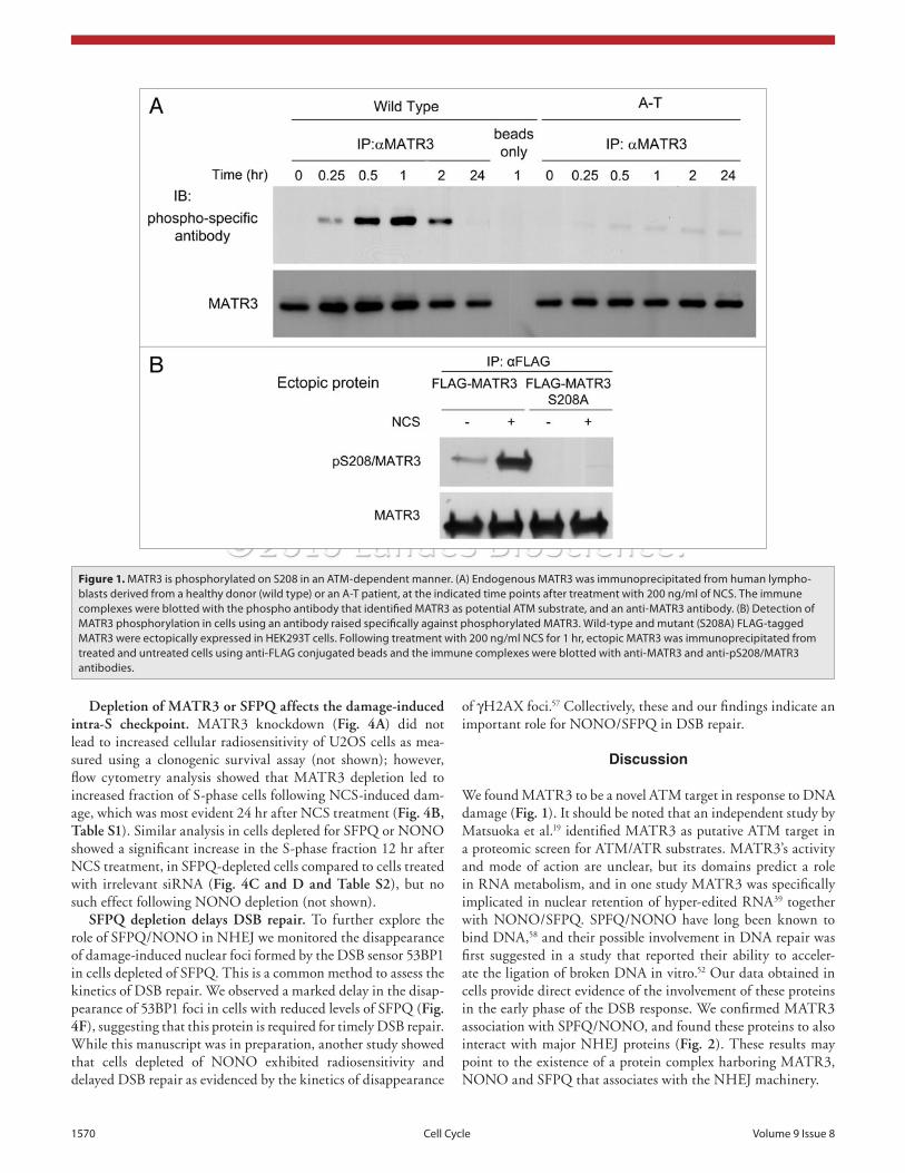
against this site was raised and used for further analysis. A vector expressing N-terminal FLAG-tagged MATR3 was constructed by subcloning full-length human MATR3 into a pCMV:FLAG2B plasmid. An S208A substitution was subsequently induced in it in order to produce a protein that cannot be phosphorylated at this site. The wild type and mutant constructs were expressed in HEK293T cells, and following treatment with 200 ng/ml of NCS, ectopic MATR3 was immunoprecipitated using an anti-FLAG antibody. Western blotting analysis of the immunopre-cipitates using the anti-pS208 antibody indicated that the ectopic MATR3 was indeed phosphorylated on S208 following DNA damage as the S208A substitution abolished this phosphorylation (Fig. 1B). The nature of MATR3’s involvement in the DDR was further explored concomitantly with that of SPFQ and NONO (see below).
Joint involvement of MATR3, SFPQ and NONO in the DDR. In view of the known interaction between MATR3, NONO and SFPQ in other contexts39 and the possible func-tional link of NONO and SFPQ to the DDR,52 we further explored the involvement of all three proteins in the cellular DSB response. We examined the interaction between these proteins in damaged and undamaged cells using co-immunoprecipitation analysis. The results (Fig. 2A) indicated that MATR3 pulled down NONO and SFPQ from cellular extracts as was previ-ously shown in vitro.39 Notably, the KU70/KU80 subunits of the DNA-PK holoenzyme were also pulled down with MATR3. When NONO was immunoprecipitated from untreated or NCS-treated cells it pulled down MATR3, SFPQ, KU70/KU80 and LIG4—a major player in the NHEJ pathway of DSB repair (Fig. 2B). These associations suggest a role for MATR3, SFPQ and NONO in DSB repair.
SFPQ and NONO are recruited to DNA damage sites and their retention is MATR3-dependent. RNAi-mediated suppres-sion of MATR3 levels in U2OS cells (Fig. 3A) was combined with ectopic expression of EGFP-tagged NONO and RFP-tagged SFPQ, and the spatial dynamics of the two proteins was monitored following the induction of localized DNA damage by laser microirradiation.12 Irradiation intensity (see Materials and Methods) was carefully calibrated to be sufficient for recruit-ment of two established DSB sensors, the MRN complex12,13,55 and 53BP1,12,13,56 while leaving the cells alive to allow subsequent cellular division. GFP-tagged constructs of Nbs1 (a member of the MRN complex) and 53BP1 were used in the calibration experiments. In control cells (siGFP) relocalization of SFPQ and NONO at damage sites was noticed as early as 2 sec following damage induction, with subsequent disappearance about 10 min later (Fig. 3B). MATR3 knockdown did not affect the recruit-ment kinetics of SFPQ and NONO but their retention at the damage sites was extended by at least 30 min (Fig. 3B). This observation placed SFPQ and NONO within a growing, hetero-geneous group of proteins that coalesce at the damage sites in the early phase of the DSB response. Surprisingly, repeated experi-ments failed to observe recruitment to damage sites of MATR3 itself (data not shown). However, collectively, the data tie SFPQ, NONO and MATR3 to the early stage of the DNA damage response.
amount decreases following activation of the NMDA receptors,34 after treatment with soy extract of homocysteine-stressed endothe-lial cells,35 in fetal Down syndrome brain,36 and its expression is downregulated by the microRNA miR-200b.37 A missense muta-tion leading to a single amino acid substitution in MATR3 was recently found to cause in humans autosomal-dominant vocal cord and pharyngeal weakness with distal myopathy (VCPDM).38
MATR3 together with the proteins SFPQ and NONO were implicated in nuclear retention of hyper-edited RNA, which prevents the translation of such RNA.39 This process occurs in nuclear bodies called paraspeckles that contain SFPQ and NONO proteins.40-42 SFPQ and NONO form a heterodimer and are involved in various aspects of RNA and DNA metabolism,43 such as transcription,44-46 pre-mRNA 3' processing,47 transcrip-tion termination48 and mRNA splicing.49,50 SFPQ was initially characterized as PSF—polypyrimidine tract-binding protein-associated splicing factor,51 but in fact it is associated mostly with the nuclear matrix. SFPQ and NONO show 71% identity, observed mainly in a common region containing two tandem RNA recognition motifs (RRM).
Both SFPQ and NONO were identified in a biochemical screen for DNA end-rejoining proteins.52 Furthermore, they were shown to facilitate in vitro the DNA binding capability of the KU70/KU80 heterodimer—a component of DNA-PK holoenzyme;52 and SFPQ was shown to bind RAD51—a central player in the HR pathway of DSB repair, and to modulate RAD51-mediated homologous pairing and strand exchange.53 Here, we provide fur-ther evidence that SFPQ/NONO heterodimer is involved in the early stage of the DSB response, and we add MATR3 as a novel player in the DDR.
Results
Identification of MATR3 as an ATM target. We carried out identification en mass of putative ATM substrates using immu-noprecipitation with anti-phospho antibodies generated against specific ATM targets (Galanty Y, Lerenthal Y, et al. unpub-lished). Despite being raised against specific phosphorylation sites, most of these antibodies cross-react with several ATM/ATR substrates due to the similarity of their core sequences, SQ or TQ.19,24,54 MATR3 was identified as a putative ATM/ATR tar-get among proteins immunoprecipitated by an antibody raised against a peptide containing the sequence SGpSQE from cells treated with 200 ng/ml of the radiomimetic drug neocarzinos-tatin (NCS). Initial validation of this finding was obtained by immunoprecipitating MATR3 from wild type and A-T lympho-blasts at different time points after treatment with 200 ng/ml NCS and blotting the immunoprecipitates with the antibody that had identified MATR3 in the screen. The antibody detected an ATM-dependent signal corresponding in size to MATR3, which peaked about 1 hr after damage induction and declined within a few hours thereafter (Fig. 1A). This time course is typical for the phosphorylation of many ATM substrates, and suggested that MATR3 could indeed be an ATM target.
There is one potential target of this antibody in MATR3 sequence, S208QE. A polyclonal antibody genuinely directed
1570 Cell Cycle Volume 9 Issue 8
of γH2AX foci.57 Collectively, these and our findings indicate an important role for NONO/SFPQ in DSB repair.
Discussion
We found MATR3 to be a novel ATM target in response to DNA damage (Fig. 1). It should be noted that an independent study by Matsuoka et al.19 identified MATR3 as putative ATM target in a proteomic screen for ATM/ATR substrates. MATR3’s activity and mode of action are unclear, but its domains predict a role in RNA metabolism, and in one study MATR3 was specifically implicated in nuclear retention of hyper-edited RNA39 together with NONO/SFPQ. SPFQ/NONO have long been known to bind DNA,58 and their possible involvement in DNA repair was first suggested in a study that reported their ability to acceler-ate the ligation of broken DNA in vitro.52 Our data obtained in cells provide direct evidence of the involvement of these proteins in the early phase of the DSB response. We confirmed MATR3 association with SPFQ/NONO, and found these proteins to also interact with major NHEJ proteins (Fig. 2). These results may point to the existence of a protein complex harboring MATR3, NONO and SFPQ that associates with the NHEJ machinery.
Depletion of MATR3 or SFPQ affects the damage-induced intra-S checkpoint. MATR3 knockdown (Fig. 4A) did not lead to increased cellular radiosensitivity of U2OS cells as mea-sured using a clonogenic survival assay (not shown); however, flow cytometry analysis showed that MATR3 depletion led to increased fraction of S-phase cells following NCS-induced dam-age, which was most evident 24 hr after NCS treatment (Fig. 4B, Table S1). Similar analysis in cells depleted for SFPQ or NONO showed a significant increase in the S-phase fraction 12 hr after NCS treatment, in SFPQ-depleted cells compared to cells treated with irrelevant siRNA (Fig. 4C and D and Table S2), but no such effect following NONO depletion (not shown).
SFPQ depletion delays DSB repair. To further explore the role of SFPQ/NONO in NHEJ we monitored the disappearance of damage-induced nuclear foci formed by the DSB sensor 53BP1 in cells depleted of SFPQ. This is a common method to assess the kinetics of DSB repair. We observed a marked delay in the disap-pearance of 53BP1 foci in cells with reduced levels of SFPQ (Fig. 4F), suggesting that this protein is required for timely DSB repair. While this manuscript was in preparation, another study showed that cells depleted of NONO exhibited radiosensitivity and delayed DSB repair as evidenced by the kinetics of disappearance
Figure 1. MAtR3 is phosphorylated on S208 in an AtM-dependent manner. (A) endogenous MAtR3 was immunoprecipitated from human lympho-blasts derived from a healthy donor (wild type) or an A-t patient, at the indicated time points after treatment with 200 ng/ml of NCS. the immune complexes were blotted with the phospho antibody that identified MAtR3 as potential AtM substrate, and an anti-MAtR3 antibody. (B) Detection of MAtR3 phosphorylation in cells using an antibody raised specifically against phosphorylated MAtR3. Wild-type and mutant (S208A) FLAG-tagged MAtR3 were ectopically expressed in HeK293t cells. Following treatment with 200 ng/ml NCS for 1 hr, ectopic MAtR3 was immunoprecipitated from treated and untreated cells using anti-FLAG conjugated beads and the immune complexes were blotted with anti-MAtR3 and anti-pS208/MAtR3 antibodies.

www.landesbioscience.com Cell Cycle 1571
phosphorylation in it are obvious future research directions. Notably, SFPQ and NONO were previ-ously found to bind poly(ADP-ribose) and physi-cally interact with poly(ADP-ribose) polymerases.64 In view of the documented role of poly(ADP)-ribo-sylation in DNA repair,65 that observation provided another indication for the emerging involvement of the two protein in the DDR.
We found an abnormally high accumulation of S-phase cells following DNA damage upon MATR3 and SFPQ depletion (Fig. 4B and D). The mechanistic aspects of the intra-S checkpoint are still largely unknown, although it is clear that, like with the other checkpoints, several pathways act in concert to regulate this checkpoint.66 Usually defects in the intra-S checkpoint lead to skipping of this checkpoint and consequently a reduction of the fraction of cells arrested at S-phase following DNA damage. Therefore, the increase in this fraction following MATR3 or SFPQ depletion is intrigu-ing and may imply defective recovery from this checkpoint.67 A possible explanation ties together two previous observations: SFPQ was found to be important for proper activity of RAD51, a central player in the HR pathway of DSB repair,53,68 and depletion of RAD51 caused S-phase arrest.44 The similar effect of MATR3 and SFPQ depletion on cell cycle progression in damaged cells suggests that MATR3 may act in a similar DDR pathway as SFPQ despite the fact that, unlike SFPQ, it is not recruited to the damaged sites.
This result, together with our recruitment data and earlier reports on physical interaction in vitro between NONO/SFPQ and NHEJ proteins52 as well as HR proteins,69 suggest that NONO/SFPQ are early response proteins in the DDR and are involved in DSB repair. Indeed we found that SFPQ depletion delayed the disappearance of 53BP1 foci that flag unrepaired DSBs (Fig. 4F), in a time course similar to that of cells treated with a
DNA-PK inhibitor.70 The emerging picture from our work and previous studies is that SFPQ/NONO are directly involved in DSB repair via the NHEJ pathway.
MATR3’s activity and mode of action are still unclear, but its domains suggest a role for this protein in RNA metabolism. The role of SFPQ/NONO in RNA metabolism is well established.44-50 Notably, proteomic19 and siRNA71 screens recently suggested a broad interface between the DDR and RNA metabolism, since proteins normally associated with RNA processing keep coming up in such screens. It is possible that proteins from the RNA arena are recruited to the DDR to serve in DDR-dedicated roles that are unrelated to their functions in RNA metabolism in unprovoked cells. Such a role change in connection with the DDR has already been noted.72 The emerging NONO/SFPQ-MATR3 axis in the DDR is a new example of the broadness of the DDR network and its implications for numerous aspects of cellular physiology.
The similar kinetics at which SFPQ and NONO are recruited to laser-induced damage (Fig. 3B) suggests that they undergo this process as an SFPQ/NONO heterodimer. This observation places SFPQ and NONO within the growing group of proteins that gather at DSB sites immediately after damage induction. The increasing number and variety of proteins in this group11-13,59-
63 attest to the complexity of the early phase of the DSB response, and the necessity for a variety of proteins to carry out the ini-tial assessment of the damage, activate the transducer and set the scene for repair. Interestingly, MATR3 depletion extended SFPQ/NONO retention at damaged sites (Fig. 3B). Since we could not detect recruitment of MATR3 itself to the damage sites we assume that binding of MATR3 to the SFPQ/NONO het-erodimer plays a role in the rapid disappearance of SFPQ/NONO from the damage sites. Understanding the mechanistic aspects of this process and investigation of the possible role of MATR3
Figure 2. NoNo and MAtR3 co-immunoprecipitate with NHeJ proteins. (A) MAtR3 was immunoprecipitated from HeK293t cells untreated or treated with 200 ng/ml of NCS. (B) NoNo was immunoprecipitated from HeK293t cells untreated or treated with 200 ng/ml of NCS. Immune complexes were blotted with the indicated antibodies.

1572 Cell Cycle Volume 9 Issue 8
Figure 3. Recruitment of SFpQ/NoNo to sites of DNA damage and MAtR3 dependence of their release. (A) Western blotting analysis of total cellular extracts of U2oS cells 96 hr after transfection with siControl (siGFp) or siMAtR3. (B) eGFp-NoNo and RFp-SFpQ were expressed in U2oS cells. Localized DNA damage was induced by a laser microbeam and accumulation of eGFp-NoNo and RFp-SFpQ was monitored using time-lapse imaging in control cells (siControl) and cells knocked-down for MAtR3 (siMAtR3).

www.landesbioscience.com Cell Cycle 1573
Figure 4. Involvement of MAtR3 and SFpQ in the intra-S checkpoint and effect of SFpQ depletion on disappearance of damage-induced 53Bp1 foci. (A) Western blotting analysis of total cellular extracts of U2oS cells 96 hr after transfection with siGFp or siMAtR3. (B) the same cells were treated with 50 ng/ml NCS 96 hr after siRNA transfection, and analyzed 24 hr later using flow cytometry. the amount of cells accumulated in S-phase is shown as a fold-change relative to untreated cells. the plot represents the mean of four independent experiments and error bars represent SD (p-value < 0.05). (C) Western blotting analysis of total cellular extracts of U2oS cells 72 hr after transfection with siGFp or siSFpQ. (D) the same cells were, treated 72 hr after siRNA transfection with 50 ng/ml NCS, and analyzed 12 h later using flow cytometry. the bar diagram was drawn as indicated in (B) and represents mean of three independent experiments (p-value < 0.05). (e) Western blotting analysis of cellular extracts of U2oS cells 72 hr after transfection with siGFp or siSFpQ. (F) the same cells were treated 72 hr after siRNA transfection with 10 ng/ml NCS, fixed and stained with anti-53Bp1 antibody. the graph presents the number of cells in which more than 5 foci were counted. Mean of three independent experiments are presented and error bars represent SD.

1574 Cell Cycle Volume 9 Issue 8
techniques: cells were harvested and lysed with RIPA lysis buf-fer, and the extracts were run on 8% SDS PAGE and transferred onto a nitrocellulose membrane. For immunoprecipitation, cells were washed twice with ice-cold PBS, harvested, and lysed for 30 min on ice in a buffer containing 0.5% NP40 150 Mm NaCl, 50 Mm Tris pH = 7.5, and 1 mM EDTA supplemented with protease and phosphatase inhibitors. Supernatants were collected after centrifugation at 21,000 g for 20 min, and protein concen-tration was determined using the Bradford method (BioRad, Hercules, CA). Antibody was added for 2 h at 4°C. Protein A and G Sepharose beads were added for and additional 1 hr. The beads were washed 4 times, boiled in sample buffer, and loaded onto the gel for analysis. The samples were subjected to standard western blotting analysis using polyvinylidene difluoride mem-branes and enhanced chemiluminescence (SuperSignal, Thermo scientific, Rockford, IL).
RNase treatment. Following protein immunoprecipitation, immune complexes bound to beads were washed twice with lysis buffer containing 0.5% NP-40 and suspended in the same buffer containing 0.1 mg/ml of RNase A for 15 min in RT.
Flow cytometry. Cells were trypsinized, washed with PBS, fixed overnight at -20°C with 70% ethanol in PBS, washed with PBS, and left for 30 min at 4°C. The cell suspension was then incubated with PBS containing 5 µg/ml DNase-free RNase and stained with propidium iodide (PI). Sorting was carried out using FACSort flow cytometry (Becton Dickinson) at 10,000 events/sample. Cell cycle analysis was performed with the ModFit software.
Immunohistochemistry. Cells were fixed in 4% paraformal-dehyde for 10 min, permeabilized with 0.5% triton and blocked with 10% BSA and immunostained. DNA was counterstained with 0.1 µg/ml DAPI. The samples were mounted using an aque-ous mounting medium (Biomeda).
Acknowledgements
We thank Y. Shav-Tal for useful advice; Y. Ziv for helpful sug-gestions, M. Lavin for a lymphoblastoid cell line, and G. Patton, T. Halazonetis, S. Jackson and S. Burma for antibodies. This research was supported by research grants from the A-T Medical Research Foundation and the Israel Cancer Research Fund (to Y.S.) and the National Institutes of Health (CA050519 and CA13499 to D.J.C.). M. Salton is a Joseph Sassoon Fellow. Y. Shiloh is a Research Professor of the Israel Cancer Research Fund.
Note
Supplementary materials can be found at:www.landesbioscience.com/supplement/SaltonCC9-8-Sup.pdf
Materials and Methods
Cell lines. HEK293T and U2OS cells were grown in DMEM with 10% fetal bovine serum at 37°C in 5% CO
2 atmosphere.
DNA transfection. RFP-SFPQ and GFP-NONO were expressed by transfection using Fugene6 (Roche). Laser mir-croirradiation and imaging were performed at 24 hour after transfection.
Induction of DNA damage using laser microirradiation. Laser-induced DNA damage was carried out as described pre-viously.73,74 Briefly, U2OS cells were plated on glass-bottomed 35-mm dishes (Matek Corp., Ashland, MA), transfected with RFP-SFPQ and GFP-NONO, and 24 hr later were irradiated with a focused 800 nm laser beam in a Zeiss LSM 510 Meta con-focal microscope equipped with a Spectra-Physics Mai-Tai multi-photon laser. Laser intensity was set to 5% for 10–20 repetitions at scan speed 9. Images were collected at 10-sec intervals.
Antibodies. The specific phospho antibodies against SGpSQE and against pS208/MATR3 were established by Bethyl Laboratories, Inc., (Montgomery, TX). A monoclonal antibody against HSC70 was obtained from Santa Cruz Biotechnology Inc., (Santa Cruz, CA). Monoclonal antibody against NONO was obtained from GenTex (Zeeland, MI). Polyclonal anti-body against LIG4 was obtained from Abcam (Cambridge, UK). Monoclonal antibody against 53BP1 was a gift from T. Halazonetis, polyclonal antibody against SFPQ was a gift from James G. Patton, polyclonal antibody against KU70 was a gift from Steve Jackson, and polyclonal antibody against KU80 was a gift from Sandeep Burma.
Chemical reagents. Neocarzinostatin was obtained from Kayaku Chemicals (Tokyo, Japan), SiImporter from Millipore (Billerica, MA), and DharmaFECT 1 transfection reagent from Dharmacon (Lafayette, CO).
Expression constructs. RFP-SFPQ and GFP-NONO were obtained from Yaron Shav-Tal. A full-length cDNA clone of MATR3, KIAA0723, was obtained from the Kazusa DNA Research Institute (Kisarazu, Japan) and cloned into pCMV:FLAG2B vector. QuikChange Site-Directed Mutagenesis kit (Stratagene, La Jolla, CA) was used to induce the S208A sequence alteration.
RNA interference. RNA duplexes of 19 nucleotides (AGA CTT CCA TGG ACT CTT A) targeting human MATR3 mRNA were designed and subsequently synthesized by Dharmacon (Lafayette, CO) with the OnTarget Plus modifica-tions. OnTarget Plus SMARTpool targeting SFPQ were obtained from Dharmacon (Lafayette, CO). U2OS cells were grown to 20%–50% confluency and transfected with siRNA using the siImporter or DharmaFECT1 reagent.
Immunoblotting and immunoprecipitation. Immunoblotting and immunoprecipitation were carried out according to standard
www.landesbioscience.com Cell Cycle 1575
References1. Hahn WC, Weinberg RA. Modelling the molecular
circuitry of cancer. Nat Rev 2002; 2:1-3.2. Murga M, Fernandez-Capetillo O. Genomic instabil-
ity: on the birth and death of cancer. Clin Transl Oncol 2007; 9:216-20.
3. Halazonetis TD, Gorgoulis VG, Bartek J. An onco-gene-induced DNA damage model for cancer develop-ment. Science 2008; 319:1352-5.
4. Harrison JC, Haber JE. Surviving the breakup: the DNA damage checkpoint. Annu Rev Genet 2006; 40:209-35.
5. Su TT. Cellular responses to DNA damage: one signal, multiple choices. Annu Rev Genet 2006; 40:187-208.
6. Bartek J, Lukas J. DNA damage checkpoints: from initiation to recovery or adaptation. Curr Opin Cell Biol 2007; 19:238-45.
7. Bassing CH, Alt FW. The cellular response to general and programmed DNA double strand breaks. DNA Repair 2004; 3:781-96.
8. Helleday T, Lo J, van Gent DC, Engelward BP. DNA double-strand break repair: from mechanistic under-standing to cancer treatment. DNA Repair (Amst) 2007; 6:923-35.
9. van Gent DC, van der Burg M. Non-homologous end-joining, a sticky affair. Oncogene 2007; 26:7731-40.
10. Wyman C, Kanaar R. DNA double-strand break repair: all’s well that ends well. Annu Rev Genet 2006; 40:363-83.
11. Lukas J, Bartek J. Watching the DNA repair ensemble dance. Cell 2004; 118:666-8.
12. Bekker-Jensen S, Lukas C, Kitagawa R, Melander F, Kastan MB, Bartek J, Lukas J. Spatial organization of the mammalian genome surveillance machinery in response to DNA strand breaks. Journ Cell Biol 2006; 173:195-206.
13. Riches LC, Lynch AM, Gooderham NJ. Early events in the mammalian response to DNA double-strand breaks. Mutagenesis 2008; 23:331-9.
14. Shiloh Y. The ATM-mediated DNA-damage response: taking shape. Trends Biochem Sci 2006; 31:402-10.
15. Shiloh Y. ATM and related protein kinases: safeguard-ing genome integrity. Nat Rev Cancer 2003; 3:155-68.
16. Lee JH, Paull TT. Activation and regulation of ATM kinase activity in response to DNA double-strand breaks. Oncogene 2007; 26:7741-8.
17. Lavin MF. ATM and the Mre11 complex combine to recognize and signal DNA double-strand breaks. Oncogene 2007; 26:7749-58.
18. Bakkenist CJ, Kastan MB. DNA damage activates ATM through intermolecular autophosphorylation and dimer dissociation. Nature 2003; 421:499-506.
19. Matsuoka S, Ballif BA, Smogorzewska A, McDonald ER, 3rd, Hurov KE, Luo J, et al. ATM and ATR substrate analysis reveals extensive protein networks responsive to DNA damage. Science 2007; 316:1160-6.
20. Mu JJ, Wang Y, Luo H, Leng M, Zhang J, Yang T, et al. A proteomic analysis of ataxia telangiectasia-mutated (ATM)/ATM-Rad3-related (ATR) substrates identi-fies the ubiquitin-proteasome system as a regulator for DNA damage checkpoints. J Biol Chem 2007; 282:17330-4.
21. Lavin MF, Kozlov S. DNA damage-induced signal-ling in ataxia-telangiectasia and related syndromes. Radiother Oncol 2007; 83:231-7.
22. Chun HH, Gatti RA. Ataxia-telangiectasia, an evolving phenotype. DNA Repair (Amst) 2004; 3:1187-96.
23. Lavin MF. Ataxia-telangiectasia: from a rare disorder to a paradigm for cell signalling and cancer. Nat Rev Mol Cell Biol 2008; 9:759-69.
24. Lovejoy CA, Cortez D. Common mechanisms of PIKK regulation. DNA Repair (Amst) 2009; 8:1004-8.
25. Weterings E, Chen DJ. DNA-dependent protein kinase in nonhomologous end joining: a lock with multiple keys? J Cell Biol 2007; 179:183-6.
26. Cimprich KA, Cortez D. ATR: an essential regulator of genome integrity. Nat Rev Mol Cell Biol 2008; 9:616-27.
27. Falck J, Coates J, Jackson SP. Conserved modes of recruitment of ATM, ATR and DNA-PKcs to sites of DNA damage. Nature 2005; 434:605-11.
28. Jazayeri A, Falck J, Lukas C, Bartek J, Smith GC, Lukas J, Jackson SP. ATM- and cell cycle-dependent regula-tion of ATR in response to DNA double-strand breaks. Nat Cell Biol 2006; 8:37-45.
29. Callen E, Jankovic M, Wong N, Zha S, Chen HT, Difilippantonio S, et al. Essential role for DNA-PKcs in DNA double-strand break repair and apoptosis in ATM-deficient lymphocytes. Mol Cell 2009; 34:285-97.
30. Belgrader P, Dey R, Berezney R. Molecular cloning of matrin 3. A 125-kilodalton protein of the nuclear matrix contains an extensive acidic domain. J Biol Chem 1991; 266:9893-9.
31. Cohen TV, Hernandez L, Stewart CL. Functions of the nuclear envelope and lamina in development and disease. Biochem Soc Trans 2008; 36:1329-34.
32. Nakayasu H, Berezney R. Nuclear matrins: identifica-tion of the major nuclear matrix proteins. Proc Natl Acad Sci USA 1991; 88:10312-6.
33. Hisada-Ishii S, Ebihara M, Kobayashi N, Kitagawa Y. Bipartite nuclear localization signal of matrin 3 is essen-tial for vertebrate cells. Biochem Biophys Res Commun 2007; 354:72-6.
34. Giordano G, Sanchez-Perez AM, Montoliu C, Berezney R, Malyavantham K, Costa LG, et al. Activation of NMDA receptors induces protein kinase A-mediated phosphorylation and degradation of matrin 3. Blocking these effects prevents NMDA-induced neuronal death. J Neurochem 2005; 94:808-18.
35. Fuchs D, Dirscherl B, Schroot JH, Daniel H, Wenzel U. Soy extract has different effects compared with the isolated isoflavones on the proteome of homocysteine-stressed endothelial cells. Mol Nutr Food Res 2006; 50:58-69.
36. Bernert G, Fountoulakis M, Lubec G. Manifold decreased protein levels of matrin 3, reduced motor protein HMP and hlark in fetal Down’s syndrome brain. Proteomics 2002; 2:1752-7.
37. Bimpaki EI, Iliopoulos D, Moraitis A, Stratakis CA. MicroRNA signature in massive macronodular adre-nocortical disease and implications for adrenocortical tumorigenesis. Clin Endocrinol (Oxf ) 2009; In press.
38. Senderek J, Garvey SM, Krieger M, Guergueltcheva V, Urtizberea A, Roos A, et al. Autosomal-dominant distal myopathy associated with a recurrent missense muta-tion in the gene encoding the nuclear matrix protein, matrin 3. Am J Human Genet 2009; 84:511-8.
39. Zhang Z, Carmichael GG. The fate of dsRNA in the nucleus: a p54(nrb)-containing complex mediates the nuclear retention of promiscuously A-to-I edited RNAs. Cell 2001; 106:465-75.
40. Bond CS, Fox AH. Paraspeckles: nuclear bodies built on long noncoding RNA. J Cell Biol 2009; 186:637-44.
41. Sasaki YT, Ideue T, Sano M, Mituyama T, Hirose T. MENepsilon/beta noncoding RNAs are essential for structural integrity of nuclear paraspeckles. Proc Natl Acad Sci USA 2009; 106:2525-30.
42. Fox AH, Lam YW, Leung AK, Lyon CE, Andersen J, Mann M, Lamond AI. Paraspeckles: a novel nuclear domain. Curr Biol 2002; 12:13-25.
43. Shav-Tal Y, Zipori D. PSF and p54(nrb)/NonO—multi-functional nuclear proteins. FEBS Lett 2002; 531:109-14.
44. Buxade M, Morrice N, Krebs DL, Proud CG. The PSF.p54nrb complex is a novel Mnk substrate that binds the mRNA for tumor necrosis factor alpha. J Biol Chem 2008; 283:57-65.
45. Hata K, Nishimura R, Muramatsu S, Matsuda A, Matsubara T, Amano K, et al. Paraspeckle protein p54nrb links Sox9-mediated transcription with RNA processing during chondrogenesis in mice. J Clin Invest 2008; 118:3098-108.
46. Dong X, Sweet J, Challis JR, Brown T, Lye SJ. Transcriptional activity of androgen receptor is modu-lated by two RNA splicing factors, PSF and p54nrb. Mol Cell Biol 2007; 27:4863-75.
47. Rosonina E, Ip JY, Calarco JA, Bakowski MA, Emili A, McCracken S, et al. Role for PSF in mediat-ing transcriptional activator-dependent stimulation of pre-mRNA processing in vivo. Mol Cell Biol 2005; 25:6734-46.
48. Kaneko S, Rozenblatt-Rosen O, Meyerson M, Manley JL. The multifunctional protein p54nrb/PSF recruits the exonuclease XRN2 to facilitate pre-mRNA 3' processing and transcription termination. Genes Dev 2007; 21:1779-89.
49. Ito T, Watanabe H, Yamamichi N, Kondo S, Tando T, Haraguchi T, et al. Brm transactivates the telomerase reverse transcriptase (TERT) gene and modulates the splicing patterns of its transcripts in concert with p54(nrb). Biochem J 2008; 411:201-9.
50. Kameoka S, Duque P, Konarska MM. p54(nrb) associ-ates with the 5' splice site within large transcription/splicing complexes. EMBO J 2004; 23:1782-91.
51. Patton JG, Porro EB, Galceran J, Tempst P, Nadal-Ginard B. Cloning and characterization of PSF, a novel pre-mRNA splicing factor. Genes Dev 1993; 7:393-406.
52. Bladen CL, Udayakumar D, Takeda Y, Dynan WS. Identification of the polypyrimidine tract binding protein-associated splicing factor.p54(nrb) complex as a candidate DNA double-strand break rejoining factor. J Biol Chem 2005; 280:5205-10.
53. Morozumi Y, Takizawa Y, Takaku M, Kurumizaka H. Human PSF binds to RAD51 and modulates its homologous-pairing and strand-exchange activities. Nucleic Acids Res 2009; 37:4296-307.
54. Mu JJ, Wang Y, Luo H, Leng M, Zhang J, Yang T, et al. A proteomic analysis of ataxia telangiectasia-mutated (ATM)/ATM-Rad3-related (ATR) substrates identi-fies the ubiquitin-proteasome system as a regulator for DNA damage checkpoints. J Biol Chem 2007; 282:17330-4.
55. Williams RS, Williams JS, Tainer JA. Mre11-Rad50-Nbs1 is a keystone complex connecting DNA repair machinery, double-strand break signaling, and the chromatin template. Biochem Cell Biol 2007; 85:509-20.
56. FitzGerald JE, Grenon M, Lowndes NF. 53BP1: func-tion and mechanisms of focal recruitment. Biochem Soc Trans 2009; 37:897-904.
57. Liu L, Lee S, Zhang J, Peters SB, Hannah J, Zhang Y, et al. CUL4A abrogation augments DNA damage response and protection against skin carcinogenesis. Mol Cell 2009; 34:451-60.
58. Zhang WW, Zhang LX, Busch RK, Farres J, Busch H. Purification and characterization of a DNA-binding heterodimer of 52 and 100 kDa from HeLa cells. Biochem J 1993; 290:267-72.
59. Harper JW, Elledge SJ. The DNA damage response: ten years after. Mol Cell 2007; 28:739-45.
60. Doil C, Mailand N, Bekker-Jensen S, Menard P, Larsen DH, Pepperkok R, et al. RNF168 binds and ampli-fies ubiquitin conjugates on damaged chromosomes to allow accumulation of repair proteins. Cell 2009; 136:435-46.
61. Galanty Y, Belotserkovskaya R, Coates J, Polo S, Miller KM, Jackson SP. Mammalian SUMO E3-ligases PIAS1 and PIAS4 promote responses to DNA double-strand breaks. Nature 2009; 462:935-9.
62. Stewart GS, Panier S, Townsend K, Al-Hakim AK, Kolas NK, Miller ES, et al. The RIDDLE syndrome protein mediates a ubiquitin-dependent signaling cas-cade at sites of DNA damage. Cell 2009; 136:420-34.

1576 Cell Cycle Volume 9 Issue 8
72. Ziv Y, Bielopolski D, Galanty Y, Lukas C, Taya Y, Schultz DC, et al. Chromatin relaxation in response to DNA double-strand breaks is modulated by a novel ATM- and KAP-1 dependent pathway. Nat Cell Biol 2006; 8:870-6.
73. Uematsu N, Weterings E, Yano K, Morotomi-Yano K, Jakob B, Taucher-Scholz G, et al. Autophosphorylation of DNA-PKCS regulates its dynamics at DNA double-strand breaks. J Cell Biol 2007; 177:219-29.
74. Dinant C, de Jager M, Essers J, van Cappellen WA, Kanaar R, Houtsmuller AB, Vermeulen W. Activation of multiple DNA repair pathways by sub-nuclear dam-age induction methods. J Cell Sci 2007; 120:2731-40.
68. Shtam TA, Varfolomeeva I, Semenova EV, Filatov MV. Human RAD51 recombinase: the role in the cell cycle checkpoint and cellular survival. Tsitologiia 2008; 50:958-63.
69. Rajesh C, Gruver AM, Basrur V, Pittman DL. The interaction profile of homologous recombination repair proteins RAD51C, RAD51D and XRCC2 as determined by proteomic analysis. Proteomics 2009; 9:4071-86.
70. Zhao Y, Thomas HD, Batey MA, Cowell IG, Richardson CJ, Griffin RJ, et al. Preclinical evalua-tion of a potent novel DNA-dependent protein kinase inhibitor NU7441. Cancer Res 2006; 66:5354-62.
71. Paulsen RD, Soni DV, Wollman R, Hahn AT, Yee MC, Guan A, et al. A genome-wide siRNA screen reveals diverse cellular processes and pathways that mediate genome stability. Mol Cell 2009; 35:228-39.
63. Pinato S, Scandiuzzi C, Arnaudo N, Citterio E, Gaudino G, Penengo L. RNF168, a new RING finger, MIU-containing protein that modifies chromatin by ubiquitination of histones H2A and H2AX. BMC Mol Biol 2009; 10:55.
64. Gagne JP, Isabelle M, Lo KS, Bourassa S, Hendzel MJ, Dawson VL, et al. Proteome-wide identification of poly(ADP-ribose) binding proteins and poly(ADP-ribose)-associated protein complexes. Nucleic Acids Res 2008; 36:6959-76.
65. Quenet D, El Ramy R, Schreiber V, Dantzer F. The role of poly(ADP-ribosyl)ation in epigenetic events. Int J Biochem Cell Biol 2009; 41:60-5.
66. Zegerman P, Diffley JF. DNA replication as a target of the DNA damage checkpoint. DNA Repair 2009; 8:1077-88.
67. Bi X, Barkley LR, Slater DM, Tateishi S, Yamaizumi M, Ohmori H, Vaziri C. Rad18 regulates DNA polymerase kappa and is required for recovery from S-phase checkpoint-mediated arrest. Mol Cell Biol 2006; 26:3527-40.