the dbhs proteins sfpq, nono and pspc1: a multipurpose
TRANSCRIPT

Published online 15 April 2016 Nucleic Acids Research, 2016, Vol. 44, No. 9 3989–4004doi: 10.1093/nar/gkw271
SURVEY AND SUMMARY
The DBHS proteins SFPQ, NONO and PSPC1: amultipurpose molecular scaffoldGavin J. Knott1, Charles S. Bond1 and Archa H. Fox2,3,*
1School of Chemistry and Biochemistry, The University of Western Australia, Crawley, Western Australia, WA 6009,Australia, 2School of Anatomy, Physiology and Human Biology, The University of Western Australia, Crawley, WesternAustralia, WA 6009, Australia and 3Harry Perkins Institute of Medical Research, QEII Medical Centre, Nedlands, WA6009, Australia
Received February 18, 2016; Revised April 04, 2016; Accepted April 05, 2016
ABSTRACT
Nuclear proteins are often given a concise title thatcaptures their function, such as ‘transcription fac-tor,’ ‘polymerase’ or ‘nuclear-receptor.’ However, formembers of the Drosophila behavior/human splicing(DBHS) protein family, no such clean-cut title exists.DBHS proteins are frequently identified engaging inalmost every step of gene regulation, including butnot limited to, transcriptional regulation, RNA pro-cessing and transport, and DNA repair. Herein, wepresent a coherent picture of DBHS proteins, inte-grating recent structural insights on dimerization,nucleic acid binding modalities and oligomerizationpropensity with biological function. The emergingparadigm describes a family of dynamic proteins me-diating a wide range of protein–protein and protein–nucleic acid interactions, on the whole acting as amultipurpose molecular scaffold. Overall, significantsteps toward appreciating the role of DBHS proteinshave been made, but we are only beginning to under-stand the complexity and broader importance of thisfamily in cellular biology.
INTRODUCTION
The control of gene expression involves the dynamic inter-play between proteins and nucleic acids. To regulate andintegrate numerous components and pathways throughoutgene regulation, the cell needs factors that can bridge DNA,RNA and protein. One such example of bridging proteinsis the ‘multifunctional’ Drosophila behavior/human splicing(DBHS) family.
The DBHS proteins are defined by highly conservedtandem N-terminal RNA recognition motifs (RRMs),
a NonA/paraspeckle domain (NOPS) and a C-terminalcoiled-coil (1) (Figure 1A). Outside this conserved re-gion, members of the family differ significantly, bothin length and sequence complexity. Found exclusivelywithin vertebrates and invertebrates; the family has ex-panded and diversified to produce multiple paralogs(2). In humans, there are three members of the fam-ily: splicing factor proline/glutamine rich (SFPQ, a.k.a.PSF), Non-POU domain-containing octamer-binding pro-tein (NONO, a.k.a. p54nrb) and paraspeckle protein com-ponent 1 (PSPC1 a.k.a. PSP1). In contrast, invertebrateshave one or two members (e.g. protein no-on-transient A(NonA) and NonA-like in Drosophila melanogaster, andNONO-1 in Caenorhabditis elegans).
DBHS proteins have a nuclear localization signal at theirC-terminus and are largely regarded as nuclear factors.DBHS proteins are found in the nucleoplasm, and undervarious conditions can be found within subnuclear bodiestermed paraspeckles, localized to chromatin, or DNA dam-age foci (3–5). In addition there is emerging evidence thatDBHS proteins function cytoplasmically and on the cellsurface in defined cell types (6,7). The function of SFPQhas been reviewed elsewhere (8); however, structural and bi-ological data suggest that DBHS proteins rarely functionalone. Here, we present a unified picture of DBHS proteinfunction by recognizing the family as dynamic factors medi-ating protein–protein and protein–nucleic acid interactions.These interactions are facilitated by novel DBHS proteinstructures and largely regulated by post-translational mod-ifications and availability of interaction partners. The cellu-lar pool of DBHS protein is thus constantly updated, reg-ulated and relocalized to facilitate dynamic and context-dependent function.
*To whom correspondence should be addressed. Tel: +61 08 6488 3297; Email: [email protected]
C© The Author(s) 2016. Published by Oxford University Press on behalf of Nucleic Acids Research.This is an Open Access article distributed under the terms of the Creative Commons Attribution License (http://creativecommons.org/licenses/by-nc/4.0/), whichpermits non-commercial re-use, distribution, and reproduction in any medium, provided the original work is properly cited. For commercial re-use, please [email protected]
Downloaded from https://academic.oup.com/nar/article-abstract/44/9/3989/2462600by gueston 09 February 2018
3990 Nucleic Acids Research, 2016, Vol. 44, No. 9
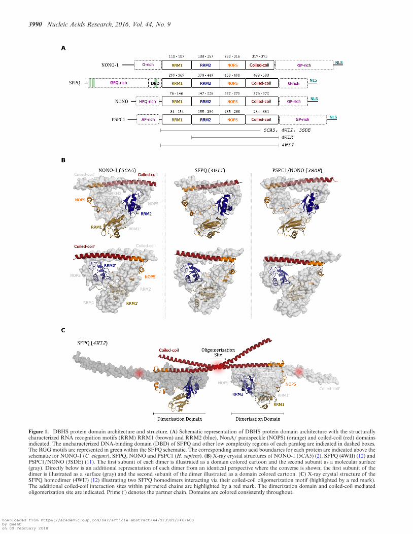
Figure 1. DBHS protein domain architecture and structure. (A) Schematic representation of DBHS protein domain architecture with the structurallycharacterized RNA recognition motifs (RRM) RRM1 (brown) and RRM2 (blue), NonA/ paraspeckle (NOPS) (orange) and coiled-coil (red) domainsindicated. The uncharacterized DNA-binding domain (DBD) of SFPQ and other low complexity regions of each paralog are indicated in dashed boxes.The RGG motifs are represented in green within the SFPQ schematic. The corresponding amino acid boundaries for each protein are indicated above theschematic for NONO-1 (C. elegans), SFPQ, NONO and PSPC1 (H. sapiens). (B) X-ray crystal structures of NONO-1 (5CA5) (2), SFPQ (4WII) (12) andPSPC1/NONO (3SDE) (11). The first subunit of each dimer is illustrated as a domain colored cartoon and the second subunit as a molecular surface(gray). Directly below is an additional representation of each dimer from an identical perspective where the converse is shown; the first subunit of thedimer is illustrated as a surface (gray) and the second subunit of the dimer illustrated as a domain colored cartoon. (C) X-ray crystal structure of theSFPQ homodimer (4WIJ) (12) illustrating two SFPQ homodimers interacting via their coiled-coil oligomerization motif (highlighted by a red mark).The additional coiled-coil interaction sites within partnered chains are highlighted by a red mark. The dimerization domain and coiled-coil mediatedoligomerization site are indicated. Prime (′) denotes the partner chain. Domains are colored consistently throughout.
Downloaded from https://academic.oup.com/nar/article-abstract/44/9/3989/2462600by gueston 09 February 2018
Nucleic Acids Research, 2016, Vol. 44, No. 9 3991
DBHS PROTEIN STRUCTURE AND FUNCTION
DBHS proteins are nucleic acid- and protein-bindingdimers capable of forming higher order oligomeric com-plexes (Figure 1). In their structured core DBHS proteinsare remarkably modular, possessing both protein–proteinand protein–nucleic acid binding sites that enable them tobehave as a ‘molecular scaffold (Figure 2). Beyond the struc-tured regions, the N- and C- terminal low-complexity re-gions contribute significantly to the functional diversity ob-served for DBHS paralogs.
DBHS domain architecture
All members of the DBHS protein family possess a con-served core of ∼300 amino acids defined as the ‘DBHSregion’ (Figure 1A). The DBHS region encompasses thetandem dissimilar RRMs, the protein–protein interactionNOPS domain and the coiled-coil domain. In DBHS pro-teins, the tandem RRMs are distinct from one another andare separated by a flexible seven-amino acid linker (2). TheRRM is one of the most abundant and well-characterizednucleic acid binding domains, present in 0.5–1.0% of hu-man genes (reviewed in (9,10). A canonical RRM has a�1�1�2�3�2�4 topology where aromatic residues on the �-sheets �-stack nucleotides with additional contacts fromcharged and sequence-specificity determining side chains(9). DBHS RRM1 is described as canonical, with con-served aromatic and charged residues exposed to the sol-vent (2,11,12). In contrast, the DBHS RRM2 lacks theconserved aromatic residues and has additional extended�-turns within loop 3 and loop 5, one of which showshigh conservation (2) and resembles a double-strandedDNA/RNA recognition motif (13). The NOPS domain def-inition, derived from a Pfam alignment, stretches from theend of RRM2 to the coiled-coil domain where it functionsalmost exclusively in mediating DBHS dimerization. How-ever, some surface-exposed basic residues within the NOPSdomain may be involved in nucleic acid binding (2). TheC-terminal end of the DBHS region features the highlycharged coiled-coil domain known to facilitate dimeriza-tion and oligomerization. The coiled-coil dimerization do-main forms an unusual right-handed antiparallel coiled-coil (12). While modular in their core domain architecture,DBHS proteins possess sequences that are likely to be in-trinsically disordered. These intrinsically disordered regionscontain low-complexity sequences (or low-complexity do-mains, LCDs) that flank the DBHS region and are sitesfor post-translational modification and potentially drive dy-namic phase separation (14).
Obligatory dimerization
The first indication that DBHS proteins function as dimerscame in 1993 with the purification of SFPQ/NONO het-erodimers from HeLa cells (1). Subsequently, yeast two hy-brid experiments, immuno-precipitation and other experi-ments on endogenous proteins, confirmed that DBHS pro-teins interact reciprocally (15–19) and others have consis-tently reported copurification and in vitro interaction toconfirm DBHS dimerization. Atomic resolution structures
for DBHS protein dimers have been determined from bothvertebrates and invertebrates (2,11,12). The structures showDBHS monomers forming a globular core with emergingantiparallel-coiled coils (Figure 1B). As a result of this, theputatively RNA-binding �-sheet surface of RRM2 faces a20-A ‘void’ in the core of the dimer (11). The obligatorydimerization is mediated by reciprocal interactions betweenRRM2 of one monomer, the partnered NOPS and the dis-tal coiled-coil domain (2,11,12). The interface involves con-tacts from across the entire DBHS region but is dominatedby a highly conserved cluster of hydrophobic interactionsbetween RRM2 and the NOPS domain. Consistent withthe role of the RRM2, NOPS and coiled-coil domains indimerization, removal of RRM1 does not hinder the abilityof SFPQ to form an obligate homodimer (12). The dimer-ization interface is highly conserved (2) such that Chirono-mus tentans Hrp65 can form stable heterodimers with hu-man SFPQ and D. melanogaster NonA (20). Consistentwith obligatory dimerization, mutation of residues withinthe NOPS-RRM2 dimerization interface results in localiza-tion and functional defects (11). Thus, the DBHS regionforms a compact and intimately intertwined core depen-dent on a complex series of contacts between RRM2, NOPSand coiled-coil domains. Unsurprisingly, deletion of eitherRRM2 and/or the NOPS domain results in a loss of func-tion, presumably due to a loss of dimer integrity. Similarly,over expression of individual parts of the proteins not capa-ble of dimerizing, such as an RRM, or coiled-coil region inisolation, should be considered dimerization incompetentand therefore functionally limited.
Recognizing SFPQ, NONO and PSPC1 as fundamen-tally dimeric means that some past literature, where theyare annotated as individual functional units, may need tobe reinterpreted. Nevertheless, we have included many suchstudies in this review as their functional insights are impor-tant. DBHS dimerization is a dynamic process whereby agiven dimer (homo or hetero) can readily exchange interac-tion partner to form an alternative dimerization state andin turn regulate function. For example, alternative dimeriza-tion between differing Hrp65 isoforms dictates their subcel-lular localization in C. tentans (20,21). Dimerization statemay also be dependent on the relative abundance of eachparalog. For example, mouse Sertoli cells have higher ex-pression of SFPQ and PSPC1 compared to NONO (18), incontrast to HeLa cells where NONO and SFPQ are moreabundant than PSPC1 (17). Sertoli cells contain all het-erodimer combinations (PSPC1/SFPQ, SFPQ/NONO andPSPC1/NONO), whereas HeLa cells predominantly con-tain SFPQ/NONO and PSPC1/NONO. Different dimersmay have different cell-type specific functions, as DBHSproteins can functionally compensate for each other in somebiological scenarios, but not others. For example, SFPQoverexpression causes increased exon inclusion in a splic-ing minigene reporter, but NONO overexpression had noeffect (22). In contrast, knockout of NONO can be com-pensated by an upregulation of PSPC1 to form a functionalheterodimer with SFPQ in DNA repair (23). However, thereare examples where SFPQ and NONO do not compensatefor the loss of PSPC1 (24) and PSPC1 and SFPQ cannotcompensate for the loss of NONO in intellectual disabilityin humans (25). Future studies cannot ignore the dynamic
Downloaded from https://academic.oup.com/nar/article-abstract/44/9/3989/2462600by gueston 09 February 2018

3992 Nucleic Acids Research, 2016, Vol. 44, No. 9
Figure 2. DBHS protein-binding sites and post-translation modifications mapped to the X-ray crystal structure of SFPQ (4WIJ). The structure illustratesa putative SFPQ/NONO heterodimer (colored surface/black cartoon, respectively) with the remaining N- and C-terminal uncharacterized and low-complexity domains modeled as flexible chains at the corresponding termini of the structure (dashed lines). Interaction sites within the X-ray crystalstructure are colored; dimerization interface (green), coiled-coil oligomerization motif (yellow), secondary oligomerization site (brown), putative RNA-binding surface of RRM1 (light blue), putative RNA-binding loop of RRM2 (dark blue). The structurally uncharacterized DNA-binding domain (12) ofSFPQ is also illustrated (purple box). Mapped as colored circles to the SFPQ and NONO chains are reported sites of post-translational modification andcorresponding amino acid number; phosphorylation (red), methylation (orange), citrullination (teal), SUMOylation (purple) and ADP-ribosylation (palegreen). Methylation sites that are also subject to citrullination are indicated with an asterisk.
expression and interplay between DBHS protein paralogs,especially given functional overlap and redundancy.
Coiled-coil mediated oligomerization
Oligomerization and functional aggregation are emergingas important to DBHS function. DBHS structures showan extended �-helical coiled-coil projecting out from thecore dimer interface (2,11,12). Truncation and mutage-nesis of this coiled-coil region resulted in aberrant sub-nuclear localization and physiological defects in severalDBHS proteins and it was postulated that these defectsresulted in perturbed coiled-coil mediated oligomerization(11,17,26,27). Recently, the SFPQ homodimer structureconfirmed that the �-helical ‘arms’ project out from the
dimerization core and provide an interface for oligomer-ization via a highly conserved motif present within the ex-tended coiled-coil domain (2,12) (Figure 1C). This inter-face takes part, in a concentration-dependent manner, in aclassical heptad-repeat coiled-coil interaction with anotherDBHS protein dimer (28). The formation of higher orderoligomers by SFPQ is not only essential for the structureof the mammalian paraspeckle, but also for the cooperativeenhancement of nucleic acid binding (11,12). C-terminal tothe coiled-coil oligomerization motif, there are regions ofhighly conserved charged residues that provide an interfacefor further coiled-coil type interactions (12), consistent withthe coiled-coil acting as a molecular ruler for DBHS proteininteractions (29). The molecular scaffolding brought about
Downloaded from https://academic.oup.com/nar/article-abstract/44/9/3989/2462600by gueston 09 February 2018

Nucleic Acids Research, 2016, Vol. 44, No. 9 3993
by combined nucleic acid binding and coiled-coil mediatedoligomerization is not uncommon, for example, coiled-coilmediated interactions feature heavily in centrosome assem-bly (30). Other aggregation-prone paraspeckle proteins alsoform higher-order oligomers to stabilize paraspeckles, al-though not through coiled-coils, instead through reversibleprion-like protein aggregation (14). It may well be thatoligomerization by coiled-coil domains and prion-like do-main interactions, are both examples of reversible and dy-namic ‘functional aggregation,’ an emerging concept in cellbiology that is driven by local concentrations of moleculessuch as DBHS proteins that readily oligomerize, or aggre-gate. This functional aggregation property, as well as theirabundance, may explain why DBHS proteins are often iden-tified in mass spectrometry/proteomic studies (31), even innegative control samples.
Sequence and structure specific RNA interaction
In spite of structural data and the presence of canonicalnucleic acid recognition motifs, precisely how DBHS pro-teins bind nucleic acids is still unknown. What is knownis that DBHS proteins recognize a broad spectrum of nu-cleic acids. In vitro, SFPQ and NONO can bind to anysingle-stranded polynucleotide (32–35) with preference forsingle-stranded RNA (ssRNA) over single-stranded DNA(ssDNA) (33,34). While both NONO and SFPQ are re-ported to have a preference for short G-rich RNA (35–40), SFPQ exhibits the highest affinity for poly-U (32) andNONO poly-G (35). NONO has also been reported to bindlong stretches of poly(ADP-ribose) (PAR) using RRM1(41) (similar to the serine/arginine-rich splicing factor 1(ASF/SF2) (42) (reviewed in (43)), an interaction poten-tially heightened by coiled-coil mediated oligomerizationgiven the preference for longer stretches of PAR. In ad-dition, DBHS proteins also bind structured nucleic acids.For example, NONO/SFPQ homo- or heterodimers inter-act with the U5 small-nuclear RNA (snRNA) stem in vitro,an interaction dependent on both the sequence and struc-ture of the target RNA (15). SFPQ and NONO also inter-act with the stem loop in the 5′-splice site of pre-mRNA(35,44,45), the terminal stem-loop of the hepatitis deltavirus RNA (46) and inverted repeat Alu elements (IRAlus)that form long dsRNA regions and can be subject to exten-sive RNA editing (38,47).
Despite this broad range of target RNAs, some degreeof sequence and/or secondary structure driven specificityis observed. It is tempting to suggest that the canonicalRRM1 facilitates interactions with unstructured nucleicacids, whilst additional complex mechanisms, likely involv-ing DBHS oligomerization and RRM2, mediate structuredRNA binding. Supporting the notion of distinct bindingmodes, DBHS binding to double-stranded nucleic acid is in-dependent of binding single stranded nucleic acids (33,34).The interaction of DBHS proteins with nucleic acid mayalso be consolidated by amino acids proximal to the highlyconserved surfaces of RRM1 and RRM2. For example, theN-terminal region preceding NONO RRM1 is implicatedin binding to the 5′-splice site of pre-mRNA (44). Simi-larly, Arginine residues in the coiled-coil region of SFPQundergo post-translation modifications influencing RNA
binding (48), albeit with an unknown mechanism. It is clearthat while we understand nucleic acid binding to some ex-tent, more data are required to deconvolute the observedpromiscuity of DBHS protein RNA interaction.
DNA interaction
SFPQ, NonA and Hrp65 all possess RGG motifs N-terminal to their first RRM, some of which constitute theputative ‘DNA-binding domain’ (Figure 1A). While theprecise role of the RGG/RG motif (reviewed in (49)) is un-clear, in the context of the N-terminal LCD of SFPQ; theyare required for the interaction with dsDNA (12,50) and anyof the five RGG motifs could also serve as a module forsensing PAR at sites of DNA damage (41,43). Indeed, therole of the RGG motifs are regulated by a swathe of post-translational modifications, including methylation, citrul-lination and ADP-ribosylation, with potential regulatoryroles discussed further below (48,51,52).
ROLES IN TRANSCRIPTIONAL REGULATION
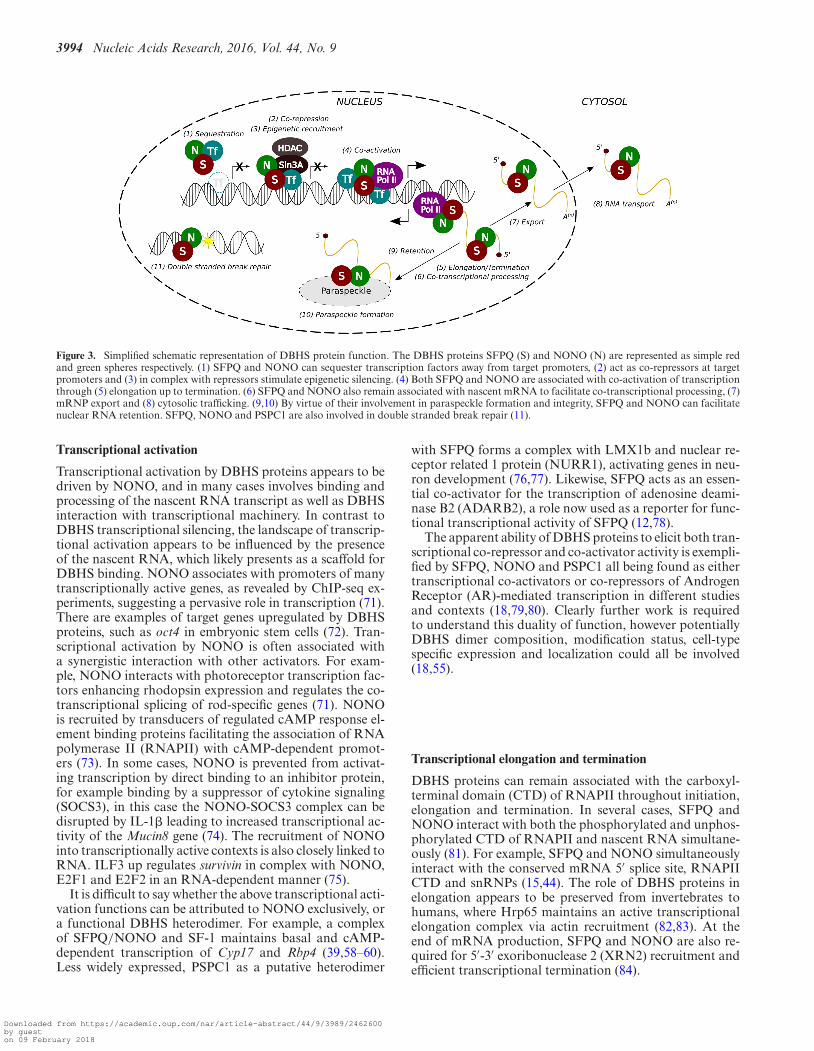
DBHS proteins mediate transcriptional repression, activa-tion, initiation, elongation and termination (Figure 3 [1–4]). Utilizing their behavior as a molecular scaffold, DBHSproteins associate synergistically with a broad spectrum oftranscription factors, DNA and RNA; acting bifunctionallyas positive and negative transcriptional regulators. Thus, aswith many transcription factors, their precise role is contextdependent (53).
Transcriptional repression
Transcriptional repression by DBHS proteins appearslargely driven by and dependent on SFPQ, either in a ho-modimer, or heterodimer context. Several studies have de-scribed how SFPQ binds directly to target gene promoters,subsequently recruiting epigenetic silencers such as Sin3Aand HDAC (54–56). Through recruitment of epigenetic reg-ulators, SFPQ/NONO can act on hormone receptors suchas the thyroid and retinoid X receptors (57), or in com-plex with steroidogenic factor 1 (SF-1), repress the humanCYP17 gene or genes involved in circadian rhythms (58–61). There is a dearth of characterized SFPQ DNA recogni-tion elements, but one example is the palindromic sequenceCTGAGTC, an insulin-like growth factor response elementin specific gene promoters (62–65). SFPQ/NONO can alsonegatively regulate transcription by sequestering activatorsaway from target promoters. For example, direct bindingof SFPQ/NONO to the progesterone receptor can preventits binding to DNA (54). In other transcriptional contexts,NONO represses genes responsive to the cAMP pathway(66). SFPQ and NONO can associate with silencer motifs inthe promoter of the phosphate carrier (PiC) gene (67); andmore recently, as transcriptional repressors of Interleukin-8 (IL8) (68). Reports suggesting that NONO can directlyinteract with promoter elements repressing transcriptionmay have over looked SFPQ heterodimerization, althoughit cannot be ruled out that NONO may possess some hith-erto uncharacterized DNA-binding ability (69,70).
Downloaded from https://academic.oup.com/nar/article-abstract/44/9/3989/2462600by gueston 09 February 2018
3994 Nucleic Acids Research, 2016, Vol. 44, No. 9
Figure 3. Simplified schematic representation of DBHS protein function. The DBHS proteins SFPQ (S) and NONO (N) are represented as simple redand green spheres respectively. (1) SFPQ and NONO can sequester transcription factors away from target promoters, (2) act as co-repressors at targetpromoters and (3) in complex with repressors stimulate epigenetic silencing. (4) Both SFPQ and NONO are associated with co-activation of transcriptionthrough (5) elongation up to termination. (6) SFPQ and NONO also remain associated with nascent mRNA to facilitate co-transcriptional processing, (7)mRNP export and (8) cytosolic trafficking. (9,10) By virtue of their involvement in paraspeckle formation and integrity, SFPQ and NONO can facilitatenuclear RNA retention. SFPQ, NONO and PSPC1 are also involved in double stranded break repair (11).
Transcriptional activation
Transcriptional activation by DBHS proteins appears to bedriven by NONO, and in many cases involves binding andprocessing of the nascent RNA transcript as well as DBHSinteraction with transcriptional machinery. In contrast toDBHS transcriptional silencing, the landscape of transcrip-tional activation appears to be influenced by the presenceof the nascent RNA, which likely presents as a scaffold forDBHS binding. NONO associates with promoters of manytranscriptionally active genes, as revealed by ChIP-seq ex-periments, suggesting a pervasive role in transcription (71).There are examples of target genes upregulated by DBHSproteins, such as oct4 in embryonic stem cells (72). Tran-scriptional activation by NONO is often associated witha synergistic interaction with other activators. For exam-ple, NONO interacts with photoreceptor transcription fac-tors enhancing rhodopsin expression and regulates the co-transcriptional splicing of rod-specific genes (71). NONOis recruited by transducers of regulated cAMP response el-ement binding proteins facilitating the association of RNApolymerase II (RNAPII) with cAMP-dependent promot-ers (73). In some cases, NONO is prevented from activat-ing transcription by direct binding to an inhibitor protein,for example binding by a suppressor of cytokine signaling(SOCS3), in this case the NONO-SOCS3 complex can bedisrupted by IL-1� leading to increased transcriptional ac-tivity of the Mucin8 gene (74). The recruitment of NONOinto transcriptionally active contexts is also closely linked toRNA. ILF3 up regulates survivin in complex with NONO,E2F1 and E2F2 in an RNA-dependent manner (75).
It is difficult to say whether the above transcriptional acti-vation functions can be attributed to NONO exclusively, ora functional DBHS heterodimer. For example, a complexof SFPQ/NONO and SF-1 maintains basal and cAMP-dependent transcription of Cyp17 and Rbp4 (39,58–60).Less widely expressed, PSPC1 as a putative heterodimer
with SFPQ forms a complex with LMX1b and nuclear re-ceptor related 1 protein (NURR1), activating genes in neu-ron development (76,77). Likewise, SFPQ acts as an essen-tial co-activator for the transcription of adenosine deami-nase B2 (ADARB2), a role now used as a reporter for func-tional transcriptional activity of SFPQ (12,78).
The apparent ability of DBHS proteins to elicit both tran-scriptional co-repressor and co-activator activity is exempli-fied by SFPQ, NONO and PSPC1 all being found as eithertranscriptional co-activators or co-repressors of AndrogenReceptor (AR)-mediated transcription in different studiesand contexts (18,79,80). Clearly further work is requiredto understand this duality of function, however potentiallyDBHS dimer composition, modification status, cell-typespecific expression and localization could all be involved(18,55).
Transcriptional elongation and termination
DBHS proteins can remain associated with the carboxyl-terminal domain (CTD) of RNAPII throughout initiation,elongation and termination. In several cases, SFPQ andNONO interact with both the phosphorylated and unphos-phorylated CTD of RNAPII and nascent RNA simultane-ously (81). For example, SFPQ and NONO simultaneouslyinteract with the conserved mRNA 5′ splice site, RNAPIICTD and snRNPs (15,44). The role of DBHS proteins inelongation appears to be preserved from invertebrates tohumans, where Hrp65 maintains an active transcriptionalelongation complex via actin recruitment (82,83). At theend of mRNA production, SFPQ and NONO are also re-quired for 5′-3′ exoribonuclease 2 (XRN2) recruitment andefficient transcriptional termination (84).
Downloaded from https://academic.oup.com/nar/article-abstract/44/9/3989/2462600by gueston 09 February 2018

Nucleic Acids Research, 2016, Vol. 44, No. 9 3995
POST-TRANSCRIPTIONAL PROCESSING AND EX-PORT
It has been suggested that DBHS proteins may couple tran-scription to post-transcriptional processing (85), namelythrough a persistent association with nascent RNA (Fig-ure 3 [5-8]). While potential binding to the majority of tran-scripts suggests nonspecific binding (80,86), it is neverthe-less clear that some substrate specificity and activity is ap-parent.
Transcript splicing, polyadenylation and stabilization
SFPQ was first identified in a stable complex withpolypyrimidine tract-binding protein (PTB), required forpre-mRNA splicing (32). Other studies have identifiedSFPQ and/or NONO as spliceosome-associated proteins(36,87,88) and shown that NONO/SFPQ associate withU5 snRNA early in formation of the spliceosome (15).NONO directly interacts with the 5′ splice site (44) andSFPQ is found in large pre-assembled spliceosomal com-plexes (89). Despite these associations, DBHS proteins arenot essential components of the spliceosomal machinery perse, but are rather involved in co-transcriptional and alter-native splicing. Specifically, SFPQ has been identified as aregulator of splicing for CD45 (90), neuronal cell-specificgenes (91), the preprotachykini (PPT) minigene (22), themicrotubule-binding protein Tau (45) and spinal muscu-lar atrophy genes SMN1/SMN2 (92). Similarly, NONOis cited in rod-specific gene expression (71), phosphodi-esterase splicing (93) and together SFPQ and NONO bindto specific A-U rich elements in pre-mRNA such as TNF-α (94). SFPQ/NONO also facilitates pre-mRNA 3′-endprocessing by promoting polyadenylation and pre-mRNAcleavage (84,95,96).
Beyond post-transcriptional processing, DBHS proteinsare thought to contribute to maintaining transcript stability.For example, the stability of some histone coding mRNAis thought to involve SFPQ through either a direct or in-direct interaction with the transcript (97). Similarly, SFPQand NONO are known to regulate the stability of non-coding RNA, such as the long non-coding RNA NEAT1(98). Given their diffuse localization and broad nucleic acidspecificity; it is highly likely that DBHS proteins functionakin to a histone in degenerately coating nascent transcriptsfor stabilization.
Regulation of RNA localization and translation
DBHS proteins can also remain associated with the pro-cessed mRNP once formed. In neuronal cells SFPQ andNONO are components of large RNA transport granules inthe neurites (99), a phenomenon important for local trans-lation at the synapse. SFPQ and NONO are also snRNAexport stimulatory factors, accelerating the recruitment ofthe phosphorylated adapter for RNA export (PHAX) forefficient nuclear export of snRNA (100). In invertebrates,NonA has been show to facilitate intranuclear mobility ofmRNP particles, where it forms a complex with nuclearexport factor 1 (NXF1) (101). Similarly, Hrp65 has beenimplicated in regulating mRNA localization and transport
(21). There is also evidence for DBHS protein function in in-ternal ribosome entry site (IRES) regulation (102,103). TheIRES can initiate translation independent of a 5′-cap by re-cruitment of specific RNA-binding proteins (104). For ex-ample, SFPQ, in complex with Annexin A2, binds directlyto the IRES of the p53 mRNA and regulates its activity(102). Similarly, NONO and hnRNPM associate with thefibroblast growth factor 1 (FGF1) IRES in differentiatingmyoblasts and this ‘loading’ may be initiated when the RNAis transcribed in the nucleus (103).
SUBNUCLEAR STRUCTURES AND COMPLEXES
DBHS proteins are highly mobile inside the cell nucleus, butthey can be triggered by binding to local high concentra-tions of various nucleic acids to form microscopically visi-ble nuclear bodies, paraspeckles or large complexes such asDNA repair foci (Figure 3 [9–11]).
Formation and function of paraspeckles
Paraspeckles are ribonucleoprotein bodies located withinthe interchromatin space of mammalian cell nuclei (98)(reviewed in (4,5,105)). Paraspeckle proteins are definedby the colocalization of SFPQ, NONO or PSPC1 withthe long noncoding RNA NEAT1 (4,17,106–108). BothSFPQ and NONO are essential for paraspeckle forma-tion and integrity, as siRNA knockdown of either pro-tein prevents paraspeckle formation (107). The DBHS pro-teins directly bind NEAT1 and likely stabilize the RNA,as loss of these proteins results in reduced NEAT1 levels(107). Furthermore, DBHS proteins are also integral to oneparaspeckle regulatory mechanism where they bind struc-tured edited RNAs derived from transcribed inverted re-peat elements, resulting in nuclear retention of these RNAsin the paraspeckle (38,47,109,110). DBHS oligomerization(12) and contacts with other paraspeckle proteins such asRBM14 (14) are also important for the paraspeckle struc-ture. While the precise functional role of the paraspeckleis unclear, a general consensus that paraspeckles fine-tunegene expression under stress conditions is emerging (re-viewed in (4,5,105,111,112)). One mechanism for gene reg-ulation is that paraspeckles sequester a subset of nuclearproteins, including DBHS proteins, effectively depleting theavailable nuclear pool of these factors with flow-on effectson the target genes of these proteins (68,78,113–115).
Localization to DNA damage sites
DBHS proteins are implicated in double-stranded break(DSB) repair where they assist in homology directed re-pair or nonhomologous end joining (NHEJ). SFPQ pro-motes homologous DNA-pairing, strand invasion, D-loopformation and topoisomerase activity in a variety of celltypes (116–119). SFPQ/NONO is found within the DSBpreligation complex with the Ku protein and substrateDNA (120) and directly interacts with RAD51 (50,121),TopBP1 (122) and Matrin3 (123), recruiting proteins tosites of DNA damage (114) and stimulating both homolo-gous and nonhomologous repair (41,50,121,123,124). Col-lectively, the DBHS proteins promote end joining of ho-mologous DNA by direct interaction with DNA ends and
Downloaded from https://academic.oup.com/nar/article-abstract/44/9/3989/2462600by gueston 09 February 2018
3996 Nucleic Acids Research, 2016, Vol. 44, No. 9
recruitment/stabilization of a preligation complex (125). Inthe context of DNA repair, DBHS proteins have redun-dant roles. For example, knockout of NONO in embry-onic fibroblasts is compensated by PSPC1 up regulation,with a subsequent involvement of PSPC1 in the DSB repairpathway (23). PSPC1 is also involved in repair of cisplatin-induced DNA damage in certain cell-types with knock-down of PSPC1 causing cell death and bypassing of theG1/S checkpoint in HeLa cells (24). Interestingly, there isan emerging theme of RNA-binding proteins playing dis-tinct roles in DNA damage responses and DBHS proteinsadd to this repertoire (reviewed in (126)).
Emerging evidence places localized poly(ADP-ribose)polymerase (PARP) activity early in the cellular response toDNA damage, where protein LCDs containing RGG mo-tifs directly associate with PAR, forming phase separatedcompartments at sites of DNA damage (127). Mechanis-tically analogous to paraspeckle nucleation through com-bined nucleic acid recognition and prion-like interactions(14); DBHS interaction with PAR may serve as a scaffold tonucleate other subnuclear or cytosolic structures (128,129).
CIRCADIAN RHYTHM AND CELL CYCLE
Circadian rhythm is the change in abundance of proteins inresponse to an ∼ 24 h cycle. DBHS proteins are involvedin coordinating cell cycle and circadian rhythm by regulat-ing different nodes of the circadian network (130,131). Inmammalian cells, the Period (Per1 and Per2) proteins con-trol a negative transcriptional feedback loop that generatesoscillations in transcript abundance (132). DBHS proteinsmodulate this by interacting with PER proteins and antag-onizing their function (133). Beyond PER binding, NONOco-activates circadian genes in a cAMP-dependent manner,by recruiting RNAPII to cAMP-dependent promoters (73).While NONO is not rhythmic in its abundance (130), SFPQprotein levels appear to oscillate with the circadian cycle(61). Akin to NONO; SFPQ can directly interact with thenuclear PER complex, moreover it can also recruit Sin3A-HDAC to drive deacetylation and repression of the Per1promoter (61). Loss of NONO does not significantly affectcircadian rhythms in mammals, suggesting perhaps com-pensation by SFPQ or PSPC1, but loss of NonA, one oftwo DBHS fly proteins, results in arrhythmic flies (133). TheNONO: PER complex, formed as a function of oscillatingPER levels, directly co-activates the promoter of the G1-S checkpoint protein p16-INK4A (130). NONO null tis-sues show increased cell proliferation, reduced expression ofINK4A, but an unaffected circadian clock (130). This cellcycle defect can be rescued with over expression of NONO,but not PSPC1 or SFPQ, suggesting that this role is exclu-sive to NONO (130). Thus, the nucleoplasmic pool of dif-ferent DBHS protein has both redundant and independentfunctions as transcriptional co-activators and co-repressorsfor circadian clock-regulated genes, which combined, con-tribute to both the cell and circadian cycles (130,134).
DBHS PROTEIN CLINICAL SIGNIFICANCE
With roles in almost every step of gene regulation, it is notsurprising that perturbation of DBHS protein function has
consequences for the cell and organism. Broadly, DBHSproteins are rapidly emerging as clinically relevant in thecontexts of development, innate immunity and cancer (Fig-ure 4). Furthermore, SFPQ, NONO and PSPC1 all belongto a class of human genes with the lowest tolerance formissense and loss of function mutations, suggesting stronginvolvement in selectable phenotypes in humans (ExomeAggregation Consortium ExAC, Cambridge, MA, URL:http://exac.broadinstitute.org, accessed March 2016).
Neurobiology and Development
Most recently, mutations in NONO have been identifiedthat lead to patients with intellectual disability, defects thatneither PSPC1 nor SFPQ can compensate for (25). Consis-tent with this, the NONO knockout mouse exhibits a sim-ilar neurological defect (23). Loss of the zebrafish SFPQortholog leads to a subset of neuronal cells failing to dif-ferentiate and arrested development in the zebrafish em-bryo due to improper brain formation (27,135,136). At themolecular level, PSPC1 and SFPQ are components of tran-scriptional and post-transcriptional complexes implicit inthe regulation of genes required for neuronal differentia-tion and development (76,77,91,99,137). SFPQ and NONOdirectly interact with c-Jun N-terminal Kinase (JNK1) inan RNA-dependent manner where they are necessary forneuronal growth (138). Similarly, in neuronal cells, SFPQand NONO directly interact with Protein degylcase-1 (DJ-1) to carry out a neuroprotective role (139). In photore-ceptor development, NONO acts as an enhancer and post-transcriptional splicing regulator for rod-specific genes suchas rhodopsin (71). Finally, via its role in progesterone signal-ing, SFPQ derepression of the PR may trigger labor (140),a function also attributed to NONO (56). Given the per-vasive role of DBHS proteins in both transcriptional andpost-transcriptional events in many cell types, not just neu-ronal cells, it is interesting to speculate that additional clin-ical roles may be masked by functional redundancy of thethree mammalian DBHS proteins.
Innate immunity
Host cells respond to viral infection by inducing innateimmunity pathways. In turn, viral systems hijack host cellcomponents for the purpose of driving viral replication, of-ten utilizing the host defense factors. DBHS proteins areheavily involved in the innate immune response to virusesand can bind directly to viral RNAs, bind to ‘decoy’ hostncRNAs, or interact with proteins to alter the transcrip-tional status of immune related genes. For example, SFPQbinds to the hepatitis delta-virus RNA and is used for theviral replication-cycle (46). SFPQ is also used for influenzaA virus transcription and post-transcriptional processing(141,142). Interestingly, knockdown of NONO had no ef-fect on influenza A viral replication (142); however, it ispossible that PSPC1 expression may compensate for theloss of NONO. SFPQ is also implicated in both the tran-scription and maturation of HIV pre-mRNA, facilitatingviral pre-mRNA nuclear export (143). NONO is also aregulator of early and late stages of HIV-1 infection in T-cells (144). As mentioned above, SFPQ can be sequestered
Downloaded from https://academic.oup.com/nar/article-abstract/44/9/3989/2462600by gueston 09 February 2018
Nucleic Acids Research, 2016, Vol. 44, No. 9 3997
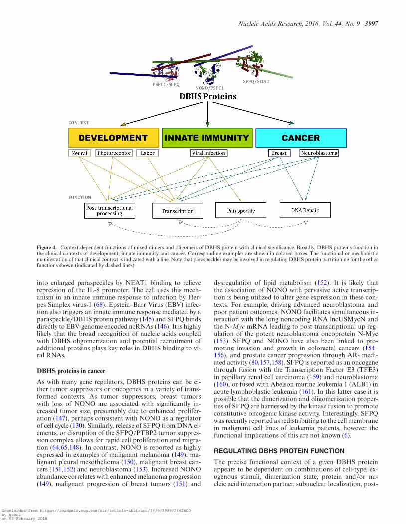
Figure 4. Context-dependent functions of mixed dimers and oligomers of DBHS protein with clinical significance. Broadly, DBHS proteins function inthe clinical contexts of development, innate immunity and cancer. Corresponding examples are shown in colored boxes. The functional or mechanisticmanifestation of that clinical context is indicated with a line. Note that paraspeckles may be involved in regulating DBHS protein partitioning for the otherfunctions shown (indicated by dashed lines).
into enlarged paraspeckles by NEAT1 binding to relieverepression of the IL-8 promoter. The cell uses this mech-anism in an innate immune response to infection by Her-pes Simplex virus-1 (68). Epstein–Barr Virus (EBV) infec-tion also triggers an innate immune response mediated by aparaspeckle/DBHS protein pathway (145) and SFPQ bindsdirectly to EBV-genome encoded ncRNAs (146). It is highlylikely that the broad recognition of nucleic acids coupledwith DBHS oligomerization and potential recruitment ofadditional proteins plays key roles in DBHS binding to vi-ral RNAs.
DBHS proteins in cancer
As with many gene regulators, DBHS proteins can be ei-ther tumor suppressors or oncogenes in a variety of trans-formed contexts. As tumor suppressors, breast tumorswith loss of NONO are associated with significantly in-creased tumor size, presumably due to enhanced prolifer-ation (147), perhaps consistent with NONO as a regulatorof cell cycle (130). Similarly, release of SFPQ from DNA el-ements, or disruption of the SFPQ/PTBP2 tumor suppres-sion complex allows for rapid cell proliferation and migra-tion (64,65,148). In contrast, NONO is reported as highlyexpressed in examples of malignant melanoma (149), ma-lignant pleural mesothelioma (150), malignant breast can-cers (151,152) and neuroblastoma (153). Increased NONOabundance correlates with enhanced melanoma progression(149), malignant progression of breast tumors (151) and
dysregulation of lipid metabolism (152). It is likely thatthe association of NONO with pervasive active transcrip-tion is being utilized to alter gene expression in these con-texts. For example, driving advanced neuroblastoma andpoor patient outcomes; NONO facilitates simultaneous in-teraction with the long noncoding RNA lncUSMycN andthe N-Myc mRNA leading to post-transcriptional up reg-ulation of the potent neuroblastoma oncoprotein N-Myc(153). SFPQ and NONO have also been linked to pro-moting invasion and growth in colorectal cancers (154–156), and prostate cancer progression through AR- medi-ated activity (80,157,158). SFPQ is reported as an oncogenethrough fusion with the Transcription Factor E3 (TFE3)in papillary renal cell carcinoma (159) and neuroblastoma(160), or fused with Abelson murine leukemia 1 (ALB1) inacute lymphoblastic leukemia (161). In this latter case it ispossible that the dimerization and oligomerization proper-ties of SFPQ are harnessed by the kinase fusion to promoteconstitutive oncogenic kinase activity. Interestingly, SFPQwas recently reported as redistributing to the cell membranein malignant cell lines of leukemia patients, however thefunctional implications of this are not known (6).
REGULATING DBHS PROTEIN FUNCTION
The precise functional context of a given DBHS proteinappears to be dependent on combinations of cell-type, ex-ogenous stimuli, dimerization state, protein and/or nu-cleic acid interaction partner, subnuclear localization, post-
Downloaded from https://academic.oup.com/nar/article-abstract/44/9/3989/2462600by gueston 09 February 2018

3998 Nucleic Acids Research, 2016, Vol. 44, No. 9
translational modification and time of day. Thus, all of theseelements have regulatory potentially in the DBHS context.
Protein interaction partner
While the dimeric and oligomeric state of a DBHS pro-tein certainly influences its role, protein interactions outsideof these also regulate their function. One such example isNONO activity in response to cAMP: at some cAMP re-sponsive promoters, NONO drives transcriptional activa-tion through interaction with the CREB/TORC complexto recruit RNAPII (73), whereas at other cAMP responsivepromoters, NONO interacts with RASD1 to instead selec-tively repress transcription (66). A similar scenario is ap-parent for AR mediated transcription (18,54). A more re-cent study showed SFPQ displays inhibited RNA bindingwhen in a complex with TRAP150, resulting in altered post-transcriptional processing (162). From a structural perspec-tive, it is not yet clear how multiple, sometimes simultane-ous, protein–protein interactions are mediated by DBHSproteins. It is likely that protein and/or nucleic acid bind-ing may induce dynamic changes in DBHS structure, thus‘revealing’ specific interaction sites that may be coupledto higher-order associations as a result of oligomerization.Furthermore, the highly variant low-complexity domainsflanking the DBHS region likely play a significant role incontributing additional protein–protein interactions.
Nucleic acid partner
Nucleic acids regulate DBHS protein function throughcompetition at mutually exclusive binding sites, allostericmodification or via delocalizing a subset of DBHS protein.The repressor activity of SFPQ can be alleviated via an al-losteric mechanism whereby binding of the murine noncod-ing RNA VL30 to SFPQ competes with its promoter bind-ing, releasing it and resulting in transcriptional activation(64,65). In a different context, SFPQ is responsible for tran-scriptional activation at the promoter of ADARB2, and thisis attenuated by NEAT1 lncRNA mediated sequestrationof SFPQ into the paraspeckle (78). Similarly, induction ofNEAT1 facilitates expression of IL-8 by relocating SFPQ toparaspeckles, relieving repression of IL-8 (68). Consistentwith lncRNA regulation of SFPQ activity, the binding ofthe lncRNA MALAT1 to SFPQ has been shown to disruptthe PTBP2: SFPQ tumor suppressor complex (148). Fur-ther, NONO interacts with the lncRNA lncUSMycN andthe N-Myc mRNA to post-transcriptionally upregulate N-Myc expression, acting as an oncogene driving neuroblas-toma progression (153). Recently, a study illustrated thatsynthetic oligonucleotides can drive degradation of nuclearDBHS protein, potentially by interrupting native dimeriza-tion (163), perhaps hinting at a regulatory role of noncodingRNA in DBHS protein degradation.
Post-translational modification
DBHS proteins are substrates for a number of post-translation modifications (Figure 2). The phosphorylationof SFPQ by Protein Kinase C inhibits its binding to RNA,but stimulates its association with ss and dsDNA promoting
D-loop formation (118). In contrast, Mnk1 and Mnk2 se-lectively phosphorylate SFPQ at Ser8 and Ser283, proximalto RRM1, enhancing RNA binding to the 3′UTR of TNF-� (94). In T-cells, GSK3 phosphorylates SFPQ at T687 pro-moting interaction with TRAP150, preventing SFPQ frombinding to CD45 pre-mRNA (90). NONO is also phospho-rylated in the region proximal to the coiled-coil domain(T412, T430 and T453) during mitosis (164). The phospho-rylation of these T–P motifs provides binding sites for thepeptidyprolyl isomerase (Pin1) that may lead to subsequentconformational changes of this region (164). Thr15 in theNONO N-terminus is also phosphorylated by CDK1 dur-ing mitosis, with consequences for RNA binding to simplesubstrates in vitro (35). Interestingly, the in vitro phospho-rylation of Thr15 disrupts binding of NONO to all homo-ribopolymers excluding poly-G (35), suggesting that the N-terminal 53 residues of NONO may allosterically regulateRNA-binding ability. Finally, NONO is also a substrate forProtein phosphatase 1 that associates with NONO RRM1and influences NONO post-transcriptional splicing (165).Phosphorylation of DBHS proteins also drives altered sub-nuclear or cellular localization. In murine neuroblastomacells, SFPQ and NONO associate with the nuclear enve-lope in response to tyrosine phosphorylation (166). Whilemechanistically unclear, the phosphorylation of SFPQ atN- and C-terminal Tyr residues proximal to the DBHS re-gion drives cytosolic localization inhibiting cell prolifera-tion (167,168). Similarly, hyperphosphorylation of the N-terminal half of SFPQ drastically alters its subnuclear lo-calization pattern in apoptosis (169).
Methylation, SUMOylation, citrullination and ADP-ribosylation of DBHS proteins also regulate their nucleicacid binding. The methylation of conserved Arg residues C-terminal to the highly charged coiled-coil oligomerizationmotif negates the binding of NONO to structured RNAssuch as mRNA containing IRAlus and dsRNA (170). Fur-thermore, the highly conserved �2-�3 Arg184 and Arg204of NONO are also reported to be methylated, however,the function of these sites are unknown (170). The N-terminal ‘DNA binding’ RGG motifs of SFPQ can alsobe mono- and di-methylated (51,171). This methylationof RGG does not perturb SFPQ dimerization, but pro-motes mRNA binding via an unknown mechanism (48).In contrast, the in vitro citrullination of SFPQ preventedRGG methylation and decreased mRNA association, po-tentially highlighting a dynamic control of SFPQ function-ality regulated by methylation and citrullination switches(48). DBHS proteins can also be post-translationally mod-ified with the addition of small ubiquitin-like modifiers(SUMO). SUMOylation of SFPQ on the surface of RRM1(residues 337–340) is required for interaction with HDAC1,promoting deacetylase activity and inhibiting activity atthe human tyrosine hydroxylase promoter (172). Finally,DBHS proteins are likely regulated by ADP-ribosylationboth within the DBHS region and adjacent LCDs. NONOand SFPQ were identified as direct substrates of PARP-1where they are modified on a series of glutamate residues(52). While these modifications are not functionally char-acterized, there is an emergent field describing ADP-ribosepolymers as important modulators of transcriptional regu-lators (128,173–176). Furthermore, in the context of DNA
Downloaded from https://academic.oup.com/nar/article-abstract/44/9/3989/2462600by gueston 09 February 2018
Nucleic Acids Research, 2016, Vol. 44, No. 9 3999
damage responses, ADP-ribosylation of SFPQ and NONOmight promote delocalization from DNA damage sites byoutcompeting DNA and PAR.
Subcellular localization
Apart from the subnuclear partitioning of DBHS proteininto paraspeckles, other foci, or the nucleoplasmic pool,DBHS proteins can also reside outside the nucleus. SFPQwas identified early on as a cell surface antigen in myoblastcells (177). Confocal microscopy experiments have shownSFPQ localization on the surface of brain microvascularendothelial cells where it is thought to be involved in in-vasive meningitis (178). Furthermore, SFPQ can be relo-cated to the cell surface membrane in multidrug-resistantcancer (6). As mentioned above, both NONO and SFPQare observed within the cytoplasm of hippocampal neu-rons associated with RNA transport granules (99). Con-sistent with cytoplasmic localization, by virtue of an in-teraction with the HERMES protein, SFPQ and NONOcan be found as components of cytoplasmic RNP granulesin retinal cells (7). Additionally, as described earlier, post-translational phosphorylation of SFPQ at C-terminal Tyrresidues drives cytoplasmic localization (168). Given theirabundance and dynamic nature, the cytoplasmic or extra-cellular role of DBHS proteins may have been underesti-mated to date.
CONCLUSION
Since the review published by Shav-Tal and Zipori in 2002(3), the body of literature on DBHS proteins has increaseddramatically, with novel contributions helping us to un-derstand their biological roles. The emerging paradigm forDBHS protein function describes a family of nuclear media-tors essential for seeding and bridging multiple nuclear pro-cesses, as well as several cytoplasmic roles. By virtue of theirmodular design, paralogs, swathe of modifications, result-ing broad nucleic acid specificity and varied protein inter-action partners; DBHS proteins are able to act as dynamicnuclear elements mediating protein–protein and protein–nucleic acid interactions in a variety of contexts. In thismanner, DBHS proteins can effectively couple gene tran-scription to post-transcriptional processing and recruit fac-tors to DNA damage foci. However, we are still lacking inour understanding of the precise mechanistic detail of theDBHS interactome, particularly beyond the core structuredregion. Nevertheless, moving forward, we now have a soundframework to reliably investigate this remarkably adaptableand versatile protein family. Further research is required toappreciate what mediates the dynamic and sometimes si-multaneous DBHS protein association with RNA, DNAand protein. Understanding the mechanistic ‘decisions’ thatare made to dictate DBHS protein partitioning will more-over be therapeutically invaluable.
FUNDING
National Health and Medical Research Council of Aus-tralia (NHMRC) Project Grants [1048659, 1050585 toC.S.B., A.H.F.]; Australian Research Council Discovery
Project [DP160102435]; University of Western AustraliaHackett Postgraduate Scholarship [to G.J.K.]. The open ac-cess publication charge for this paper has been waived byOxford University Press - NAR.Conflict of interest statement. None declared.
REFERENCES1. Dong,B., Horowitz,D.S., Kobayashi,R. and Krainer,A.R. (1993)
Purification and cDNA cloning of HeLa cell p54nrb, a nuclearprotein with two RNA recognition motifs and extensive homologyto human splicing factor PSF and Drosophila NONA/BJ6. NucleicAcids Res., 21, 4085–4092.
2. Knott,G.J., Lee,M., Passon,D.M., Fox,A.H. and Bond,C.S. (2015)Caenorhabditis elegans NONO-1: Insights into DBHS proteinstructure, architecture and function. Protein Sci., 24, 2033–2043.
3. Shav-Tal,Y. and Zipori,D. (2002) PSF and p54(nrb)/NonO -multi-functional nuclear proteins. FEBS Lett., 531, 109–114.
4. Bond,C.S. and Fox,A.H. (2009) Paraspeckles: nuclear bodies builton long noncoding RNA. J. Cell. Biol., 186, 637–644.
5. Fox,A.H. and Lamond,A.I. (2010) Paraspeckles. Cold SpringHarbor Perspect. Biol., 2,a000687.
6. Ren,S., She,M., Li,M., Zhou,Q., Liu,R., Lu,H., Yang,C. andXiong,D. (2014) The RNA/DNA-binding protein PSF relocates tocell membrane and contributes cells’ sensitivity to antitumor drug,doxorubicin. Cytometry A, 85, 231–241.
7. Furukawa,M.T., Sakamoto,H. and Inoue,K. (2015) Interaction andcolocalization of HERMES/RBPMS with NonO, PSF, and G3BP1in neuronal cytoplasmic RNP granules in mouse retinal line cells.Genes Cells, 20, 257–266.
8. Yarosh,C.A., Iacona,J.R., Lutz,C.S. and Lynch,K.W. (2015) PSF:nuclear busy-body or nuclear facilitator? Wiley Interdiscip, Rev.RNA, 6, 351–367.
9. Clery,A., Blatter,M. and Allain,F.H. (2008) RNA recognitionmotifs: boring? Not quite. Curr. Opin. Struct. Biol., 18, 290–298.
10. Daubner,G.M., Clery,A. and Allain,F.H. (2013) RRM-RNArecognition: NMR or crystallography...and new findings. Curr. Opin.Struct. Biol., 23, 100–108.
11. Passon,D.M., Lee,M., Rackham,O., Stanley,W.A., Sadowska,A.,Filipovska,A., Fox,A.H. and Bond,C.S. (2012) Structure of theheterodimer of human NONO and paraspeckle protein component1 and analysis of its role in subnuclear body formation. Proc. Natl.Acad. Sci. U.S.A., 109, 4846–4850.
12. Lee,M., Sadowska,A., Bekere,I., Ho,D., Gully,B.S., Lu,Y.,Iyer,K.S., Trewhella,J., Fox,A.H. and Bond,C.S. (2015) Thestructure of human SFPQ reveals a coiled-coil mediated polymeressential for functional aggregation in gene regulation. Nucleic AcidsRes., 43, 3826–3840.
13. Skrisovska,L., Bourgeois,C.F., Stefl,R., Grellscheid,S.N., Kister,L.,Wenter,P., Elliott,D.J., Stevenin,J. and Allain,F.H.T. (2007) Thetestis-specific human protein RBMY recognizes RNA through anovel mode of interaction. EMBO Rep., 8, 372–379.
14. Hennig,S., Kong,G., Mannen,T., Sadowska,A., Kobelke,S.,Blythe,A., Knott,G.J., Iyer,K.S., Ho,D., Newcombe,E.A. et al.(2015) Prion-like domains in RNA binding proteins are essential forbuilding subnuclear paraspeckles. J. Cell Biol., 210, 529–539.
15. Peng,R., Dye,B.T., Perez,I., Barnard,D.C., Thompson,A.B. andPatton,J.G. (2002) PSF and p54nrb bind a conserved stem in U5snRNA. RNA, 8, 1334–1347.
16. Myojin,R., Kuwahara,S., Yasaki,T., Matsunaga,T., Sakurai,T.,Kimura,M., Uesugi,S. and Kurihara,Y. (2004) Expression andfunctional significance of mouse paraspeckle protein 1 onspermatogenesis. Biol. Reprod., 71, 926–932.
17. Fox,A.H., Bond,C.S. and Lamond,A.I. (2005) P54nrb forms aheterodimer with PSP1 that localizes to paraspeckles in anRNA-dependent manner. Mol. Biol Cell, 16, 5304–5315.
18. Kuwahara,S., Ikei,A., Taguchi,Y., Tabuchi,Y., Fujimoto,N.,Obinata,M., Uesugi,S. and Kurihara,Y. (2006) PSPC1, NONO, andSFPQ are expressed in mouse Sertoli cells and may function ascoregulators of androgen receptor-mediated transcription. Biol.Reprod., 75, 352–359.
19. Lee,M., Passon,D.M., Hennig,S., Fox,A.H. and Bond,C.S. (2011)Construct optimization for studying protein complexes: obtaining
Downloaded from https://academic.oup.com/nar/article-abstract/44/9/3989/2462600by gueston 09 February 2018

4000 Nucleic Acids Research, 2016, Vol. 44, No. 9
diffraction-quality crystals of the pseudosymmetric PSPC1-NONOheterodimer. Acta Crystallogr. Sect. D Biol. Crystallogr., 67,981–987.
20. Kiesler,E., Miralles,F., Ostlund Farrants,A.K. and Visa,N. (2003)The Hrp65 self-interaction is mediated by an evolutionarilyconserved domain and is required for nuclear import of Hrp65isoforms that lack a nuclear localization signal. J. Cell Sci., 116,3949–3956.
21. Miralles,F. and Visa,N. (2001) Molecular characterization ofCt-hrp65: identification of two novel isoforms originated byalternative splicing. Exp. Cell Res., 264, 284–295.
22. Marko,M., Leichter,M., Patrinou-Georgoula,M. and Guialis,A.(2010) hnRNP M interacts with PSF and p54(nrb) and co-localizeswithin defined nuclear structures. Exp. Cell Res., 316, 390–400.
23. Li,S., Li,Z., Shu,F.J., Xiong,H., Phillips,A.C. and Dynan,W.S.(2014) Double-strand break repair deficiency in NONO knockoutmurine embryonic fibroblasts and compensation by spontaneousupregulation of the PSPC1 paralog. Nucleic Acids Res., 42,9771–9780.
24. Gao,X., Kong,L., Lu,X., Zhang,G., Chi,L., Jiang,Y., Wu,Y., Yan,C.,Duerksen-Hughes,P., Zhu,X. et al. (2014) Paraspeckle protein 1(PSPC1) is involved in the cisplatin induced DNA damageresponse–role in G1/S checkpoint. PLoS One, 9, e97174.
25. Mircsof,D., Langouet,M., Rio,M., Moutton,S., Siquier-Pernet,K.,Bole-Feysot,C., Cagnard,N., Nitschke,P., Gaspar,L., Znidaric,M.et al. (2015) Mutations in NONO lead to syndromic intellectualdisability and inhibitory synaptic defects. Nat. Neurosci., 18,1731–1736.
26. Rendahl,K.G., Jones,K.R., Kulkarni,S.J., Bagully,S.H. and Hall,J.C.(1992) The dissonance mutation at the No-on-transient-a locus ofDrosophila-melanogaster - genetic-control of courtship song andvisual behaviors by a protein with putative Rna-binding motifs. J.Neurosci., 12, 390–407.
27. Lowery,L.A., Rubin,J. and Sive,H. (2007) Whitesnake/sfpq isrequired for cell survival and neuronal development in the zebrafish.Dev. Dyn., 236, 1347–1357.
28. Lupas,A.N. and Gruber,M. (2005) The structure of alpha-helicalcoiled coils. Adv. Protein Chem., 70, 37–78.
29. Dobson,L., Nyitray,L. and Gaspari,Z. (2015) A conserved chargedsingle alpha-helix with a putative steric role in paraspeckleformation. RNA, 21, 2023–2029.
30. Salisbury,J.L. (2003) Centrosomes: coiled-coils organize the cellcenter. Curr. Biol., 13, R88–R90.
31. Trinkle-Mulcahy,L., Boulon,S., Lam,Y.W., Urcia,R., Boisvert,F.M.,Vandermoere,F., Morrice,N.A., Swift,S., Rothbauer,U.,Leonhardt,H. et al. (2008) Identifying specific protein interactionpartners using quantitative mass spectrometry and bead proteomes.J. Cell Biol., 183, 223–239.
32. Patton,J.G., Porro,E.B., Galceran,J., Tempst,P. andNadal-Ginard,B. (1993) Cloning and characterization of PSF, anovel pre-mRNA splicing factor. Genes Dev., 7, 393–406.
33. Yang,Y.S., Hanke,J.H., Carayannopoulos,L., Craft,C.M.,Capra,J.D. and Tucker,P.W. (1993) NonO, anon-POU-domain-containing, octamer-binding protein, is themammalian homolog of Drosophila nonAdiss. Mol. Cell. Biol., 13,5593–5603.
34. Zhang,W.W., Zhang,L.X., Busch,R.K., Farres,J. and Busch,H.(1993) Purification and characterization of a DNA-bindingheterodimer of 52 and 100 kDa from HeLa cells. Biochem. J., 290,267–272.
35. Bruelle,C., Bedard,M., Blier,S., Gauthier,M., Traish,A.M. andVincent,M. (2011) The mitotic phosphorylation of p54(nrb)modulates its RNA binding activity. Biochem. Cell Biol., 89,423–433.
36. Hallier,M., Tavitian,A. and Moreau-Gachelin,F. (1996) Thetranscription factor Spi-1/PU.1 binds RNA and interferes with theRNA-binding protein p54nrb. J. Biol. Chem., 271, 11177–11181.
37. Basu,A., Dong,B., Krainer,A.R. and Howe,C.C. (1997) Theintracisternal A-particle proximal enhancer-binding proteinactivates transcription and is identical to the RNA- andDNA-binding protein p54nrb/NonO. Mol. Cell. Biol., 17, 677–686.
38. Zhang,Z. and Carmichael,G.G. (2001) The fate of dsRNA in thenucleus: A p54(nrb)-containing complex retention of promiscuouslymediates the nuclear A-to-I edited RNAs. Cell, 106, 465–475.
39. Bianconcini,A., Lupo,A., Capone,S., Quadro,L., Monti,M.,Zurlo,D., Fucci,A., Sabatino,L., Brunetti,A., Chiefari,E. et al.(2009) Transcriptional activity of the murine retinol-binding proteingene is regulated by a multiprotein complex containing HMGA1,p54 nrb/NonO, protein-associated splicing factor (PSF) andsteroidogenic factor 1 (SF1)/liver receptor homologue 1 (LRH-1).Int. J. Biochem. Cell Biol., 41, 2189–2203.
40. Murthy,U.M. and Rangarajan,P.N. (2010) Identification of proteininteraction regions of VINC/NEAT1/Men epsilon RNA. FEBSLett., 584, 1531–1535.
41. Krietsch,J., Caron,M.C., Gagne,J.P., Ethier,C., Vignard,J.,Vincent,M., Rouleau,M., Hendzel,M.J., Poirier,G.G. andMasson,J.Y. (2012) PARP activation regulates the RNA-bindingprotein NONO in the DNA damage response to DNAdouble-strand breaks. Nucleic Acids Res., 40, 10287–10301.
42. Malanga,M., Czubaty,A., Girstun,A., Staron,K. and Althaus,F.R.(2008) Poly(ADP-ribose) binds to the splicing factor ASF/SF2 andregulates its phosphorylation by DNA topoisomerase I. J. Biol.Chem., 283, 19991–19998.
43. Krietsch,J., Rouleau,M., Pic,E., Ethier,C., Dawson,T.M.,Dawson,V.L., Masson,J.Y., Poirier,G.G. and Gagne,J.P. (2013)Reprogramming cellular events by poly(ADP-ribose)-bindingproteins. Mol. Aspects Med., 34, 1066–1087.
44. Kameoka,S., Duque,P. and Konarska,M.M. (2004) P54(nrb)associates with the 5 ’ splice site within large transcription/splicingcomplexes. EMBO J., 23, 1782–1791.
45. Ray,P., Kar,A., Fushimi,K., Havlioglu,N., Chen,X. and Wu,J.Y.(2011) PSF suppresses tau exon 10 inclusion by interacting with astem-loop structure downstream of exon 10. J. Mol. Neurosci., 45,453–466.
46. Greco-Stewart,V.S., Thibault,C.S.L. and Pelchat,M. (2006) Bindingof the polypyrimidine tract-binding protein-associated splicingfactor (PSF) to the hepatitis delta virus RNA. Virology, 356, 35–44.
47. Elbarbary,R.A., Li,W., Tian,B. and Maquat,L.E. (2013) STAU1binding 3’ UTR IRAlus complements nuclear retention to protectcells from PKR-mediated translational shutdown. Genes Dev., 27,1495–1510.
48. Snijders,A.P., Hautbergue,G.M., Bloom,A., Williamson,J.C.,Minshull,T.C., Phillips,H.L., Mihaylov,S.R., Gjerde,D.T.,Hornby,D.P., Wilson,S.A. et al. (2015) Arginine methylation andcitrullination of splicing factor proline- and glutamine-rich(SFPQ/PSF) regulates its association with mRNA. RNA, 21,347–359.
49. Thandapani,P., O’Connor,T.R., Bailey,T.L. and Richard,S. (2013)Defining the RGG/RG Motif. Mol. Cell, 50, 613–623.
50. Rajesh,C., Baker,D.K., Pierce,A.J. and Pittman,D.L. (2011) Thesplicing-factor related protein SFPQ/PSF interacts with RAD51Dand is necessary for homology-directed repair and sister chromatidcohesion. Nucleic Acids Res., 39, 132–145.
51. Snijders,A.P., Hung,M.L., Wilson,S.A. and Dickman,M.J. (2010)Analysis of arginine and lysine methylation utilizing peptideseparations at neutral pH and electron transfer dissociation massspectrometry. J. Am. Soc. Mass Spectrom., 21, 88–96.
52. Zhang,Y., Wang,J., Ding,M. and Yu,Y. (2013) Site-specificcharacterization of the Asp- and Glu-ADP-ribosylated proteome.Nat. Methods, 10, 981–984.
53. Stampfel,G., Kazmar,T., Frank,O., Wienerroither,S., Reiter,F. andStark,A. (2015) Transcriptional regulators form diverse groups withcontext-dependent regulatory functions. Nature, 528, 147–151.
54. Dong,X., Shylnova,O., Challis,J.R. and Lye,S.J. (2005) Identificationand characterization of the protein-associated splicing factor as anegative co-regulator of the progesterone receptor. J. Biol. Chem.,280, 13329–13340.
55. Dong,X., Sweet,J., Challis,J.R., Brown,T. and Lye,S.J. (2007)Transcriptional activity of androgen receptor is modulated by twoRNA splicing factors, PSF and p54nrb. Mol. Cell. Biol., 27,4863–4875.
56. Dong,X., Yu,C., Shynlova,O., Challis,J.R., Rennie,P.S. and Lye,S.J.(2009) p54nrb is a transcriptional corepressor of the progesteronereceptor that modulates transcription of the labor-associated gene,connexin 43 (Gja1). Mol. Endocrinol., 23, 1147–1160.
57. Mathur,M., Tucker,P.W. and Samuels,H.H. (2001) PSF is a novelcorepressor that mediates its effect through Sin3A and the DNA
Downloaded from https://academic.oup.com/nar/article-abstract/44/9/3989/2462600by gueston 09 February 2018

Nucleic Acids Research, 2016, Vol. 44, No. 9 4001
binding domain of nuclear hormone receptors. Mol. Cell. Biol., 21,2298–2311.
58. Sewer,M.B., Nguyen,V.Q., Huang,C.J., Tucker,P.W., Kagawa,N. andWaterman,M.R. (2002) Transcriptional activation of human CYP17in H295R adrenocortical cells depends on complex formationamong p54(nrb)/NonO, protein-associated splicing factor, andSF-1, a complex that also participates in repression of transcription.Endocrinology, 143, 1280–1290.
59. Sewer,M.B. and Waterman,M.R. (2002) Transcriptional complexesat the CYP17 CRS. Endocr. Res., 28, 551–558.
60. Sewer,M.B. and Waterman,M.R. (2002) Adrenocorticotropin/cyclicadenosine 3’,5’-monophosphate-mediated transcription of thehuman CYP17 gene in the adrenal cortex is dependent onphosphatase activity. Endocrinology, 143, 1769–1777.
61. Duong,H.A., Robles,M.S., Knutti,D. and Weitz,C.J. (2011) Amolecular mechanism for circadian clock negative feedback.Science, 332, 1436–1439.
62. Urban,R.J., Bodenburg,Y., Kurosky,A., Wood,T.G. and Gasic,S.(2000) Polypyrimidine tract-binding protein-associated splicingfactor is a negative regulator of transcriptional activity of theporcine p450scc insulin-like growth factor response element. Mol.Endocrinol., 14, 774–782.
63. Urban,R.J., Bodenburg,Y.H. and Wood,T.G. (2002) NH2 terminusof PTB-associated splicing factor binds to the porcine P450sccIGF-I response element. Am. J. Physiol. Endocrinol. Metab., 283,E423–427.
64. Song,X., Sui,A. and Garen,A. (2004) Binding of mouse VL30retrotransposon RNA to PSF protein induces genes repressed byPSF: effects on steroidogenesis and oncogenesis. Proc. Natl. Acad.Sci. U.S.A., 101, 621–626.
65. Song,X., Sun,Y. and Garen,A. (2005) Roles of PSF protein andVL30 RNA in reversible gene regulation. Proc. Natl. Acad. Sci.U.S.A., 102, 12189–12193.
66. Ong,S.A., Tan,J.J., Tew,W.L. and Chen,K.S. (2011) Rasd1modulates the coactivator function of NonO in the cyclic AMPpathway. PLoS One, 6, e24401.
67. Iacobazzi,V., Infantino,V., Costanzo,P., Izzo,P. and Palmieri,F.(2005) Functional analysis of the promoter of the mitochondrialphosphate carrier human gene: identification of activator andrepressor elements and their transcription factors. Biochem. J., 391,613–621.
68. Imamura,K., Imamachi,N., Akizuki,G., Kumakura,M.,Kawaguchi,A., Nagata,K., Kato,A., Kawaguchi,Y., Sato,H.,Yoneda,M. et al. (2014) Long noncoding RNA NEAT1-dependentSFPQ relocation from promoter region to paraspeckle mediates IL8expression upon immune stimuli. Mol. Cell, 53, 393–406.
69. Zhang,C., Zhang,M.X., Shen,Y.H., Burks,J.K., Zhang,Y., Wang,J.,LeMaire,S.A., Yoshimura,K., Aoki,H., Coselli,J.S. et al. (2007)TNF-alpha suppresses prolyl-4-hydroxylase alpha1 expression viathe ASK1-JNK-NonO pathway. Arterioscler. Thromb. Vasc. Biol.,27, 1760–1767.
70. Zhang,C., Zhang,M.X., Shen,Y.H., Burks,J.K., Li,X.N.,LeMaire,S.A., Yoshimura,K., Aoki,H., Matsuzaki,M., An,F.S. et al.(2008) Role of NonO-histone interaction in TNFalpha-suppressedprolyl-4-hydroxylase alpha1. Biochim. Biophys. Acta, 1783,1517–1528.
71. Yadav,S.P., Hao,H., Yang,H.J., Kautzmann,M.A., Brooks,M.,Nellissery,J., Klocke,B., Seifert,M. and Swaroop,A. (2014) Thetranscription-splicing protein NonO/p54nrb and threeNonO-interacting proteins bind to distal enhancer region andaugment rhodopsin expression. Hum. Mol. Genet., 23, 2132–2144.
72. Park,Y., Lee,J.M., Hwang,M.Y., Son,G.H. and Geum,D. (2013)NonO binds to the CpG island of oct4 promoter and functions as atranscriptional activator of oct4 gene expression. Mol. Cell, 35,61–69.
73. Amelio,A.L., Miraglia,L.J., Conkright,J.J., Mercer,B.A., Batalov,S.,Cavett,V., Orth,A.P., Busby,J., Hogenesch,J.B. and Conkright,M.D.(2007) A coactivator trap identifies NONO (p54nrb) as a componentof the cAMP-signaling pathway. Proc. Natl. Acad. Sci. U.S.A., 104,20314–20319.
74. Song,K.S., Kim,K., Chung,K.C., Seol,J.H. and Yoon,J.H. (2008)Interaction of SOCS3 with NonO attenuates IL-1beta-dependentMUC8 gene expression. Biochem. Biophys. Res. Commun., 377,946–951.
75. Yamauchi,T., Nakamura,N., Hiramoto,M., Yuri,M., Yokota,H.,Naitou,M., Takeuchi,M., Yamanaka,K., Kita,A., Nakahara,T. et al.(2012) Sepantronium bromide (YM155) induces disruption of theILF3/p54(nrb) complex, which is required for survivin expression.Biochem. Biophys. Res. Commun., 425, 711–716.
76. Jacobs,F.M., van Erp,S., van der Linden,A.J., von Oerthel,L.,Burbach,J.P. and Smidt,M.P. (2009) Pitx3 potentiates Nurr1 indopamine neuron terminal differentiation through release ofSMRT-mediated repression. Development, 136, 531–540.
77. Hoekstra,E.J., Mesman,S., de Munnik,W.A. and Smidt,M.P. (2013)LMX1B is part of a transcriptional complex with PSPC1 and PSF.PLoS One, 8, e53122.
78. Hirose,T., Virnicchi,G., Tanigawa,A., Naganuma,T., Li,R.,Kimura,H., Yokoi,T., Nakagawa,S., Benard,M., Fox,A.H. et al.(2014) NEAT1 long noncoding RNA regulates transcription viaprotein sequestration within subnuclear bodies. Mol. Biol. Cell, 25,169–183.
79. Ishitani,K., Yoshida,T., Kitagawa,H., Ohta,H., Nozawa,S. andKato,S. (2003) p54nrb acts as a transcriptional coactivator foractivation function 1 of the human androgen receptor. Biochem.Biophys. Res. Commun., 306, 660–665.
80. Adegbola,O. and Pasternack,G.R. (2005) A pp32-retinoblastomaprotein complex modulates androgen receptor-mediatedtranscription and associates with components of the splicingmachinery. Biochem. Biophys. Res. Commun., 334, 702–708.
81. Emili,A., Shales,M., McCracken,S., Xie,W.J., Tucker,P.W.,Kobayashi,R., Blencowe,B.J. and Ingles,C.J. (2002) Splicing andtranscription-associated proteins PSF and p54(nrb)/NonO bind tothe RNA polymerase II CTD. RNA, 8, 1102–1111.
82. Percipalle,P., Fomproix,N., Kylberg,K., Miralles,F., Bjorkroth,B.,Daneholt,B. and Visa,N. (2003) An actin-ribonucleoproteininteraction is involved in transcription by RNA polymerase II. Proc.Natl. Acad. Sci. U.S.A., 100, 6475–6480.
83. Sjolinder,M., Bjork,P., Soderberg,E., Sabri,N., Farrants,A.K.O. andVisa,N. (2005) The growing pre-mRNA recruits actin andchromatin-modifying factors to transcriptionally active genes. GenesDev., 19, 1871–1884.
84. Kaneko,S., Rozenblatt-Rosen,O., Meyerson,M. and Manley,J.L.(2007) The multifunctional protein p54nrb/PSF recruits theexonuclease XRN2 to facilitate pre-mRNA 3’ processing andtranscription termination. Genes Dev., 21, 1779–1789.
85. Montes,M., Becerra,S., Sanchez-Alvarez,M. and Sune,C. (2012)Functional coupling of transcription and splicing. Gene, 501,104–117.
86. Rosonina,E., Ip,J.Y., Calarco,J.A., Bakowski,M.A., Emili,A.,McCracken,S., Tucker,P., Ingles,C.J. and Blencowe,B.J. (2005) Rolefor PSF in mediating transcriptional activator-dependentstimulation of pre-mRNA processing in vivo. Mol. Cell. Biol., 25,6734–6746.
87. Gozani,O., Patton,J.G. and Reed,R. (1994) A novel set ofspliceosome-associated proteins and the essential splicing factorPSF bind stably to pre-mRNA prior to catalytic step II of thesplicing reaction. EMBO J., 13, 3356–3367.
88. Lutz,C.S., Cooke,C., O’Connor,J.P., Kobayashi,R. and Alwine,J.C.(1998) The snRNP-free U1A (SF-A) complex(es): identification ofthe largest subunit as PSF, the polypyrimidine-tract bindingprotein-associated splicing factor. RNA, 4, 1493–1499.
89. Peng,R., Hawkins,I., Link,A.J. and Patton,J.G. (2006) The splicingfactor PSF is part of a large complex that assembles in the absenceof pre-mRNA and contains all five snRNPs. RNA Biol., 3, 69–76.
90. Heyd,F. and Lynch,K.W. (2010) Phosphorylation-dependentregulation of PSF by GSK3 controls CD45 alternative splicing. Mol.Cell, 40, 126–137.
91. Kim,K.K., Kim,Y.C., Adelstein,R.S. and Kawamoto,S. (2011)Fox-3 and PSF interact to activate neural cell-specific alternativesplicing. Nucleic Acids Res., 39, 3064–3078.
92. Cho,S., Moon,H., Loh,T.J., Oh,H.K., Williams,D.R., Liao,D.J.,Zhou,J., Green,M.R., Zheng,X. and Shen,H. (2014) PSF contactsexon 7 of SMN2 pre-mRNA to promote exon 7 inclusion. Biochim.Biophys. Acta, 1839, 517–525.
93. Lu,J.Y. and Sewer,M.B. (2015) p54nrb/NONO regulates cyclicAMP-dependent glucocorticoid production by modulatingphosphodiesterase mRNA splicing and degradation. Mol. Cell.Biol., 35, 1223–1237.
Downloaded from https://academic.oup.com/nar/article-abstract/44/9/3989/2462600by gueston 09 February 2018

4002 Nucleic Acids Research, 2016, Vol. 44, No. 9
94. Buxade,M., Morrice,N., Krebs,D.L. and Proud,C.G. (2008) ThePSF.p54nrb complex is a novel Mnk substrate that binds the mRNAfor tumor necrosis factor alpha. J. Biol. Chem., 283, 57–65.
95. Liang,S. and Lutz,C.S. (2006) p54nrb is a component of thesnRNP-free U1A (SF-A) complex that promotes pre-mRNAcleavage during polyadenylation. RNA, 12, 111–121.
96. Hall-Pogar,T., Liang,S., Hague,L.K. and Lutz,C.S. (2007) Specifictrans-acting proteins interact with auxiliary RNA polyadenylationelements in the COX-2 3’-UTR. RNA, 13, 1103–1115.
97. Heyd,F. and Lynch,K.W. (2011) PSF controls expression of histonevariants and cellular viability in thymocytes. Biochem. Biophys. Res.Commun., 414, 743–749.
98. Fox,A.H., Lam,Y.W., Leung,A.K.L., Lyon,C.E., Andersen,J.,Mann,M. and Lamond,A.I. (2002) Paraspeckles: A novel nucleardomain. Curr. Biol., 12, 13–25.
99. Kanai,Y., Dohmae,N. and Hirokawa,N. (2004) Kinesin transportsRNA: isolation and characterization of an RNA-transportinggranule. Neuron, 43, 513–525.
100. Izumi,H., McCloskey,A., Shinmyozu,K. and Ohno,M. (2014)p54nrb/NonO and PSF promote U snRNA nuclear export byaccelerating its export complex assembly. Nucleic Acids Res., 42,3998–4007.
101. Kozlova,N., Braga,J., Lundgren,J., Rino,J., Young,P.,Carmo-Fonseca,M. and Visa,N. (2006) Studies on the role of NonAin mRNA biogenesis. Exp. Cell Res., 312, 2619–2630.
102. Sharathchandra,A., Lal,R., Khan,D. and Das,S. (2012) Annexin A2and PSF proteins interact with p53 IRES and regulate translation ofp53 mRNA. RNA Biol., 9, 1429–1439.
103. Ainaoui,N., Hantelys,F., Renaud-Gabardos,E., Bunel,M., Lopez,F.,Pujol,F., Planes,R., Bahraoui,E., Pichereaux,C., Burlet-Schiltz,O.et al. (2015) Promoter-Dependent Translation Controlled by p54nrband hnRNPM during Myoblast Differentiation. PLoS One, 10,e0136466.
104. Jopling,C.L., Spriggs,K.A., Mitchell,S.A., Stoneley,M. andWillis,A.E. (2004) L-Myc protein synthesis is initiated by internalribosome entry. RNA, 10, 287–298.
105. Nakagawa,S. and Hirose,T. (2012) Paraspeckle nuclearbodies–useful uselessness? Cell. Mol. Life Sci., 69, 3027–3036.
106. Clemson,C.M., Hutchinson,J.N., Sara,S.A., Ensminger,A.W.,Fox,A.H., Chess,A. and Lawrence,J.B. (2009) An architectural rolefor a nuclear noncoding RNA: NEAT1 RNA Is essential for thestructure of paraspeckles. Mol. Cell, 33, 717–726.
107. Sasaki,Y.T.F., Ideue,T., Sano,M., Mituyama,T. and Hirose,T. (2009)MEN epsilon/beta noncoding RNAs are essential for structuralintegrity of nuclear paraspeckles. Proc. Natl. Acad. Sci. U.S.A., 106,2525–2530.
108. Sunwoo,H., Dinger,M.E., Wilusz,J.E., Amaral,P.P., Mattick,J.S. andSpector,D.L. (2009) MEN epsilon/beta nuclear-retained non-codingRNAs are up-regulated upon muscle differentiation and areessential components of paraspeckles. Genome Res., 19, 347–359.
109. Prasanth,K.V., Prasanth,S.G., Xuan,Z., Hearn,S., Freier,S.M.,Bennett,C.F., Zhang,M.Q. and Spector,D.L. (2005) Regulating geneexpression through RNA nuclear retention. Cell, 123, 249–263.
110. Chen,L.L., DeCerbo,J.N. and Carmichael,G.G. (2008) Aluelement-mediated gene silencing. EMBO J., 27, 1694–1705.
111. Sasaki,Y.T.F. and Hirose,T. (2009) How to build a paraspeckle.Genome Biol., 10, 227.
112. Scadden,D. (2009) A NEAT way of regulating nuclear export ofmRNAs. Mol. Cell, 35, 395–396.
113. Cardinale,S., Cisterna,B., Bonetti,P., Aringhieri,C., Biggiogera,M.and Barabino,S.M. (2007) Subnuclear localization and dynamics ofthe Pre-mRNA 3’ end processing factor mammalian cleavage factorI 68-kDa subunit. Mol. Biol. Cell, 18, 1282–1292.
114. Ha,K., Takeda,Y. and Dynan,W.S. (2011) Sequences in PSF/SFPQmediate radioresistance and recruitment of PSF/SFPQ-containingcomplexes to DNA damage sites in human cells. DNA Rep., 10,252–259.
115. Naganuma,T., Nakagawa,S., Tanigawa,A., Sasaki,Y.F., Goshima,N.and Hirose,T. (2012) Alternative 3’-end processing of longnoncoding RNA initiates construction of nuclear paraspeckles.EMBO J., 31, 4020–4034.
116. Akhmedov,A.T., Bertrand,P., Corteggiani,E. and Lopez,B.S. (1995)Characterization of two nuclear mammalian homologous
DNA-pairing activities that do not require associated exonucleaseactivity. Proc. Natl. Acad. Sci. U.S.A., 92, 1729–1733.
117. Straub,T., Grue,P., Uhse,A., Lisby,M., Knudsen,B.R., Tange,T.O.,Westergaard,O. and Boege,F. (1998) The RNA-splicing factorPSF/p54 controls DNA-topoisomerase I activity by a directinteraction. J. Biol. Chem., 273, 26261–26264.
118. Akhmedov,A.T. and Lopez,B.S. (2000) Human 100-kDahomologous DNA-pairing protein is the splicing factor PSF andpromotes DNA strand invasion. Nucleic Acids Res., 28, 3022–3030.
119. Straub,T., Knudsen,B.R. and Boege,F. (2000) PSF/p54(nrb)stimulates ”jumping” of DNA topoisomerase I between separateDNA helices. Biochemistry, 39, 7552–7558.
120. Bladen,C.L., Udayakumar,D., Takeda,Y. and Dynan,W.S. (2005)Identification of the polypyrimidine tract binding protein-associatedsplicing factor.p54(nrb) complex as a candidate DNA double-strandbreak rejoining factor. J. Biol. Chem., 280, 5205–5210.
121. Morozumi,Y., Takizawa,Y., Takaku,M. and Kurumizaka,H. (2009)Human PSF binds to RAD51 and modulates itshomologous-pairing and strand-exchange activities. Nucleic AcidsRes., 37, 4296–4307.
122. Kuhnert,A., Schmidt,U., Monajembashi,S., Franke,C., Schlott,B.,Grosse,F., Greulich,K.O., Saluz,H.P. and Hanel,F. (2012) Proteomicidentification of PSF and p54(nrb) as TopBP1-interacting proteins.J. Cell. Biochem., 113, 1744–1753.
123. Salton,M., Lerenthal,Y., Wang,S.Y., Chen,D.J. and Shiloh,Y. (2010)Involvement of Matrin 3 and SFPQ/NONO in the DNA damageresponse. Cell Cycle, 9, 1568–1576.
124. Li,S., Kuhne,W.W., Kulharya,A., Hudson,F.Z., Ha,K., Cao,Z. andDynan,W.S. (2009) Involvement of p54(nrb), a PSF partner protein,in DNA double-strand break repair and radioresistance. NucleicAcids Res., 37, 6746–6753.
125. Udayakumar,D. and Dynan,W.S. (2015) Characterization of DNAbinding and pairing activities associated with the nativeSFPQ.NONO DNA repair protein complex. Biochem. Biophys. Res.Commun., 463, 473–478.
126. Shkreta,L. and Chabot,B. (2015) The RNA Splicing Response toDNA Damage. Biomolecules, 5, 2935–2977.
127. Altmeyer,M., Neelsen,K.J., Teloni,F., Pozdnyakova,I., Pellegrino,S.,Grofte,M., Rask,M.B., Streicher,W., Jungmichel,S., Nielsen,M.L.et al. (2015) Liquid demixing of intrinsically disordered proteins isseeded by poly(ADP-ribose). Nat. Commun., 6, 8088.
128. Leung,A., Todorova,T., Ando,Y. and Chang,P. (2012)Poly(ADP-ribose) regulates post-transcriptional gene regulation inthe cytoplasm. RNA Biol., 9, 542–548.
129. Leung,A.K. (2014) Poly(ADP-ribose): an organizer of cellulararchitecture. J. Cell. Biol., 205, 613–619.
130. Kowalska,E., Ripperger,J.A., Hoegger,D.C., Bruegger,P., Buch,T.,Birchler,T., Mueller,A., Albrecht,U., Contaldo,C. and Brown,S.A.(2013) NONO couples the circadian clock to the cell cycle. Proc.Natl. Acad. Sci. U.S.A., 110, 1592–1599.
131. Maier,B. and Kramer,A. (2013) A NONO-gate times the cell cycle.Proc. Natl. Acad. Sci. U.S.A., 110, 1565–1566.
132. Albrecht,U. and Eichele,G. (2003) The mammalian circadian clock.Curr. Opin. Genet. Dev., 13, 271–277.
133. Brown,S.A., Ripperger,J., Kadener,S., Fleury-Olela,F., Vilbois,F.,Rosbash,M. and Schibler,U. (2005) PERIOD1-associated proteinsmodulate the negative limb of the mammalian circadian oscillator.Science, 308, 693–696.
134. Kowalska,E., Ripperger,J.A., Muheim,C., Maier,B., Kurihara,Y.,Fox,A.H., Kramer,A. and Brown,S.A. (2012) Distinct roles ofDBHS family members in the circadian transcriptional feedbackloop. Mol. Cell. Biol., 32, 4585–4594.
135. Jiang,Y.J., Brand,M., Heisenberg,C.P., Beuchle,D.,Furutani-Seiki,M., Kelsh,R.N., Warga,R.M., Granato,M.,Haffter,P., Hammerschmidt,M. et al. (1996) Mutations affectingneurogenesis and brain morphology in the zebrafish, Danio rerio.Development, 123, 205–216.
136. Schier,A.F., Neuhauss,S.C., Harvey,M., Malicki,J.,Solnica-Krezel,L., Stainier,D.Y., Zwartkruis,F., Abdelilah,S.,Stemple,D.L., Rangini,Z. et al. (1996) Mutations affecting thedevelopment of the embryonic zebrafish brain. Development, 123,165–178.
137. Tapia-Paez,I., Tammimies,K., Massinen,S., Roy,A.L. and Kere,J.(2008) The complex of TFII-I, PARP1, and SFPQ proteins regulates
Downloaded from https://academic.oup.com/nar/article-abstract/44/9/3989/2462600by gueston 09 February 2018
Nucleic Acids Research, 2016, Vol. 44, No. 9 4003
the DYX1C1 gene implicated in neuronal migration and dyslexia.FASEB J., 22, 3001–3009.
138. Sury,M.D., McShane,E., Hernandez-Miranda,L.R., Birchmeier,C.and Selbach,M. (2015) Quantitative proteomics reveals dynamicinteraction of c-Jun N-terminal kinase (JNK) with RNA transportgranule proteins splicing factor proline- and glutamine-rich (Sfpq)and non-POU domain-containing octamer-binding protein (Nono)during neuronal differentiation. Mol. Cell. Proteomics, 14, 50–65.
139. Xu,J., Zhong,N., Wang,H., Elias,J.E., Kim,C.Y., Woldman,I.,Pifl,C., Gygi,S.P., Geula,C. and Yankner,B.A. (2005) TheParkinson’s disease-associated DJ-1 protein is a transcriptionalco-activator that protects against neuronal apoptosis. Hum. Mol.Genet., 14, 1231–1241.
140. Xie,N., Liu,L., Li,Y., Yu,C., Lam,S., Shynlova,O., Gleave,M.,Challis,J.R., Lye,S. and Dong,X. (2012) Expression and function ofmyometrial PSF suggest a role in progesterone withdrawal and theinitiation of labor. Mol. Endocrinol., 26, 1370–1379.
141. Emmott,E., Wise,H., Loucaides,E.M., Matthews,D.A., Digard,P.and Hiscox,J.A. (2010) Quantitative proteomics using SILACcoupled to LC-MS/MS reveals changes in the nucleolar proteome ininfluenza A virus-infected cells. J. Proteome Res., 9, 5335–5345.
142. Landeras-Bueno,S., Jorba,N., Perez-Cidoncha,M. and Ortin,J.(2011) The splicing factor proline-glutamine rich (SFPQ/PSF) isinvolved in influenza virus transcription. PLoS Pathog., 7, e1002397.
143. Kula,A., Gharu,L. and Marcello,A. (2013) HIV-1 pre-mRNAcommitment to Rev mediated export through PSF and Matrin 3.Virology, 435, 329–340.
144. Gelais,C.S., Roger,J. and Wu,L. (2015) Non-POUDomain-Containing Octamer-Binding Protein Negatively RegulatesHIV-1 Infection in CD4(+) T Cells. Aids Res. Hum. Retrov., 31,806–816.
145. Cao,S., Moss,W., O’Grady,T., Concha,M., Strong,M.J., Wang,X.,Yu,Y., Baddoo,M., Zhang,K., Fewell,C. et al. (2015) NewNoncoding Lytic Transcripts Derived from the Epstein-Barr VirusLatency Origin of Replication, oriP, Are Hyperedited, Bind theParaspeckle Protein, NONO/p54nrb, and Support Viral LyticTranscription. J. Virol., 89, 7120–7132.
146. Lee,N., Yario,T.A., Gao,J.S. and Steitz,J.A. (2016) EBV noncodingRNA EBER2 interacts with host RNA-binding proteins to regulateviral gene expression. Proc. Natl. Acad. Sci. U.S.A., 113, 3221–3226.
147. Traish,A.M., Huang,Y.H., Ashba,J., Pronovost,M., Pavao,M.,McAneny,D.B. and Moreland,R.B. (1997) Loss of expression of a55 kDa nuclear protein (nmt55) in estrogen receptor-negativehuman breast cancer. Diagn. Mol. Pathol., 6, 209–221.
148. Ji,Q., Zhang,L., Liu,X., Zhou,L., Wang,W., Han,Z., Sui,H.,Tang,Y., Wang,Y., Liu,N. et al. (2014) Long non-coding RNAMALAT1 promotes tumour growth and metastasis in colorectalcancer through binding to SFPQ and releasing oncogene PTBP2from SFPQ/PTBP2 complex. Br. J. Cancer, 111, 736–748.
149. Schiffner,S., Zimara,N., Schmid,R. and Bosserhoff,A.K. (2011)p54nrb is a new regulator of progression of malignant melanoma.Carcinogenesis, 32, 1176–1182.
150. Vavougios,G.D., Solenov,E.I., Hatzoglou,C., Baturina,G.S.,Katkova,L.E., Molyvdas,P.A., Gourgoulianis,K.I. andZarogiannis,S.G. (2015) Computational genomic analysis of PARK7interactome reveals high BBS1 gene expression as a prognosticfactor favoring survival in malignant pleural mesothelioma. Am. J.Physiol. Lung Cell. Mol. Physiol., 309, L677–L686.
151. Pavao,M., Huang,Y.H., Hafer,L.J., Moreland,R.B. and Traish,A.M.(2001) Immunodetection of nmt55/p54nrb isoforms in humanbreast cancer. BMC Cancer, 1, 15.
152. Zhu,Z., Zhao,X., Zhao,L., Yang,H., Liu,L., Li,J., Wu,J., Yang,F.,Huang,G. and Liu,J. (2015) p54/NONO regulates lipid metabolismand breast cancer growth through SREBP-1A. Oncogene, 35,1399–1410.
153. Liu,P.Y., Erriquez,D., Marshall,G.M., Tee,A.E., Polly,P., Wong,M.,Liu,B., Bell,J.L., Zhang,X.D., Milazzo,G. et al. (2014) Effects of anovel long noncoding RNA, lncUSMycN, on N-Myc expression andneuroblastoma progression. J. Natl. Cancer Inst., 106, pii: dju113.
154. Larriba,M.J., Casado-Vela,J., Pendas-Franco,N., Pena,R., Garcia deHerreros,A., Berciano,M.T., Lafarga,M., Casal,J.I. and Munoz,A.(2010) Novel snail1 target proteins in human colon cancer identifiedby proteomic analysis. PLoS One, 5, e10221.
155. Cristobo,I., Larriba,M.J., de los Rios,V., Garcia,F., Munoz,A. andCasal,J.I. (2011) Proteomic analysis of 1alpha,25-dihydroxyvitaminD3 action on human colon cancer cells reveals a link to splicingregulation. J. Proteomics, 75, 384–397.
156. Tsofack,S.P., Garand,C., Sereduk,C., Chow,D., Aziz,M., Guay,D.,Yin,H.H. and Lebel,M. (2011) NONO and RALY proteins arerequired for YB-1 oxaliplatin induced resistance in colonadenocarcinoma cell lines. Mol. Cancer, 10, 145.
157. Ishiguro,H., Uemura,H., Fujinami,K., Ikeda,N., Ohta,S. andKubota,Y. (2003) 55 kDa nuclear matrix protein (nmt55) mRNA isexpressed in human prostate cancer tissue and is associated with theandrogen receptor. Int. J. Cancer, 105, 26–32.
158. Nonomura,N., Takayama,H., Nakayama,M., Nakai,Y.,Kawashima,A., Mukai,M., Nagahara,A., Aozasa,K. andTsujimura,A. (2011) Infiltration of tumour-associated macrophagesin prostate biopsy specimens is predictive of disease progression afterhormonal therapy for prostate cancer. BJU Int., 107, 1918–1922.
159. Mathur,M. and Samuels,H.H. (2007) Role of PSF-TFE3oncoprotein in the development of papillary renal cell carcinomas.Oncogene, 26, 277–283.
160. Tanaka,M., Kato,K., Gomi,K., Matsumoto,M., Kudo,H.,Shinkai,M., Ohama,Y., Kigasawa,H. and Tanaka,Y. (2009)Perivascular epithelioid cell tumor with SFPQ/PSF-TFE3 genefusion in a patient with advanced neuroblastoma. Am. J. Surg.Pathol., 33, 1416–1420.
161. Duhoux,F.P., Auger,N., De Wilde,S., Wittnebel,S., Ameye,G.,Bahloula,K., Van den Berg,C., Libouton,J.M., Saussoy,P.,Grand,F.H. et al. (2011) The t(1;9)(p34;q34) fusing ABL1 withSFPQ, a pre-mRNA processing gene, is recurrent in acutelymphoblastic leukemias. Leuk. Res., 35, e114–e117.
162. Yarosh,C.A., Tapescu,I., Thompson,M.G., Qiu,J., Mallory,M.J.,Fu,X.D. and Lynch,K.W. (2015) TRAP150 interacts with theRNA-binding domain of PSF and antagonizes splicing of numerousPSF-target genes in T cells. Nucleic Acids Res., 43, 9006–9016.
163. Shen,W., Liang,X.H., Sun,H. and Crooke,S.T. (2015)2’-Fluoro-modified phosphorothioate oligonucleotide can causerapid degradation of P54nrb and PSF. Nucleic Acids Res., 43,4569–4578.
164. Proteau,A., Blier,S., Albert,A.L., Lavoie,S.B., Traish,A.M. andVincent,M. (2005) The multifunctional nuclear protein p54nrb ismultiphosphorylated in mitosis and interacts with the mitoticregulator Pin1. J. Mol. Biol., 346, 1163–1172.
165. Liu,L., Xie,N., Rennie,P., Challis,J.R., Gleave,M., Lye,S.J. andDong,X. (2011) Consensus PP1 binding motifs regulatetranscriptional corepression and alternative RNA splicing activitiesof the steroid receptor coregulators, p54nrb and PSF. Mol.Endocrinol., 25, 1197–1210.
166. Otto,H., Dreger,M., Bengtsson,L. and Hucho,F. (2001)Identification of tyrosine-phosphorylated proteins associated withthe nuclear envelope. Eur. J. Biochem., 268, 420–428.
167. Galietta,A., Gunby,R.H., Redaelli,S., Stano,P., Carniti,C.,Bachi,A., Tucker,P.W., Tartari,C.J., Huang,C.J., Colombo,E. et al.(2007) NPM/ALK binds and phosphorylates theRNA/DNA-binding protein PSF in anaplastic large-cell lymphoma.Blood, 110, 2600–2609.
168. Lukong,K.E., Huot,M.E. and Richard,S. (2009) BRKphosphorylates PSF promoting its cytoplasmic localization and cellcycle arrest. Cell Signal., 21, 1415–1422.
169. Shav-Tal,Y., Cohen,M., Lapter,S., Dye,B., Patton,J.G.,Vandekerckhove,J. and Zipori,D. (2001) Nuclear relocalization ofthe pre-mRNA splicing factor PSF during apoptosis involveshyperphosphorylation, masking of antigenic epitopes, and changesin protein interactions. Mol. Biol. Cell., 12, 2328–2340.
170. Hu,S.B., Xiang,J.F., Li,X., Xu,Y., Xue,W., Huang,M., Wong,C.C.,Sagum,C.A., Bedford,M.T., Yang,L. et al. (2015) Protein argininemethyltransferase CARM1 attenuates the paraspeckle-mediatednuclear retention of mRNAs containing IRAlus. Genes Dev., 29,630–645.
171. Ong,S.E., Mittler,G. and Mann,M. (2004) Identifying andquantifying in vivo methylation sites by heavy methyl SILAC. Nat.Methods, 1, 119–126.
172. Zhong,N., Kim,C.Y., Rizzu,P., Geula,C., Porter,D.R., Pothos,E.N.,Squitieri,F., Heutink,P. and Xu,J. (2006) DJ-1 transcriptionallyup-regulates the human tyrosine hydroxylase by inhibiting the
Downloaded from https://academic.oup.com/nar/article-abstract/44/9/3989/2462600by gueston 09 February 2018

4004 Nucleic Acids Research, 2016, Vol. 44, No. 9
sumoylation of pyrimidine tract-binding protein-associated splicingfactor. J. Biol. Chem., 281, 20940–20948.
173. Kraus,W.L. (2008) Transcriptional control by PARP-1: chromatinmodulation, enhancer-binding, coregulation, and insulation. Curr.Opin. Cell. Biol., 20, 294–302.
174. Ji,Y. and Tulin,A.V. (2013) Post-transcriptional regulation bypoly(ADP-ribosyl)ation of the RNA-binding proteins. Int. J. Mol.Sci., 14, 16168–16183.
175. Kraus,W.L. and Hottiger,M.O. (2013) PARP-1 and gene regulation:progress and puzzles. Mol. Aspects Med., 34, 1109–1123.
176. Bock,F.J., Todorova,T.T. and Chang,P. (2015) RNA regulation bypoly(ADP-Ribose) polymerases. Mol. Cell, 58, 959–969.
177. Gower,H.J., Moore,S.E., Dickson,G., Elsom,V.L., Nayak,R. andWalsh,F.S. (1989) Cloning and characterization of a myoblast cellsurface antigen defined by 24.1D5 monoclonal antibody.Development, 105, 723–731.
178. Zou,Y., He,L., Wu,C.H., Cao,H., Xie,Z.H., Ouyang,Y., Wang,Y.,Jong,A. and Huang,S.H. (2007) PSF is an IbeA-binding proteincontributing to meningitic Escherichia coli K1 invasion of humanbrain microvascular endothelial cells. Med. Microbiol. Immunol.,196, 135–143.
Downloaded from https://academic.oup.com/nar/article-abstract/44/9/3989/2462600by gueston 09 February 2018