investigation of the efficacy of passive ultrasonic
TRANSCRIPT
Investigation of the Efficacy of Passive Ultrasonic IrrigationVersus Irrigation with Reciprocating Activation: AnEnvironmental Scanning Electron Microscopic StudyAugusto Shoji Kato, DDS, MSc, PhD,* Rodrigo Sanches Cunha, DDS, MSc, PhD,
†
Carlos Eduardo da Silveira Bueno, DDS, MSc, PhD,* Rina Andrea Pelegrine, DDS, MSc, PhD,*
Carlos Eduardo Fontana, DDS, MSc, PhD,* and Alexandre Sigrist de Martin, DDS, MSc, PhD*
Abstract
Introduction: The objective of this ex vivo study wasto compare the efficacy of passive ultrasonic irrigation(PUI) versus a new activation system using reciprocatingmotion (EasyClean [EC]; Easy Equipamentos Odon-tol�ogicos, Belo Horizonte, Brazil) to remove debrisfrom the root canal walls at 6 predetermined apicallevels using environmental scanning electron micro-scopy. Methods: Mesiobuccal root canals of 10mandibular molars were prepared with a 30/.05 final in-strument. The specimens were embedded in flasks con-taining heavy body silicone, cleaved longitudinally, and6 round indentations were made into the apical regionof the buccal half at 1-mm intervals. The same speci-mens were used to prepare a blank control group (nodebris), a negative control group (completely coveredby debris), and 2 experimental groups: PUI and irrigationwith reciprocating activation. Standardized images ofthe indentations were obtained under environmentalscanning electron microscopy and assessed by 2 exam-iners. The amount of debris was then classified using a4-category scoring system. The kappa test was appliedto determine interexaminer agreement, whereas theKruskal-Wallis, Dunn, and Friedman tests were used tocompare scores. Results: The EC group had results sta-tistically similar to those of the blank control group forall 6 root levels examined. The PUI group had results sta-tistically similar to those of the negative control groupfor the 3 most apical levels and similar to those of theblank control group for the 3 most cervical levels. Con-clusions: Activating the irrigant with a reciprocatingsystem (EC) promoted more effective debris removalfrom the more apical regions of the root canal whencompared with PUI. (J Endod 2016;-:1–5)
Key WordsDebris, endodontic irrigation, environmental scanningelectron microscopy, reciprocating motion, ultrasonics
The presence of debris adhered to root canal walls after endodontic instrumentation,particularly in the apical third, can be detrimental to subsequent steps of the end-
odontic treatment, leading to microleakage through the filling materials (1, 2) andfailure of the disinfection process (3). Debris removal increases dentinal permeability,improving the effectiveness of the disinfection process (4).
Irrigation with activation using ultrasonic tips is a widely cited technique in thecurrent literature. This technique is based on the premise that energy released bythe instrument enhances the properties of the irrigation solution (5, 6) by cavitationand acoustic streaming (7–9). However, the effective occurrence of thesephenomena is highly dependent on the power intensity of the device, the free spacewithin the canal, and the total absence of interference on the tip (10). Because ofthe anatomic characteristics of the root canal, ultrasonic activation is less effective inthe apical region than in the cervical region (11–16).
The introduction of mechanical agitation of the irrigant using electric motor-driven instruments with reciprocating motion provided a new option for debris removalin the root canal system, particularly in the apical third. Adopting the same principles ofoptimizing the action of chemical agents using instruments unaffected by contact withcanal walls, by the space in which they operate or by the dispersion of forces within thecanal, an acrylonitrile butadiene styrene (ABS) plastic instrument called EasyClean [EC]was developed (Easy Equipamentos Odontol�ogicos, Belo Horizonte, Brazil [US patentpending 61/849,608]). The instrument has a size of 25/.04 and an ‘‘aircraft wing’’–shaped cross section and operates with a reciprocating motion (ie, a 180� clockwiseturn followed by a 90� counterclockwise turn).
Therefore, the objective of this ex vivo study was to compare the efficacy of passiveultrasonic irrigation (PUI) versus irrigation with reciprocating activation (EC) in theremoval of debris from root canal walls at 6 predetermined apical levels during finalirrigation of the canals determined by environmental scanning electron microscopy.The null hypothesis was that there would be no significant differences between the irri-gation techniques tested.
Materials and MethodsThe study protocol (no. 694.151) was approved by the Research Ethics Committee
of the S~ao Leopoldo Mandic Center for Dental Research, Campinas, S~ao Paulo, Brazil.Ten human mandibular molars with completely formed roots and distinct mesial
canal ends and without any vertical fracture or root resorption (whether internal orexternal) were selected from the Tooth Bank of the S~ao Leopoldo Mandic School ofDentistry, Campinas, S~ao Paulo, Brazil. The selected teeth were stored in a 0.1% thymolsolution until use in the experiment. Based on the 4 study groups involved, the
From the *Department of Endodontics, S~ao Leopoldo Mandic Center for Dental Research, Campinas, S~ao Paulo, Brazil; and †Division of Endodontics, University ofManitoba, Winnipeg, Manitoba, Canada.
Address requests for reprints to Dr Augusto Shoji Kato, R Paraiso, 139 cj 126, S~ao Paulo, Brazil. E-mail address: [email protected]/$ - see front matter
Copyright ª 2016 American Association of Endodontists.http://dx.doi.org/10.1016/j.joen.2016.01.016
Basic Research—Technology
JOE — Volume -, Number -, - 2016 PUI vs Irrigation with Reciprocating Activation 1
minimally required sample size was 8 (17). Therefore, the 10 speci-mens used in each group were deemed sufficient.
After coronal access, a #10 Flex-R file (Miltex Inc, York, PA) wasintroduced into the canal using an oscillating motion until its tipbecame visible at the apical foramen. Buccolingual and mesiodistalradiographs were taken to determine the extent of the canal’s curva-ture according to Pruett et al (18). Canals with a curvature between15� and 20� were selected.
After establishing canal curvature, the real length of the specimenwas determined with the aid of an endodontic ruler and rubber stop.The crowns were abraded from the occlusal surface down using a dia-mond disk (Horico Dental Hopf, Ringleb & Co GmbH & Cie, Berlin, Ger-many) until the rubber stop met the occlusal edge at 19.0 mm, thusstandardizing the length of each specimen. One millimeter was thensubtracted from this measurement to obtain a working length of18.0 mm for all of the specimens. The mesiobuccal canal of each toothwas instrumented using the ProDesign Logic rotary system (Easy Equi-pamentos Odontol�ogicos). The #10 Flex-R file was inserted up to theapical foramen followed by a 25/.01 file to achieve patency. A 30/.05file was then introduced using an ‘‘in-and-out’’ motion up to the work-ing length (18.0mm). At each instrument change during the procedure,a #10 Flex-R file was used to confirm patency, and the canals were irri-gated with 3mL distilled water using a syringe and a 30-G NaviTip needle(Ultradent Products Inc, South Jordan, UT) positioned at the workinglength. The distal root was removed after instrumentation.
After preparation, a fine-medium gutta-percha cone (Odos deDeus, Belo Horizonte, Brazil) adjusted to a 0.30 tip diameter using agutta-percha gauge ruler (Dentsply Maillefer, Ballaigues, Switzerland)was inserted into the mesiobuccal canal up to the working length.Two longitudinal grooves running the whole length of the mesiobuccalcanal were cut into themesial and distal walls using a 0.08 diamond disc(Horico Dental Hopf, Ringleb & Co GmbH & Cie) under a dental oper-ating microscope (DF Vasconcelos, S~ao Paulo, Brazil) at 8� magnifi-cation. The resultant grooves reached a depth close to the root canalyet without communicating with the main canal. After grooving, theroots were washed in running water to remove debris.
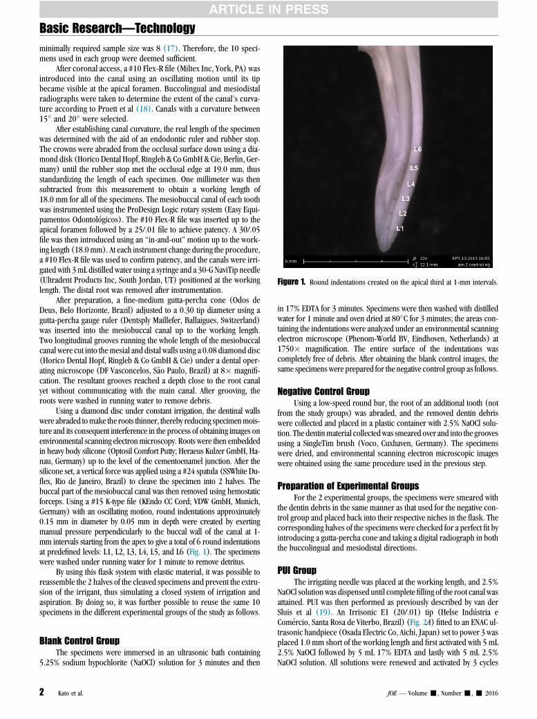
Using a diamond disc under constant irrigation, the dentinal wallswere abraded tomake the roots thinner, thereby reducing specimenmois-ture and its consequent interference in the process of obtaining images onenvironmental scanning electronmicroscopy. Roots were then embeddedin heavy body silicone (Optosil Comfort Putty; Heraeus Kulzer GmbH, Ha-nau, Germany) up to the level of the cementoenamel junction. After thesilicone set, a vertical force was applied using a #24 spatula (SSWhite Du-flex, Rio de Janeiro, Brazil) to cleave the specimen into 2 halves. Thebuccal part of the mesiobuccal canal was then removed using hemostaticforceps. Using a #15 K-type file (KEndo CC Cord; VDW GmbH, Munich,Germany) with an oscillating motion, round indentations approximately0.15 mm in diameter by 0.05 mm in depth were created by exertingmanual pressure perpendicularly to the buccal wall of the canal at 1-mm intervals starting from the apex to give a total of 6 round indentationsat predefined levels: L1, L2, L3, L4, L5, and L6 (Fig. 1). The specimenswere washed under running water for 1 minute to remove detritus.
By using this flask system with elastic material, it was possible toreassemble the 2 halves of the cleaved specimens and prevent the extru-sion of the irrigant, thus simulating a closed system of irrigation andaspiration. By doing so, it was further possible to reuse the same 10specimens in the different experimental groups of the study as follows.
Blank Control GroupThe specimens were immersed in an ultrasonic bath containing
5.25% sodium hypochlorite (NaOCl) solution for 3 minutes and then
in 17% EDTA for 3 minutes. Specimens were then washed with distilledwater for 1 minute and oven dried at 80�C for 3 minutes; the areas con-taining the indentations were analyzed under an environmental scanningelectron microscope (Phenom-World BV, Eindhoven, Netherlands) at1750� magnification. The entire surface of the indentations wascompletely free of debris. After obtaining the blank control images, thesame specimens were prepared for the negative control group as follows.
Negative Control GroupUsing a low-speed round bur, the root of an additional tooth (not
from the study groups) was abraded, and the removed dentin debriswere collected and placed in a plastic container with 2.5% NaOCl solu-tion. The dentinmaterial collectedwas smeared over and into the groovesusing a SingleTim brush (Voco, Cuxhaven, Germany). The specimenswere dried, and environmental scanning electron microscopic imageswere obtained using the same procedure used in the previous step.
Preparation of Experimental GroupsFor the 2 experimental groups, the specimens were smeared with
the dentin debris in the same manner as that used for the negative con-trol group and placed back into their respective niches in the flask. Thecorresponding halves of the specimens were checked for a perfect fit byintroducing a gutta-percha cone and taking a digital radiograph in boththe buccolingual and mesiodistal directions.
PUI GroupThe irrigating needle was placed at the working length, and 2.5%
NaOCl solution was dispensed until complete filling of the root canal wasattained. PUI was then performed as previously described by van derSluis et al (19). An Irrisonic E1 (20/.01) tip (Helse Ind�ustria eCom�ercio, Santa Rosa de Viterbo, Brazil) (Fig. 2A) fitted to an ENAC ul-trasonic handpiece (Osada Electric Co, Aichi, Japan) set to power 3 wasplaced 1.0 mm short of the working length and first activated with 5 mL2.5% NaOCl followed by 5 mL 17% EDTA and lastly with 5 mL 2.5%NaOCl solution. All solutions were renewed and activated by 3 cycles
Figure 1. Round indentations created on the apical third at 1-mm intervals.
Basic Research—Technology
2 Kato et al. JOE — Volume -, Number -, - 2016

of 20 seconds. Finally, the canals were flushed using a 30-G needle andsyringe containing 20 mL distilled water.
The specimens were then removed from their niches, processed,and analyzed adopting the same procedure used for the control groups.The second experimental group was prepared by resmearing the samespecimens with dentinal debris.
Reciprocating Activation Group Using ECEC (Fig. 2B–D) was introduced up to the working length and oper-
ated with a reciprocating motion of a 180� clockwise followed by a 90�
counterclockwise turn using an EasyEndo Motor (Easy EquipamentosOdontol�ogicos). The sequence of the solutions and irrigation timewere the same as those used for the PUI group and likewise for imageprocessing and obtaining procedures.
Assessment CriteriaThe images were saved in a digital file, analyzed, and classified us-
ing a 4-category scoring system adapted from Gambarini and Laszkie-wicz (20) as follows: score 1, open dentinal tubules, with no debris;score 2, open dentinal tubules, with debris covering less than 50% ofthe area; score 3, open dentinal tubules, with debris covering morethan 50% of the area; and score 4, dentinal tubules covered by debrisin 100% of the area examined.
Data AnalysisEach image obtained was coded according to the group (negative
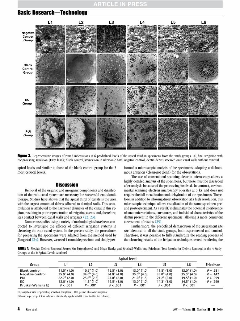
control, blank control, PUI group, or EC group), the tooth (from 1 to10), and the level at which the reading was taken (L1, L2, L3, L4, L5, orL6) (Fig. 3).
All of the images from the control and experimental groups for thesame level were loaded into the Microsoft Office PowerPoint application(Microsoft Corporation, Redmond, WA) and displayed in slide formaton an LCD monitor. Two independent examiners, previously calibratedand blind to the study, scored the images according to the assessmentcriteria outlined previously.
Statistical AnalysisThe level of interexaminer agreement was determined using the
kappa test. The Kruskal-Wallis test was used to compare data oncleansing efficacy. Multiple comparisons were performed using theDunn test when applicable. The Friedman test was applied to detect dif-ferences in cleansing promoted by each irrigation system at the differentapical levels examined.
All statistical calculations were performed using the SPSS 20 (SPSSInc, Chicago, IL) and BioEstat 5.0 (Fundac~ao Mamirau�a, Bel�em, PA,Brazil) software programs. The level of significance adopted was 5%.
ResultsThe level of interexaminer agreement was excellent, attaining a
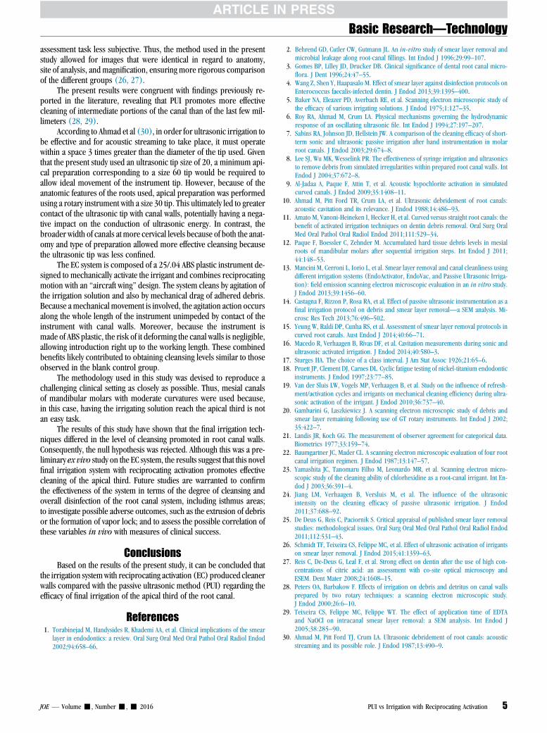
kappa value of 0.88 (21). The median debris removal scores for thespecimens from the study groups at the 6 apical levels examined anda statistical analysis comparing mean ranks and results of Kruskal-Wallis and Friedman tests are shown in Table 1.
A statistically significant difference was found between the irriga-tion techniques assessed in debris removal for the 3 most apical roundindentations (P < .05). No significant difference in debris removal wasfound between the reciprocating activation group and the blank controlgroup for all apical levels analyzed (P< .05). The PUI group had resultsstatistically similar to those of the negative control group for the 3 most
Figure 2. (A) Irrisonic tip; (B) EC tip, frontal view; (C) EC tip, lateral view; and (D) EC tip, cross-sectional view.
Basic Research—Technology
JOE — Volume -, Number -, - 2016 PUI vs Irrigation with Reciprocating Activation 3
apical levels and similar to those of the blank control group for the 3most cervical levels.
DiscussionRemoval of the organic and inorganic components and disinfec-
tion of the root canal system are necessary for successful endodontictherapy. Studies have shown that the apical third of canals is the areawith the largest amount of debris adhered to dentinal walls. This accu-mulation is attributed to the narrower diameter of the canal in this re-gion, resulting in poorer penetration of irrigating agents and, therefore,less contact between canal walls and irrigants (22, 23).
Numerous studies using a variety of methodologies have been con-ducted to investigate the efficacy of different irrigation systems incleansing the root canal system. In the present study, the proceduresfor preparing the specimens were adapted from the method used byJiang et al (24). However, we used 4 round depressions and simply per-
formed a microscopic analysis of the specimens, adopting a dichoto-mous criterion (clean/not clean) for the observations.
The use of conventional scanning electron microscopy allows ahighly detailed analysis of the specimens, but these must be discardedafter analysis because of the processing involved. In contrast, environ-mental scanning electron microscopy operates at 5 kV and does notrequire the full metallization and dehydration of the specimens. There-fore, in addition to allowing direct observation at a high resolution, thismicroscopic technique allows visualization of the same specimen pre-and postexperiment. As a result, it eliminates the potential interferenceof anatomic variations, curvatures, and individual characteristics of thedentin present in the different specimens, allowing a more consistentassessment of results (25).
Furthermore, the predefined demarcation of the assessment sitewas identical in all the study groups, both experimental and control.Therefore, it was possible to fully standardize the reading process ofthe cleansing results of the irrigation techniques tested, rendering the
Figure 3. Representative images of round indentations at 6 predefined levels of the apical third in specimens from the study groups. EC, final irrigation withreciprocating activation (EasyClean); blank control, immersion in ultrasonic bath; negative control, dentin debris smeared onto canal walls without removal.
TABLE 1. Median Debris Removal Scores (in Parentheses) and Mean Ranks and Kruskall-Wallis and Friedman Test Results for Debris Removal in the 4 StudyGroups at the 6 Apical Levels Analyzed
Group
Apical level
FriedmanL1 L2 L3 L4 L5 L6
Blank control 11.5a (1.0) 10.5a (1.0) 12.5a (1.0) 13.0a (1.0) 11.5a (1.0) 13.0a (1.0) P = .981Negative control 35.0b (4.0) 34.0b (4.0) 34.0b (4.0) 35.0b (4.0) 35.0b (4.0) 35.0b (4.0) P = .142PUI 22.7b (2.0) 25.8b (2.5) 23.0b (2.0) 21.0a (1.5) 21.2a (2.0) 19.5a (1.0) P > .999EC 12.8a (1.0) 11.8a (1.0) 12.5a (1.0) 13.0a (1.0) 14.3a (1.0) 14.5a (1.0) P > .999Kruskal-Wallis (a b) P < .001 P < .001 P < .001 P < .001 P < .001 P < .001 —
EC, irrigation with reciprocating activation (EasyClean); PUI, passive ultrasonic irrigation.
Different superscript letters indicate a statistically significant difference (within the column).
Basic Research—Technology
4 Kato et al. JOE — Volume -, Number -, - 2016
assessment task less subjective. Thus, the method used in the presentstudy allowed for images that were identical in regard to anatomy,site of analysis, and magnification, ensuring more rigorous comparisonof the different groups (26, 27).
The present results were congruent with findings previously re-ported in the literature, revealing that PUI promotes more effectivecleaning of intermediate portions of the canal than of the last few mil-limeters (28, 29).
According to Ahmad et al (30), in order for ultrasonic irrigation tobe effective and for acoustic streaming to take place, it must operatewithin a space 3 times greater than the diameter of the tip used. Giventhat the present study used an ultrasonic tip size of 20, a minimum api-cal preparation corresponding to a size 60 tip would be required toallow ideal movement of the instrument tip. However, because of theanatomic features of the roots used, apical preparation was performedusing a rotary instrument with a size 30 tip. This ultimately led to greatercontact of the ultrasonic tip with canal walls, potentially having a nega-tive impact on the conduction of ultrasonic energy. In contrast, thebroader width of canals at more cervical levels because of both the anat-omy and type of preparation allowed more effective cleansing becausethe ultrasonic tip was less confined.
The EC system is composed of a 25/.04 ABS plastic instrument de-signed to mechanically activate the irrigant and combines reciprocatingmotion with an ‘‘aircraft wing’’ design. The system cleans by agitation ofthe irrigation solution and also by mechanical drag of adhered debris.Because a mechanical movement is involved, the agitation action occursalong the whole length of the instrument unimpeded by contact of theinstrument with canal walls. Moreover, because the instrument ismade of ABS plastic, the risk of it deforming the canal walls is negligible,allowing introduction right up to the working length. These combinedbenefits likely contributed to obtaining cleansing levels similar to thoseobserved in the blank control group.
The methodology used in this study was devised to reproduce achallenging clinical setting as closely as possible. Thus, mesial canalsof mandibular molars with moderate curvatures were used because,in this case, having the irrigating solution reach the apical third is notan easy task.
The results of this study have shown that the final irrigation tech-niques differed in the level of cleansing promoted in root canal walls.Consequently, the null hypothesis was rejected. Although this was a pre-liminary ex vivo study on the EC system, the results suggest that this novelfinal irrigation system with reciprocating activation promotes effectivecleaning of the apical third. Future studies are warranted to confirmthe effectiveness of the system in terms of the degree of cleansing andoverall disinfection of the root canal system, including isthmus areas;to investigate possible adverse outcomes, such as the extrusion of debrisor the formation of vapor lock; and to assess the possible correlation ofthese variables in vivo with measures of clinical success.
ConclusionsBased on the results of the present study, it can be concluded that
the irrigation systemwith reciprocating activation (EC) produced cleanerwalls compared with the passive ultrasonic method (PUI) regarding theefficacy of final irrigation of the apical third of the root canal.
References1. Torabinejad M, Handysides R, Khademi AA, et al. Clinical implications of the smear
layer in endodontics: a review. Oral Surg Oral Med Oral Pathol Oral Radiol Endod2002;94:658–66.
2. Behrend GD, Cutler CW, Gutmann JL. An in-vitro study of smear layer removal andmicrobial leakage along root-canal fillings. Int Endod J 1996;29:99–107.
3. Gomes BP, Lilley JD, Drucker DB. Clinical significance of dental root canal micro-flora. J Dent 1996;24:47–55.
4. Wang Z, Shen Y, Haapasalo M. Effect of smear layer against disinfection protocols onEnterococcus faecalis-infected dentin. J Endod 2013;39:1395–400.
5. Baker NA, Eleazer PD, Averbach RE, et al. Scanning electron microscopic study ofthe efficacy of various irrigating solutions. J Endod 1975;1:127–35.
6. Roy RA, Ahmad M, Crum LA. Physical mechanisms governing the hydrodynamicresponse of an oscillating ultrasonic file. Int Endod J 1994;27:197–207.
7. Sabins RA, Johnson JD, Hellstein JW. A comparison of the cleaning efficacy of short-term sonic and ultrasonic passive irrigation after hand instrumentation in molarroot canals. J Endod 2003;29:674–8.
8. Lee SJ, Wu MK, Wesselink PR. The effectiveness of syringe irrigation and ultrasonicsto remove debris from simulated irregularities within prepared root canal walls. IntEndod J 2004;37:672–8.
9. Al-Jadaa A, Paque F, Attin T, et al. Acoustic hypochlorite activation in simulatedcurved canals. J Endod 2009;35:1408–11.
10. Ahmad M, Pitt Ford TR, Crum LA, et al. Ultrasonic debridement of root canals:acoustic cavitation and its relevance. J Endod 1988;14:486–93.
11. Amato M, Vanoni-Heineken I, Hecker H, et al. Curved versus straight root canals: thebenefit of activated irrigation techniques on dentin debris removal. Oral Surg OralMed Oral Pathol Oral Radiol Endod 2011;111:529–34.
12. Paque F, Boessler C, Zehnder M. Accumulated hard tissue debris levels in mesialroots of mandibular molars after sequential irrigation steps. Int Endod J 2011;44:148–53.
13. Mancini M, Cerroni L, Iorio L, et al. Smear layer removal and canal cleanliness usingdifferent irrigation systems (EndoActivator, EndoVac, and Passive Ultrasonic Irriga-tion): field emission scanning electron microscopic evaluation in an in vitro study.J Endod 2013;39:1456–60.
14. Castagna F, Rizzon P, Rosa RA, et al. Effect of passive ultrasonic instrumentation as afinal irrigation protocol on debris and smear layer removal—a SEM analysis. Mi-crosc Res Tech 2013;76:496–502.
15. Yeung W, Raldi DP, Cunha RS, et al. Assessment of smear layer removal protocols incurved root canals. Aust Endod J 2014;40:66–71.
16. Macedo R, Verhaagen B, Rivas DF, et al. Cavitation measurements during sonic andultrasonic activated irrigation. J Endod 2014;40:580–3.
17. Sturges HA. The choice of a class interval. J Am Stat Assoc 1926;21:65–6.18. Pruett JP, Clement DJ, Carnes DL. Cyclic fatigue testing of nickel-titanium endodontic
instruments. J Endod 1997;23:77–85.19. Van der Sluis LW, Vogels MP, Verhaagen B, et al. Study on the influence of refresh-
ment/activation cycles and irrigants on mechanical cleaning efficiency during ultra-sonic activation of the irrigant. J Endod 2010;36:737–40.
20. Gambarini G, Laszkiewicz J. A scanning electron microscopic study of debris andsmear layer remaining following use of GT rotary instruments. Int Endod J 2002;35:422–7.
21. Landis JR, Koch GG. The measurement of observer agreement for categorical data.Biometrics 1977;33:159–74.
22. Baumgartner JC, Mader CL. A scanning electron microscopic evaluation of four rootcanal irrigation regimen. J Endod 1987;13:147–57.
23. Yamashita JC, Tanomaru Filho M, Leonardo MR, et al. Scanning electron micro-scopic study of the cleaning ability of chlorhexidine as a root-canal irrigant. Int En-dod J 2003;36:391–4.
24. Jiang LM, Verhaagen B, Versluis M, et al. The influence of the ultrasonicintensity on the cleaning efficacy of passive ultrasonic irrigation. J Endod2011;37:688–92.
25. De Deus G, Reis C, Paciornik S. Critical appraisal of published smear layer removalstudies: methodological issues. Oral Surg Oral Med Oral Pathol Oral Radiol Endod2011;112:531–43.
26. Schmidt TF, Teixeira CS, Felippe MC, et al. Effect of ultrasonic activation of irrigantson smear layer removal. J Endod 2015;41:1359–63.
27. Reis C, De-Deus G, Leal F, et al. Strong effect on dentin after the use of high con-centrations of citric acid: an assessment with co-site optical microscopy andESEM. Dent Mater 2008;24:1608–15.
28. Peters OA, Barbakow F. Effects of irrigation on debris and detritus on canal wallsprepared by two rotary techniques: a scanning electron microscopic study.J Endod 2000;26:6–10.
29. Teixeira CS, Felippe MC, Felippe WT. The effect of application time of EDTAand NaOCl on intracanal smear layer removal: a SEM analysis. Int Endod J2005;38:285–90.
30. Ahmad M, Pitt Ford TJ, Crum LA. Ultrasonic debridement of root canals: acousticstreaming and its possible role. J Endod 1987;13:490–9.
Basic Research—Technology
JOE — Volume -, Number -, - 2016 PUI vs Irrigation with Reciprocating Activation 5