investigation of estuarine sediment as a … · investigation of estuarine sediment as a reservoir...
TRANSCRIPT

INVESTIGATION OF ESTUARINE SEDIMENT AS A RESERVOIR FOR SEWAGE ASSOCIATED BACTERIA
A Final Report of the Tibor T. Polgar Fellowship Program
Erin Schneider Polgar Fellow
School for Earth and Environmental Studies CUNY Queens College
Queens, NY 11367
Project Advisor: Dr. Gregory D. O’Mullan
School for Earth and Environmental Studies Queens College, City University of New York
Queens, NY 11367 Schneider, E. and G. D. O’Mullan. 2013. Investigation of estuarine sediment as a reservoir for sewage associated bacteria. Section II: 1-22 pp. In S.H. Fernald, D.J. Yozzo and H. Andreyko (eds.), Final Reports of the Tibor T. Polgar Fellowship Program 2012. Hudson River Foundation.
II-1
ABSTRACT
The Fecal Indicator Bacteria (FIB), Escherichia. coli and Enterococci, are
commonly used by Hudson River monitoring programs to quantify the level of sewage
pollution in estuarine water and to provide information about the health risk to recreators
from sewage associated bacteria. It is generally assumed that these FIB do not persist in
the water for extended periods of time and that their presence represents a recent release
of sewage into the environment. However, much less is known about the abundance and
environmental persistence of FIB in sediments, as opposed to the water column, of the
Hudson River Estuary. In this study, FIB were quantified, using cultivation-based
techniques, in water and sediment samples collected from six locations in the estuary, and
the persistence of FIB in sediment was investigated in laboratory incubation experiments.
FIB were found to be widely distributed in both sediment and water from the estuary. E.
coli and Enterococci displayed correlated abundances in sediment, consistent with
sewage pollution as a shared source for both FIB in the environment. However, the
levels of FIB were not correlated in paired water and sediment samples collected
simultaneously from the same sites, suggesting that environmental persistence of these
FIB differs in water versus sediment. Enterococci concentrations were found to decrease
over time in laboratory incubations of estuarine sediment but remained at detectable
levels for weeks after collection. In order to confirm the presence of Enterococci, and
rule out the possibility of false positives from the cultivation-based assay, isolated
bacterial colonies were characterized using molecular genetic techniques and the vast
majority (96%) were confirmed as Enterococci.
II-2

TABLE OF CONTENTS
Abstract ................................................................................................................ II-2
Table of Contents ................................................................................................. II-3
Lists of Figures and Tables .................................................................................. II-4
Introduction .......................................................................................................... II-5
Methods................................................................................................................ II-8
Results .................................................................................................................. II-12
Discussion ............................................................................................................ II-16
Conclusions .......................................................................................................... II-18
Acknowledgements .............................................................................................. II-18
References ............................................................................................................ II-19
II-3
LIST OF FIGURES AND TABLES
Figure 1 – Map of Sampling Sites ....................................................................... II-9
Figure 2 – Enterococci Concentrations in Water and Sediment .......................... II-13
Figure 3 – Enterococci and E. coli Correlation in Sediment ............................... II-14
Figure 4 – Enterococci in Paired Water and Sediment ........................................ II-14
Figure 5 – Sediment FIB Decay Curves .............................................................. II-15
Table 1 – FIB samples processed from the Hudson River Estuary ..................... II-13
II-4

INTRODUCTION
Fifty years ago, the lower Hudson River and connected waterways surrounding
New York City were commonly considered to be inappropriate for any recreational
contact due to extensive pollution, with the river often acting as an open sewer for the
surrounding population. Enforcement of the Clean Water Act and major investment in
wastewater treatment facilities since the 1970s initiated a trend of improving water
quality (Steinberg et al. 2004; NYC DEP 2009). Long-term environmental monitoring
data from New York Harbor clearly document the prior history of poor water quality and
the resulting improvements in mean seasonal water quality in recent decades (Brosnan
and O’Shea 1996; Hetling et al. 2003; Brosnan et al. 2006; NYC DEP 2009). Along with
improvements in water quality, there has been a widespread increase in recreational use
of the Hudson at official and “unofficial” swimming beaches (Lawler, Matusky, and
Skelley Engineers 2005), and management action aimed at re-claiming the waterfront and
increasing public access to the river (New York-New Jersey Harbor and Estuary Program
2013; City of New York 2013).
Despite improvement in wastewater treatment infrastructure and in seasonal water
quality, raw and partially treated sewage continues to enter the river and continues to be a
management challenge. It is estimated that approximately 27 billion gallons of
stormwater, mixed with raw sewage, are still released each year into New York Harbor
through Combined Sewer Overflow (CSO) events (NYC DEP 2010). Numerous studies
from other aquatic systems have demonstrated that waterways contaminated with sewage
contain potentially pathogenic strains of microbes such as Salmonella, Campylobacter,
and Escherichia coli 0157:H7 (Obiri–Danso and Jones 2000; Walters et al. 2007).
II-5
Similarly, a recent study in the Hudson River Estuary found that the abundance of
antibiotic resistant microbes was correlated to the concentration of Fecal Indicator
Bacteria (FIB) and that levels of these bacteria increased following rainfall (Young et al.
2013), presumably due to sewage discharge from CSOs.
Increased public access to the waterfront and improved mean seasonal water
quality has led to a widespread demand from the public for more detailed water quality
data. Enterococcus is an Environmental Protection Agency (EPA) approved FIB used for
recreational water quality management whose presence in water has been shown to
correlate with the occurrence of gastrointestinal illness in recreators (US-EPA 2004).
Multiple monitoring programs in the lower Hudson River (NYC DEP 2013; New York
Water Trails Association 2013; Riverkeeper 2013) now collect data on the concentration
of FIB, and distribute these data to the public. Enterococci are commonly detected in the
Hudson River Estuary (HRE) at elevated levels, with 21% of water samples (from 75
locations in the lower HRE) tested from 2006 through 2010 deemed unacceptable by
EPA standards for primary contact recreation (Riverkeeper 2011).
Microorganisms released into the coastal environment are subjected to numerous
stressors such as temperature change (Davies et al. 1995; Thomas et al. 1998), salinity
(Mezrioui et al. 1995), nutrient deficiencies (Ozkanca and Flint 1997), and sunlight
(Sinton et al. 1999). As a result, FIB and other sewage-associated bacteria are generally
not thought to live for very long in the water column. In fact, short environmental
persistence is a desired characteristic of FIB, because their detection is intended to reflect
the recent input of sewage to the environment. However, in the HRE water column a
high percentage of Enterococci are attached to particles, (Suter et al. 2011) and settle out
II-6

of the water column to the underlying sediment more quickly than “free-living” bacteria.
Environmental conditions in the sediment are quite different than in the water column,
including reduced sunlight, protect against predators, increased nutrient and organic
carbon availability, and increased colonizable surfaces (Brettar and Holfe 1992; Davies et
al. 1995; Blumenroth and Wagner-Dobler 1998; Sinton et al. 1999). In combination,
sediment conditions may lead to increased environmental persistence for FIB, as
compared to the water column (Lee et al. 2006).
For the past two years, data on the persistence of sediment associated Enterococci
and E. coli in the Hudson River has been collected and analyzed in the O’Mullan
laboratory at Queens College and the Juhl laboratory at Columbia University. Sediment
samples collected from the environment in areas impacted by sewage pollution and
incubated under a range of temperatures in the laboratory suggest that cultured FIB can
remain at detectable levels for weeks to months in Hudson sediment (O’Mullan and Juhl,
unpublished data).
Although local monitoring programs typically only measure FIB in water,
recreators may also come into contact with contaminated sediment. In addition, the
turbulence from large storm events and disturbance from boats or recreators may
reintroduce sediment FIB, and associated pathogens, back into the water column creating
a connection between water quality and sediment quality. Therefore, studying the
persistence patterns of FIB in the all compartments of the environment, including
sediment, is imperative to understanding the ecology of sewage associated bacteria in the
environment and to interpreting water quality patterns from local monitoring programs.
II-7

The goals of this study were to: 1) quantify FIB abundance in sediment and water
from six locations in the estuary, using cultivation-based approaches; 2) to determine if
FIB abundance would be correlated in paired water and sediment samples; and 3) to use
DNA-based assays to confirm that FIB enumerated, with cultivation-based methods, from
sediment in laboratory persistence experiments were correctly identified as Enterococci.
The hypothesis was tested that FIB would be widely distributed in sediment samples
from the estuary, but that their abundance would not be well correlated in paired water
and sediment samples due to longer FIB persistence in sediment relative to water. In
addition, the hypothesis was tested that the majority of isolated colonies characterized
using DNA based assays would be confirmed as Enterococci and that FIB cultivation-
based methods applied to sediment would not be prone to false positives.
METHODS
Paired Water and Sediment Field Sampling
Water and sediment samples for FIB analyses were collected from six field
locations in Flushing Bay, Sparkill Creek, and the lower Hudson River (Figure 1)
between late May and mid-July in 2012. Samples were collected four to six times from
each of the six field sites. Approximately 40 ml of surface water was collected from
along the shoreline (depth of less than 0.3 m), just above the paired sediment sample that
was also collected (see below), into sterile 50ml plastic tubes that had been triple rinsed
with water from the environment. A surface sediment core (approximately 20 ml from
the top 3 cm of sediment) was collected using a modified sterile 60ml syringe barrel as a
coring device, along with a metal putty knife to help retain the core in the syringe barrel
II-8

during collection. The sediment was then extruded into a sterile 50ml tube for storage
and transport to the laboratory. Both water and sediment sampling tubes were placed into
a cooler, away from light, and transported to the laboratory for processing within six
hours of collection.
Figure 1. Map of sampling sites. Tappan Zee region stations: 1- Stony Point, 2- Piermont Pier, 3- Sparkill Creek; Flushing Bay region stations: 4- Flushing Bay Boat Launch, 5- Flushing Bay Marina, 6- Flushing River-Corona Park.
Laboratory Persistence Experiments with Sediment
Bulk samples of sediment, scraped from the top 3 cm using a cleaned trowel, were
removed from the estuary and transferred to clean plastic incubation chambers
(approximately 20cm long x 10cm wide x 10cm tall). Two samples were collected in late
May 2012; one from a muddy, organic rich, site near the public boat launch in Flushing
Bay, and one from a sandy site also in Flushing Bay but closer to Flushing Bay Marina
II-9

(Figure 1). A third sample was collected, also from the sandy Flushing Bay Marina site
but in late June 2012. All samples were immediately transported to the laboratory and
incubated at 4oC in the dark for approximately five weeks. This incubation temperature
was selected based upon prior research (O’Mullan and Juhl, unpublished) to allow for
weeks of FIB persistence in order to test for false positives after an extended incubation.
Sediment samples were collected from each incubation chamber periodically over
the five weeks to quantify the persistence of FIB in the sediment sample, using the same
procedures for Enterococcus, as described below, for field sediment samples. It is worth
noting that other samples from the O’Mullan and Juhl labs have been used to more
completely characterize persistence rates of Enterococci in laboratory incubations using a
variety of conditions (e.g., variable temperature). The purpose of the persistence
incubations was to obtain cultured isolates of Enterococci that could be processed for
DNA based identification using the 16S rRNA genes, to confirm that cultivation based
approaches were actually quantifying Enterococci and were not prone to false positive
results. The rates of decay are not reported quantitatively in this report, as this was not a
goal of the study and is best estimated with a more complete data set spanning more
persistence experiments.
FIB Enumeration Procedures
Microbes were extracted from sediment samples, using a method modified from
Van Elsas et al. (2002), by mixing 10ml of sediment with 100ml of extraction buffer
containing 0.1% sodium pyrophosphate and 0.1 mM EDTA in a sterile, sealed 500ml
container shaken at 200 rpm for 30 minutes. Mass of replicate sediment samples was
II-10

recorded before and after drying at 60oC for two days to determine wet and dry sediment
mass and to allow normalization of microbial counts per gram dry weight of sediment.
After extracting sediment samples, microbial processing of sediment and water
samples for FIB was identical, although only Enterococci was measured from water,
while both Enterococci and E. coli were measured from sediment. Enterococci and E.
coli were enumerated using the IDEXX Enterolert and Colilert methodology
(www.Idexx.com). A 10% dilution of sample water in sterile water and growth media
was sealed into a quanti-tray 2000 (IDEXX) vessel and incubating at 41oC
(Enterococcus) and 37oC (E. coli) for 24 hours. After incubation, samples were exposed
to UV light and the Most Probably Number (MPN) of Enterococci or E. coli cells was
calculated per 100 ml (for water samples) or per gram of dry sediment weight (for
sediment). In addition, a subset of samples were also processed using the EPA approved
membrane filtration technique (US-EPA 2007) so that the isolated colonies could be used
for DNA based taxonomic identification (described below) to confirm that the cultivation
based technique was not prone to false positive results.
DNA Characterization of Sediment FIB and Statistical Analyses
Isolated Enterococci colonies from membrane filtration based enumeration of
laboratory persistence samples were picked off petri dishes using sterile pipette tips and
transferred into tubes with 40 µl of sterile water for molecular analysis. Colonies were
then heated to 95oC for 5 minutes using an Eppendorf mastercycler to lysis cells and the
16S rRNA gene was amplified from the released DNA using universal primers 8F and
1492R, followed by gene sequencing by SeqWright Inc. using the conditions described
II-11

by Young et al. (2013). The resulting gene sequences were taxonomically classified
using the Ribosomal Database Project (RDP; http://rdp.cme.msu.edu/) and searched
against the Genbank database (www.ncbi.nlm.nih.gov/genbank/) to confirm species
identification. Non-parametric tests, including the Spearman’s coefficient, were
preformed using the GraphPad Prism (Version 4C, May 2005) statistical analysis
software.
RESULTS
Paired Water and Sediment Field Sampling
Enterococci were detected in both water and sediment at all of the six sampling
sites (Table 1). Only one water sample, out of 30 total samples enumerated, was found to
be below detection for Enterococci (Stony Point, MPN <10/100ml). All 30 sediment
samples were found to have detectable levels of FIB. Stony Point, in the Tappan Zee,
had by far the lowest water and sediment FIB concentrations compared to all other sites
(Table 1, Figure 2) with a maximum Enterococci MPN of only 113/100ml in the water,
more than four times lower than any other site; and a maximum Enterococci MPN of
only 19.7/g in sediment, more than an order of magnitude lower than any other site.
Levels of Enterococci and E. coli, the two common FIB used in water quality
monitoring programs, were positively correlated (Spearman r = 0.622; p < 0.001) when
sediment samples were compared among all sites (Figure 3). In contrast, levels of
Enterococci in paired water and sediment samples, collected at the same site and at the
same time, were not correlated (Spearman r = 0.124; p = 0.515) (Figure 4).
II-12

Site # of samples
Enterococci water MPN/100ml
Enterococci sediment MPN/gram dry wt.
E. coli sediment MPN/gram dry wt
Minimum maximum minimum maximum minimum maximum 1) Stony Point 4 <10
113 5 20 2 12
2) Piermont Pier 4 63 471 46 3788 274 3170
3) Sparkill Creek 4 121 >24196
12
2377 188 2378
4) Flushing Bay Boat Launch, muddy
6 10 >24196
134 4327 484 4327
5) Flushing Bay Marina, sandy
6 20 >24196
97
2866 143 2296
6) Flushing River- Corona Park
6 20 >24196
76
2818 112 2818
Table 1. FIB samples processed from the Hudson River Estuary.
Figure 2. Enterococci Concentrations in Water and Sediment. A) water concentration and B) sediment concentration from the six spatial sampling sites. Stony Point, in the Tappan Zee, had the lowest concentrations of Enterococci for both water and sediment.
II-13

Figure 3. Enterococci and E. coli Correlation in Sediment. Concentrations of Enterococci and E. coli measured from the same sediment samples were found to have a significant positive correlation.
Figure 4. Enterococci in Paired Water and Sediment. Enterococci concentrations in paired water and sediment samples were not significantly correlated.
II-14
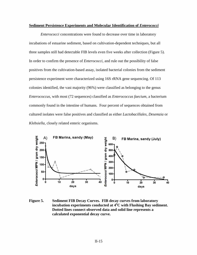
Sediment Persistence Experiments and Molecular Identification of Enterococci
Enterococci concentrations were found to decrease over time in laboratory
incubations of estuarine sediment, based on cultivation-dependent techniques, but all
three samples still had detectable FIB levels even five weeks after collection (Figure 5).
In order to confirm the presence of Enterococci, and rule out the possibility of false
positives from the cultivation-based assay, isolated bacterial colonies from the sediment
persistence experiment were characterized using 16S rRNA gene sequencing. Of 113
colonies identified, the vast majority (96%) were classified as belonging to the genus
Enterococcus, with most (72 sequences) classified as Enterococcus faecium, a bacterium
commonly found in the intestine of humans. Four percent of sequences obtained from
cultured isolates were false positives and classified as either Lactobacillales, Desemzia or
Klebsiella, closely related enteric organisms.
Figure 5. Sediment FIB Decay Curves. FIB decay curves from laboratory incubation experiments conducted at 4oC with Flushing Bay sediment. Dotted lines connect observed data and solid line represents a calculated exponential decay curve.
II-15
DISCUSSION
Paired Water and Sediment Field Sampling
Despite the improving water quality of Hudson River, FIB were widely
distributed, detected in both water and sediment at every sampling site and within every
individual sediment sample analyzed (Table 1, and Figure 2). These data suggest that
sediment in the HRE act as a significant reservoir for FIB, as has been found in other
similar systems (Anderson et al. 2005; Boehm et al. 2005; Bonilla et al. 2007). The
correlated abundances of Enterococci and E. coli in sediment samples provides added
confidence in the use of each indicator and would indicate that both FIB share a common
delivery mechanism (Nobel et al. 2003), consistent with sewage as a source for both FIB
to the Hudson sediment. Given the correlations of FIB to known pathogens (e.g. Walters
et al. 2007), recreator illness (e.g. Haile et al. 1999), and antibiotic resistant bacteria
(Young et al. 2013) from prior studies, the current FIB results are also strong evidence
that other microbial agents of concern, beyond the FIB themselves, are likely to be
widely distributed in Hudson sediment.
Recreators could be exposed to sewage-associated pathogens residing in sediment
through activities along the shoreline, for example wading (Phillip et al. 2009), when
sediment is directly contacted, but also through other activities when sediment becomes
resuspended into the water column. Boat traffic (Pettibone et al. 1996), wave-shore
interactions (LeFevre and Lewis 2003), high flow conditions in rivers and estuaries
(Jamieson et al. 2005; Wilkinson et al. 2006), and even high winds (Roslev et al. 2008)
can cause sediment re-suspension events that may negatively impact water quality. Some
II-16
water quality models are now attempting to incorporate FIB resuspension as a central
factor controlling water quality (Liu et al. 2006).
Sediment Persistence Experiments and Molecular Identification of Enterococci
FIB in Hudson sediments from this study were found to persist for more than five
weeks at detectable levels (Figure 5), a similar time scale found in some other aquatic
systems (e.g. Haller et al. 2009). Extended persistence is not only expected in sediments,
but there is also some evidence that particle-associated microbes in the water column
may persist for longer periods than free-living FIB (Fries et al. 2008). This may be
significant in the Hudson, where a high percentage of FIB are found to be particle
associated (Suter et al. 2011).
A recent review (Jamieson et al. 2005) of water quality modeling studies
identified gaps in the understanding of FIB and enteric ecological behavior within the
environment as a significant obstacle to the generation of improved prediction systems.
Variable persistence in high versus low nutrient environments, the significance of particle
attachment for transport, and interactions between sediment and water column associated
microbes are all important areas of continued research toward the goal of next generation
water quality models (US-EPA 2007; Surbeck 2009; Kim et al. 2010).
Finally, the molecular genetic characterization of FIB isolates suggest that the
vast majority (96%) of isolates obtained using cultivation based approaches were
Enterococci. This finding is significant because it supports the use of cultivation-based
approaches to quantify FIB in estuarine sediments and it confirms the long persistence of
FIB in estuarine sediment, as suggested by cultivation based approaches.
II-17

CONCLUSION
The results from this study demonstrated that FIB are widely distributed in
Hudson River sediment and appear to act as a reservoir for sewage associated pathogens.
The microbes within this reservoir can persist for weeks, complicating the interpretation
of FIB monitoring data. The high FIB content in sediments and attached to particles
suggestions that additional research is required to understand the ecology of FIB in the
Hudson and to allow improved approaches to water quality monitoring and modeling.
Finally, the molecular genetic results from this study confirm that cultivation based
approaches can be confidently used to enumerate FIB from sediments, supporting this
method for studying FIB ecology in the environment.
ACKNOWLEDGEMENTS
We would like to thank Andrew Juhl, Suzanne Young, Roman Reichert, and Eli
Deuker for their assistance, and the Hudson River Foundation Tibor T. Polgar Fellowship
for funding for this project.
II-18

REFERENCES
Anderson, K.L., J.E. Whitlock, and V.J. Harwood. 2005. Persistence and differential survival of fecal indicator bacteria in subtropical waters and sediments. Applied and Environmental Microbiology 71:3041-3048.
Blumenroth, P., and I. Wagner-Dobler. 1998. Survival of inoculants in polluted
sediments: effect of strain origin and carbon source competition. Microbial Ecology 35:279-288.
Boehm, A.B., D.P. Keymer, and G.G. Shellenbarger. 2005. An analytical model of enterococci inactivation, grazing, and transport in the surf zone of a marine beach. Water Research 39:3565-3578.
Bonilla, T.D., K. Nowolsielski, M. Cuvelier, A. Hartz, M. Green, N. Esiobu, D.S.
McCorquodale, J.M. Fleisher, and A. Rogerson. 2007. Prevalence and distribution of fecal indicator organisms in South Florida beach sand and preliminary assessment of health effects associated with beach sand exposure. Marine Pollution Bulletin 54:1472-1482.
Brettar, I., and M.G. Holfe. 1992. Influence of ecosystematic factors on survival of
Escherichia coli after large-scale release into lake water mesocosms. Applied and Environmental Microbiology 58:2201-2210.
Brosnan, T.M., and M.L. O’Shea. 1996. Long-term improvements in water quality due to sewage abatement in the lower Hudson River. Estuaries 19:890-900.
Brosnan, T.M., A. Stoddard, and L.J. Hetling. 2006. Hudson River sewage inputs and
impacts: past and present. pp. 335-348 in Levinton, J. S., and J.R. Waldman (Eds.), The Hudson River Estuary. Cambridge University Press, New York.
City of New York. 2013. PLANYC 2030 www.nyc.gov/planyc Davies, C.M., J.A. Long, M. Donald, and N.J. Ashbolt. 1995. Survival of fecal
microorganisms in marine and freshwater sediments. Applied and Environmental Microbiology 61:1888-1896.
Fries J.S., G.W. Characklis, and R.T. Noble. 2008. Sediment-water exchange of Vibrio
sp. and fecal indicator bacteria: Implications for persistence and transport in the Neuse River Estuary, North Carolina, USA. Water Research 42:941-950.
Haile, R.W., J.S. Witte, M. Gold, R. Cressey, C. McGee, R.C. Millikan, A. Glasser, N.
Harawa, C. Ervin, P. Harmon, J. Harper, J. Dermand, J. Alamillo, K. Barrett, M. Nides, and G. Wang. 1999. The health effects of swimming in ocean water contaminated by storm drain runoff. Epidemiology 10:355–363.
II-19

Haller, L., E. Amedegnato, J. Pote, and W. Wildi. 2009. Influence of freshwater sediment characteristics on persistence of fecal indicator bacteria. Water, Air and Soil Pollution 203:217-227.
Hetling, L.J., A. Stoddard, T.M. Brosnan, D.A. Hammerman, and T.M. Norris. 2003.
Effect of water quality management efforts on wastewater loadings during the past century. Water Environment Research 75:30-38.
Jamieson R.C., D.M. Joy, H. Lee, R. Kostaschuk, and R.J. Gordon. 2005. Resuspension
of sediment-associated Escherichia coli in a natural stream. Journal of Environmental Quality 34:581-589.
Kim J.-W., Y.A. Pachepsky, D.R. Shelton, and C. Coppock. 2010. Effect of streambed
bacteria release on E. coli concentrations: Monitoring and modeling with the modified SWAT. Ecological Modeling 221:1592-1604.
Lawler, Matusky, and Skelley Engineers. 2005. Swimming in the Hudson River,
Feasibility Report on Potential Sites. www.dec.ny.gov/lands/5452.htm Lee, C.M., T.Y. Lin, C.C. Lin, G.A. Kohbodi, A. Bhatt, R. Lee, and J.A. Jay. 2006.
Persistence of fecal indicator bacteria in Santa Monica Bay beach sediments. Water Research 40:2593-2602.
LeFevre, N.M., and G.D. Lewis. 2003. The role of resuspension in Enterococci
distribution in water at an urban beach. Water Science and Technology 47:205-210.
Liu, L., M.S. Phanikumar, S.L. Molloy, R.L. Whitman, D.A. Shivley, M.B. Nevers, D.J.
Schwab, and J.B. Rose. 2006. Modeling the transport and inactivation of E. coli and Enterococci in the near-shore region of Lake Michigan. Environmental Science and Technology 40:5022-5028.
Mezrioui, N., B. Baleux, and M. Troussellier. 1995. A microcosm study of the survival of
Escherichia coli and Salmonella typhimurium in brackish water. Water Research 29: 459-465.
Nobel, R.T., D.F. Moore, M.K. Leecaster, C.D. McGee, and S.B. Weisberg. 2003.
Comparison of total coliform, fecal coliform, and Enterococcus bacterial indicator response for ocean recreational water quality testing. Water Research 37:1637 – 1643.
New York-New Jersey Harbor and Estuary Program. 2013.
www.harborestuary.org/publicaccess.htm New York Water Trails Association. 2013. 2013 Citizens’ Water Quality Testing
Program. http://www.nycwatertrail.org/water_quality.html
II-20

NYC DEP. 2009. New York Harbor Survey Program, celebrating one hundred years,
1909-2009. www.nyc.gov/html/dep/pdf/hwqs_centennial.pdf NYC DEP. 2010. NYC Green Infrastructure Plan: A Sustainable Strategy For Clean
Waterways.” New York City Department of Environmental Protection, New York, NY, 2010 p. 8.
NYC DEP. 2013. New York City Department of Environmental Protection Harbor
Water Sampling Data. http://www.nyc.gov/html/dep/html/harborwater/ harbor_water_sampling_results.shtml
Obiri-Danso, K., and K. Jones. 2000. Intertidal sediments as reservoirs for hippurate
negative campylobacters, Salmonellae and fecal indicators in three EU recognised bathing waters in north west England. Water Research 34:519-527.
Ozkanca, R., and K.P. Flint. 1997. Relationship between respiratory enzymes and
survival of Escherichia coli under starvation stress in lake water. Journal of Applied Microbiology. 82:301-309.
Pettibone, G.W., K.N. Irvine, and K.M. Monahan. 1996. Impact of ship passage on
bacteria levels and suspended sediment characteristics in the Buffalo River, New York. Water Research 30:2517-2521.
Phillip, D.A., P. Antoine, V. Cooper, L. Francis, E. Mangal, N. Seepersad, R. Ragoo, S.
Ramsaran, I. Singh, and A. Ramsubhag. 2009. Impact of recreation on recreational water quality of a small tropical stream. Journal of Environmental Monitoring 11: 1192-1198
Riverkeeper. 2011. How is the Water? 2012. Sewage Contamination in the Hudson River
Estuary. http://www.riverkeeper.org/wp-content/uploads/2012/12/RvK_How-Is-the-Water-2012.pdf
Riverkeeper. 2013. Hudson River Water Quality. http://www.riverkeeper.org/water-
quality/ Roslev, P., S. Bastholm, and N. Iversen. 2008. Relationship between fecal indicators in
sediment and recreational waters in a Danish estuary. Water, Air, and Soil Pollution 194:13-21.
Sinton, L.W., R.K. Finaly, and P.A. Lynch. 1999. Sunlight inactivation of fecal
bacteriophages and bacteria in sewage-polluted seawater. Applied and Environmental Microbiology. 65:3605-3613.
II-21

Steinberg N., D.J. Suszkowski, L. Clark, and J. Way. 2004. Health of the Harbor: The first comprehensive look at the state of the NY/NJ Harbor Estuary: A report to the NY/NJ Harbor Estuary Program. Hudson River Foundation, New York, NY. 82 pp.
Surbeck, C.Q. 2009. Factors influencing the challenges of modeling and treating fecal
indicator bacteria in surface waters. Ecohydrology 2:399-403. Suter, E., A.R. Juhl, and G.D. O’Mullan. 2011. Particle association of Enterococcus and
total bacteria in the lower Hudson River Estuary, USA. Journal of Water Resource and Protection. 3:715-725.
Thomas, C., H. Gibson, D.J. Hill, and M. Mabey. 1998. Campylobacter epidemiology: an
aquatic perspective. Journal of Applied Microbiology 85:168S-177S. U.S. Environmental Protection Agency (U.S. EPA). 2004. Method 1600: Enterococci in
water by membrane filtration using membrane-Enterococcus Indoxyl-B-D Glucoside Agar (mEI). EPA-821-R-06-009.
U.S. Environmental Protection Agency (U.S. EPA). 2007. Report of the experts scientific
workshop on critical research needs for the development of new or revised recreational water quality criteria. EPA 823-R-07-006.
Van Elsas J.D., K. Smalla, A.K. Lilley, and M.J. Bailey. 2002. Methods for sampling soil
microbes. pp. 505-515 in Hurst, C.J., R.L. Crawford, G.R. Knudsen, M.J. McInerney, and L.D. Stetzenbach (Eds), Manual of Environmental Microbiology 2nd Ed. ASM Press, Washington, D.C.
Walters S.P., V.P. Gannon, and K.G. Field. 2007. Detection of Bacteroidales fecal
indicators and the zoonotic pathogens E. coli 0157:H7, Salmonella, and Campylobacter in river water. Environmental Science and Technology 41:1856-1862.
Wilkinson J., D. Kay, M. Wyer, and A. Jenkins. 2006. Processes driving the episodic flux
of faecal indicator organisms in streams impacting on recreational and shellfish harvesting waters. Water Research 40:153-161.
Young, S., A. Juhl, and G.D. O’Mullan. 2013. Antibiotic-resistant bacteria in the Hudson
River Estuary linked to wet weather sewage contamination. Journal of Water and Health 11:297-310.
II-22