introduction & review of literatureshodhganga.inflibnet.ac.in/bitstream/10603/31614/8/08_chapter...
TRANSCRIPT

CChhaapptteerr 11 IInnttrroodduuccttiioonn && RReevviieeww ooff
LLiitteerraattuurree

Chapter 1: Introduction and Review of Literature
Ph.D. Thesis 2013 University of Delhi 1
CHAPTER 1
INTRODUCTION AND REVIEW OF LITERATURE 1.1 INTRODUCTION Diabetes mellitus (DM) is syndrome characterized by high levels of blood glucose resulting from defect in production of insulin or due to ineffectiveness of the insulin produced (Bloomgarden and Einhorn, 2012). This results in increased concentration of glucose in blood, which may lead to polyurea, polyphagia, thirst and weight loss. Diabetes is progressive disease and is associated with many complications like neuropathy, retinopathy, nephropathy and cardiovascular disease. It is one of the most common metabolic disorders. The global prevalence of diabetes in 2010 was 284 million people worldwide constituting around 6.4% of the world’s population, which is higher than was projected in earlier studies. Furthermore, the projections for 2030 show the prevalence to reach 439 million, comprising ~7.7% of the world population (Farag and Gaballa, 2011). Until 1995 insulin and sulphonylureas were the only treatment available for diabetes. In recent times the numbers of drugs for treatment of diabetes has increased substantially viz. metformin, thiazolidinediones, α-glucosidase inhibitors, repaglinide etc. There are many side effects associated with prolonged use of insulin and other hypoglycemic drugs (Watkins,2003). There is an increased demand for natural especially herbal drugs for treatment of diabetes. Plant drugs are frequently considered less toxic and relatively free from side effects. (Shukla et al, 2000; Grover et al, 2002)
Plants have been widely used for the treatment of diabetes mellitus in various traditional systems of medicine, worldwide. Ayurveda and other Indian traditional approaches have described more than 800 plants, found in Indian subcontinent to possess anti-diabetic potential. (Rawi et al, 2011). Only a few of them have been scientifically evaluated for their antidiabetic potential. Work has been carried out in our laboratory to assess antidiabetic potential and isolate antidiabetic component from medicinal plants like Ficus bangalensis, Trigonella foenum greacum, Eugenia jambolana, Withania caogulans, Cassia auriculata,. (Shukla et al, 2004; Puri et al, 2002;Sharma et al, 2011;Shukla et al, 2012; Gupta et al, 2010)

Chapter 1: Introduction and Review of Literature
Ph.D. Thesis 2013 University of Delhi 2
The plant of interest in this study is Musa sapientum linn. It is commonly known as kela in Hindi, banana in English & kadali in Tamil. It is used in Indian folk medicine for treatment of diabetes. Musa sapientum is a herbaceous perennial plant of the Musacae family that grow in 5-9 mt. in height. It has tuberous sub tarranean rhizome from which the leaves emerge. The lower part of the leaves is folded with in each other producing false stem from which long narrow blades protrude &spread out. In the centre of the folded leaf sheath,a growing point form the tip of rhizome grows up stem and emerges as an over hanging inflorescence with a succession of reddish brown bracts. Different part of the Musa sapientum are used in wide array of human diseases. The ripe fruit is laxative when eaten early in the morning. An excellent food for those anemic persons having general weakness, jaundice, nervous breakdown, obesity, weak digestion and vitamin deficiency. Decoction of the unripe fruit is good for diarrhea and scurvy. The juice of the plant is taken to cure hemorrhages, cholera, epilepsy and hysteria. An extract of the trunk's juice can be used to massage scalp to promote healthy growth of hair and preventing hair loss. The pounded inflorescence "puso" is used as poultice for skin ulcers and wounds (Ross, 2000). Some parts of Musa sapientum plants are used for treatment of diabetes.
Pectin from inflorescence stalk of Musa sapientum are reported to decrease the blood glucose level in diabetic rats significantly (Gomathy et al, 1990). Feeding dietary fiber of banana (M. Sapientum) significantly decreased levels of fasting blood glucose and increased of liver glycogen (Usha et al, 1989). Oral administration of chloroform extract of the Musa sapientum flowers for 30 days resulted in a significant reduction in blood glucose, glycosylated haemoglobin and increased total haemoglobin in rats (Pari and Uma, 1999). Root extract of Musa sapientum caused significant remedial effect on blood glucose level as well as on carbohydrate metabolic enzymes in rats (Mallick et al, 2007). Fruit extract of Musa sapientum caused significant regeneration of pancreatic islets of diabetic rats (Kaimal et al, 2010). Among active hypoglycemic compounds isolated from different part of banana plant (Different species of genus Musa) are flavonoids, triterpenoids, pectins and fibers. Flavanoids (Naringenin, 7-O-β-D-glucoside) isolated from banana flowers have been reported to act by increasing tyrosine kinase activity of insulin receptors which have beneficial effect in curing the diabetes (Ganugapati et al, 2012).
Chapter 1: Introduction and Review of Literature
Ph.D. Thesis 2013 University of Delhi 3
The trunk, or the pseudostem, of the banana plant is not a true stem, but rather a cluster of cylindrical aggregations of leaf stalk bases. The true stem begins as an underground corm, which grows upwards, pushing its way through the center of the pseudostem, this we are referring to as central stem of Musa sapientum. Literature survey revealed that central stem of Musa sapientum plant has not been studied for its hypoglycemic effect. This part of plant is used for making edible preparation like curry and chutney in south india. In our intial studies we found this part of the plant to possess significant hypoglycemic activity. Therefore this work has been planned to study antidiabetic effect of central stem of Musa sapientum. 1.2 HISTORICAL PERSPECTIVE OF DIABETES Diabetes has been recognized as a disease since antiquity. The earliest mention of diabetes like illness characterized by polyurea can be traced in Eygptian Papyrus dating back to around 1550 BC. The papyrus was discovered by German Egyptologist George Ebers in 1862 (Lister,1959). In ancient Ayurvedic system of India, Charak and Sushurta supplemented the earlier information and presented a comprehensive picture of diabetes (called Madhumeha in sanskrit), its possible predisposing factors, clinical features course and complications along with principles of medical care (Nadkarni, 1954).The term “diabetes” conveying the meaning ‘flowing through a siphon’ was coined by Areteaus of Cappadocia in the 2nd century AD (Lister,1959). Subsquently, Cullen added the term mellitus (mel means honey) to diabetes to constitute the full name that the disease bears so far. The association of polyurea with a sweet substance in the urine, first reported by Charak and Sushruta, was rediscovered in the 17th century by Thomas Willis (1674). Nearly a century later in 1776, Mathew Dobson showed that the sweetness in both urine and serum was due to presence of sugar. 1.3 PREVALENCE OF DIABETES Diabetes, for the whole world is not an epidemic anymore but has turned into pandemic. It is one of the most common metabolic disorders. The global prevalence of diabetes in 2010 was 284 million people worldwide constituting around 6.4% of
Chapter 1: Introduction and Review of Literature
Ph.D. Thesis 2013 University of Delhi 4
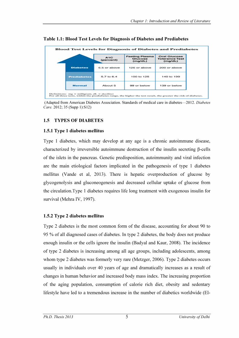
the world population, which is higher than was projected in earlier studies. Furthermore, the projections for 2030 show the prevalence to reach 439 million, comprising ~7.7% of the world’s population (Farag and Gaballa, 2011).India has a high prevalence of diabetes and the numbers are increasing at an alarming rate. In India alone, number of diabetics is expected to increase from 40.6 million in 2006 to 79.4 million by 2030 (Singh et al, 2012). 1.3.1 Pre-diabetes Pre-diabetes is a recently designated term by ADA which includes individuals who have impaired fasting plasma glucose (FPG 100-125mg /dL) and/or impaired glucose tolerance (plasma glucose level varies from 140 -199 mg / dL during OGTT) (Nicholas et al, 2007). Beyond the epidemiological evidence, the concept of pre-diabetes has potential relevance to clinical practice: at some early and reasonable point in the pathogenesis of type 2 DM, health care providers can prevent or postpone the development of this harmful condition. (Table 1.1) 1.4 CRITERIA FOR DIAGNOSIS OF DIABETES MELLITUS According to American diabetic association for diagnosis of diabetes, a diabetic will have one of these findings:
Symptoms of diabetes plus plasma glucose concentration > 200 mg/dl (11.1 mmol/l). Casual is defined as any time of day without regard to time since last meal. The classic symptoms of diabetes incluse polyuria, polydipsia and unexplained weight loss.
Or
FPG >126 mg/dl (7.0 mmol/l). Fasting is defined as no calorie intake for at least 8 hr.
Or
2h post load glucose > 200 mg/dl (11.1 mmol/l) during OGTT. The test should be performed as described by World Health Organisation (WHO) using glucose load containing the equivalent of 75 gm anhydrous glucose dissolved in water.
Chapter 1: Introduction and Review of Literature
Ph.D. Thesis 2013 University of Delhi 5
Table 1.1: Blood Test Levels for Diagnosis of Diabetes and Prediabetes
(Adapted from American Diabetes Association. Standards of medical care in diabetes—2012. Diabetes Care. 2012; 35 (Supp 1):S12)
1.5 TYPES OF DIABETES 1.5.1 Type 1 diabetes mellitus Type 1 diabetes, which may develop at any age is a chronic autoimmune disease, characterized by irreversible autoimmune destruction of the insulin secreting β-cells of the islets in the pancreas. Genetic predisposition, autoimmunity and viral infection are the main etiological factors implicated in the pathogenesis of type 1 diabetes mellitus (Vande et al, 2013). There is hepatic overproduction of glucose by glycogenolysis and gluconeogenesis and decreased cellular uptake of glucose from the circulation.Type 1 diabetes requires life long treatment with exogenous insulin for survival (Mehra IV, 1997). 1.5.2 Type 2 diabetes mellitus Type 2 diabetes is the most common form of the disease, accounting for about 90 to 95 % of all diagnosed cases of diabetes. In type 2 diabetes, the body does not produce enough insulin or the cells ignore the insulin (Badyal and Kaur, 2008). The incidence of type 2 diabetes is increasing among all age groups, including adolescents, among whom type 2 diabetes was formerly very rare (Metzger, 2006). Type 2 diabetes occurs usually in individuals over 40 years of age and dramatically increases as a result of changes in human behavior and increased body mass index. The increasing proportion of the aging population, consumption of calorie rich diet, obesity and sedentary lifestyle have led to a tremendous increase in the number of diabetics worldwide (El-
Chapter 1: Introduction and Review of Literature
Ph.D. Thesis 2013 University of Delhi 6
Shenawy and Abdel-Nabi, 2006). This form of diabetes, previously referred to as non–insulin dependent diabetes, type 2 diabetes, or adult-onset diabetes, encompasses individuals who have insulin resistance and usually have relative (rather than absolute) insulin deficiency. At least initially and often throughout their lifetime, these individuals do not need insulin treatment to survive. Most patients with this form of diabetes are obese and obesity itself causes some degree of insulin resistance. This form of diabetes frequently goes undiagnosed for many years (American Diabetes Association, 2010a). Impaired intrauterine growth / nutrition results in the programming of systems that regulate insulin sensitivity, insulin secretion and energy storage and utilization throughout the lifetime of the individual (Metzger, 2006). The familial predisposition to type 2 diabetes is mediated by both genetic and intrauterine environmental factors (Seshiah et al, 2008). 1.5.3 Type 1.5 diabetes Type 1.5 diabetes is usually diagnosed after 35 years of age and there is no immediate need for insulin (Goel et al, 2007).Type 1.5 diabetics are phenotypically similar to type 2 diabetic patients but they are also positive for the autoantibody commonly seen in type 1 diabetes. Approximately 10% to 30% of adults with type 2 diabetes test positive for autoantibodies, depending on the age and ethnicity of the study group (Stenstrom et al, 2005). This group of phenotypic adult type 2 diabetic patients (approximately 10%) are said to have latent autoimmune diabetes in adults (LADA) or type 1.5 diabetes or type 1. diabetes (Palmer et al, 2005). Patients with type 1.5 diabetes have an autoimmune process similar to that found with type 1 diabetes. Though patients with type 1.5 diabetes possess genes such as HLA DR2, DQB1*0602, which appear to protect an individual from developing diabetes and the beta-cells become so inflamed by repetitive environmental insults that they begin to succumb to autoimmune destruction within the beta-cells. This immune-mediated destruction of beta-cells in type 1.5 diabetics leads to insulin dependency more rapidly than in type 2 diabetes, but the more attenuated genetic and immune factors
Chapter 1: Introduction and Review of Literature
Ph.D. Thesis 2013 University of Delhi 7
associated with type 1.5 diabetes as compared with type 1 diabetes lead to an older age at onset and a slower progression to insulin dependency (Unger, 2008a)
1.6 INSULIN RESISTANCE American diabetes association (2005) suggested that people with diabetes mellitus were hyperglycemic because of resistance to action of insulin to remove glucose from the blood rather than insulin deficiency. The multiple defects in the insulin signaling cascade have been identified. There do not appear any measure disturbance in insulin receptor-binding, the measure decrease in the tyrosine kinase activity of receptor’s β subunit is responsible for the attenuation of the downstream signaling. Normally, insulin receptor phosphorylates insulin receptor substrate (IRS) proteins which are linked to the activation of phosphatidylinositol 3 kinase (PI3K)/ Protein kinase B (AkT pathway). During diabetes, reduced expression of PI3K and consequent reduced phosphorylation and activation of AKT arrests insulin actions (Folli et al, 2011). Myocytes, adipocytes and hepatocytes are the main organs of insulin resistance. Insulin resistance in adipocytes leads to failure of basal insulin levels to suppress lipolysis resulting in increased non-esterified fatty acids (NEFA) concentrations. Raised NEFA levels may stimulate gluconeogenesis and triglyceride synthesis in the liver, inhibit glucose uptake and utilization by skeletal muscle and reduce the extraction of insulin by the liver. Raised NEFA levels, like hyperglycemia, may also impair β cell function and inhibit insulin secretion (Tschritter et al, 2009). The liver is also resistant to insulin in type 2 diabetes and therefore, insulin does not suppress hepatic glucose production as effectively as in normal subjects. Hyperglycemia itself normally inhibits hepatic glucose production and this action is also impaired in type 2 diabetes. Obesity is especially trunkal distribution with increased intra-abdominal fat mass is an important determinant of insulin resistance, especially in skeletal muscle. Possible mechanisms include enhanced lipolysis generating excessive amount of NEFA and perhaps increased expression of tumor necrosis factor-α (TNF-α) by adipocytes and skeletal muscles which can induced insulin resistance by inhibiting by tyrosine kinase activity of insulin receptor (Ruan, 2002).
Chapter 1: Introduction and Review of Literature
Ph.D. Thesis 2013 University of Delhi 8
1.7 INSULIN 1.7.1 Insulin structure Insulin was the first peptide hormone discovered. With the elucidation of the primary sequence of insulin by Sanger in the mid 1950's (Sanger and Tupphy,1951) it became known that insulin was a two chain heterodimer consisting of a 21 amino acid A-chain linked to a 30 residue B chain by two disulfide bonds derived from cysteine residues (A7-B7 and A20-B19). An intrachain disulfide bond also exists in the A-chain (A6-A11). (Figure 1.1) 1.7.2 Insulin Biosynthesis In human the gene encoding preproinsulin, the ultimate precursor of insulin, is located on the short arm of chromosome 11(Bell et al, 1980). It is 1355 base pairs in the length and its encoding region consists of 3 exons, the first encodes the signal peptide at the N-terminus of preproinsulin, second, B and the part of the C-peptide and the third, the rest of C peptide and the A chains. Preproinsulin, an 11 KDa polypeptide is rapidly (with in 1 min) discharged into the cisternal space of the rough endoplasmic reticulum, where proteolytic enzymes immediately cleave in proinsulin, removing the signal peptide. Proinsulin is a 9 KDa peptide, containing A and B chain of insulin (with 21 AA and 30 AA). Proinsulin is then transported by microvesicles to glolgi apparatus (Steiner et al, 1972). The conversion of proinsulin to insulin is initiated in the Golgi complex and continues with in the maturing secretory granules through the sequential action of two endopeptidases and carboxypeptidases H (Hutton,1994). These enzymes act to remove the C peptide chain, liberating to cleavage dipeptides and finally yielding insulin. Insulin has a lower solubility and therefore, coprecipitated with Zinc ions to form microcrystal with in the secretory granules (Orci et al, 1986). Insulin and C peptide are stored in the granule sac and are ultimately released in equimolar amounts, under normal condition. This pathway of secretion via storage granules is termed ‘regulated pathway’.(Figure 1.2)

Figure 1.1 (Insulin structure)
Figure 1.2 (Insulin synthesis)
Chapter 1: Introduction and Review of Literature
Ph.D. Thesis 2013 University of Delhi 9
1.7.3 Mechanism and regulation of insulin secretion The secretion of insulin from pancreatic beta cells is a complex process involving the integration and interaction of multiple external and internal stimuli. Thus, nutrients, hormones, neurotransmitters, and drugs all activate -or inhibit - insulin release. The primary stimulus for insulin secretion is the beta-cell response to changes in ambient glucose. Normally, glucose induces a biphasic pattern of insulin release. First-phase insulin release occurs within the first few minutes after exposure to an elevated glucose level; this is followed by a more enduring second phase of insulin release. Of particular importance is the observation that first-phase insulin secretion is lost in patients with type 2 diabetes. Thus, molecular mechanisms involved in phasic insulin secretion are important. (Seino,2002)
A widely accepted sequence of events involved in glucose-induced insulin secretion is as follows:
i.) Glucose is transported into beta cells through facilitated diffusion of GLUT2 glucose transporters.
ii.) Intracellular glucose is metabolized to ATP. iii.) Elevation in the ATP/ADP ratio induces closure of cell-surface ATP-sensitive
K+ (K+ ATP) channels, leading to cell membrane depolarization. iv.) Cell-surface voltage-dependent Ca2+ channels (VDCC) are opened, facilitating
extracellular Ca2+ influx into the beta cell. v.) A rise in free cytosolic Ca2+ triggers the exocytosis of insulin. (Figure 1.3) 1.7.4 The Importance of Potassium ATP (K+ ATP) Channels The K+ ATP channels play an integral role in glucose-stimulated insulin secretion by serving as the transducer of a glucose-generated metabolic signal (ATP) to cell electrical activity (membrane depolarization). Thus, like neurons, beta cells are electrically excitable and capable of generating Ca2+ action potentials that are important in synchronizing islet cell activity and insulin release. In addition to being signal targets for glucose, K+ATP channels are the targets for sulfonylureas, which are

Chapter 1: Introduction and Review of Literature
Ph.D. Thesis 2013 University of Delhi 10
commonly prescribed oral agents in the treatment of type 2 diabetes. The sulfonylureas, like glucose, induce closure of K+ATP channels and stimulate insulin secretion. The beta-cell K+ ATP channel is a complex octameric unit of 2 different proteins: the sulfonylurea receptor (SUR-1) and an inward rectifier (Kir6.2). The sulfonylurea receptor belongs to a superfamily of ATP-binding cassette proteins and contains the binding site for sulfonylurea drugs and nucleotides. The inward rectifier represents the K+ conducting pore and is also regulated by ATP. It is interesting that K+ATP channels are present in other tissues of the body, including heart (SUR-2A/Kir 6.2), smooth muscle (SUR-2B/Kir 6.2), and brain (SUR-1/Kir 6.2). Glucose sensing in the brain during hypoglycemia may be mediated by K+ATP channels located in brain hypothalamic neurons (Evans,2002). Thus, these molecules may also serve as new therapeutic targets for the restoration of impaired hypoglycemia awareness and glucose counter regulation in type 1 diabetes. 1.7.5 Role of Incretins in Release of Insulin The incretins are polypeptide hormones released in the gut after a meal that potentiate insulin secretion in a glucose-dependent manner. They are important hormonal regulators of insulin secretion. (Holst, 2002) Due to their dependence on ambient glucose for action, they are emerging as important new therapeutic agents to promote insulin secretion without accompanying hypoglycemia (a common complication of sulfonylurea treatment).Unlike sulfonylureas, incretins act by activating Gs (a G-protein that activates adenylyl cyclase) to increase cAMP in beta cells. cAMP, like ATP, is an important signal that regulates insulin release. Typically, the main mechanism of action of cAMP is by activation of an enzyme called protein kinase A (PKA) that, in turn, phosphorylates other substrates to turn on (or off) vital cell functions. Some identified a novel protein, cAMP-GEF II, a cAMP sensor (cAMP) that forms a complex with other intracellular proteins (Rim2 and Rab3) to directly regulate insulin exocytosis. Then, using molecular reagents that antagonize the effects of cAMP, they observed that incretin-potentiated insulin secretion is attenuated. These results provide a mechanism whereby cAMP can directly promote exocytosis of insulin granules without activation of PKAs (a PKA-independent pathway), and thereby provide additional molecular targets for therapeutic intervention.(Seino and Shibasaki, 2005).
Chapter 1: Introduction and Review of Literature
Ph.D. Thesis 2013 University of Delhi 11
1.7.5 Voltage-Dependent Ca2+ Channels: Novel Regulators of insulin Release Extracellular Ca2+ influx through L-type voltage-dependent Ca2+ channels (VDCC) raises free cytoplasmic Ca2+ levels and triggers insulin secretion. The structure of the VDCC is complex and consists of 5 subunits: alpha1, alpha2, beta, gamma, and delta units. The alpha subunit constitutes the ion-conducting pore, whereas the other units serve a regulatory role. Previous work has identified that isoforms of alpha1 subunits interact with exocytotic proteins. (Findlay et al, 1985) More recently, using the yeast hybrid screening method, a novel protein, Kir-GEM, interacting with the beta 3 isoform of the VDCC, has been identified by Seino and colleagues (2002). Furthermore, it has been determined that Kir-GEM inhibits alpha ionic activity and prevents cell-surface expression of alpha subunits. The investigators have proposed that in the presence of Ca2+, Kir-GEM binds to the beta isoform, and this interaction interferes in the trafficking or translocation of alpha subunits to the plasma membrane. The relevance of Kir-GEM in insulin secretion was made evident by its attenuation of glucose-stimulated Ca2+ increases and C-peptide secretion in an insulin-secreting cell line.The potential therapeutic role of Kir-GEM lies in the inhibitory effects on VDCC activity that may serve to protect beta cells from overstimulation and subsequent failure, which is part of the disease etiology of type 2 diabetes. 1.7.7 Mechanism of action of Insulin The diverse effects of insulin are mediated through a multicomponent signalling complex that is strongly conserved across a wide range of species (Withers and white, 2000). Binding of insulin to its receptor triggers a cascade of signalling events that ultimately leads to modifications in a number of biological processes. When insulin binds to its receptor, resultant activation of the insulin signaling cascade leads to multiple effects on several biological processes, including glucose and lipid uptake/metabolism, gene expression/ protein synthesis and cell growth, division and survival.So insulin receptor having an important role in signaling mechanism. Insulin receptors are ubiquitous in vertebrate cells although expression varies significantly between cell types, from as few as 40 receptors per cell on erythrocytes to more than 200000 on adipocytes and hepatocytes. The insulin receptor is a large transmembrane
Chapter 1: Introduction and Review of Literature
Ph.D. Thesis 2013 University of Delhi 12
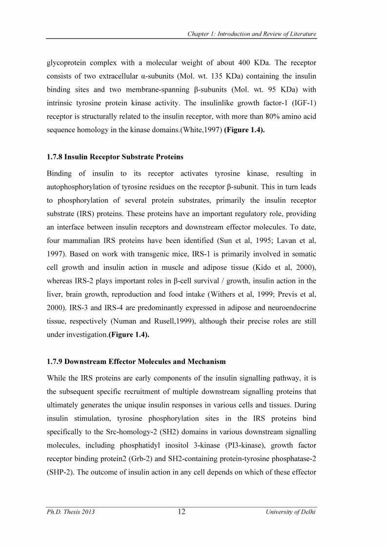
glycoprotein complex with a molecular weight of about 400 KDa. The receptor consists of two extracellular α-subunits (Mol. wt. 135 KDa) containing the insulin binding sites and two membrane-spanning β-subunits (Mol. wt. 95 KDa) with intrinsic tyrosine protein kinase activity. The insulinlike growth factor-1 (IGF-1) receptor is structurally related to the insulin receptor, with more than 80% amino acid sequence homology in the kinase domains.(White,1997) (Figure 1.4). 1.7.8 Insulin Receptor Substrate Proteins Binding of insulin to its receptor activates tyrosine kinase, resulting in autophosphorylation of tyrosine residues on the receptor β-subunit. This in turn leads to phosphorylation of several protein substrates, primarily the insulin receptor substrate (IRS) proteins. These proteins have an important regulatory role, providing an interface between insulin receptors and downstream effector molecules. To date, four mammalian IRS proteins have been identified (Sun et al, 1995; Lavan et al, 1997). Based on work with transgenic mice, IRS-1 is primarily involved in somatic cell growth and insulin action in muscle and adipose tissue (Kido et al, 2000), whereas IRS-2 plays important roles in β-cell survival / growth, insulin action in the liver, brain growth, reproduction and food intake (Withers et al, 1999; Previs et al, 2000). IRS-3 and IRS-4 are predominantly expressed in adipose and neuroendocrine tissue, respectively (Numan and Rusell,1999), although their precise roles are still under investigation.(Figure 1.4). 1.7.9 Downstream Effector Molecules and Mechanism While the IRS proteins are early components of the insulin signalling pathway, it is the subsequent specific recruitment of multiple downstream signalling proteins that ultimately generates the unique insulin responses in various cells and tissues. During insulin stimulation, tyrosine phosphorylation sites in the IRS proteins bind specifically to the Src-homology-2 (SH2) domains in various downstream signalling molecules, including phosphatidyl inositol 3-kinase (PI3-kinase), growth factor receptor binding protein2 (Grb-2) and SH2-containing protein-tyrosine phosphatase-2 (SHP-2). The outcome of insulin action in any cell depends on which of these effector

Figure 1.3 (Mechanism of Insulin release)
Figure 1.4 (Insulin receptor and its autophosphorylation sites)

Chapter 1: Introduction and Review of Literature
Ph.D. Thesis 2013 University of Delhi 13
molecules are expressed and recruited into the signalling complex and the pathways that are activated as a result (Withers et al, 2000). In skeletal muscle and adipose tissue, insulin stimulation of the PI 3-kinase pathway enhances glucose utilization by regulating the expression or sub-cellular localization of glucose transporters, GLUT4 and GLUT1, and stimulates the storage of glucose as glycogen or fat (Clarke et al, 1994; Okada et al, 1994). In pancreatic β cells, the PI 3- kinase cascade probably romotes survival of β cells (Cho et al, 2001; Tuttle et al, 2001). Moreover, insulin stimulation of PI 3-kinase strongly activates total protein synthesis in most cell types that are regulated by the mammalian target of rapamycin (mTOR) pathway. Alternatively, activation of the adapter protein Grb-2 stimulates gene transcription through the mitogen-activated protein kinase (MAPK) cascade. (Figure 1.5). 1.8 KEY PARTICIPANTS OF EUKARYOTIC INSULIN SIGNALING 1.8.1 Tyrosine kinase activity Insulin is the major hormone controlling critical energy functions such as glucose and lipid metabolism. Insulin activates the insulin receptor tyrosine kinase (IR), which phosphorylates and recruits different substrate adaptors such as the IRS family of proteins.(Siddle et al, 2011) Tyrosine phosphorylated IRS then displays binding sites for numerous signaling partners. Among them, PI3K has a major role in insulin function, mainly via the activation of the Akt/PKB and the PKCζ cascades. Activated Akt induces glycogen synthesis through inhibition of GSK-3; protein synthesis via mTOR and downstream elements; and cell survival through inhibition of several pro-apoptotic agents (Bad, Forkhead family transcription factors, GSK-3). Insulin stimulates glucose uptake in muscle and adipocytes via translocation of GLUT4 vesicles to the plasma membrane. GLUT4 translocation involves the PI3K/Akt pathway and IR-mediated phosphorylation of CAP, and formation of the CAP:Cbl:CrkII complex. Insulin signaling also has growth and mitogenic effects, which are mostly mediated by the Akt cascade as well as by activation of the Ras/ MAPK pathway. In addition, insulin signaling inhibits gluconeogenesis in the liver, through disruption of CREB/CBP/Torc2 binding. Insulin signaling also promotes fatty acid synthesis through activation of SREBP-1C, USF1, and LXR. A negative
Chapter 1: Introduction and Review of Literature
Ph.D. Thesis 2013 University of Delhi 14
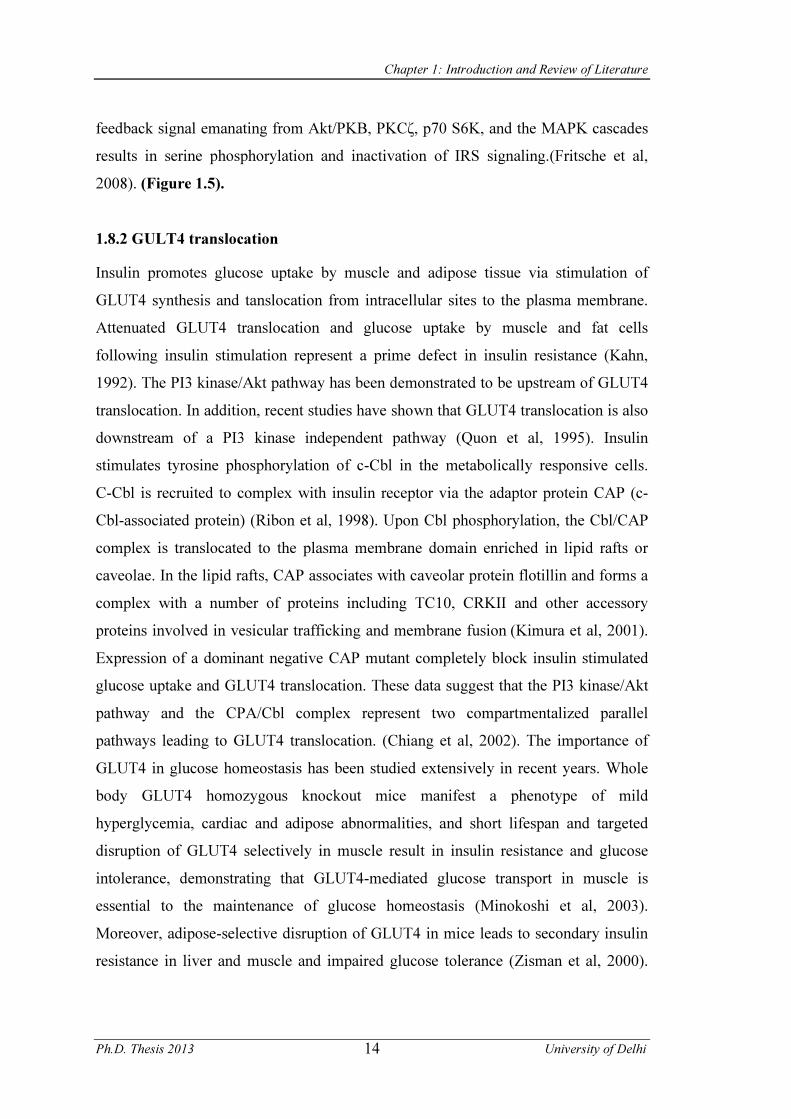
feedback signal emanating from Akt/PKB, PKCζ, p70 S6K, and the MAPK cascades results in serine phosphorylation and inactivation of IRS signaling.(Fritsche et al, 2008). (Figure 1.5). 1.8.2 GULT4 translocation Insulin promotes glucose uptake by muscle and adipose tissue via stimulation of GLUT4 synthesis and tanslocation from intracellular sites to the plasma membrane. Attenuated GLUT4 translocation and glucose uptake by muscle and fat cells following insulin stimulation represent a prime defect in insulin resistance (Kahn, 1992). The PI3 kinase/Akt pathway has been demonstrated to be upstream of GLUT4 translocation. In addition, recent studies have shown that GLUT4 translocation is also downstream of a PI3 kinase independent pathway (Quon et al, 1995). Insulin stimulates tyrosine phosphorylation of c-Cbl in the metabolically responsive cells. C-Cbl is recruited to complex with insulin receptor via the adaptor protein CAP (c-Cbl-associated protein) (Ribon et al, 1998). Upon Cbl phosphorylation, the Cbl/CAP complex is translocated to the plasma membrane domain enriched in lipid rafts or caveolae. In the lipid rafts, CAP associates with caveolar protein flotillin and forms a complex with a number of proteins including TC10, CRKII and other accessory proteins involved in vesicular trafficking and membrane fusion (Kimura et al, 2001). Expression of a dominant negative CAP mutant completely block insulin stimulated glucose uptake and GLUT4 translocation. These data suggest that the PI3 kinase/Akt pathway and the CPA/Cbl complex represent two compartmentalized parallel pathways leading to GLUT4 translocation. (Chiang et al, 2002). The importance of GLUT4 in glucose homeostasis has been studied extensively in recent years. Whole body GLUT4 homozygous knockout mice manifest a phenotype of mild hyperglycemia, cardiac and adipose abnormalities, and short lifespan and targeted disruption of GLUT4 selectively in muscle result in insulin resistance and glucose intolerance, demonstrating that GLUT4-mediated glucose transport in muscle is essential to the maintenance of glucose homeostasis (Minokoshi et al, 2003). Moreover, adipose-selective disruption of GLUT4 in mice leads to secondary insulin resistance in liver and muscle and impaired glucose tolerance (Zisman et al, 2000).
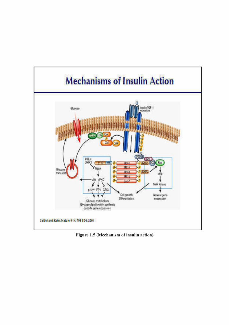
Figure 1.5 (Mechanism of insulin action)

Chapter 1: Introduction and Review of Literature
Ph.D. Thesis 2013 University of Delhi 15
Taken together, these imply that alteration of GLUT4 expression and/or function could contribute to the development of insulin resistance and diabetes. 1.9 METABOLIC EFFECT OF INSULIN Insulin play a key role in glucose homeostasis and also play a primary role in control of lipid and protein metabolism. 1.9.1 Effect of Insulin on Carbohydrate Metabolism Elevated concentration of glucose in the blood stimulus release of insulin and insulin acts on cells through out the body to stimulate uptake, utilization and storage of glucose. The effects of insulin on glucose metabolism vary depending on the target tissues. Its important effects are:- 1.9.1.1 Insulin Facilitates Entry of Glucose into Muscle, Adipose and Several
Other Tissues Insulin enhances the transport of glucose across the cell membrane in muscle (Geiger et al, 2005) and adipose tissue (Crofford and renold, 1965). Cells can take up glucose by facilitated diffusion through a family hexose transporters abbreviated as GLUT. There are 12 isoforms of these transporters, GLUT1-12. Among these, GLUT4 is an insulin responsive glucose transporter found in skeletal muscles, cardiac muscle and adipose tissue which made available in the plasma membrane through the action of insulin. In the absence of insulin GLUT4 are present in cytoplasmic vesicles where they are useless for transporting glucose. Binding of insulin to its receptors leads to fusion of these vesicles with the plasma membrane and insertion of Glucose tranporters, there by giving the cell and ability to efficiently take up the glucose. When insulin level decrease and insulin receptors are no longer occupied GLUT4 are recycled back into the cytoplasm. During Diabetes, reduced translocation of GLUT4 as well as its expression leads to impaired glucose disposal due to insulin insufficiency (Karnieli and Armoni,2008).
Chapter 1: Introduction and Review of Literature
Ph.D. Thesis 2013 University of Delhi 16
1.9.1.2 Insulin Stimulates Glycolysis and Glycogenesis, where as Inhibits Gluconeogenesis and Glycogenolysis
Insulin stimulates the synthesis of enzyme glucokinase, a key enzyme of glycolysis, which phosphorylates glucose and trapping it with in the cell, thereby increase its utilization (Pilkis et al, 1988). Other important enzymes of glycolysis i.e. pyruvate kinase and phosphofructokinase are also regulated by insulin (Sillero et al, 1969).
Insulin enhances glycogen synthesis by increasing the activity of glycogen synthase (Salas and larner,1975), and prevents glycogenolysis by bringing down cyclic AMP level as result of stimulation of a phosphodiesterase (Zinman and Hollenberg,1974). Hence during insulin deficient state, decreased glucose utilization and increased glycogen breakdown lead to hyperglycemia. Insulin has been found to be a suppressor of biosynthesis of key enzymes of gluconeogenesis such as glucose 6 phosphatase and fructose-1,6-biphosphatase.(Weber et al, 1965). In diabetes, gluconeogenesis is increased and glucose is synthesized from amino acids, glycerol and lactate which further aggravating hyperglycemia. 1.9.2 Effect of Insulin on lipid metabolism i.) Insulin is lipogenic in action. When the liver is saturated with glycogen, any
additional glucose taken up by hepatocytes is shunted into pathways leading to synthesis to fatty acid. Insulin stimulates the enzyme ATP citrate lyase (Ramakrishna and Benjamin,1981), which provides NADP+ for the fatty acid biosynthesis. It increases activity of acetyl Co A carboxylase, which catalyzes first step of fatty acid biosynthesis. These fatty acids are exported from the liver as lipoproteins. The lipoproteins are ripped a part in circulation, providing the free fatty acids to other tissues, including adipocytes, which use it to synthesize triglycerides. Insulin activates the enzyme lipoprotein lipase, which breakdown lipoproteins in blood provide free fatty acids to tissues.
ii.) One of the most potent action of insulin is the suppression of lipolysis in the adipose tissue (Jensen et al, 1989). Insulin inhibits the enzyme hormone sensitive lipase by reducing cyclic AMP level in adipocytes (Soderling et al, 1973). This enzyme hydrolysis TG to release fatty acid. Insulin facilitates entry

Chapter 1: Introduction and Review of Literature
Ph.D. Thesis 2013 University of Delhi 17
of glucose in adipocytes and within those cells where glucose can be used to synthesize glycerol. In diabetes the abnormal high concentration of serum lipids is mainly due to the increased mobilization of free fatty acids from peripheral depots which is due to the activation of hormones sensitive lipase during insulin deficiency (Agradh et al, 1999). Dysfunction of lipoprotein lipase in insulin deficiency present in lipoproteins in blood contributes to hypertriglyceridemia due to impaired catabolism of triglyceride- rich paticles (Neimeijer-Kanters et al, 2001).
iii.) Insulin increases the receptor-mediated removal of LDL-C and hence insulin insufficiency during diabetes causes hypercholesterolemia of LDL-C. Diabetes is also characterized by low level of HDL-C (Garber, 2002). Reduction in the production of HDL-C is due to the impaired metabolism of TG-rich lipoproteins which provide a significant portion of HDL-C. (Taskinen,1987).
1.9.3 Effect of Insulin on protein metabolism i.) Insulin increases uptake of amino acids by various tissues through facilitating
their transport across the cell membrane (Wool et al, 1972). ii.) Insulin also increases the formation of aminoacyl t-RNA (Manchester,1970). In
muscles, insulin has been shown to increase no. of ribosomes as well as their translational efficiency (Jefferson et al, 1980). In general, insulin stimulates protein synthesis.
iii) In diabetes lack of insulin leads to decrease in protein synthesis and increase in protein breakdown. Diabetic patients with negligible plasma insulin concentrations have increased whole body protein catabolism. Insulin therapy diminishes catabolism of protein in type 1 diabetic patients (Carlson and Campbell,1993).
1.10 BIOCHEMICAL ROLE OF C-PEPTIDE ACTIVITY C-peptide has been widely accepted as the most appropriate measure of residual β-cell function because it is secreted on a equimolar basis to insulin and unlike the latter, is not removed in the first pass through the liver (Panero et al., 2009). Because

Chapter 1: Introduction and Review of Literature
Ph.D. Thesis 2013 University of Delhi 18
C-peptide is secreted from islet cells into the circulation in equimolar concentrations with insulin and is not extracted by the liver, many investigators have used C-peptide levels as a biomarker of β-cell function (Ko et al, 2009). Fasting C-peptide level < 0.6 ng/ml is considered as an indicator of poor insulin reserve. Hence, C-peptide is a useful guide in initiating therapy to prevent complications (Abdullah et al., 2010). The cellular signaling effects of C-peptide is depicted in Figure 1.6. While the physiological functions of insulin are well established and understood, the functions of C-peptide are still being established and investigated. Recently it was found that C-peptide acts through G-protein-coupled receptors to activate calcium-dependent signaling pathways via the C-terminal pentapeptide portion of the molecule. C-peptide activation of calcium signaling is thought to increase the activity of Na+ K+ ATPase, which has been found to be reduced in patients with the advanced microvascular complications of diabetes mellitus (nephropathy and retinopathy).(Marques et al, 2004) The middle segment of the peptide has also been shown to activate Na+K+ATPase, but not as strongly as the full peptide or the pentapeptide sequence.(Wahren et al, 2000) C-peptides beneficial affects on the microvascular complications of diabetes mellitus are thought to be mediated through endothelial nitric oxide synthase.(Marques et al, 2004) In addition to receptor-mediated effects, C-peptide has been shown to produce non-receptor- mediated effects. (Ido et al, 1997) 1.11 COMPLICATIONS OF DIABETES The chronic hyperglycemia of diabetes is associated with long-term damage, dysfunction and failure of various organs especially the eyes, kidneys, nerves, heart and blood vessels (Pandolfi and De Filippis,2007).Generally, the injurious effects of hyperglycemia are separated into macrovascular complications (coronary artery disease, peripheral arterial disease and stroke) and microvascular complications (diabetic nephropathy, neuropathy, and retinopathy) (Fowler, 2008). 1.11.1 Causes of Diabetic Complications The complications of diabetes are the result of multiple factors in particular, cellular pathways that lead to diabetes. The complications of diabetes have been tied to
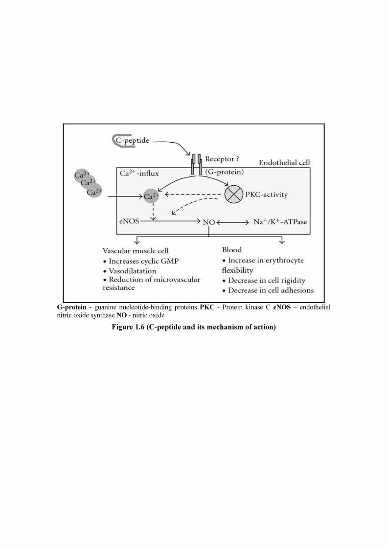
G-protein - guanine nucleotide-binding proteins PKC - Protein kinase C eNOS – endothelial nitric oxide synthase NO - nitric oxide
Figure 1.6 (C-peptide and its mechanism of action)

Chapter 1: Introduction and Review of Literature
Ph.D. Thesis 2013 University of Delhi 19
oxidant stress (Szabo, 2009). Studies with diabetic animals have shown that oxidative stress leads to DNA damage in renal cortical cells. Although early effects of elevated glucose may increase the presence of potentially protective pathways, more prolonged exposure of elevated glucose with the rise in insulin levels can lead to reactive oxygen species (ROS) and can be detrimental even if glucose levels are controlled (Barbosa et al. 2008 ). 1.11.2 Mechanisms Leading to Complications i.) Polyol pathway, focuses on the enzyme aldose reductase. Aldose reductase
normally has the function of reducing toxic aldehydes in the cell to inactive alcohols, but when the glucose concentration in the cell becomes too high, aldose reductase also reduces that glucose to sorbitol, which is later oxidized to fructose. In the process of reducing high intracellular glucose to sorbitol, the aldose reductase consumes the cofactor NADPH (Lee and chung,1999). But as shown in figure 1.8, NADPH is also the essential cofactor for regenerating a critical intracellular antioxidant, reduced glutathione. By reducing the amount of reduced glutathione, the polyol pathway increases susceptibility to intracellular oxidative stress.
ii.) The second mechanism is increased flux through the hexosamine pathway. As shown in figure 1.7,when glucose is high inside a cell, most of that glucose is metabolized through glycolysis, going first to glucose-6 phosphate, then fructose-6 phosphate, and then on through the rest of the glycolytic pathway. However, some of that fructose-6-phosphate gets diverted into a signaling pathway in which an enzyme called GFAT (glutamine: fructose-6 phosphate amidotransferase) converts the fructose-6 phosphate to glucosamine-6 phosphate and finally to UDP (uridine diphosphate) N-acetyl glucosamine. N-acetyl glucosamine gets attached onto serine and threonine residues of transcription factors, just like the more familiar process of phosphorylation, and overmodification by this glucosamine often results in pathologic changes in gene expression (Kolm et al, 1998; Wells et al, 2003). For example, in Fig 1.7, increased modification of the transcription factor Sp1 results in increased
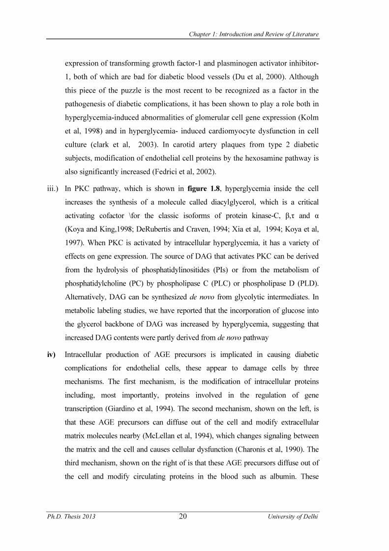
Chapter 1: Introduction and Review of Literature
Ph.D. Thesis 2013 University of Delhi 20
expression of transforming growth factor-1 and plasminogen activator inhibitor-1, both of which are bad for diabetic blood vessels (Du et al, 2000). Although this piece of the puzzle is the most recent to be recognized as a factor in the pathogenesis of diabetic complications, it has been shown to play a role both in hyperglycemia-induced abnormalities of glomerular cell gene expression (Kolm et al, 1998) and in hyperglycemia- induced cardiomyocyte dysfunction in cell culture (clark et al, 2003). In carotid artery plaques from type 2 diabetic subjects, modification of endothelial cell proteins by the hexosamine pathway is also significantly increased (Fedrici et al, 2002).
iii.) In PKC pathway, which is shown in figure 1.8, hyperglycemia inside the cell increases the synthesis of a molecule called diacylglycerol, which is a critical activating cofactor \for the classic isoforms of protein kinase-C, β,τ and α (Koya and King,1998; DeRubertis and Craven, 1994; Xia et al, 1994; Koya et al, 1997). When PKC is activated by intracellular hyperglycemia, it has a variety of effects on gene expression. The source of DAG that activates PKC can be derived from the hydrolysis of phosphatidylinositides (PIs) or from the metabolism of phosphatidylcholine (PC) by phospholipase C (PLC) or phospholipase D (PLD). Alternatively, DAG can be synthesized de novo from glycolytic intermediates. In metabolic labeling studies, we have reported that the incorporation of glucose into the glycerol backbone of DAG was increased by hyperglycemia, suggesting that increased DAG contents were partly derived from de novo pathway
iv) Intracellular production of AGE precursors is implicated in causing diabetic complications for endothelial cells, these appear to damage cells by three mechanisms. The first mechanism, is the modification of intracellular proteins including, most importantly, proteins involved in the regulation of gene transcription (Giardino et al, 1994). The second mechanism, shown on the left, is that these AGE precursors can diffuse out of the cell and modify extracellular matrix molecules nearby (McLellan et al, 1994), which changes signaling between the matrix and the cell and causes cellular dysfunction (Charonis et al, 1990). The third mechanism, shown on the right of is that these AGE precursors diffuse out of the cell and modify circulating proteins in the blood such as albumin. These
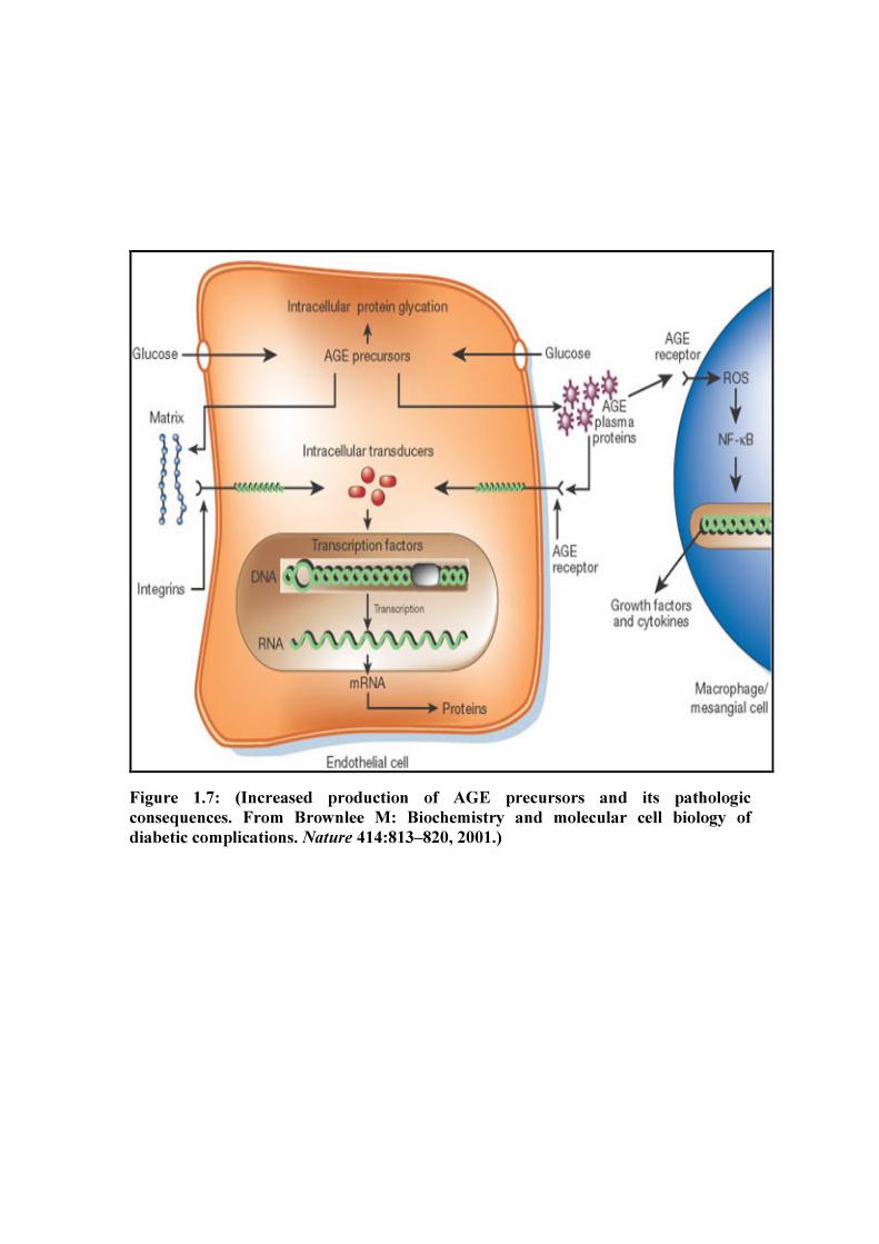
Figure 1.7: (Increased production of AGE precursors and its pathologic consequences. From Brownlee M: Biochemistry and molecular cell biology of diabetic complications. Nature 414:813–820, 2001.)
Figure 1.8 (Pathways associated with diabetic complications)
Chapter 1: Introduction and Review of Literature
Ph.D. Thesis 2013 University of Delhi 21
modified circulating proteins can then bind to AGE receptors and activate them, thereby causing the production of inflammatory cytokines and growth factors, which in turn cause vascular pathology (Li et al, 1996; Viassara et al, 1988). From many animal studies such as one done by Hans-Peter Hammes et al 1998, showing that that pharmacologic inhibition of AGEs prevents late structural changes of experimental diabetic retinopathy(Figure 1.7).
1.12 OXIDATIVE STRESS AND DIABETES Oxidative stress may promote the onset of diabetes by decreasing insulin sensitivity and destroying the insulin-producing cells. ROS can penetrate through cell membranes and cause damage to β-cells of pancreas (Lightfoot et al, 2012). A high fat diet or free fatty acids also has been shown to release ROS and contribute to mitochondrial DNA damage and impaired pancreatic β-cell function (Graciano et al, 2011). Oxidant stress and ROS exposure can result in the opening of the mitochondrial membrane permeability transition pore, reduce mitochondrial NAD+ stores and result in apoptotic cell injury. The development of diabetes has been associated with a decrease in the levels of mitochondrial proteins and mitochondrial DNA. Cellular pathways in diabetes are closely associated to cellular energy maintenance and intact mitochondrial function (Newsholme et al., 2007). 1.12.1 Lipid Peroxidation Lipids when react with free radicals, they undergo peroxidation to form lipid peroxides. Lipid peroxides decompose to form numerous products including malondialdehyde. (Rashmi et al, 2007) The toxicity of oxygen, or of its radical derivatives, is often accompanied by the peroxidation of lipids. Lipid peroxidation as induced by low-level exposures to nitrogen dioxide appears to proceed either by hydrogen atom abstraction or by nitrogen dioxide addition to the olefin. The most common way to measure lipid peroxides is to estimate malondialdehyde (MDA) content. MDA is formed during lipid peroxidation after rupture of the carbon chain of unsaturated fatty acids. The amount of malondialdehyde is then determined colorometrically after reaction with thiobarbituric acid. (Jamienson et al, 1986) (Figure 1.9a).
Chapter 1: Introduction and Review of Literature
Ph.D. Thesis 2013 University of Delhi 22
1.12.2 Superoxide Dismutase (SOD) The primary ROS produced in the course of oxygen metabolism is superoxide, which is a highly reactive, cytotoxic ROS. Superoxide is dismutated to a far less reactive product, hydrogen peroxide (H2O2), by a family of metalloenzymes known as superoxide dismutase (SOD).(Nosratola et al, 2003) Superoxide dismutase (SODs) catalyze the superoxide anion to molecular oxygen and H2O2 and thus are critical for protecting the cell against the toxic products of aerobic respiration. The primary ROS produced in the course of oxygen metabolism is superoxide, which is a highly reactive, cytotoxic ROS. O2- is commonly produced within aerobic biological systems, and superoxide dismutases (SODs) provide an important defense against it. Thus, SOD is the front line of defense against ROS-mediated injury (Figure 1.9b). 1.12.3 Glutathione Levels GSH is by far the most important antioxidant in most mammalian cells. This ubiquitous tripeptide, γ-Glu-Cys- Gly, performs many cellular functions. In particular, the thiol containing moiety is a potent reducing agent. Intracellular GSH is converted to GSSG by selenium-containing GSH peroxidase, which catalyzes the reduction of H2O2 in the presence of GSH and GSH peroxidase is coupled with oxidation of glucose-6-phosphate and of 6-phosphogluconate, which provides NADPH for reduction of GSSG by GSSG reductase. This is a major pathway of H2O2 metabolism in many cells. It is thus important for the protection of membrane lipids against oxidation. GSH has the important function of destroying reactive oxygen intermediates and free radicals that are constantly formed in metabolism (Figure 1.9c). 1.12.4 Glutathione Peroxidase and Glutathione Reductase Glutathione peroxidase and reductase are two enzymes that are found in the cytoplasm, mitochondria, and nucleus. Glutathione peroxidase metabolizes hydrogen peroxide to water by using reduced glutathione as a hydrogen donor (Sies, 1993). Glutathione disulfide is recycled back to glutathione by glutathione reductase, using the cofactor NADPH generated by glucose 6- phosphate dehydrogenase. (Figure 1.9b).

Figure 1.9a.(Reaction of TBA with MDA forming the 532 nm chromophore)
Figure 1.9b (Superoxide dismutase, Catalase and Glutathione peroxidase)
Figure1.9c (Reduced Glutathione)
Chapter 1: Introduction and Review of Literature
Ph.D. Thesis 2013 University of Delhi 23
1.12.5 Catalase Catalase (CAT, H2O2: H2O2 oxidoreductase) is an enzyme that decompose hydrogen peroxide (H2O2) to molecular oxygen (O2) and water (H2O). It also exhibits peroxidatic activity and catalyses the oxidation of various hydrogen donors in the presence of relatively lower concentrations of hydrogen peroxide (Figure 1.9b). 1.13 EXPERIMENTAL DIABETES Apart from the surgical removal of pancreas, there are two distinct methods for producing experimental diabetes: • Chemically induced diabetes • Spontaneous diabetes mellitus 1.13.1 Chemically Induced Diabetes Mellitus: Chemical agents used to induce diabetes in animals are alloxan, streptozotocin,N-nitrosomethyl urea, N-nitroso moietyuria (Cooperstein and Watkins,1977).
A) Alloxan: it was first found to cause hyperglycemia in rats (Dunn et al, 1944) and rabbits (Bailey et al, 1949), and in other species like dog, cat, hamsters, sheep and monkey (Luken,1948). Chemically, it is 2,4,5,6 tetraoxohexahydropyrimide (Fig.) a highly unstable and reactive substance, which exists in tautomeric forms. (Figure 1.10a) i.) Course of action after giving injection of alloxan: Single injection of
appropriate amount of alloxan can cause selective necrosis of beta cells. Three distinct phases are seen after giving injections of alloxan. Initially there is hyperglycemia during 2-4 hours after giving injections. This is said to be due to sudden cessation of insulin release together with liberation of epinephrine which increases the glucose level. This is followed by hypoglycemia for the next 6 hours and during this phase, there is uncontrolled release of insulin from damaged islets cells. Severe hypoglycemia, often lethal, may occur which can be prevented by administering glucose. Third is permanent chronic diabetic phase which
Chapter 1: Introduction and Review of Literature
Ph.D. Thesis 2013 University of Delhi 24
begins 18-24 hours after injection. By this time degenerative changes in islet cells are complete, resulting in diabetes.
B) Streptozotocin (STZ):-It is a broad spectrum antibiotic and antitumor agent from Streptomyces achromogens. It was found to produce diabetes mellitus in rats and dogs (Rakieten et al ,1963) and monkeys (Schein et al, 1973). Chemically STZ is 2-deoxy-2-[( methyl-nitrosoamino) carbonyl)-amino]-D-glucopyranose. The nitrourea moiety provides its cytotoxic effect whereas deoxyglucose moiety facilitate its entry into islet cells (Like and Rossini,1976) (Figure 1.10b)
C) Mode of action of alloxan and streptozotocin: Studies with both drugs indicate rapid and specific uptake by beta-cell (Cooperstein and Watkins,1981; Malaisse,1982). In an environment rich in thiol groups, alloxan is reduced to dialuric acid and during autooxidation back to alloxan, superoxide anions are formed. Superoxide radicals can dismutate spontaneously or by the action of superoxide dismutase, to form hydrogenperoxide. Hydroxy radicals, the most reactive oxygen containing species are then generated from the superoxide dismutase, to form hydrogen peroxide. Hydroxyl radicals, the most reactive oxygen containing species are then generated from the superoxide radical and hydrogen peroxide, by the iron catalyzed Haber Weiss reaction. Hydroxy radicals may reduce the activity of enzymes by interaction with their protein moiety (Crouch et al, 1981). The hydroxyl radicals produce membrane accounts for impairment of mitochondrial function and membrane ion permeability (Gandy et al, 1981). Effect of streptozotocin may be due to its alkylating properties. Both alloxan and streptozotocin cause DNA stran breaks in beta cells. The activate polyp (ADP ribose) synthetase. It initiates DNA repair and depletes the cell of NAD+, leading to beta cell dysfunction and possible necrosis (Okamoto and Yamamoto,1983). (Figure 1.10c) Diabetogenic dose of alloxan produces mild and mostly reversible changes in the kidney. If more than the diabetogenic dose of alloxan is given, many histological changes are seen in kidneys (Brunschwig,1944).Diabetogenic dose of streptozotocin does not produce any toxic symptom on kidneys.(Arison and
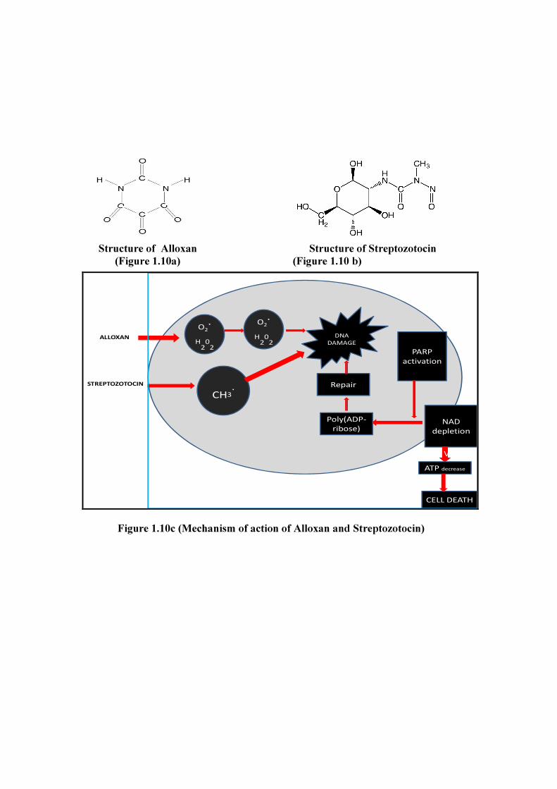
Structure of Alloxan Structure of Streptozotocin (Figure 1.10a) (Figure 1.10 b)
ALLOXAN
STREPTOZOTOCIN
O2.
H202
CH3.
O2.
H202
DNA DAMAGE
PARP activation
NAD depletion
Poly(ADP-ribose)
Repair
VATP decrease
CELL DEATH
Figure 1.10c (Mechanism of action of Alloxan and Streptozotocin)

Chapter 1: Introduction and Review of Literature
Ph.D. Thesis 2013 University of Delhi 25
Feudale,1967) Higher doses have been shown to affect the kidney tubules, liver and exocrine portion of pancreas.
1.13.2 Spontaneous Diabetes Mellitus Spontaneous diabetes mellitus has been shown to occur in a number of animal species without the use of any chemical agent. It might be due to an inherent defect in the insulin release or in its action in the peripheral tissues. Chinese hamster (inbred) (Meier and Yerganian,1959), KK mouse (Nakamura and Yamada, 1967) and Eygptian sand rat are some of the animals widely used as a model for experimental diabetes. These are now a days recommended for experiments on diabetes. As they are expensive and limited in number, only few scientists use them. They give valuable insight into the early phases of human diabetes. 1.14 MANAGEMENT OF DIABETES 1.14.1 Diet Diet therapy is the corner stone of treatment in diabetes, especially for type 2 diabetes patients. It is difficult to maintain dietary control for long periods, but dietary control is important and necessary. With more sedentary lives and more available food, our waistlines are growing and chronic diseases related to nutrition – like diabetes and cardiovascular disease are on the rise. Our diets, although abundant, are relatively less healthy than in the past (Livesey and Taylor, 2008). Nutrition therapy is an essential component of successful diabetes management and carbohydrate accounts for the largest percentage of energy intake (Wylie-Rosett et al, 2007). Different carbohydrate foods have different effects on blood glucose and can be ranked by the overall effect on the blood glucose levels using the glycemic index (Thomas and Elliott, 2009). The glycemic index (GI) is a system for ranking carbohydrates according to their effects on postprandial glucose concentrations. Although low-GI foods are known to produce less postprandial hyperglycemia and hyperinsulinemia than are high- GI foods, the role of low-GI foods in the prevention and treatment of diabetes remains unclear (Miles, 2008). The glycemic index reflects the glycemic response for a fixed amount of carbohydrate, while the glycemic load reflects the total glycemic response by accounting for the quantity and type of carbohydrate consumed. Glycemic index may
Chapter 1: Introduction and Review of Literature
Ph.D. Thesis 2013 University of Delhi 26
be beneficial in improving weight regulation, postprandial glucose level, insulin action and risk for cardiovascular disease Both the amount and the type of carbohydrate induce distinct plasma glucose and insulin responses that are quantified by the glycemic index (Miller et al, 2009). It is now accepted and recommended by diabetic associations that 60-70% of the calories in a diabetic diet should be provided by carbohydrate and that carbohydrate should be in the form of complex polysaccharides (starch) and non starch polysaccharide (dietary fiber). Intake of food high in dietary fiber (such as whole grain, unrefined cereals and legumes) instead of more rapidly digested forms of carbohydrates improve glycemic control because of the slow release of carbohydrate due to the high fiber content (Weickert et al, 2006). Fiber, particularly soluble fiber, has repeatedly been shown to decrease postprandial blood glucose and insulin response, both in persons with diabetes and in those without the disease. 1.14.2 Physical Activity During the past 50 years several studies have underlined the central role of physical exercise in the management of patients with both type 1 and type 2 diabetes mellitus. Children, adolescents and young adults with diabetes must be educated on the metabolic changes occurring during physical activity in order to acquire the ability to individually modulate their diet and insulin therapy before and after exercise (Giannini et al, 2007). Physical activity has acute and chronic effects on glucose, lipid and protein metabolism. In type 1 diabetic subjects, the lack of physiological inhibition of insulin secretion during exercise results in a potential risk of hypoglycemia. On the other hand, exercise-induced activation of counter regulatory hormones might trigger an acute metabolic derangement in severe insulin deficient subjects. Long-term effects of regular exercise are particularly advantageous for type 2 diabetic patients. Regular aerobic exercise reduces visceral fat mass and body weight without decreasing lean body mass, ameliorates insulin sensitivity, glucose and blood pressure control, lipid profile and reduces the cardiovascular risk (Feo et al, 2006).

Chapter 1: Introduction and Review of Literature
Ph.D. Thesis 2013 University of Delhi 27
1.14.3 Oral Antidiabetic Drugs Pharmacologic treatment of type 2 diabetes to improve glycemic control, to control hypertension and to reduce blood lipid concentrations reduces the occurrence and progression of diabetes complications (Wolever et al, 2008). With a better understanding of the molecular mechanisms of diabetes, patients with genetic defects encoding the β-cell pathways were found to be more responsive to sulphonylurea therapy than to metformin treatment. 1.14.3.1 α- Glucosidase Inhibitors Alpha- glucosidase inhibitors (AGIs) such as voglibose are known to inhibit disaccharide hydrolysis in intestinal mucosa, thereby reducing the hydrolysis of disaccharides to monosaccharides. This impedes absorption of carbohydrate and therefore reduces glucose levels in type 2 diabetes patients. Voglibose treatment was found to prevent the increase in body weight (Negishi et al, 2008). Alpha- glucosidase inhibitors (acarbose, miglitol, voglibose) are widely used in the treatment of patients with type 2 diabetes (Van de Laar et al, 2005) (Figure 1.11a). 1.14.3.2 Biguanides Metformin, a biguanide, is one of the most commonly used first-line antihyperglycemic agents in the treatment of type 2 diabetes. Metformin has been available since the 1950s. Its historic roots and origin can be traced back to the guanidine-rich Galega officinalis (goat's rue or French lilac) which has traditionally been used in Europe to treat diabetes. Metformin has a variety of clinical actions that extend beyond just the glucose lowering effects such as weight reduction, improving lipid profiles and vascular effects, which includes improving endothelial function, as well as decreasing PAI-1 levels. The mainstay of action of this class of drug can be attributed to its hepatic effects. Hepatic sensitivity to insulin is increased, thereby reducing gluconeogenesis as well as glycogenolysis, which contributes to the post-prandial plasma glucose lowering effects. Skeletal muscle and adipocytes undergo up-regulation of the insulin-sensitive GLUT- 4 and GLUT-1 transporters to the cell membranes, thereby increasing glucose uptake. Glucose metabolism in the splanchnic
Chapter 1: Introduction and Review of Literature
Ph.D. Thesis 2013 University of Delhi 28
bed also increases. Further metabolic effects include suppression of fatty acid oxidation as well as triglyceride lowering (Charbonnel et al, 2006). (Figure 1.11 b)
1.14.3.3 Third Generation Sulphonylurea Drug The sulfonylureas stimulate insulin release from pancreatic β cells and have been a cornerstone of type 2 diabetes pharmacotherapy for over 50 years. (Aquilante, 2010). The third generation of sulphonylurea for example glibenclamide. Glibenclamide is commonly used to treat type II diabetes. The blood glucose decrease provoked by glibenclamide can be explained by a combination of stimulation of insulin release from the pancreas and direct enhancement, as well as potentiation of the insulin response of glucose utilization in peripheral tissues only. The underlying molecular mechanisms seemed to rely on cells on a sulfonylurea receptor protein, SURX, associated with the ATP-sensitive potassium channel (K+ ATP) and different from SUR1 for glibenclamide, and in muscle and adipose cells on: (a) the increased production of diacylglycerol and activation of protein kinase C; (b) the enhanced expression of glucose transporter isoforms; and (c) the insulin receptor-independent activation of the insulin receptor substrate/phosphatidylinositol 3-kinase pathway. (III) The latter mechanism involved a nonreceptor tyrosine kinase and a number of components, such as caveolin and glycosylphos phatidylinositol structures, which are assembled in caveolae/detergent-insoluble glycolipid-enriched rafts of the target cell plasma membrane. (Muller, 2000) (Figure 1.11c)
1.14.3.4 Thiazolidinediones Pioglitazone, a member of the thiazolidinedione drug family, is widely used for the treatment of type 2 diabetic patients. This antihyperglycemic drug is a selective ligand of the nuclear transcription factor, peroxisome proliferator-activated receptor (PPAR-γ). It interacts with PPAR-γ receptors that are located predominantly on adipose, hepatic and skeletal muscle cells. Modulation of these receptors adjusts the regulation of genes involved in metabolic control and also reduces insulin resistance (Smith et al., 2005). PPAR-γ receptor activation increases glucose and lipid uptake, increases glucose oxidation, decreases free fatty acid concentration and decreases insulin resistance. PPAR-γ receptor activation also stimulates adipocyte differentiation resulting in more and smaller fat cells. Hepatic fat is significantly decreased with improvements in glycemic control and correction of dyslipidemia. Insulin action is improved by various
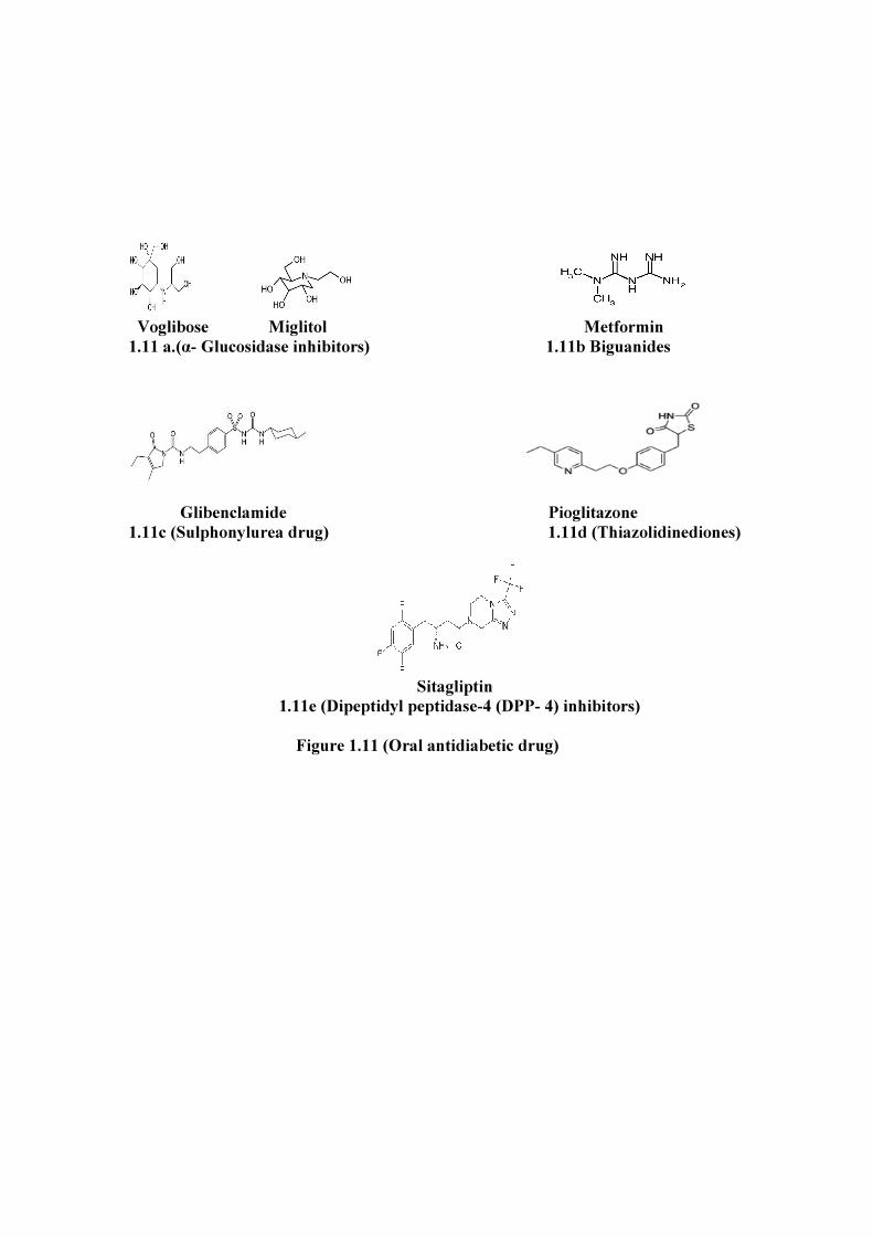
Voglibose Miglitol Metformin 1.11 a.(α- Glucosidase inhibitors) 1.11b Biguanides
Glibenclamide Pioglitazone 1.11c (Sulphonylurea drug) 1.11d (Thiazolidinediones)
Sitagliptin 1.11e (Dipeptidyl peptidase-4 (DPP- 4) inhibitors) Figure 1.11 (Oral antidiabetic drug)
Chapter 1: Introduction and Review of Literature
Ph.D. Thesis 2013 University of Delhi 29
mechanisms: increasing expression, synthesis and release of adiponectin from fat cells; increasing expression of genes that increase glucose oxidation and lowering plasma free-fatty acid levels (Gupta et al, 2009).(Figure 1.11d). 1.14.3.5 Dipeptidyl Peptidase-4 (DPP- 4) Inhibitors DPP-4 inhibitors offer a new therapeutic approach for the management of patients with type 2 diabetes (Charbonnel et al, 2006).Sitagliptin is a oncedaily, orally active, competitive and fully reversible inhibitor of dipeptidyl peptidase-4, the enzyme that is responsible for the rapid degradation of the incretin hormone glucagon-like peptide-1. It is the first in this new class of antihyperglycemic agents to gain regulatory approval for the treatment of type 2 diabetes, both as a monotherapy and for use in combination with metformin or a thiazolidinedione. Sitagliptin improves glycemic control by reducing both fasting and postprandial glucose concentrations, leading to clinically meaningful reductions in glycosylated hemoglobin levels (Deacon, 2007). (Figure 1.11e). 1.15 MEDICINAL PLANTS WITH ANTIDIABETIC EFFECT Despite considerable progress in the treatment of diabetes by oral hypoglycemic agents, search for newer drugs continues because the existing synthetic drugs have several limitations. The herbal drugs with antidiabetic activity are yet to be commercially formulated as modern medicines, even though they have been acclaimed for their therapeutic properties in the traditional systems of medicine (Wadkar et al, 2008). The plants provide a potential source of hypoglycemic drugs because many plants and plant derived compounds have been used in the treatment of diabetes. Many Indian plants have been investigated for their beneficial use in different types of diabetes and reports occur in numerous scientific journals. Ayurveda and other traditional medicinal system for the treatment of diabetes describe a number of plants used as herbal drugs. Hence, they play an important role as alternative medicine due to less side effects and low cost. Hyperglycemia is involved in the etiology of development of diabetic complications. Hypoglycemic herbs increase insulin secretion, enhance glucose uptake by adipose or muscle tissues and inhibit glucose absorption from intestine and glucose production from liver (Hongxiang et al, 2009). Insulin and oral hypoglycemic agents like sulphonylureas
Chapter 1: Introduction and Review of Literature
Ph.D. Thesis 2013 University of Delhi 30
and biguanides are still the major players in the management but there is quest for the development of more effective anti-diabetic agents. I. Aegle marmelose (L) (Bottle guard) It belonged to Rutaceae family. A species of tree native to India, it is present throughout Southeast Asia. A significant decrease in liver glycogen of diabetic rats is reversed to almost the normal level by the leaf extract. An active principle i.e. Aegeline and Fagarine in A. marmelos leaf extract has hypoglycemic activity similar to insulin treatment (Ponnachan et al, 1993). It also decreases the blood urea and serum cholesterol.
II. Annona squamosa L (Sweet sop) It belonged to Annonaceae family. It is a small well-branched tree or shrub, grows at lower altitudes. Administration of 15 mg/kg/day of isolated quercetin-3-O-glucoside from Annona squamosa leaves for 10 consecutive days to the hyperglycemic animals reverse these effects and simultaneously inhibits the activity of hepatic GIucose-6-phosphatase. It further decreases the hepatic and renal lipid peroxidation with a concomitant increase in the activities of antioxidative enzymes, such as Catalase and Superoxide dismutase as well as glutathione content, indicating its safe and antiperoxidative effects (Panda and Kar, 2007). III. Cassia Auriculata L.(Tanners) It belonged to Caesalpiniaceae family. It occurs in the dry regions of India and Sri Lanka. Oral administration of cassia auriculata leaf extract (CLEt) to mildly diabetic (MD) and severely diabetic (SD) rats at a dose of 400 mg/kg once a day for 15 days shows significant reduction in fasting blood glucose, also enhances the activity of hepatic hexokinase, phosphofructokinase, suppresses glucose-6-phosphatase and fructose-l,6-bisphosphatase in both MD and SD rats. Histopathological examination of pancreatic sections reveals increased number of islets and beta-cells in CLEt-treated MD as well as SD rats (Gupta et al, 2010).

Chapter 1: Introduction and Review of Literature
Ph.D. Thesis 2013 University of Delhi 31
IV. Ficus bengalensis L. (Banyan tree) It belonged to Moraceae family. At a dose of 100 mg/kg for one month, there is significant decrease in blood and urine sugar, certainm lipid components in serum, tissues and glucose-6- phosphatase activity in liver, but increase in body weight, the activities of hexokinase and HMG-COA reductase in tissues as compared to diabetic control. The mechanism of action of the principle (Leucopelargonidin, β sitosterol α-D glucose) may be related to its protective/inhibitory action against the insulin degradative processes (Shukla et al, 2004; Augusti et al, 1994). V. Trigonella foenum graecum L. (Fenugreek) It belonged to Moraceae family. Common name is ‘Fenugreek’. Used both as an herb (the leaves) and as a spice (the seed) and cultivated worldwide as a semi-arid crop. Oral administration of 50 mg/kg of its hypoglycemic principle (Flavonoid–C-Glycosides) isolated from seeds, produces fall in the blood glucose levels in both normal as well as diabetic rats (Shan et al, 2008). VI. Psidium guajava L. (Guava) It belonged to Myrtaceae family. An indigenous medicinal plant used to control diabetes in Indian System of Medicine. Ethanol stem bark extract and its hypoglycemic principle (quercetin-3-glycoside) exhibits statistically significant hypoglycemic activity in alloxan-induced hyperglycemic rats but devoid of hypoglycemic effect in normal and glucose loaded rats (OGTT) (Mukhtar et al, 2006). VII. Aloe vera (L) Burm. (Aloe) It belonged to Asphodelceae family. It grows in arid climates and is widely distributed in Africa, India and other arid areas. Aloe vera gel (phytosterols) at 200 mg/kg possesses significant antidiabetic activity.(Misawa et al, 2008) It has also cardioprotective activity, reduces the increased TBARS, maintains the Superoxide dismutase and Catalase activity up to the normal level and increases reduced glutathione by four times in diabetic rats (Jain et al, 2010).

Chapter 1: Introduction and Review of Literature
Ph.D. Thesis 2013 University of Delhi 32
VIII. Andrographis paniculata Burm. (Indian Echinacea) It belonged to Canthaceae family. It is a herbaceous plant native to India, Sri Lanka and widely cultivated in southern Asia. Oral administration of andrographis (andographolide) significantly decreases blood glucose. It increases the activity of SOD and Catalase. (Dandu and Inamdar, 2009). IX. Ocimum sanctum L. (Tulsi) It belonged to Lamiaceae family. Since ancient times, this plant is known for its medicinal properties. The aqueous extract of leaves shows significant reduction in blood sugar level in both normal and alloxan induced diabetic rats (Vats et al, 2002). Significant reduction in fasting blood glucose, uronic acid, total amino acid, total cholesterol, triglyceride and total lipid indicate the hypoglycemic and hypolipidemic effects of Eugenol (which is purified from Ocimum sanctum) in diabetic rats (Rai et al, 1997). X. Panax ginseng C. Meyer. (Chienese ginseng) It belonged to Araliaceae family. The roots are taken orally in the treatment of type II diabetes. Ginseng species (panaxosid) shows antihyperglycemic activity associated with increased peroxisome proliferator-activated receptor gamma expression and adenosine monophosphate-activated protein kinase phosphorylation in liver and muscle (Lim et al, 2009). XI. Phyllanthus amarus (Bhojpatra) It belonged to Euphobiaceae family. A traditional Ayurveda herb used in southern India. Ellagic acid which purified from of P. amarus has potential antidiabetic activity. This plant extract also anti-oxidant activity as it could inhibit lipid peroxidation, and scavenge hydroxyl and superoxide radicals in vitro. (Raphel et al, 2002). XII. Telfaria occidentalis Hook. (African vine plant) It belonged to Cucurbitaceae family. It is a tropical vine grown in West Africa as a leaf vegetable and for its edible seeds. The aqueous extract given orally in 1 g/kg to the mice 60 minutes before glucose administration reduces the blood glucose level from day two when compared with that of chlorpropamide (200 mg/kg) under the

Chapter 1: Introduction and Review of Literature
Ph.D. Thesis 2013 University of Delhi 33
same conditions. The results of this study indicates that the aqueous extract of the leaves of T. occidentalis possess hypoglycemic activity (Aderibigbe et al, 1999). XIII. Lepidium sativum L. (Fluted pumpkin) It belonged to Brassicaceae family. It is a fast-growing, edible herb. The aqueous extract at a dose of 10 mg/kg/h causes a potent inhibition of renal glucose reabsorption which in turn reduces blood sugar. This renal effect is at least one mechanism explaining the hypoglycemic activity of this plant in normal and diabetic rats (Eddouks et al, 2008). XIV. Mangifera indica L.(Mango) It belonged to Anacardiaceae family. The mangiferin isolated compound from mangifera indica produces reduction of blood glucose level in normoglycemic and glucose-induced hyperglycemia,but does not have any effect on streptozotocin-induced diabetic mice under the same conditions when compared with that of an oral dose of chlorpropamide. The result indicates that the aqueous extract of the leaves of M. indica possess hypoglycemic activity (Aderibigbe et al 2001). XV. Pterocarpus Marsupium Roxb.(Indian Kino Tree) An aqueous extract of wood shows hypoglycemic activity in alloxan induced diabetic rats at oral dose of 250 mg/kg. The butanol subfraction of alcohol extract of bark exhibits significant antidiabetic activity by ameliorating blood glucose and lipid parameters in alloxan induced diabetic rats (Mukhtar et al, 2005). XVI. Eugenia Jambolana (Jamuna) Its botanical name is myrtaceae. The water and ethanolic extracts of the fruit-pulp of E. jambolana elicit antihyperglycemic effect. Water extract was found to be more effective than the ethanolic extract in reducing fasting blood glucose and improving blood glucose in glucose tolerance test. Arayne et al reported that, the methanolic extract of E. jambolana inhibited glucose diffusion in vitro.(Sharma et al, 2011) The flavonoid rich extract from E. jambolana seeds elicits both hypoglycemic effects by stimulating increase in insulin release in vitro from pancreatic islets and antihyperlipidemic effects in streptozotocin induced diabetic rats(Ravi et al, 2005).

Chapter 1: Introduction and Review of Literature
Ph.D. Thesis 2013 University of Delhi 34
Table 1.2: List of antidiabetic plants and their mode of actions S. No. Botanical name
and family Common
name Parts used Active Principle Therapeutic action
1 Agele Marmelose (Rutaceae) Bottle guard Leaf Ageline and Fargerin Lowers blood glucose via increase insulin secretion (Ponnachan et al, 1993) 2 Annona Squamosa (Annonaceae) Sweetsop Leaf Quercetin-3-o glucoside Decrease in blood glucose Level via inhibit the glucose-6-phosphatase activity
improved carbohydrate metabolism(Panda and Kar, 2007) 3 Cassia auriculata (Caesalpiniaceae) Tanners Leaf - It have pancreatic effect and improve blood glucose levels.(Gupta et al, 2010) 5 Ficus bengalensis (Moraceae) Banyan tree
Bark Leucopelargonidin, β sitosterol α-D glucose
Inhibits activity from liver and kidney, stimulates insulin secretion (Shukla et al, 2004)
6 Trigonella fenugraceum (Moraceae) Fenugreek Leaves Flavonoid–C-
glycosides Stimulates synthesis and/or release of insulin from -cells (Shan et al, 2008) 7 Psidium guajava L. (Myrtaceae) Guava Bark and leaf Quercetin-3-o glucoside Antihyperglycemic, glucose metabolism (Mukhtar et al, 2006) 8 Aloe vera (L) (Asphodelaceae) Aloe Leaf Aloe Gel (Phytosterols) Antidiabetic, cardioprotective and antioxidant activity. (Mishra et al ,2007) 9
Andrographis paniculata (Canthaceae)
Indian Echinacea
Fruit Andographolide Antidiabetic and antioxidant (Dandu and Inamdar, 2009)
10 Ocimum sanctum L. (Lamiaceae) Tulsi Leaves, Seeds Eugenol Hypoglycemic and Hypolipidemic action(Rai et al, 1997)
11 Panax ginseng L (Araliaceae)
Chinese ginseng
Roots Panaxoid Decreased glucose level, Increased PPAR Y expression and PKC activity(Lim
et al, 2009)
12 Phyllanthus amarus Schum (Euphorbiaceae) Bhojpatra Roots and
fruits
Ellagic acid Antidiabetic and antioxidant activity (Raphel et al, 2002)
13 Telfaria Occidentalis (Cucurbitaceae)
African vine plant Fruit
Inhibits intestinal glucose absorption, en-hancement of mobility. (Aderibigbe et al, 1999)
14 Lepidium setivum (Brassicaceae) Fluted Pumpkin’
Leaf Inhibition of renal absorption and decreased glucose levels (Eddouks et al,
2008) 15 Mangifera Indica (Anacardiaceae) Mango Leaf and bark Mangiferin glucoside Antidiabetic and Hypolipidemic activity (Aderibigbe et al, 2001) 16 Eugenia Jambolana (Myrtaceae) Jamuna Fruit Pulp,
Seed Hypoglycemic due to Insulin release from pancreatic islets (Sharma et al, 2011)
17 Petrocarpus Marsupium (Fabaceae) Indian Kino Tree Wood Increased Blood glucose absorption and decreased glucose value (Mukhtar et
al, 2005) 18 Momordica charantia
(Cucurbiteceae) Bitter melon Fruit Known as ‘Vegetable insulin’ decreased post prandial glucose and increased α glucosidase activity (Shih et al, 2009)

Chapter 1: Introduction and Review of Literature
Ph.D. Thesis 2013 University of Delhi 35
XVII. Momordica Charantia (Bitter Melon) M. charantia is commonly known as vegetable insulin. An oral sucrose tolerance test reveals that administration of aqueous extract (AE), methanol fraction (MF) or methanol insoluble fraction (MIF) each significantly suppresses plasma glucose levels at 30 min as compared with control. In addition, the plasma insulin level at 30 min also lowers after MF administration than the control in the oral sucrose tolerance test, these results demonstrates that bitter melon suppresses postprandial hyperglycemia by inhibition of á-glucosidase activity. (Shih et al, 2009). 1.16. MUSA SAPIENTUM VAR. PARADISIACA Banana is major food crop grown globally and consumed in more than 100 countries through out tropics and subtropics (INFOMUSA, 2000). In devoloping countries it is the fourth most important crop after rice, wheat and maize. Total world production of banana in 2005 was over 100 milllion tones. Two major sweet banana producing countries in the world are India and Brazil. Commercially all banana parts have several uses. Banana fruit can be eaten raw or cooked (deep fried, dehydrated, baked in skin, steamed) can be processed into flour and can be fermented for the production of beverages such as banana juice, vinegar, wine. Other parts of banana like pseudostem is used for cooking, flower is eaten in southeast asia. Banana leaves are used for wrapping foods.
Musa sapientum are large, herbaceous monocots belonging to the musaceae family with a robust tree like pseudostem, a crown of large elongated oval deep green leaves (up to 365 cm in length and 61 cm in width), with a prominent midrib. The "trunk" or pseudostem is not a true stem, but only the clustered, cylindrical aggregation of leaf stalk bases. Leaves are among the largest of all plants. Margins are entire and venation is pinnate; leaves tear along the veins in windy conditions, giving a feathered or tattered look. There are 5-15 leaves on each plant, with 10 considered the minimum for properly maturing a bunch of fruit. The perennial portion of the plant is the rhizome, which may weigh several pounds. It is often called a corm. It produces suckers, or vegetative shoots, which are thinned to 2 per plant - one "parent" sucker
Chapter 1: Introduction and Review of Literature
Ph.D. Thesis 2013 University of Delhi 36
for fruiting and one "follower" to take the place of the parent after it fruits and dies back. It also produces roots and serves as a storage organ for the plant (Figure 1.12). 1.16.1 Phytochemistry of Musa Sapientum Var. Paradisiaca One medium banana contains 1.29 grams of protein, 105 calories and 3.1 grams of dietary fiber. Potassium - 422 mg, Phosphorus - 26 mg, Magnesium - 32 mg, Calcium - 6 mg, Sodium - 1 mg, Iron-0.31mg, Selenium-1.2mcg, Manganese-0.319mg, Copper-0.092mg and Zinc-0.18mg. (Deborah, Eldad, & Guy,2011)
Flowers consist of tannins, saponins, reducing and non reducing sugars, sterols and triterpenes (Pradeep et al., 1986). The pulp protein is rich in arginine, aspartic acid, glutamic acid, methionine and tryptophan ( Adegboyega and ketiku, 2006). A new 1,7 bis(4-hydroxy phenyl) hepta- 4(E),6(E)-diene-3-one(5)were isolated from ethyl acetate soluble fraction of the methanolic extract of fruits( Jang et al., 2002). From peeled fruits of Musa Sapientum two new acyl steryl glycosides Sitoindoside-III and Sitosterol myo-inosityl-beta-D-glucoside have been isolated by gradient solvent extraction and extensive chromatography(CC, GC, TLC and HPLC) ( Ghosal,1985). Three forms of α-glucan phosphorylase from mature banana fruit pulp separated by DEAE-cellulose chromatography (Singh and Sanwal,1975). Two forms (A and B) of starch phosphorylase were found in the mature banana leaf by polyacrylamide gel electrophoresis and DEAE-cellulose chromatography. 1.16.2 Pharmacological Activities The various effects of Musa Sapientum are documented in traditional as well as in recent scientific literature. The main pharmacological effects of this plant are antiulcer, wound healing, antioxidant, antidote for snake bite, hypoglycemic, atherogenic, augmentation of skeletal muscles. Antiulcer activity orally administered banana pulp powder was shown to have significant antiulcerogenic activity in rats subjected to Aspirin, Indomethacin, Phenyl butazone, Prednisolone, Cysteamine and in guinea pigs subjected to hisamine. Banana pulp powder not only increased mucosal thickness but also significantly increases thymidine (incorporation into mucosal DNA). This study suggests that banana powder treatment not only strenghthens

Figure:-1.12: Musa sapientum Var. Paradisiaca
Chapter 1: Introduction and Review of Literature
Ph.D. Thesis 2013 University of Delhi 37
mucosal resistance against ulcerogens but also promotes healing by inducing cellular proliferation (Goel et al, 1986).Methanolic extracts of plantain banana pulp was evaluated for its antiulcer and antioxidant activities in 2 hr cold restraint stress and anti H.pylori activity in vitro. The extract (50mg/kg twice daily for 5 days) showed significant antiulcer effect and antioxidant activity in gastric mucosa homogenates where it reversed the increase in ulcer index, lipid peroxidation and superoxide dismutase values induced by stress (Goel et al, 2001). An extract obtained from stem juice induces twitch augmentation in skeletal muscles. The mechanism of action was investigated in the mouse hemi-diaphragm preparation. The results are consistent with an action of banana tree juice on the molecule responsible for excitation-contraction coupling in skeletal muscle resulting in mobilization of intracellular Ca2+ (Singh and Drydem, 1990). The fresh juice of Musa stem (Puttubale) was tested for its antilithiatic activity. Musa stem juice (3ml rat/day, orally) was found to be effective in reducing the stone formation and also in dissolving the pre-formed stones (Prasad et al, 1993). Musa sapientum var. paradisiaca extract does not show protection against the toxic effects of snake venoms in vivo but it was very effective when the experiment were done in vitro (Borges et al,2005). 1.16.3 Antidiabetic Activity The green fruit of M. paradisiaca has been reported to have hypoglycemic effect due to stimulation of insulin production and glucose utilization (Ojewole and Adewunmi, 2003). Fibers from M. paradisiacal fruit increased glycogenesis in the liver and lowered fasting blood glucose (Usha et al., 1989). Antihyperglycemic effect of the hydromethanolic extract of M. paradisiaca root has been found significant ( Mallick et al., 2007). Musa sapientum showed antihyperglycemic effect in hyperglycemic rabbit (Alarcon-Aguilara et al., 1998). The chloroform extract of flowers of M. sapientum showed blood glucose and Glycosylated haemoglobin reduction and total hemoglobin increase after oral administration in rats (Pari and Maheshwari, 1999). It also controls lipid peroxidation in diabetes (Pari and Maheshwari, 2000). Isolated pectin from the juice of the inflorescence stalk of M. sapientum increases the glycogen synthesis, decreases glycogenolysis and gluconeogenesis (Gomathy et al, 1990).
Chapter 1: Introduction and Review of Literature
Ph.D. Thesis 2013 University of Delhi 38
Literature survey revealed that central stem of Musa sapientum plant has not been studied for its hypoglycemic effect. This part of plant is used for making edible preparation like curry and chutney in south india. In our intial studies we found this part of the plant to possess significant hypoglycemic activity. Therefore this work has been planned to study antihyperglycemic effect of central stem of Musa sapientum with following aims and objectives.