introduction - nimhans
TRANSCRIPT

Dept of Neuropathology, NIMHANS
Instructions for clinicians, patients and users
Document ID: NP/HP/INS
Page 1 of 26
Prepared by: Dr. Anita Mahadevan
Approved by: Quality Manager
Issued by: Head of Dept/ Lab Director Dr Yasha TC
Amendment No:
Amendment Date:
Issue No: 02 Issue Date: 20-04-18 Copy No: Page 1 of 26
Introduction
What is this manual about?
This manual is designed to provide an overview of the services available in the
Neuropathology Department and serve as a quick reference guide for all users.
Laboratory Management is committed to:
Ensure stringent adherence to quality in all laboratory procedures that meet requirements
of internal and external quality assessment tests.
Ensure appropriate collection, transport and processing of samples to ensure optimal
performance of all the tests required.
Ensure reports are accurate, timely and clinically useful.
Ensure use of procedures and methods that are up to date with current practices.
Ensure continued staff training at all levels to keep them updated with recent advances in
the field.
Periodically assess user satisfaction by feedback forms.
Copies of this manual are available in specimen collection centres, wards, OTs, casualty,
short stay wards. Electronic version is available on the NIMHANS website.
Contact details:
Location: Department of Neuropathology, First floor,
Administrative block, NIMHANS
Postal address: Department of Neuropathology,
NIMHANS, Hosur Road, Bangalore 560 029
Phone: Off: 080-26995130
(for report enquiries, call between 9.30-4.30pm on working days
only)
Email: [email protected]
Website: www.nimhans.kar.nic.in/neuropathology/.default.htm

Dept of Neuropathology, NIMHANS
Instructions for clinicians, patients and users
Document ID: NP/HP/INS
Page 2 of 26
Prepared by: Dr. Anita Mahadevan
Approved by: Quality Manager
Issued by: Head of Dept/ Lab Director Dr Yasha TC
Amendment No:
Amendment Date:
Issue No: 02 Issue Date: 20-04-18 Copy No: Page 2 of 26
Table of Contents
OUR SERVICES .......................................................................................................... 3
GETTING STARTED ................................................................................................ 4
Location of the department & its sections ................................................... 4
Department working hours ............................................................................. 5
Departmental faculty & contact numbers.................................................... 5,6
List of tests offered & turn-around time (overview) ............................... 7
GENERAL INSTRUCTIONS FOR ALL SAMPLES………………………………..8
Intraoperative frozen…………………………………………………………………....….9
Endoscopic/Stereotactic biopsy…………………………………………………9, 10
Brain biopsies/Epilepsy surgery………………………………………………...11
Cyst fluid/abscess aspirate ....................................................................... ….11
Muscle biopsy .....................................................................................................11,12
Nerve Biopsy ......................................................................................................13
Skin biopsy ..........................................................................................................14
SAMPLE RECEIPT .................................................................................................15
Sample acceptance criteria ............................................................................15
Sample “non-compliance” criteria............................................................... 16
Precautions for biopsy of suspected “Prion” /CJD diseases ..............16
REPORTING OF RESULTS .................................................................................17
Release of results ..............................................................................................17
Telephonic enquiries for reports……………………………………………… 17
Intraoperative frozen reports…………………………………………………….17
Reporting of emergency results(“critical alert”) .................................... 17
Retention of specimens/blocks/reports after reporting.....................18
Review of reports……………………………………………………………………... 18
SUMMARY OF TESTS……………………………………………………………………19
Histopathology Request form………………………………………………………..21
Sample Transport requirements……………………………………………22
Dept of Neuropathology, NIMHANS
Instructions for clinicians, patients and users
Document ID: NP/HP/INS
Page 3 of 26
Prepared by: Dr. Anita Mahadevan
Approved by: Quality Manager
Issued by: Head of Dept/ Lab Director Dr Yasha TC
Amendment No:
Amendment Date:
Issue No: 02 Issue Date: 20-04-18 Copy No: Page 3 of 26
Our Services
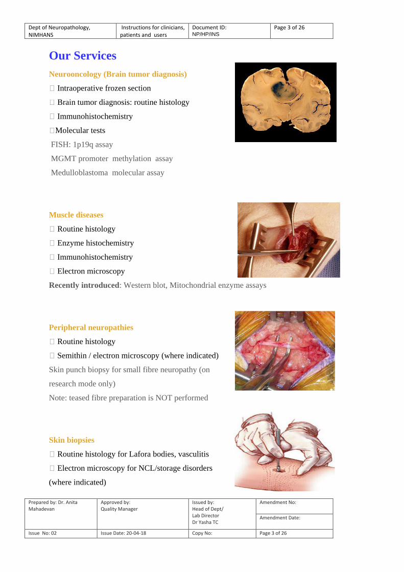
Neurooncology (Brain tumor diagnosis)
Intraoperative frozen section
Brain tumor diagnosis: routine histology
Immunohistochemistry
Molecular tests
FISH: 1p19q assay
MGMT promoter methylation assay
Medulloblastoma molecular assay
Muscle diseases
Routine histology
Enzyme histochemistry
Immunohistochemistry
Electron microscopy
Recently introduced: Western blot, Mitochondrial enzyme assays
Peripheral neuropathies
Routine histology
Semithin / electron microscopy (where indicated)
Skin punch biopsy for small fibre neuropathy (on
research mode only)
Note: teased fibre preparation is NOT performed
Skin biopsies
Routine histology for Lafora bodies, vasculitis
Electron microscopy for NCL/storage disorders
(where indicated)
Dept of Neuropathology, NIMHANS
Instructions for clinicians, patients and users
Document ID: NP/HP/INS
Page 4 of 26
Prepared by: Dr. Anita Mahadevan
Approved by: Quality Manager
Issued by: Head of Dept/ Lab Director Dr Yasha TC
Amendment No:
Amendment Date:
Issue No: 02 Issue Date: 20-04-18 Copy No: Page 4 of 26
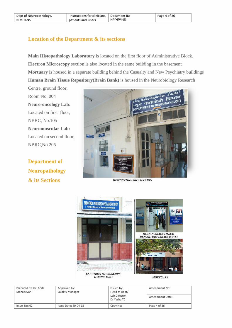
Location of the Department & its sections
Main Histopathology Laboratory is located on the first floor of Administrative Block.
Electron Microscopy section is also located in the same building in the basement
Mortuary is housed in a separate building behind the Casualty and New Psychiatry buildings
Human Brain Tissue Repository(Brain Bank) is housed in the Neurobiology Research
Centre, ground floor,
Room No. 004
Neuro-oncology Lab:
Located on first floor,
NBRC, No.105
Neuromuscular Lab:
Located on second floor,
NBRC,No.205
Department of
Neuropathology
& its Sections

Dept of Neuropathology, NIMHANS
Instructions for clinicians, patients and users
Document ID: NP/HP/INS
Page 5 of 26
Prepared by: Dr. Anita Mahadevan
Approved by: Quality Manager
Issued by: Head of Dept/ Lab Director Dr Yasha TC
Amendment No:
Amendment Date:
Issue No: 02 Issue Date: 20-04-18 Copy No: Page 5 of 26
Departmental working hours
SECTION Opening hours Closing
time
Histopathology
Weekdays (Mon-Sat)
Second Saturday, & all closed Govt
holidays declared by Institute
(Sunday holiday)
9.00 am
9.30am
4.30pm
3.30pm
Electron Microscopy
Weekdays (Mon-Sat) Only
Closed on second Saturday, all
Sundays, & Govt holidays declared
by the Institute
9.00 am
4.30 pm
Mortuary
Staff and faculty are on call (in
campus)
and functions 24x7
9.00am
(all days including
Sundays & holidays)
4.30pm

Dept of Neuropathology, NIMHANS
Instructions for clinicians, patients and users
Document ID: NP/HP/INS
Page 6 of 26
Prepared by: Dr. Anita Mahadevan
Approved by: Quality Manager
Issued by: Head of Dept/ Lab Director Dr Yasha TC
Amendment No:
Amendment Date:
Issue No: 02 Issue Date: 20-04-18 Copy No: Page 6 of 26
Departmental faculty& contact numbers
SECTION Faculty Contact No.s
Histopathology
Dr. Yasha TC(HOD)
Dr. Vani Santosh
Dr. N. Gayathri
Dr. Anita Mahadevan
Dr. Nandeesh BN
(2699)5132
(2699)5133
(2699)5131
(2699)5137
(2699)5596
Electron Microscopy Dr. BK. Chandrashekar Sagar
Dr. N. Gayathri (2699)5738
(2699)5131
Mortuary Dr. Anita Mahadevan
Dr. Yasha TC
Dr. Vani Santosh
Dr. Nandeesh BN
(2699)5137
(2699)5133
(2699)5132
(2699)5596
Human BrainTissue
Repository Dr. Anita Mahadevan (2699)5786
(2699)5137 Neurooncology lab Dr Vani Santosh
Dr. Yasha TC
Dr Nandeesh BN
(2699)5583
Neuromuscular lab Dr. N Gayathri
Dr. Anita Mahadevan
(2699)5094
Section Phone number
SECTION Contact No.s
Office (2699)5130
Residents Room ( DM and Post Doctoral Fellows) (2699)5134
Discussion Room (2699)5135
Histopathology Laboratory-1 (2699)5136
Histopathology Laboratory-2 (2699)5790
Immunohistochemistry laboratory (2699)5736
Electron microscopy (2699)5138
Mortuary (2699)5213
Brain Bank (Neurobiology Research Centre) (2699)5786
Museum(Neurobiology Research Centre) (2699)5582
Neurooncology Lab (2699)5583
Neuromuscular Lab (2699)5094
Dept of Neuropathology, NIMHANS
Instructions for clinicians, patients and users
Document ID: NP/HP/INS
Page 7 of 26
Prepared by: Dr. Anita Mahadevan
Approved by: Quality Manager
Issued by: Head of Dept/ Lab Director Dr Yasha TC
Amendment No:
Amendment Date:
Issue No: 02 Issue Date: 20-04-18 Copy No: Page 7 of 26
LIST OF TESTS & TURN AROUND TIME
Sl
No
Tissue type Specific Test Turn around
Time*,#
1 Tumor tissue (Formalin
fixed )
Histopathology
Special stains or Immunohistochemsitry
4 days
7 days
2 Fresh tissue Intra-operative smear/squash 30 mins after
sample receipt
3 Epilepsy surgery Histopathology
& Immunohistochemistry
15 days
4 Fresh Muscle
Histopathology & enzyme histochemistry
Immunohistochemistry
4-7days
4 weeks
5 Nerve biopsy
(gluteraldehyde)
Histopathology
examination
4-7 days
6 Skin (formalin/
glutaraldehyde)
Routine histopathology Examination
Electron microscopy
4-7 days
6 weeks
7 Liver biopsy
(formalin/ alcohol)
Histopathology with special stains
4-7days
8 Brain biopsy
(Fresh )
Histopathology &
immunohistochemsitry
10 days
9 Paraffin block
Routine
Immunohistochemistry
4days
7 days
10 Slides Histopathology 2 days
*In working days
# General guidelines: Routine HPE - 4 days
Special stains/Immunohistochemistry - 7 days

Dept of Neuropathology, NIMHANS
Instructions for clinicians, patients and users
Document ID: NP/HP/INS
Page 8 of 26
Prepared by: Dr. Anita Mahadevan
Approved by: Quality Manager
Issued by: Head of Dept/ Lab Director Dr Yasha TC
Amendment No:
Amendment Date:
Issue No: 02 Issue Date: 20-04-18 Copy No: Page 8 of 26
GENERAL INSTRUCTIONS FOR ALL SAMPLES
All samples and requests are screened for the following:
(i) All samples must be accompanied by a histopathology request form, completely filled with
all relevant details. The request form for inpatient NIMHANS cases and outside Referral cases
are different and non exchangeable.
(ii) Request should be from treating physician. Requests from patient party are not entertained
(iii) All samples should have at least two identifiers – patient name, and hospital number,
hospital name/referring doctor (if from outside) with age and gender.
(iv) Personal identifiers of the sample and the request form should match
(v) Appropriate fixative/ container must be used; and transported/ stored in appropriate
conditions to ensure stability of sample and optimum results
Sample receipt timings:
Mon-Sat: 9.00am – 4.30pm
Second Saturdays, Institute holidays: 9.00am-3.30pm
Sundays are holidays.

Dept of Neuropathology, NIMHANS
Instructions for clinicians, patients and users
Document ID: NP/HP/INS
Page 9 of 26
Prepared by: Dr. Anita Mahadevan
Approved by: Quality Manager
Issued by: Head of Dept/ Lab Director Dr Yasha TC
Amendment No:
Amendment Date:
Issue No: 02 Issue Date: 20-04-18 Copy No: Page 9 of 26
Neurosurgical biopsies
Intraoperative frozen – facility available ONLY for hospitals located close to NIMHANS
Caution
1. Inform pathologist prior to sending sample for frozen
2. Samples should be sent immersed in sufficient volumes of saline to prevent drying up in
transit.
3. Samples SHOULD reach the Lab within 30mins-1hour during lab working hours.
4. Intraoperative frozen requests after lab working hours (4.30 pm) or holidays CANNOT be
performed.
5. Any sample associated infection risk SHOULD BE CLEARLY STATED ON THE
REQUEST FORM (eg., retroviral/HBsAg/suspected prion disease) and marked
„BIOHAZARD‟
Neurosurgical biopsies
Surgical samples should be sent in 10% formalin
Ensure sample is labelled correctly, placed in sufficient volumes of formalin and is
accompanied by complete clinical details
All sample transport requirements (see below for details) should be strictly adhered to.
Small biopsies (stereotactic/CT/MR guided/endoscopic)
Samples to be sent in 10% FORMALIN.
If tissues are collected, in multiple bottles, the bottles to be labelled
serially clearly INDICATING sites of biopsy

Dept of Neuropathology, NIMHANS
Instructions for clinicians, patients and users
Document ID: NP/HP/INS
Page 10 of 26
Prepared by: Dr. Anita Mahadevan
Approved by: Quality Manager
Issued by: Head of Dept/ Lab Director Dr Yasha TC
Amendment No:
Amendment Date:
Issue No: 02 Issue Date: 20-04-18 Copy No: Page 10 of 26
CAUTION
Diagnostic brain biopsy is a special test and requires extensive co-ordination between clinician
and pathologist BEFORE biopsy is done for optimal yield. Hence strictly follow the
instructions below:
1. Brief the pathologist as to complete clinical details prior to biopsy
2. Confirm date of biopsy previous day
3. Timings: Ensure samples reach within working hours
4. Indicate clearly if risk of infection (eg., retroviral status/HBsAg/prion etc) asit requires special
precautions from our side. Label biopsy as ‘BIOHAZARD’
Tests performed:
Routine and special stains;
Immunohistochemistry
EM and molecular tests if required
Neurosurgical biopsies
Diagnostic brain biopsies: In formalin
Biopsy site: based on clinical/imaging findings
Biopsy size: 1x1cm cube with overlying meninges (+ dura if pachymeningeal disease)
Fixative: 10% formalin
Note: Send in FRESH STATE for hospitals within Bangalore ONLY.
Please discuss with pathologists PRIOR to sending.
Epilepsy surgeries: In formalin
Label each sample clearly as to site
Fixative: 10% formalin
Aspirates
Caution: Any sample associated infection risk SHOULD
BECLEARLY STATED ON THE REQUEST FORM (eg., retroviral/ HbsAg/suspected prion
disease) and marked ‘BIOHAZARD.
Dept of Neuropathology, NIMHANS
Instructions for clinicians, patients and users
Document ID: NP/HP/INS
Page 11 of 26
Prepared by: Dr. Anita Mahadevan
Approved by: Quality Manager
Issued by: Head of Dept/ Lab Director Dr Yasha TC
Amendment No:
Amendment Date:
Issue No: 02 Issue Date: 20-04-18 Copy No: Page 11 of 26
Neurosurgical biopsies
Cyst fluid aspirates:
1. Fluids for pathological examination should be sent in
10% formalin
2. Additional sample for microbiological studies need to be submitted to the concerned lab*
lab in fresh state depending on diagnostic requirements (eg., for cytopsin/culture)
INFECTIVE LESIONS (EG., ABSCESS WITH PUS)
1. Send one sample in 10% formalin to Neuropathology
2. Send one sample (fresh) to concerned lab*.
* Microbiology lab for additional studies like culture (esp if fungal infection is suspected)
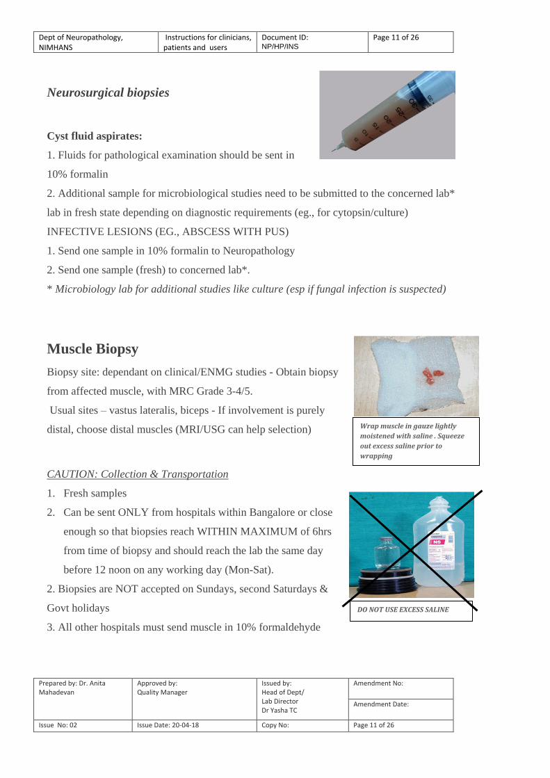
Muscle Biopsy
Biopsy site: dependant on clinical/ENMG studies - Obtain biopsy
from affected muscle, with MRC Grade 3-4/5.
Usual sites – vastus lateralis, biceps - If involvement is purely
distal, choose distal muscles (MRI/USG can help selection)
CAUTION: Collection & Transportation
1. Fresh samples
2. Can be sent ONLY from hospitals within Bangalore or close
enough so that biopsies reach WITHIN MAXIMUM of 6hrs
from time of biopsy and should reach the lab the same day
before 12 noon on any working day (Mon-Sat).
2. Biopsies are NOT accepted on Sundays, second Saturdays &
Govt holidays
3. All other hospitals must send muscle in 10% formaldehyde
DO NOT USE EXCESS SALINE
Wrap muscle in gauze lightly
moistened with saline . Squeeze
out excess saline prior to
wrapping
Dept of Neuropathology, NIMHANS
Instructions for clinicians, patients and users
Document ID: NP/HP/INS
Page 12 of 26
Prepared by: Dr. Anita Mahadevan
Approved by: Quality Manager
Issued by: Head of Dept/ Lab Director Dr Yasha TC
Amendment No:
Amendment Date:
Issue No: 02 Issue Date: 20-04-18 Copy No: Page 12 of 26
Tests possible ONLY on fresh samples
a. Enzyme histochemistry
b. Immunohistochemistry , Western blot for muscular dystrophies
c. Respiratory chain enzymes (for mitochondrial disorders)
Formalin Fixed samples:
Only Routine histology possible
For electron microscopy: muscle should be sent in 2.5%
glutaraldehyde
If paraffin blocks are sent:
1. Only routine H&E and special stains are possible
2. Enzyme histochemistry/immunohistochemistry/electron microscopy not possible
Muscle Biopsy
Biopsy instructions (for fresh samples)
Biopsy specifications
- Dissect with minimum trauma along the long axis of the muscle fibers
- Do not use electrocautery or a muscle clamp
- Additional bits if more tests are needed (eg., culture/enzyme studies for mitochondrial or
rare genetic disorders)
- Provide site, date and time of biopsy
- State what tests are required
- AVOID severely affected muscle with MRC grade <3
- AVOID muscle traumatized by IM injections or by EMG.
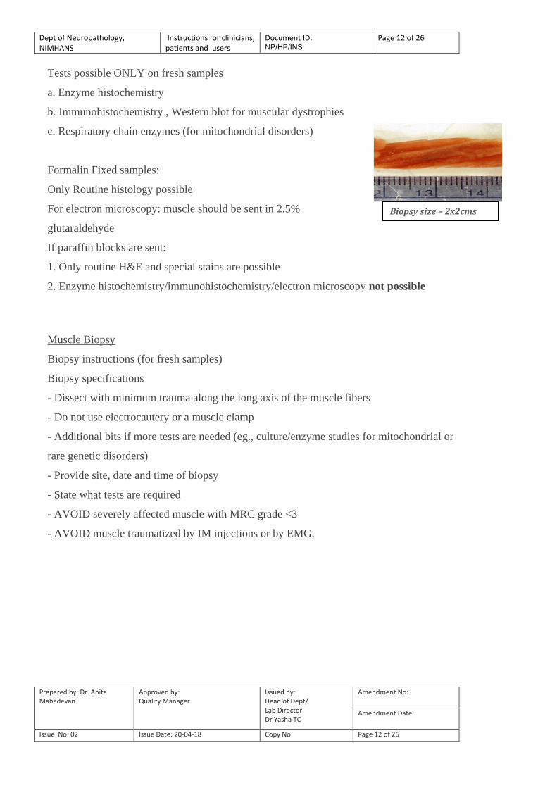
Biopsy size – 2x2cms

Dept of Neuropathology, NIMHANS
Instructions for clinicians, patients and users
Document ID: NP/HP/INS
Page 13 of 26
Prepared by: Dr. Anita Mahadevan
Approved by: Quality Manager
Issued by: Head of Dept/ Lab Director Dr Yasha TC
Amendment No:
Amendment Date:
Issue No: 02 Issue Date: 20-04-18 Copy No: Page 13 of 26
Nerve Biopsies
Biopsy site: dependant on clinical/nerve conductions
- Clinically & electrophysiologically involved nerve
- Sural nerve/superficial peroneal nerve/dorsal cutaneous nerve/superficial radial (commonly
biopsied)
Fixative:
- USE ONLY 2.5% gluteraldehyde
- DO NOT USE 10% buffered formalin
- DO NOT SEND FRESH (even for
metachromatic stains)
Tests performed
a. Routine Histopathology
b. Special stains:
- Myelin stains (Kulchitsky pal)
- Masson trichrome for collagen
- Cresyl violet for metachromatic granules
- PAS-diastase stain & Lugol‟s iodine (polyglucosan bodies)
- Fite Ferraco (for lepra bacilli)
- Congo red (for amyloid) etc
c. Immunohistochemistry (eg., ubiquitin for
polyglucosan bodies, neurofilament for axons etc)
d. Electron microscopy (USE ONLY 2.5% glutaraldehyde)
Nerve biopsies
CAUTION: BIOPSY SPECIFICATIONS
- Take a minimum length of 2 cms biopsy
- AVOID pinching/crushing nerve during procedure
- DO NOT tie nerve with thread
- AVOID COMMERCIALLY AVAILABLE CIDEX
- Use 2.5% glutaraldehyde
Dept of Neuropathology, NIMHANS
Instructions for clinicians, patients and users
Document ID: NP/HP/INS
Page 14 of 26
Prepared by: Dr. Anita Mahadevan
Approved by: Quality Manager
Issued by: Head of Dept/ Lab Director Dr Yasha TC
Amendment No:
Amendment Date:
Issue No: 02 Issue Date: 20-04-18 Copy No: Page 14 of 26
- Avoid 10% formalin as fixative
Skin Biopsies
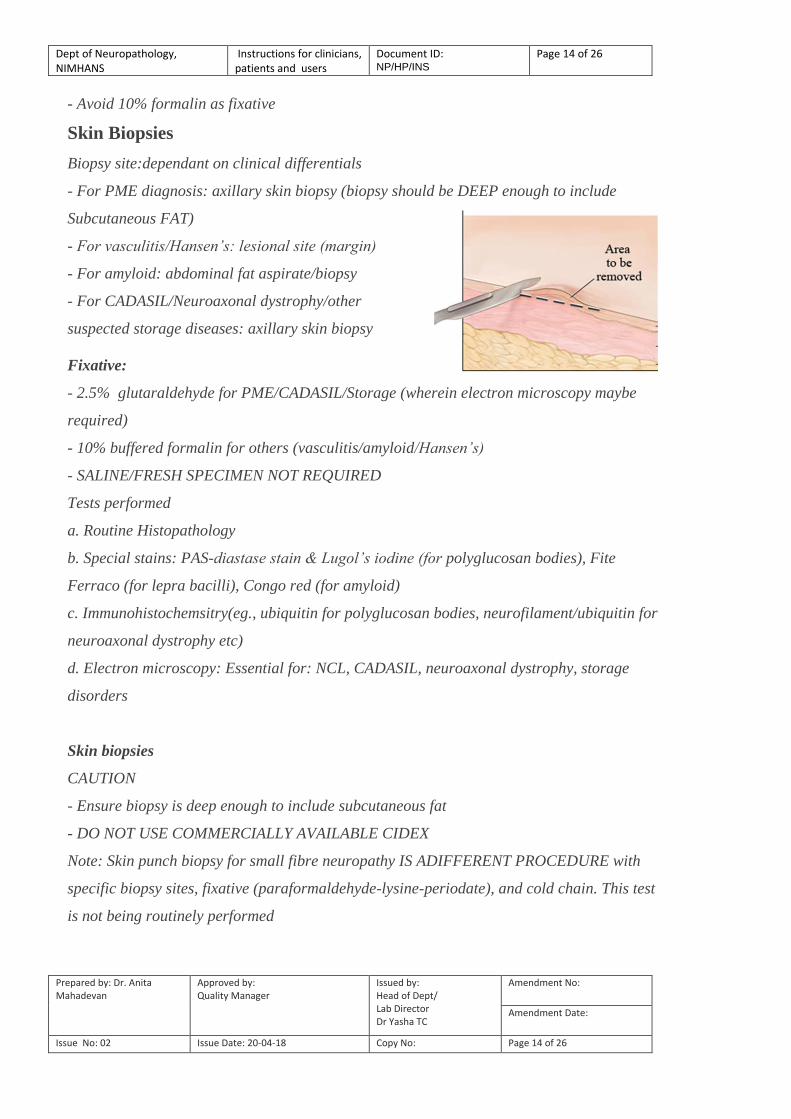
Biopsy site:dependant on clinical differentials
- For PME diagnosis: axillary skin biopsy (biopsy should be DEEP enough to include
Subcutaneous FAT)
- For vasculitis/Hansen’s: lesional site (margin)
- For amyloid: abdominal fat aspirate/biopsy
- For CADASIL/Neuroaxonal dystrophy/other
suspected storage diseases: axillary skin biopsy
Fixative:
- 2.5% glutaraldehyde for PME/CADASIL/Storage (wherein electron microscopy maybe
required)
- 10% buffered formalin for others (vasculitis/amyloid/Hansen’s)
- SALINE/FRESH SPECIMEN NOT REQUIRED
Tests performed
a. Routine Histopathology
b. Special stains: PAS-diastase stain & Lugol’s iodine (for polyglucosan bodies), Fite
Ferraco (for lepra bacilli), Congo red (for amyloid)
c. Immunohistochemsitry(eg., ubiquitin for polyglucosan bodies, neurofilament/ubiquitin for
neuroaxonal dystrophy etc)
d. Electron microscopy: Essential for: NCL, CADASIL, neuroaxonal dystrophy, storage
disorders
Skin biopsies
CAUTION
- Ensure biopsy is deep enough to include subcutaneous fat
- DO NOT USE COMMERCIALLY AVAILABLE CIDEX
Note: Skin punch biopsy for small fibre neuropathy IS ADIFFERENT PROCEDURE with
specific biopsy sites, fixative (paraformaldehyde-lysine-periodate), and cold chain. This test
is not being routinely performed

Dept of Neuropathology, NIMHANS
Instructions for clinicians, patients and users
Document ID: NP/HP/INS
Page 15 of 26
Prepared by: Dr. Anita Mahadevan
Approved by: Quality Manager
Issued by: Head of Dept/ Lab Director Dr Yasha TC
Amendment No:
Amendment Date:
Issue No: 02 Issue Date: 20-04-18 Copy No: Page 15 of 26
SAMPLE ACCEPTANCE CRITERIA
Samples should be accompanied by Histopathology request form from TREATING
PHYSICIAN (not patient party), completed in all respects with Priority Status of Request
(Urgent, intraoperative frozen requirement or Routine) CLEARLY INDICATED ON THE
REQUEST FORM
The following information must be documented in a LEGIBLE manner on the Request
form.
1. *Patient‟s Full Name (legible, preferably in CAPITALS).
2. *Patient‟s Age/Sex
3. Consultant name / address/ phone number/email address
4. *Specimen Type
5. Date and time of collection
6. Complete Clinical Details with results of relevant investigations and Clinical Diagnoses
considered
7. Test requested
8. Priority Status of Request (Urgent or Routine)
9. Details of any sample associated Infection Risk
Items marked with an * are minimum identifiers and failure to provide the data required
may lead to delay in processing of the sample and release of reports.
ALL Samples should be labelled with the following
1. *Patients‟ Full name (legible, preferably IN CAPITALS)
2. *Age/Sex
3. *Hospital name/referring doctor(with telephone number/email)
4. *Specimen Type
Paraffin blocks/slides submitted for review:
Request form should contain block/slide identification number
and number of blocks/slides submitted in addition to complete
clinical details and diagnoses considered.

Dept of Neuropathology, NIMHANS
Instructions for clinicians, patients and users
Document ID: NP/HP/INS
Page 16 of 26
Prepared by: Dr. Anita Mahadevan
Approved by: Quality Manager
Issued by: Head of Dept/ Lab Director Dr Yasha TC
Amendment No:
Amendment Date:
Issue No: 02 Issue Date: 20-04-18 Copy No: Page 16 of 26
NON-COMPLIANCE WITH ACCEPTANCE CRITERIA
Compliance with acceptance criteria is mandatory for the referring clinicians to ensure
accuracy of results.
Patient samples are precious and hence will not be rejected.
However, „non compliance with the above requirements will interfere with the correctness
of results or lead to delay/withholding of results/ inability to perform certain tests.
The laboratory management does not bear responsibility for this.
Examples of non compliance:
1. Samples unaccompanied by completed Histopathology request form
2. Mismatch of information on the label and the request
3. Inappropriate transport temperature particularly for muscle biopsies
4. Excessive delay in transportation in case of fresh samples (muscle biopsies after 12.00pm)
5. Inappropriate transport medium
a. specimen received in saline and autolysed at time of receipt
b. dried specimen.
c. leakage of fixative due to breakage of container
Safety precautions for suspected “CJD” /prion diseases
1. Operative procedure: Follow universal precautions
2. Send Biopsy Tissue fixed in 10% formalin to our lab, well sealed and double bagged to
prevent leakage. DO NOT SEND FRESH
3. DECONTAMINATION PROCEDURES (after biopsy)
Disposable clothing /instruments/ gloves/ cotton/gauze etc – double bag, immerse in conc
hypochlorite and mark for incineration
Disposable sharps – immerse in sharps can with concentrate hypochlorite (DO NOT
DILUTE) for 30 mins, mark for incineration
Non-disposable instruments - Steam autoclave at134-1360C for 60mins (gravity
displacement) or immerse in 2M NaOH
Work surfaces, glassware – 2M NaOH
Note: 2M NaOH is prepared by dissolving 80gms NaOH in 1 litre H2O. Reaction is
exothermic (Ref: CJD Surveillance Unit, Edinburgh).

Dept of Neuropathology, NIMHANS
Instructions for clinicians, patients and users
Document ID: NP/HP/INS
Page 17 of 26
Prepared by: Dr. Anita Mahadevan
Approved by: Quality Manager
Issued by: Head of Dept/ Lab Director Dr Yasha TC
Amendment No:
Amendment Date:
Issue No: 02 Issue Date: 20-04-18 Copy No: Page 17 of 26
Release of laboratory reports
Telephonic reports
REPORTS WILL NOT BE GIVEN ON TELEPHONE TO PATIENTS/RELATIVES
- Reports over telephone is usually AVOIDED except for intraoperative frozen reports
- Reports maybe provided on telephone only to the treating physician by faculty/resident
- WORKING hours (9.00 am– 4.30pm) on working days (Mon-Sat)
- No enquiries possible on second Saturdays/holidays and Sundays as office is not
manned
Reports of intraoperative frozen sections
- Report will be informed telephonically to the clinician by the resident/faculty after
verifying name of patient and operating surgeon
- Entry of the person‟s name (resident/surgeon/anaesthetist/OT sister/OT technician)
receiving the report, date and time will be recorded on the reverse side of the HP request
form.
- Inability to reach anyone in OT to give reports will also be recorded.
Release of laboratory reports
- All reports typed in by the office staff will be checked for typographic errors and patient
demographic details by residents/faculty
- A printed copy of reports will be sent by post (NOT courier) and a copy by e-mail to the
referring clinician ONLY (NOT patient party), if email id is provided
- For local cases, copies of reports will be handed over to patient party on producing
payment receipt.
Reports of test results considered “Critical Alert”
In consultation with clinicians, following have been considered as requiring “critical alert”
for immediate notification to treating physician
- Diagnosis of fungal infection, viral infections (particularly HSV) and protozoal infection
(toxoplasmosis)
- CNS lymphoma, germinoma (radio/chemosensitive)

Dept of Neuropathology, NIMHANS
Instructions for clinicians, patients and users
Document ID: NP/HP/INS
Page 18 of 26
Prepared by: Dr. Anita Mahadevan
Approved by: Quality Manager
Issued by: Head of Dept/ Lab Director Dr Yasha TC
Amendment No:
Amendment Date:
Issue No: 02 Issue Date: 20-04-18 Copy No: Page 18 of 26
- CSF cytopsin positive for malignant cells/cryptococci
Reports are immediately informed (telephonically) to treating physician/resident in charge of
the case. The clinician informed including date and time is documented on the request form.
A final written report is sent subsequently.
Retention time of specimens/blocks/reports after reporting
Retention time of specimens/blocks/reports after reporting
1. Tissues remaining after grossing and generation of reports will be reviewed by reporting
faculty. Those of research/teaching interest will be retained, and others will be discarded
within 6-8 months from the date of reporting
2. All paraffin blocks and slides will be preserved under appropriate conditions,
chronologically numbered and will not be discarded.
3. Printed copies of all reports are bound and preserved protecting from any accident/loss
due to fire etc.
4. Frozen muscle bits are retained for 10 years. Extra unstained cryosections for
immunohistochemistry are retained for 1 year.
Additional tests that can be performed on paraffin blocks can be requested at any time.
Additional tests on frozen muscle cryosections possible only if requested within the retention
period.
Review of reports
Review of previously reported slides can be undertaken at any time as paraffin blocks are
not discarded.
All requests should be accompanied by request form with clinical details and specifying
reasons for requesting review.
Request for duplicate slides
Duplicate slides for referral to oncology centres/ to obtain second opinion can be provided
Requests should be from treating physician only.
Dept of Neuropathology, NIMHANS
Instructions for clinicians, patients and users
Document ID: NP/HP/INS
Page 19 of 26
Prepared by: Dr. Anita Mahadevan
Approved by: Quality Manager
Issued by: Head of Dept/ Lab Director Dr Yasha TC
Amendment No:
Amendment Date:
Issue No: 02 Issue Date: 20-04-18 Copy No: Page 19 of 26
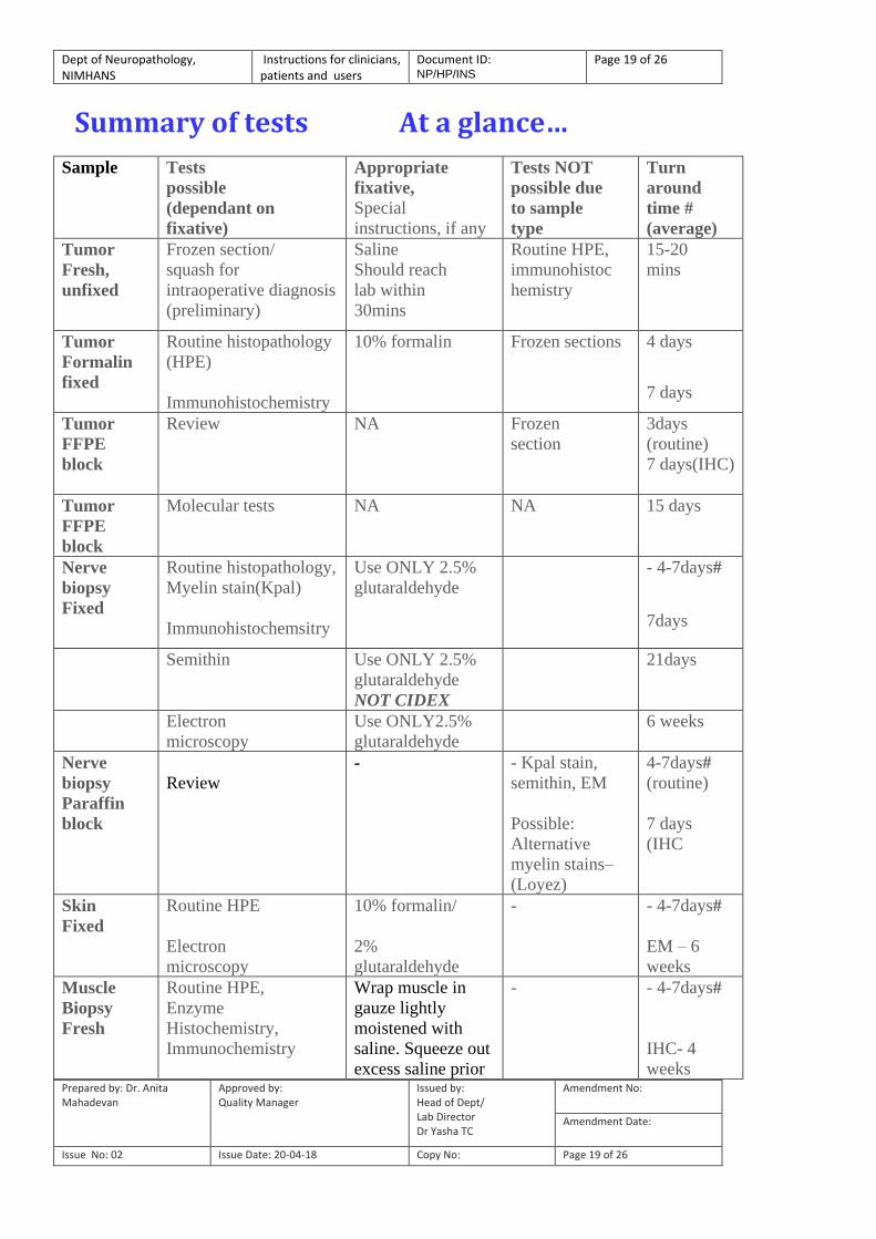
Summary of tests At a glance…
Sample Tests
possible
(dependant on
fixative)
Appropriate
fixative,
Special
instructions, if any
Tests NOT
possible due
to sample
type
Turn
around
time #
(average)
Tumor
Fresh,
unfixed
Frozen section/
squash for
intraoperative diagnosis
(preliminary)
Saline
Should reach
lab within
30mins
Routine HPE,
immunohistoc
hemistry
15-20
mins
Tumor
Formalin
fixed
Routine histopathology
(HPE)
Immunohistochemistry
10% formalin Frozen sections
4 days
7 days
Tumor
FFPE
block
Review NA
Frozen
section
3days
(routine)
7 days(IHC)
Tumor
FFPE
block
Molecular tests NA NA 15 days
Nerve
biopsy
Fixed
Routine histopathology,
Myelin stain(Kpal)
Immunohistochemsitry
Use ONLY 2.5%
glutaraldehyde
- 4-7days#
7days
Semithin
Use ONLY 2.5%
glutaraldehyde
NOT CIDEX
21days
Electron
microscopy
Use ONLY2.5%
glutaraldehyde
6 weeks
Nerve
biopsy
Paraffin
block
Review
- - Kpal stain,
semithin, EM
Possible:
Alternative
myelin stains–
(Loyez)
4-7days#
(routine)
7 days
(IHC
Skin
Fixed
Routine HPE
Electron
microscopy
10% formalin/
2%
glutaraldehyde
- - 4-7days#
EM – 6
weeks
Muscle
Biopsy
Fresh
Routine HPE,
Enzyme
Histochemistry,
Immunochemistry
Wrap muscle in
gauze lightly
moistened with
saline. Squeeze out
excess saline prior
- - 4-7days#
IHC- 4
weeks
Dept of Neuropathology, NIMHANS
Instructions for clinicians, patients and users
Document ID: NP/HP/INS
Page 20 of 26
Prepared by: Dr. Anita Mahadevan
Approved by: Quality Manager
Issued by: Head of Dept/ Lab Director Dr Yasha TC
Amendment No:
Amendment Date:
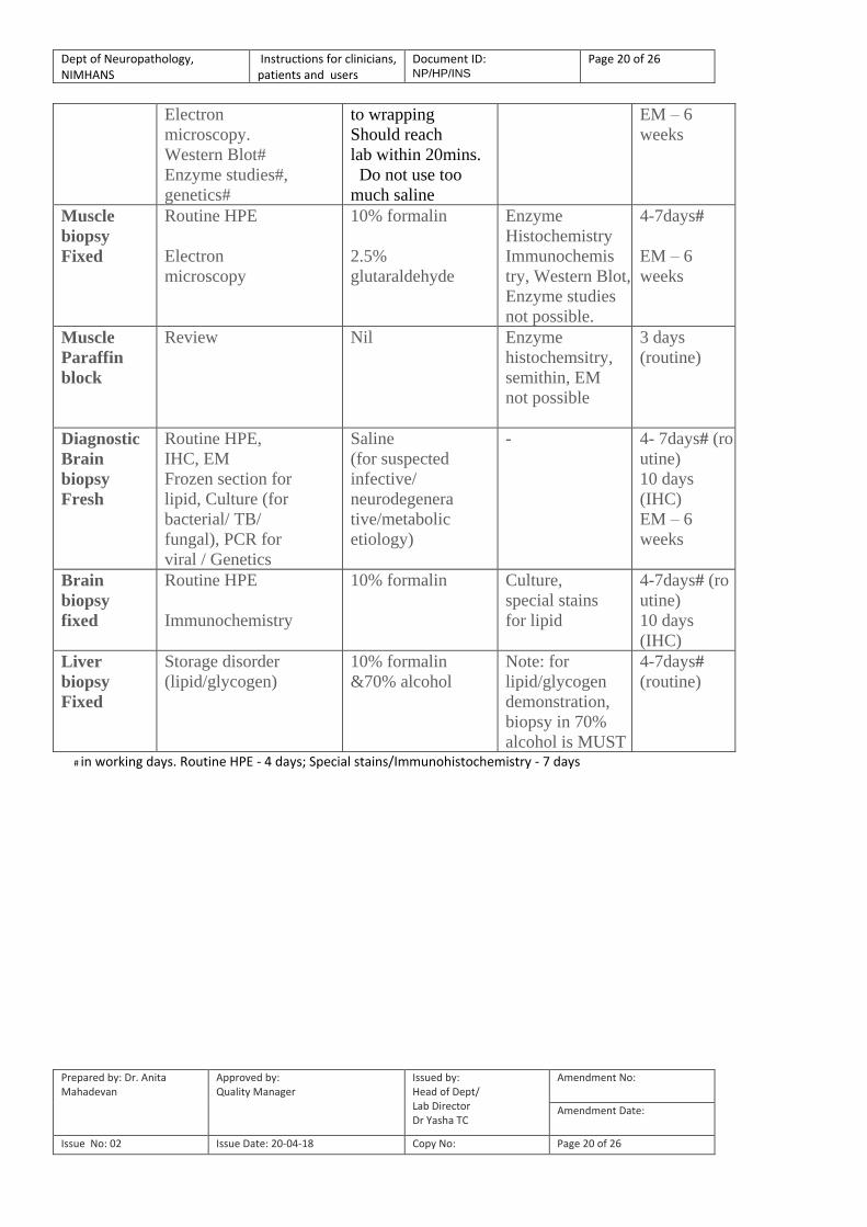
Issue No: 02 Issue Date: 20-04-18 Copy No: Page 20 of 26
Electron
microscopy.
Western Blot#
Enzyme studies#,
genetics#
to wrapping
Should reach
lab within 20mins.
Do not use too
much saline
EM – 6
weeks
Muscle
biopsy
Fixed
Routine HPE
Electron
microscopy
10% formalin
2.5%
glutaraldehyde
Enzyme
Histochemistry
Immunochemis
try, Western Blot,
Enzyme studies
not possible.
4-7days#
EM – 6
weeks
Muscle
Paraffin
block
Review Nil Enzyme
histochemsitry,
semithin, EM
not possible
3 days
(routine)
Diagnostic
Brain
biopsy
Fresh
Routine HPE,
IHC, EM
Frozen section for
lipid, Culture (for
bacterial/ TB/
fungal), PCR for
viral / Genetics
Saline
(for suspected
infective/
neurodegenera
tive/metabolic
etiology)
- 4- 7days# (ro
utine)
10 days
(IHC)
EM – 6
weeks
Brain
biopsy
fixed
Routine HPE
Immunochemistry
10% formalin Culture,
special stains
for lipid
4-7days# (ro
utine)
10 days
(IHC)
Liver
biopsy
Fixed
Storage disorder
(lipid/glycogen)
10% formalin
&70% alcohol
Note: for
lipid/glycogen
demonstration,
biopsy in 70%
alcohol is MUST
4-7days#
(routine)
# in working days. Routine HPE - 4 days; Special stains/Immunohistochemistry - 7 days
Dept of Neuropathology, NIMHANS
Instructions for clinicians, patients and users
Document ID: NP/HP/INS
Page 21 of 26
Prepared by: Dr. Anita Mahadevan
Approved by: Quality Manager
Issued by: Head of Dept/ Lab Director Dr Yasha TC
Amendment No:
Amendment Date:
Issue No: 02 Issue Date: 20-04-18 Copy No: Page 21 of 26
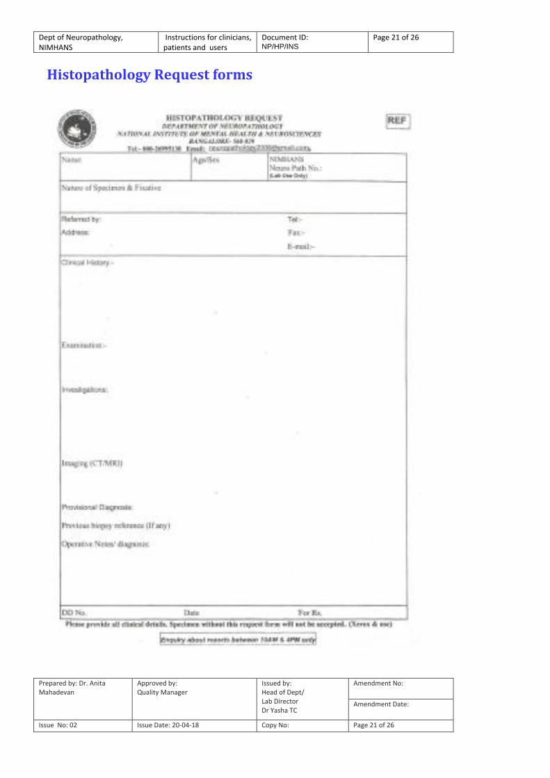
Histopathology Request forms
Dept of Neuropathology, NIMHANS
Instructions for clinicians, patients and users
Document ID: NP/HP/INS
Page 22 of 26
Prepared by: Dr. Anita Mahadevan
Approved by: Quality Manager
Issued by: Head of Dept/ Lab Director Dr Yasha TC
Amendment No:
Amendment Date:
Issue No: 02 Issue Date: 20-04-18 Copy No: Page 22 of 26
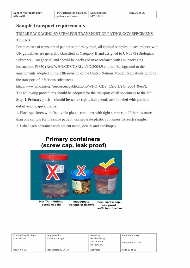
Sample transport requirements
TRIPLE PACKAGING SYSTEM FOR TRANSPORT OF PATHOLOGY SPECIMENS
TO LAB
For purposes of transport of patient samples by road, all clinical samples, in accordance with
UN guidelines are generally classified as Category B and assigned to UN3373 (Biological
Substance, Category B) and should be packaged in accordance with UN packaging
instructions PI650 (Ref: WHO/CDS/CSRL/LYO/2004.9 entitled Background to the
amendments adopted in the 13th revision of the United Nations Model Regulations guiding
the transport of infectious substances
http://www.who.int/csr/resources/publications/WHO_CDS_CSR_LYO_2004_9/en/).
The following procedures should be adopted for the transport of all specimens to the lab:
Step 1:Primary pack – should be water tight, leak proof, and labeled with patient
detail and hospital name.
1. Place specimen with fixative in plastic container with tight screw cap. If there is more
than one sample for the same patient, use separate plastic containers for each sample.
2. Label each container with patient name, details and site/biopsy

Dept of Neuropathology, NIMHANS
Instructions for clinicians, patients and users
Document ID: NP/HP/INS
Page 23 of 26
Prepared by: Dr. Anita Mahadevan
Approved by: Quality Manager
Issued by: Head of Dept/ Lab Director Dr Yasha TC
Amendment No:
Amendment Date:
Issue No: 02 Issue Date: 20-04-18 Copy No: Page 23 of 26
Step 2: Secondary pack – encloses primary pack(s), leak proof and water tight and
protects the primary sample (see picture below)
1. Place the primary samples container in sealable plastic bag/ziplock cover/plastic container
with screw cap.
2. Place large amounts of absorbent material (cotton/filter paper) between primary and
secondary receptacle to absorb all spills, if any.
Step 3:Outer most pack: sealable & strong enough to protect contents from physical
damage during transport.
1. Place secondary pack in a strong cardboard carton/ thermocol box (ideal)/plastic box. The
smallest overall external dimension shall be 10x10 cm.
2. Place clinical datasheet/request form securely enclosed within a plastic cover to prevent it
from getting soiled with leakage of sample contents
LABELING OF OUTER PACKAGE
Biohazard Label for Category B as per UN recommendations
should be affixed

Dept of Neuropathology, NIMHANS
Instructions for clinicians, patients and users
Document ID: NP/HP/INS
Page 24 of 26
Prepared by: Dr. Anita Mahadevan
Approved by: Quality Manager
Issued by: Head of Dept/ Lab Director Dr Yasha TC
Amendment No:
Amendment Date:
Issue No: 02 Issue Date: 20-04-18 Copy No: Page 24 of 26
TRIPLE PACKING SYSTEM MANDATORY FOR SAMPL E TRANSPORTATION TO NEUROPATHOLOGY LAB
PACKAGING FOR FRESH MUSCLE BIOPSIES (without fixative)
Step 1:Primary pack – should be water tight, leak proof, and labeled with patient
detail and hospital name.
1. Place muscle biopsy wrapped in gauze lightly moistened with saline (to prevent drying) in
zip lock cover and seal securely.
2. Label container with patient name, details and site/biopsy type.
Step 2: Secondary pack – encloses primary pack(s), leak proof and water tight
3. Place the zip lock cover in wide mouthed flask OR Thermocol box.
4. Place large quantities of ice (sprinkle cooking salt over ice) to maintain cold chain in
transit.
5. Seal securely
Dept of Neuropathology, NIMHANS
Instructions for clinicians, patients and users
Document ID: NP/HP/INS
Page 25 of 26
Prepared by: Dr. Anita Mahadevan
Approved by: Quality Manager
Issued by: Head of Dept/ Lab Director Dr Yasha TC
Amendment No:
Amendment Date:
Issue No: 02 Issue Date: 20-04-18 Copy No: Page 25 of 26
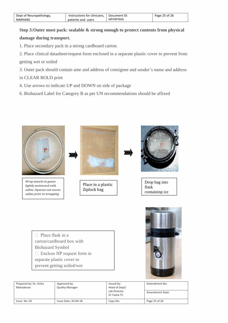
Step 3:Outer most pack: sealable & strong enough to protect contents from physical
damage during transport.
1. Place secondary pack in a strong cardboard carton.
2. Place clinical datasheet/request form enclosed in a separate plastic cover to prevent from
getting wet or soiled
3. Outer pack should contain ame and address of consignee and sender‟s name and address
in CLEAR BOLD print
4. Use arrows to indicate UP and DOWN on side of package
6. Biohazard Label for Category B as per UN recommendations should be affixed
Wrap muscle in gauze
lightly moistened with
saline .Squeeze out excess
saline prior to wrapping
Place in a plastic Ziplock bag
Drop bag into
flask
containing ice
Place flask in a
carton/cardboard box with
Biohazard Symbol
Enclose HP request form in
separate plastic cover to
prevent getting soiled/wet

Dept of Neuropathology, NIMHANS
Instructions for clinicians, patients and users
Document ID: NP/HP/INS
Page 26 of 26
Prepared by: Dr. Anita Mahadevan
Approved by: Quality Manager
Issued by: Head of Dept/ Lab Director Dr Yasha TC
Amendment No:
Amendment Date:
Issue No: 02 Issue Date: 20-04-18 Copy No: Page 26 of 26
ADDITIONAL INSTRUCTIONS FOR TRANSPORTATION OF FRESH
MUSCLE
1. Samples SHOULD be hand delivered and MUST NOT be transported by courier as it
should reach the lab within max of 12 hours of biopsy
2. Samples should reach BEFORE 12 noon on working days (second Saturdays and Govt
holidays, lab is open to receive samples)
3. Samples should NOT be sent on Second Saturdays, Sundays or Govt holidays
4. For muscle fixed in 10% formalin/2.5% glutaraldehyde, please follow the instructions as
for “fixed” samples