intoduction to endocrine
TRANSCRIPT

Introduction to Introduction to EndocrinologyEndocrinology
Physiology of the endocrine system by
Dr Ehsan Saboory
Professor of physiology
Department of physiology, Faculty of medicine, Urmia University of Medical Sciences

Endocrine System: OverviewEndocrine System: Overview
• Endocrine system – the body’s second great controlling system which influences metabolic activities of cells by means of hormones
• Endocrine glands – pituitary, thyroid, parathyroid, adrenal, pineal, and thymus glands
• The pancreas and gonads produce both hormones and exocrine products
• The hypothalamus has both neural functions and releases hormones
• Other tissues and organs that produce hormones – adipose cells, pockets of cells in the walls of the small intestine, stomach, kidneys, and heart
HormonesHormones
• Hormones – chemical substances secreted by cells into the extracellular fluids
• Regulate the metabolic function of other cells
• Have lag times ranging from seconds to hours
• Tend to have prolonged effects
• Are classified as amino acid-based hormones, or steroids
• Eicosanoids – biologically active lipids with local hormone–like activity

Types of HormonesTypes of Hormones
• Amino acid–based – most hormones belong to this class, including:
• Amines, thyroxine, peptide, and protein hormones
• Steroids – gonadal and adrenocortical hormones
• Eicosanoids – leukotrienes and prostaglandins

Hormone ActionHormone Action
• Hormones alter cell activity by one of the following mechanisms:
• Direct changes in cell membrane permeability
• Second messengers involving:
• Regulatory G proteins (amino acid–based hormones)
• Direct gene activation involving steroid hormones
• The precise response depends on the type of the target cell and its receptor
Mechanism of Hormone ActionMechanism of Hormone Action
• Hormones produce one or more of the following cellular changes:
• Alter plasma membrane permeability
• Stimulate protein synthesis
• Activate or deactivate enzyme systems
• Induce secretory activity
• Stimulate mitosis

• Hormone (first messenger) binds to its receptor, which then binds to a G protein
• The G protein is then activated as it binds GTP, displacing GDP
• Activated G protein activates the effector enzyme adenylate cyclase
• Adenylate cyclase generates cyclic AMP (cAMP) (second messenger ) from ATP
• cAMP activates protein kinases, which then cause cellular effects
Amino Acid–Based Hormone Action: cAMPAmino Acid–Based Hormone Action: cAMP
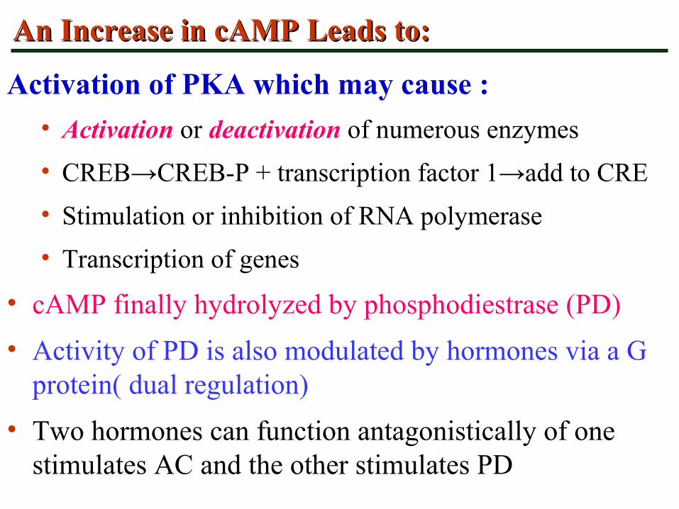
An Increase in cAMP Leads to:An Increase in cAMP Leads to:
Activation of PKA which may cause :
• Activation or deactivation of numerous enzymes
• CREB→CREB-P + transcription factor 1→add to CRE
• Stimulation or inhibition of RNA polymerase
• Transcription of genes
• cAMP finally hydrolyzed by phosphodiestrase (PD)
• Activity of PD is also modulated by hormones via a G protein( dual regulation)
• Two hormones can function antagonistically of one stimulates AC and the other stimulates PD
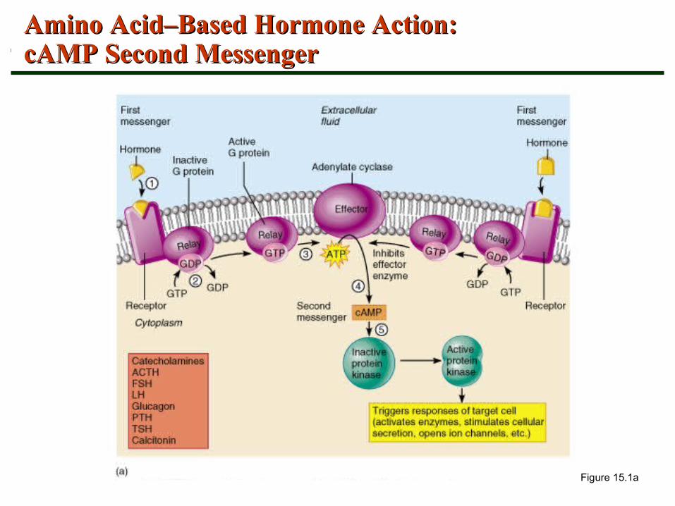
Figure 15.1a
Amino Acid–Based Hormone Action: Amino Acid–Based Hormone Action: cAMP Second MessengercAMP Second Messenger


• H binds to the receptor and activates G protein
• G protein binds and activates a PLC enzyme
• PLC splits the PIP2 into DAG and IP3 (both act as second messengers)
• DAG activates protein kinase C; IP3 triggers release of Ca2+ stores (PKC is Ca2+ dependent)
• Ca2+ (third messenger) alters cellular responses
• Arachidonic acid is derived from hydrolysis of DAG serve as a substrate of prostaglandins (PG)
• The PGs are also modulators of hormonal response
Amino Acid–Based Hormone Action: Amino Acid–Based Hormone Action: PIP–CalciumPIP–Calcium
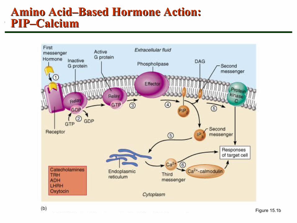
Figure 15.1b
Amino Acid–Based Hormone Action: Amino Acid–Based Hormone Action: PIP–CalciumPIP–Calcium


Other types of Other types of Signal transductionSignal transduction ( (STST))
• 2 other mechanisms of ST from surface R are known in which the transducers lie in the cytoplasmic tail of the R
• In one of these, H+R→ autophosphorylation of R→ the R itself becomes a tyrosine kinase and P the tyrosine residues on intracellular protein. Tyrosine P initiates a cascade of serine and threonine P of enzymes
• Causes multiple intracellular events (Metabolism, proliferation and differentiation), e.g. Insulin
• In the second one H+R→ conformational changes in R which attracts and docks tyrosine kinases e.g. GH
• Another second messenger is cGMP→ activates PKG

Tyrosine kinase ( Tyrosine kinase ( insulin & GHinsulin & GH ) )

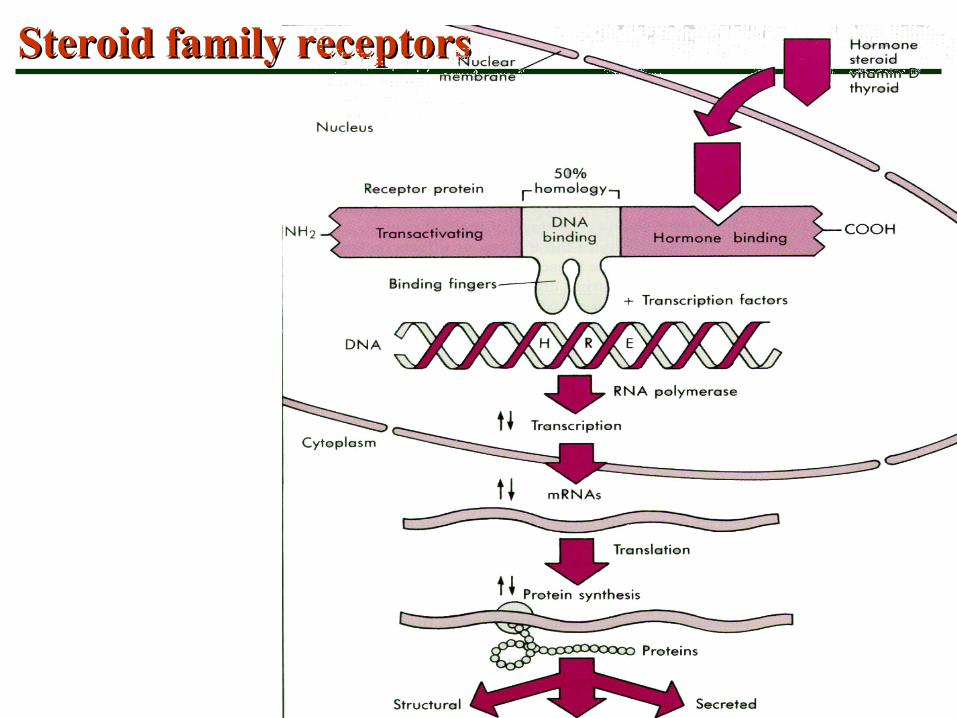
Steroid HormonesSteroid Hormones
• Steroid hormones and thyroid hormone diffuse easily into their target cells
• Once inside, they bind and activate a specific intracellular receptor
• The hormone-receptor complex travels to the nucleus and binds a DNA-associated receptor
• This interaction prompts DNA transcription to produce mRNA
• The mRNA is translated into proteins, which bring about a cellular effect

Steroid HormonesSteroid Hormones
Figure 15.2
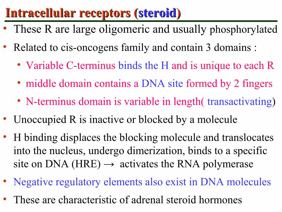
Intracellular receptors (Intracellular receptors (steroidsteroid))• These R are large oligomeric and usually phosphorylated
• Related to cis-oncogens family and contain 3 domains :
• Variable C-terminus binds the H and is unique to each R
• middle domain contains a DNA site formed by 2 fingers
• N-terminus domain is variable in length( transactivating)
• Unoccupied R is inactive or blocked by a molecule
• H binding displaces the blocking molecule and translocates into the nucleus, undergo dimerization, binds to a specific site on DNA (HRE) → activates the RNA polymerase
• Negative regulatory elements also exist in DNA molecules
• These are characteristic of adrenal steroid hormones

Intracellular receptors (Intracellular receptors (thyroxinthyroxin))
• In another general model of activation which is characteristic of thyroid hormones and Vit D the unoccupied R is already attached to DNA binding sites and prevents gene transcription
• H binds R and relieves the suppressive effect of the R
• In a variant of this model, H binding causes dissociation of the two identical receptor monomers constituting the homodimer, formation of heterodimer which activates the gene transcription
Steroid family receptorsSteroid family receptors
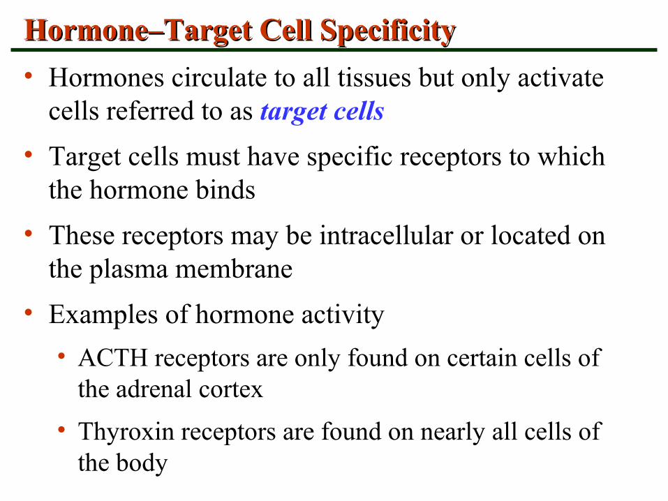
Hormone–Target Cell SpecificityHormone–Target Cell Specificity
• Hormones circulate to all tissues but only activate cells referred to as target cells
• Target cells must have specific receptors to which the hormone binds
• These receptors may be intracellular or located on the plasma membrane
• Examples of hormone activity
• ACTH receptors are only found on certain cells of the adrenal cortex
• Thyroxin receptors are found on nearly all cells of the body
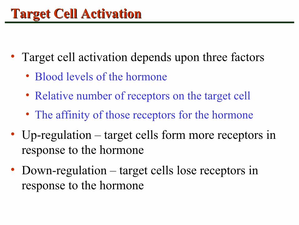
Target Cell ActivationTarget Cell Activation
• Target cell activation depends upon three factors
• Blood levels of the hormone
• Relative number of receptors on the target cell
• The affinity of those receptors for the hormone
• Up-regulation – target cells form more receptors in response to the hormone
• Down-regulation – target cells lose receptors in response to the hormone
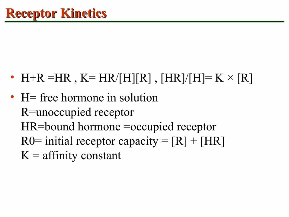
Receptor KineticsReceptor Kinetics
• H+R =HR , K= HR/[H][R] , [HR]/[H]= K × [R]
• H= free hormone in solution R=unoccupied receptor HR=bound hormone =occupied receptor R0= initial receptor capacity = [R] + [HR] K = affinity constant

Schatchard plot for HR kineticSchatchard plot for HR kinetic
A linear plot results when the H reacts with a single R class and no cooperativity is present. The negative of the K equals the slope of the line. The R number,R0, equals the intercept with the X axis.

Schatchard plot for HR kineticSchatchard plot for HR kinetic An exponential
plot results when the H occupancy of one R molecule alters the local affinity of a second nearby molecule for the H. This phenomenon called negative cooperativity.
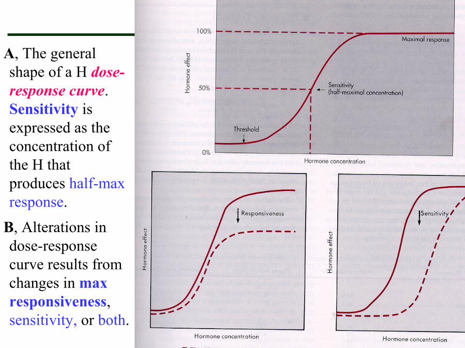
A, The general shape of a H dose-response curve. Sensitivity is expressed as the concentration of the H that produces half-max response.
B, Alterations in dose-response curve results from changes in max responsiveness, sensitivity, or both.
Hormone Concentrations in the BloodHormone Concentrations in the Blood
• Concentrations of circulating hormone reflect:
• Rate of release
• Speed of inactivation and removal from the body
• Hormones are removed from the blood by:
• Degrading enzymes
• The kidneys
• Liver enzyme systems

Control of Hormonal SecretionControl of Hormonal Secretion
• Feedback control• Positive feedback:
• Stimulus detected, H released, H reaches target cell, H binds R, Effect produced, Feedback to endocrine structure, More H released
• Negative feedback:• Stimulus detected, H released, reaches target cell, binds R,
Effect produced, Feedback to endocrine structure, Hormone release shut down
• Direct nerve control• Autonomic nervous system
• Inhibiting hormones or Releasing hormones
• Chronotropic control

Feedback
Neural
Chronal

Control of Hormone Synthesis and ReleaseControl of Hormone Synthesis and Release
• Blood levels of hormones:
• Are controlled by negative feedback systems
• by positive feedback systems (seldom)
• Vary only within a narrow desirable range
• Hormones are synthesized and released in response to:
• Humoral stimuli (substrate or mineral- hormone)
• Neural stimuli
• Hormonal stimuli (hormone- hormone)

Humoral StimuliHumoral Stimuli
• Humoral stimuli – secretion of hormones in direct response to changing blood levels of ions and nutrients
• Example: Declining blood Ca2+ concentration stimulates the parathyroid glands to secrete PTH (parathyroid hormone)
• PTH causes Ca2+ concentrations to rise and the stimulus is removed Figure 17.3a
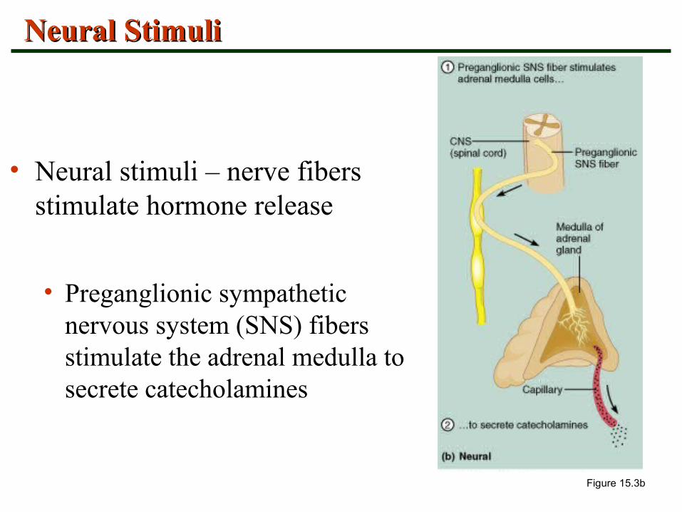
Neural StimuliNeural Stimuli
• Neural stimuli – nerve fibers stimulate hormone release
• Preganglionic sympathetic nervous system (SNS) fibers stimulate the adrenal medulla to secrete catecholamines
Figure 15.3b

Hormonal StimuliHormonal Stimuli
• Hormonal stimuli – release of hormones in response to hormones produced by other endocrine organs
• The hypothalamic hormones stimulate the anterior pituitary
• In turn, pituitary hormones stimulate targets to secrete still more hormones
Figure 15.3c

Nervous System ModulationNervous System Modulation
• The nervous system modifies the stimulation of endocrine glands and their negative feedback mechanisms
• The nervous system can override normal endocrine controls
For example, control of blood glucose levels
• Normally the endocrine system maintains blood glucose
• Under stress, the body needs more glucose
• The hypothalamus and the sympathetic nervous system are activated to supply ample glucose

Chronal control (the circadian rhythms=CR)Chronal control (the circadian rhythms=CR)
The origin of CR in H secretion, behavioral and metabolic activity.
A clock with a 24-25h cycle is located in the SCN, this free running clock is entrained by environmental light signals to the external 24h day.
It has bidirectional relationship with the sleep-wake cycle,too.

Type of cell to cell signalingType of cell to cell signaling
• Endocrine: hormone enters the blood stream
• Neurocrine (neuroendocrine): also enters the blood
• Paracrine:through Int. fluid or GJ to another cell type
• Autocrine: through Int. fluid or gap junction and act on neighboring identical cells or back to the cell of origin
• Juxtacrine ?
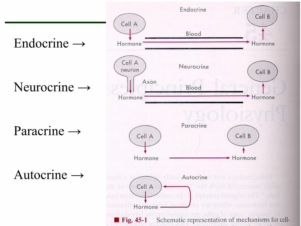
Endocrine →
Neurocrine →
Paracrine →
Autocrine →
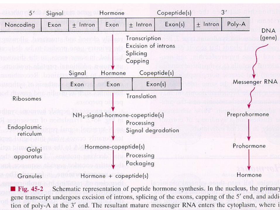
Types of hormone synthesis Types of hormone synthesis
• Protein or peptide hormone synthesis
• Aminoacid based hormone synthesis
• Steroid hormone synthesis
• Eicosanoids synthesis
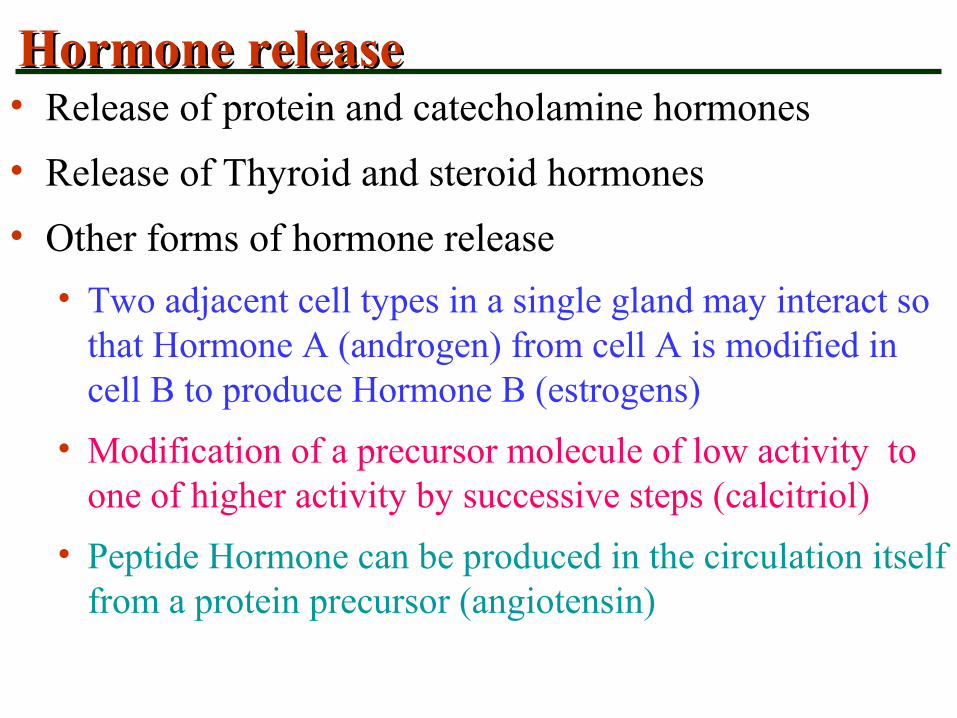
Hormone release Hormone release • Release of protein and catecholamine hormones
• Release of Thyroid and steroid hormones
• Other forms of hormone release
• Two adjacent cell types in a single gland may interact so that Hormone A (androgen) from cell A is modified in cell B to produce Hormone B (estrogens)
• Modification of a precursor molecule of low activity to one of higher activity by successive steps (calcitriol)
• Peptide Hormone can be produced in the circulation itself from a protein precursor (angiotensin)


Hormone TransportHormone Transport
• Free
• Bound to plasma proteins

Hormone DisposalHormone Disposal• Irreversible removal of H is a result of:
• Target cell uptake
• Metabolic degradation
• Urinary excretion
• Biliary excretion
• The sum of all removal processes is expressed as Metabolic Clearance Rate (MCR)
• MCR= mg/min removed / mg/ml of plasma
• K= MCR / volume of distribution
• K is the fractional turnover rate
• The plasma half-life is inversely related to K

Correlation of tCorrelation of t1/21/2 and MCR and MCR
Hormone measurementHormone measurement
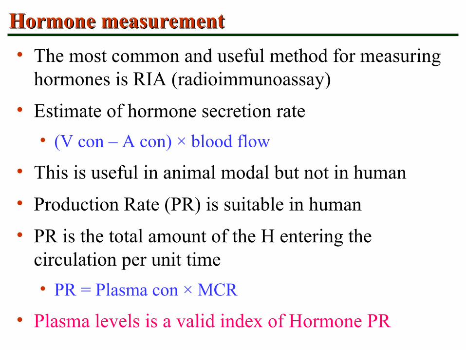
• The most common and useful method for measuring hormones is RIA (radioimmunoassay)
• Estimate of hormone secretion rate
• (V con – A con) × blood flow
• This is useful in animal modal but not in human
• Production Rate (PR) is suitable in human
• PR is the total amount of the H entering the circulation per unit time
• PR = Plasma con × MCR
• Plasma levels is a valid index of Hormone PR

Location of the Major Endocrine GlandsLocation of the Major Endocrine Glands
• The major endocrine glands include:
• Pineal gland, hypothalamus, and pituitary
• Thyroid, parathyroid, and thymus
• Adrenal glands and pancreas
• Gonads – male testes and female ovaries
Figure 15.4