interventional radiology for primary post-partum...
TRANSCRIPT
11. Interventional radiology for primary post-partum hemorrhage (Reiko Woodhams, MD, PhD)
62
Interventional radiology for primary post-partum hemorrhage Reiko Woodhams, MD, PhD Department of Diagnostic Radiology, Kitasato University School of Medicine, Japan Introduction
Postpartum hemorrhage (PPH) consists of primary PPH which occurs within 24 hours after delivery and secondary PPH which occurs between 24 hours and 6 weeks. The bleeding amount, bleeding speed and level of coagulopathy at primary PPH is more severe than those at secondary PPH. The delayed and inappropriate initial treatment can be life threatening at primary PPH. Interventional radiology (IR) has been focused as a useful treatment of primary PPH because of its high success rate, low invasiveness, and preservation of fertility 1, 2). This paper will discuss the clinical implication and utility of IR at primary PPH.
Management of primary PPH
The treatment of primary PPH needs multidisciplinary approach3). The obstetrician, emergency physician, interventional radiologist, staff of blood transfusion department, clinical laboratory technician and nurses have to work together as a team. The bleeding etiology is evaluated along with aggressive fluid and blood replacement, followed by the initial treatment to remove the causative factors.
The principal of management for primary PPH is rapid and effective control of bleeding. The delay of hemostasis could cause disseminating intravascular coagulopathy (DIC) and increase morbidity.
Technique of IR
IR ideally should be performed with a person of experience to achieve quick access to the targeted artery followed by embolization. Technique of IR for PPH consists of arterial balloon occlusion (ABO) and transcatheter arterial embolization (TAE). ABO may be chosen at the critical situation because of its quick access into the targeted artery and prompted hemostatic effect soon after inflation of balloon catheter. Resuscitative endovascular balloon occlusion of the Aorta (REBOA) has the most powerful hemostatic effect because it occludes almost all possible collateral pathways into the pelvis. However, there is risk of ischemic complication with prolonged REBOA. Therefore definite hemostatic method should be performed during REBOA as soon as possible.
TAE achieves hemostasis by introducing embolic material through the catheter at the bleeding arteries. Gelatin sponge is the most frequently used embolic material. Most of primary PPH can be controlled with gelatin sponge. N-buchyl cyanoacrirate (NBCA) may be selected at the traumatic bleeding including laceration and

11. Interventional radiology for primary post-partum hemorrhage (Reiko Woodhams, MD, PhD)
63
pseudoaneurysm of the proximal artery, and severe DIC 4). NBCA is mixed with Lipiodol to change the polymerization time. The mixing ratios is determined by calculating the polymerization times of NBCA and the time taken for contrast material to travel from the microcatheter tip to the destination. The preparation and introduction of NBCA requires certain level of skill and experience to avoid proximal embolization which may cause re-bleeding from the collateral pathway, untargeted embolization, and adhesion of catheter to the vessel wall.
The use of metallic coil should be avoided because of the possibility of recanalization via the collateral pathway, unless injury like rupture or pseudoaneurysm of major artery.
Indication of IR
IR can be indicated to all etiologies of primary PPH 5). However, the priority, surgical operation or IR, is decided based on the comprehensive assessment of bleeding etiology, hemodynamics, availability of operating theater or angiographic room, and human recourses and experiences. Prompt surgical repair is required for uterine inversion and rupture. However, the primary introduction of REBOA before surgery will help to reduce the bleeding speed and will recover and maintain the hemodynamic condition, and clear the visual field at the surgery.
Collateral pathway
Table 1 shows the possible extrapelvic collateral pathway into the pelvis. The bleeding from the collateral pathway has to be considered at persistent bleeding after pelvic embolization, ligation of internal iliac artery or uterine artery, or hysterectomy. In this case, contrast enhanced CT scan before angiography may be useful to detect the bleeding origin.
Procedure of IR for primary PPH Firstly, 5F sheath introducer is placed at unilateral common femoral artery. If the
hemodynamics is unstable or critical, bilateral femoral access is recommended so that you can perform quick embolization bilaterally at the same time, or you can perform IABO and TAE simultaneously. Pelvic arteriogram may be omitted when the bleeding origin has already been determined 6). Selective catheterization of the internal iliac artery is done with a 5-F catheter, such as a Cobra, Multipurpose, Roberts, or Mohri catheter guided by hydrophilic 0.035-inch angled guidewire. If engagement into the targeted artery with 5F catheter is difficult, coaxial method using microcatheter may be chosen, though selectivity is highly dependent on the patient’s hemodynamics

11. Interventional radiology for primary post-partum hemorrhage (Reiko Woodhams, MD, PhD)
64
status and bleeding etiology. Bilateral embolization is principal at embolization of pelvic artery because of the bilateral communication (Figure 1).
Atonic bleeding is the most common etiology of primary PPH (Figure 2). TAE can be a definite treatment for atonic bleeding. The high success rate of IR for atonic bleeding encourages the primary choice of IR. Hysterectomy as a first choice of treatment should be avoided if immediate TAE is available. The embolization can be performed from bilateral anterior division of the internal iliac arteries, not necessarily from the uterine artery. However, the hemostasis with TAE for the patient with severe DIC is challenging. The mechanism of bleeding at atonic bleeding is functional, which means that the bleeding occurs diffusely. Even if NBCA is used in expectation of robust embolic effect for DIC patient, it may not work because NBCA does not occlude the very distal end of capitally artery. In this situation, the treatment priority is quick and massive introduction of fresh frozen plasma (FFP) for coagulation factor replacement.
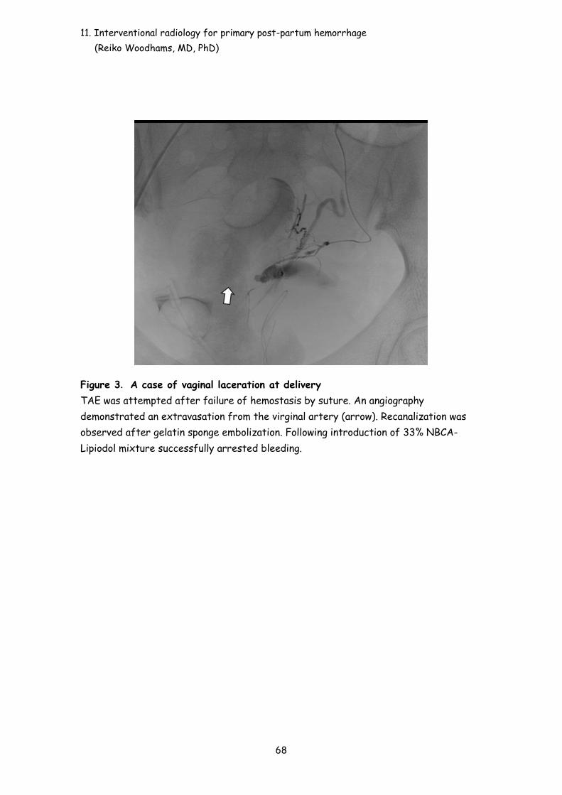
Surgical repair is recognized to be the first choice for birth canal laceration. However, primary introduction of IR before surgery is considered if IR can be started immediately because TAE will provide better visibility of the surgical field and stability of secure surgical hemostatic procedure. When massive extravasation or pseudoaneurysm is observed from the particular point due to genital tract laceration, superselective catheterization as close as possible to the bleeding point using microcatheter followed by NBCA embolization is ideal (Figure 3). Gelatin sponge may be useful for multiple slow bleeding due to crushed wound.
The massive bleeding due to placental accreta is life threatening because of difficulty of preoperative diagnosis, massive bleeding speed, and difficulty of surgical hemostatic procedure. Infrarenal IABO is the most secure and prompt hemostatic method for this critical situation. The immediate surgical hemostasis should be performed during IABO, as IABO is a temporal hemostatic method and has a risk of ischemic complication.
Complication
Overall complication rates for obstetric transcatheter embolization procedures are reported to be 6%–9% 6). The untargeted embolization may cause buttock claudication, skin necrosis, vesical necrosis, rectal necrosis 6, 7). Ovarian dysfunction is caused by migration of embolic material though communication between uterine artery and ovarian artery. The risks of such occurrences are decreased by knowledge of the vascular anatomy and meticulous attention to embolization technique.
Conclusion

11. Interventional radiology for primary post-partum hemorrhage (Reiko Woodhams, MD, PhD)
65
The appropriate strategy depending on the etiology and hemodynamics, speed and reliability of interventional procedure is essential for successful and low invasive hemostasis at IR for primary PPH. References 1) Pelage JP, Le Dref O, Mateo J, et al. Life-threatening primary postpartum
hemorrhage: treatment with emergency selective arterial embolization. Radiology. 1998;208(2):359-362.
2) Lee HY, Shin JH, Kim J, et al. Primary postpartum hemorrhage: outcome of pelvic arterial embolization in 251 patients at a single institution. Radiology. 2012;264(3):903-909.
3) Oyelese Y, Scorza WE, Mastrolia R, Smulian JC. Postpartum hemorrhage. Obstet Gynecol Clin North Am. 2007;34(3):421-441, x.
4) Kim GM, Yoon CJ, Seong NJ, Kang SG, Kim YJ. Postpartum haemorrhage from ruptured pseudoaneurysm: efficacy of transcatheter arterial embolisation using N-butyl-2-cyanoacrylate. Eur Radiol. 2013;23(8):2344-2349.
5) Soyer P, Dohan A, Dautry R, et al. Transcatheter Arterial Embolization for Postpartum Hemorrhage: Indications, Technique, Results, and Complications. Cardiovasc Intervent Radiol. 2015;38(5):1068-1081.
6) Salazar GM, Petrozza JC, Walker TG. Transcatheter endovascular techniques for management of obstetrical and gynecologic emergencies. Tech Vasc Interv Radiol. 2009;12(2):139-147.
7) Cheong JY, Kong TW, Son JH, Won JH, Yang JI, Kim HS. Outcome of pelvic arterial embolization for postpartum hemorrhage: A retrospective review of 117 cases. Obstet Gynecol Sci. 2014;57(1):17-27.

11. Interventional radiology for primary post-partum hemorrhage (Reiko Woodhams, MD, PhD)
66
Table 1
Figure 1. A case of atonic bleeding An angiography from the left uterine artery shows an extravasation from the right side through a communication between bilateral uterine arteries.
Ovarian artery
Pubic branch of inferior epigastric artery
Medial circumflex femoral artery
Middle sacral artery
Lumber artery
Artery of round ligament
Superior rectal artery
Circumflex iliac artery

11. Interventional radiology for primary post-partum hemorrhage (Reiko Woodhams, MD, PhD)
67
Figure 2. A case of atonic bleeding Bilateral uterine angiography shows dilated uterine arteries, though there is no extravasation observed (a, b). Hemostasis was successfully achieved with embolization of bilateral uterine arteries and the ovarian artery (c).
a b
c
11. Interventional radiology for primary post-partum hemorrhage (Reiko Woodhams, MD, PhD)
68
Figure 3.A case of vaginal laceration at delivery TAE was attempted after failure of hemostasis by suture. An angiography demonstrated an extravasation from the virginal artery (arrow). Recanalization was observed after gelatin sponge embolization. Following introduction of 33% NBCA-Lipiodol mixture successfully arrested bleeding.