diagnostic and interventional radiology · interventional radiology in this subchapter, information...
TRANSCRIPT

CONTENTS INTRODUCTION GENERAL INFORMATION
SURVEY QUESTIONNAIRES
ONLINE PLATFORM REFERENCES
U N S C E A R G L O B A L S U R V E Y O N M E D I C A L E X P O S U R E : A U S E R M A N U A L
DIAGNOSTIC AND INTERVENTIONAL
RADIOLOGY
In this subchapter, information is provided to facilitate the completion of the diagnostic and interventional radiology questionnaire. The information requested is organized according to the four main sections (sheets) of the questionnaire, namely:
• General information: for general information about the country, its population, the survey period and contact details of contributors.
• Staff and devices: for information on staffing levels and the equipment available.
• Frequency: for data on frequency of examinations and on optional data regarding the age and the sex distribution of the population undergoing these examinations.
• Dose: for information on mean doses from different examinations.
SURVEY QUESTIONNAIRES | DIAGNOSTIC AND INTERVENTIONAL RADIOLOGY |

CONTENTS INTRODUCTION GENERAL INFORMATION
SURVEY QUESTIONNAIRES
ONLINE PLATFORM REFERENCES
U N S C E A R G L O B A L S U R V E Y O N M E D I C A L E X P O S U R E : A U S E R M A N U A L
SURVEY QUESTIONNAIRES | DIAGNOSTIC AND INTERVENTIONAL RADIOLOGY | GENERAL INFORMATION
General information
In this section, contributors are requested to fill in their contact details and general information about their country such as:
• Year: Indicate the year (or period) to which the data refers.
• Population (inhabitants): Indicate the total population size in number of inhabitants of the country for the year the data refers to.
• Population (survey base): Indicate the size of population in number of inhabitants if the data collected are for a particular region, city or hospital only. Provide the total population size of the country if the information provided is for the whole country. When a smaller fraction of the population is used as a basis to assess the situation in the whole country, an estimation of how representative the data is with regard to the entire country should be provided for each examination separately. Such information is requested in the frequency sheet.
• Contact information: The data submitted should be signed off by the national contact person (NCP), registered on the UNSCEAR online platform, including name, institution and contact details (e-mail/phone) to facilitate any feedback. Please, indicate contact details of any other persons who have provided information and who should be acknowledged in the final report. Contact details for at least one person are required as this information will be transferred to the UNSCEAR database.
The country code is provided automatically, and the date of submission will be generated during the upload of the file.
DIAGNOSTIC AND INTERVENTIONAL
RADIOLOGY

CONTENTS INTRODUCTION GENERAL INFORMATION
SURVEY QUESTIONNAIRES
ONLINE PLATFORM REFERENCES
U N S C E A R G L O B A L S U R V E Y O N M E D I C A L E X P O S U R E : A U S E R M A N U A L
SURVEY QUESTIONNAIRES | DIAGNOSTIC AND INTERVENTIONAL RADIOLOGY | STAFF AND DEVICES
Staff and devices
Staff
Information about the number and type of professionals working in diagnostic and interventional radiology should be provided in this sheet. The numbers should reflect the situation in the entire country as accurately as possible and could be based on data originating from national registries or professional associations. Data should reflect the situation regarding professionals working in both hospital and private practice. Please note that:
• Only practising professionals should be included and their actual occupancy time should be taken roughly into account. For example, two part-time nurses, each working 50% of the normal working hours, count as one full-time professional.
• Persons working in two areas or more should be counted according to their main activity.
• Some data about professionals not working in diagnostic or interventional radiology are also requested. For example the number of general practitioners.
The number of practising individuals working in the following categories is sought in the RD questionnaire:
• Physicians: All physicians in the country irrespective of their medical specialization.
• General practitioners: Medical doctors usually acting as primary care providers at an early stage of a disease’s onset. In some countries they are called family doctors.
• Dentists: Only physicians specialized in dental medicine. In some countries they are also known as dental surgeons.
• Radiologists: Only medical doctors specialized in radiology, excluding interventional radiologists.
• Other physicians conducting radiological examinations: All other physicians using X-rays (e.g. orthopaedic surgeons, gynaecologists, gastroenterologists).
• Interventional radiologists: Only radiologists with additional specialization in performing interventional procedures.
• Interventional cardiologists: Only cardiologists who are specialized or licensed to perform interventional cardiology procedures.
• Other physicians conducting interventional procedures: Physicians who are not radiologists or cardiologists but are specialized or licensed to perform interventional procedures: e.g. urologists, vascular surgeons or general surgeons.
• Medical physicists in radiology/imaging: Medical physicists licensed or authorized to practise in diagnostic and interventional radiology.
• Radiation technologists in radiology/imaging: Radiation technologists licensed or authorized to perform examinations or procedures in diagnostic and interventional radiology.
• Nurses in radiology/imaging: Nurses practising in diagnostic or interventional radiology or working as radiation technologists.
Collected information on staffing and equipment may be
correlated with frequency and dose information and provide
useful insights on the effect of equipment and staffing levels
on population doses from medical radiation.
DIAGNOSTIC AND INTERVENTIONAL
RADIOLOGY

CONTENTS INTRODUCTION GENERAL INFORMATION
SURVEY QUESTIONNAIRES
ONLINE PLATFORM REFERENCES
U N S C E A R G L O B A L S U R V E Y O N M E D I C A L E X P O S U R E : A U S E R M A N U A L
SURVEY QUESTIONNAIRES | DIAGNOSTIC AND INTERVENTIONAL RADIOLOGY | STAFF AND DEVICES
Staff and devices
Devices
A wide range of different radiological devices are used in diagnostic and interventional radiology. Some general information and guidelines on how to list the different devices are provided here:
• Only data on the numbers of actually operating equipment are required.
• Data about analogue and digital diagnostic radiology systems should be listed separately. Only systems with a fully digital image receptor, such as the ones comprising flat panel detectors, are defined as digital devices.
• Computed radiography systems are not considered full digital systems and are categorized under “image processing modalities”.
• Systems with multiple X-ray tubes are counted as one device.
• CT system components of nuclear medicine hybrid systems should not be listed in the diagnostic and interventional radiology questionnaire.
The number of MRI scanners is requested even though MRI does not use ionizing radiation for imaging. Collecting data about the number of MRI machines is important to provide a more complete overview of the level of medical imaging in a country.
Note that equipment that should be listed in the radiotherapy questionnaire also includes imaging devices. These may be different from the ones used in the radiology department and may be for exclusive use for radiotherapy purposes. If this is not the case and the same equipment (e.g. CT) is also used in diagnostic radiology, then it should be listed only once in the radiology section of the questionnaire. Patient doses resulting from CT uses for radiotherapy planning are not taken into account in the UNSCEAR survey.
Table 1 describes the different types of equipment used in diagnostic and interventional radiology.
DIAGNOSTIC AND INTERVENTIONAL
RADIOLOGY

U N S C E A R G L O B A L S U R V E Y O N M E D I C A L E X P O S U R E : A U S E R M A N U A L
CONTENTS INTRODUCTION GENERAL INFORMATION
SURVEY QUESTIONNAIRES
ONLINE PLATFORM REFERENCES
PAGE 1 of 5
SURVEY QUESTIONNAIRES | DIAGNOSTIC AND INTERVENTIONAL RADIOLOGY | STAFF AND DEVICES
Diagnostic radiological systems
Radiography systems Systems which consist of one or more X-ray tubes and use two-dimensional image receptors for image acquisition. The image receptors may be based on radiographic film usually used in conjunction with intensifying screen cassettes or on computed radiography cassettes containing a receptor which is read out by a digital reader.
Digital systems comprise fully digital image receptors capable of directly producing two-dimensional radiographic images. However, computed radiography is categorized as an image processing modality.
Fluoroscopy systems Systems that comprise a C-shaped arm (C-arm). On the one end of the arm there is an X-ray tube and on the other end an image receptor. Analogue systems sport image intensifiers which resemble bulky cylindrical devices. Digital systems comprise flat panel detectors similar to the ones used in digital radiography systems.
Some radiography systems have fluoroscopy capabilities, mostly used for patient positioning. Such systems do not include a C-arm and should be listed under radiography systems and not under fluoroscopy systems.
A bi-plane system sporting two C-arms should also be considered as one system.
Mammography systems Mammography systems comprise an X-ray tube capable of producing a low-energy X-ray beam. These systems include a breast-compressing device in order to even out the thickness of the breast tissue. The console is shielded by an appropriate shielding wall and is usually in the same room as the X-ray tube. There are digital and analogue mammography systems depending on the type of the detector.
Table 1. Diagnostic and interventional radiological devices listed in the RD questionnaire
Descriptions and images are provided in order to help recognize and categorize the different devices.

U N S C E A R G L O B A L S U R V E Y O N M E D I C A L E X P O S U R E : A U S E R M A N U A L
CONTENTS INTRODUCTION GENERAL INFORMATION
SURVEY QUESTIONNAIRES
ONLINE PLATFORM REFERENCES
PAGE 2 of 5
Dental X-ray systems (a) Plain dental X-ray systems: usually consist of a flexible arm with an X-ray generator at its end. The image receptors may be films or digital devices placed into the mouth.
(b) Orthopantomographs: devices comprising an X-ray source that spins around the patient’s head with the synchronous movement of an image receptor.
Please list all dental imaging devices in this category except dental cone beam CTs. These are listed separately in their own category.
Angiography systems Angiography systems are dedicated fluoroscopy systems used for vascular imaging. These systems produce images through the use of contrast agents injected into the patient’s blood vessels.
Angiography systems are used for all vascular interventions, including cardiac ones.
Bone densitometry systems
Bone densitometry systems are used for the evaluation of patient bone density. Reduced bone density is linked with the onset of osteoporosis. Bone densitometry devices comprise a patient bed and a moving X-ray source under the patient’s body. This source produces a pencil or fan beam which is detected by a detector arm moving simultaneously over the patient’s body.
SURVEY QUESTIONNAIRES | DIAGNOSTIC AND INTERVENTIONAL RADIOLOGY | STAFF AND DEVICES
Table 1. Diagnostic and interventional radiological devices listed in the RD questionnaire

U N S C E A R G L O B A L S U R V E Y O N M E D I C A L E X P O S U R E : A U S E R M A N U A L
CONTENTS INTRODUCTION GENERAL INFORMATION
SURVEY QUESTIONNAIRES
ONLINE PLATFORM REFERENCES
PAGE 3 of 5
Image processing modalitiesChemical development systems
Chemical development systems are used to develop radiographic film. After image acquisition, the radiological technologist enters the dark room and removes the film from its cassette. The film is immediately inserted into the receiver of the development system, located inside the dark room. After a while, a developed film comes out of the system. The exit point of the films is usually outside the dark room.
Development systems that can be used in any lighting conditions are called daylight developers and contain mechanisms that automatically unload the radiographic film from the cassette inside the housing of the machine avoiding exposure to natural light. Similar to dark room developing systems, they use chemicals for the development of X-ray films.
Computed radiography systems
Computed radiography systems resemble chemical development systems. However, the development is not chemical and does not need a dark room. The CR cassettes are fed directly into the CR reader and the image is read out. The cassette emerges from the system erased and ready for reuse.
Please count only CR readers in this category. X-ray machines should be counted under radiography systems. CR systems are readers only and are not connected to X-ray tubes.
Table 1. Diagnostic and interventional radiological devices listed in the RD questionnaire
SURVEY QUESTIONNAIRES | DIAGNOSTIC AND INTERVENTIONAL RADIOLOGY | STAFF AND DEVICES

U N S C E A R G L O B A L S U R V E Y O N M E D I C A L E X P O S U R E : A U S E R M A N U A L
CONTENTS INTRODUCTION GENERAL INFORMATION
SURVEY QUESTIONNAIRES
ONLINE PLATFORM REFERENCES
PAGE 4 of 5
SURVEY QUESTIONNAIRES | DIAGNOSTIC AND INTERVENTIONAL RADIOLOGY | STAFF AND DEVICES
Table 1. Diagnostic and interventional radiological devices listed in the RD questionnaire
Computed tomography scannersSingle slice CT Cross-sectional imaging devices with only one row of imaging
detectors. These devices produce only one cross-sectional image per rotation.
They comprise a gantry wide enough for a patient to be placed inside. The detector rows and the X-ray tube are placed opposite each other and move around the patient together in order to acquire images.
Multi-slice CT Cross-sectional imaging devices with multiple rows of imaging detectors. These devices may produce more than 300 images per rotation (slices) and cover wide ranges of human anatomy, such as whole organs.
They comprise a gantry wide enough for a patient to be placed inside. The detector rows and the X-ray tube are placed opposite each other and move around the patient together in order to acquire images.
Dual source CT CT machines that comprise two X-ray tubes in 90-degree angle configuration. The two tubes produce different energy photon spectra and may help in image enhancement and quicker image acquisition, especially for cardiac imaging.
Dental CT Dental CTs are usually cone beam CT systems with smaller fields of view suited for dental imaging. The machines comprise a patient chair and a head immobilization device. The X-ray tube and the digital image receptor are opposite each other and spin around the patient’s head in order to acquire images. These devices often resemble the extremity or head and neck CBCT systems.
In this category, please list only CBCT used in dental practice.

U N S C E A R G L O B A L S U R V E Y O N M E D I C A L E X P O S U R E : A U S E R M A N U A L
CONTENTS INTRODUCTION GENERAL INFORMATION
SURVEY QUESTIONNAIRES
ONLINE PLATFORM REFERENCES
PAGE 5 of 5
SURVEY QUESTIONNAIRES | DIAGNOSTIC AND INTERVENTIONAL RADIOLOGY | STAFF AND DEVICES
Table 1. Diagnostic and interventional radiological devices listed in the RD questionnaire
Cone beam CT Cone beam CT machines may look a lot like digital fluoroscopy or angiography machines or comprise a full 360-degree gantry. In fact, some modern fluoroscopy and angiography machines may be used for CBCT image reconstruction. Such machines usually contain a digital image receptor and may perform a 180-360 degree rotation around the patient.
Extremity and head and neck CBCT systems may resemble dental CBCTs but usually have wider fields of vision.
Flat panel detectors for cone beam CTs have been mounted on CT or CT-like gantries. This is sometimes called flat panel CT.
Please only include systems that are dedicated to CBCT imaging in this category.
MRI scanners1.5 Tesla
> 1.5 Tesla
They comprise gantries with bore holes similar to CT scanners. The gantries are usually more bulky than the CT ones. MRI scanners use strong magnetic fields to investigate the anatomy and physiology of the body.

CONTENTS INTRODUCTION GENERAL INFORMATION
SURVEY QUESTIONNAIRES
ONLINE PLATFORM REFERENCES
U N S C E A R G L O B A L S U R V E Y O N M E D I C A L E X P O S U R E : A U S E R M A N U A L
Frequency
This questionnaire section (sheet) is for information collection on the frequency of various diagnostic examinations. This information is very relevant in order to assess levels of practice in a country as the frequency of radiological examinations is the most important factor in determining medical radiation exposure for a population. Examinations have been categorized by modality and anatomical regions.
Categorization of examinations
The categorization of examinations in this survey follows the four main modalities used in radiology:
• Projection radiography (without contrast media)
• Radiography and fluoroscopy (mostly with contrast media)
• Computed tomography (CT)
• Image-guided interventional procedures (IGIP)
The categorization of radiological diagnostic examinations used in this survey considers the anatomical regions that might have been exposed. For a single radiological examination a different number of projections may be used. Such differences in clinical practice make it difficult to define all radiological examinations clearly. Thus, for this survey “an X-ray examination or interventional procedure is defined as one or a series of X-ray exposures of one anatomical region/organ/organ system, using a single imaging modality (i.e. radiography/fluoroscopy or CT), needed to answer a specific diagnostic problem or clinical question, during one visit to the radiology department, hospital or clinic” [E1].
The examination categories used in this survey follow the DOSE DATAMED approach which has identified more than 200 radiological examinations and has grouped them into about 70 categories [E1]. The categories and the specific examinations are listed in the following table 2.
SURVEY QUESTIONNAIRES | DIAGNOSTIC AND INTERVENTIONAL RADIOLOGY | FREQUENCY
DIAGNOSTIC AND INTERVENTIONAL
RADIOLOGY

U N S C E A R G L O B A L S U R V E Y O N M E D I C A L E X P O S U R E : A U S E R M A N U A L
CONTENTS INTRODUCTION GENERAL INFORMATION
SURVEY QUESTIONNAIRES
ONLINE PLATFORM REFERENCES
PAGE 1 of 11
Modality Examination category Specific examinations Comments
Projection radiography (without contrast media)
Head (skull and facial bones) Skull and facial bones, head
– Orbits
– Temporal bones
– Petrous bone
– Mastoids
– Sphenoid bone
– Sella turcica
– Sphenoid fissures
Facial bones
– Nose
– Sinuses
– Zygomas
– Temporo-mandibular joint
– Cervico-occipital hinge
– Maxilla
– Mandible
– Cephalometry
If only one head examination category is available, please provide the data in this category.
Head (soft tissue) Dacryocystography (tear ducts)
Sialography (salivary glands)
Eyes/orbits
Neck (cervical spine) Cervical spine Common techniques: AP and LAT/Oblique.
If only one neck examination category is available, please provide the data in this category.
Neck (soft tissue) Larynx
Pharynx
Trachea
Table 2. Categorization of specific radiological examinations used in the UNSCEAR Global Survey
SURVEY QUESTIONNAIRES | DIAGNOSTIC AND INTERVENTIONAL RADIOLOGY | STAFF AND DEVICES

U N S C E A R G L O B A L S U R V E Y O N M E D I C A L E X P O S U R E : A U S E R M A N U A L
CONTENTS INTRODUCTION GENERAL INFORMATION
SURVEY QUESTIONNAIRES
ONLINE PLATFORM REFERENCES
PAGE 2 of 11
Modality Examination category Specific examinations Comments
Projection radiography (without contrast media)
Chest/Thorax (lungs PA and LAT) Lung
Thoracic inlet
Bronchography
Common techniques: PA and LAT
If only one chest examination category is available, please provide the data in this category.
Chest (thoracic spine) Thoracic spine Common techniques: AP and LAT
Chest (shoulder girdle & ribs) Shoulder blades/scapulae
Collar bone(s)/clavicle(s)
Acromio-clavicular joint
Sterno-clavicular joint
Manubrio-sternal joint
Sternum
Ribs
Mammography Symptomatic:
One or two views of one or two breasts
A mammography examination (bilateral) consists of two views for each breast. Thus, the number of unilateral examinations should be divided by two and included in this category.
If different, explain it in the comment field.
Common techniques: medio-lateral oblique and cranio-caudal for one or two breasts.
If only one mammography examination category is available, please provide the data in this category.
Mammography (screening) One or two views of one or two breasts
Common technique: A screening mammography examination (bilateral) consists of two views for each breast.
Lumbar spine Lumbar spine Common technique: AP and LAT
SURVEY QUESTIONNAIRES | DIAGNOSTIC AND INTERVENTIONAL RADIOLOGY | STAFF AND DEVICES
Table 2. Categorization of specific radiological examinations used in the UNSCEAR Global Survey

U N S C E A R G L O B A L S U R V E Y O N M E D I C A L E X P O S U R E : A U S E R M A N U A L
CONTENTS INTRODUCTION GENERAL INFORMATION
SURVEY QUESTIONNAIRES
ONLINE PLATFORM REFERENCES
PAGE 3 of 11
SURVEY QUESTIONNAIRES | DIAGNOSTIC AND INTERVENTIONAL RADIOLOGY | STAFF AND DEVICES
Modality Examination category Specific examinations Comments
Projection radiography (without contrast media)
Lumbo-sacral joint only Lumbo-sacral joint
Abdomen Abdomen (plain film, patient supine or erect)
Common technique: AP
Pelvis and hips (bone) Pelvic bones
– Ilium/ischium/pubis
– Sacrum
– Sacro-iliac joint
– Coccyx
– Pelvimetry (obstetric)
Hips
– One or both hips
Common technique: AP only or AP and LAT
If only one pelvis examination category is available, please provide the data in this category
Pelvis (soft tissue) Pelvis (soft tissue)
Limbs and joints Elbow
Forearm (radius and ulna)
Wrist (scaphoid)
Hand
– Fingers and thumbs
Femur
Knee
Knee cap (patella)
Lower leg (tibia and fibula)
Ankle
Foot
Calcaneum (heel)
Toes
Whole leg
Whole spine (trunk) Scoliosis
Table 2. Categorization of specific radiological examinations used in the UNSCEAR Global Survey

U N S C E A R G L O B A L S U R V E Y O N M E D I C A L E X P O S U R E : A U S E R M A N U A L
CONTENTS INTRODUCTION GENERAL INFORMATION
SURVEY QUESTIONNAIRES
ONLINE PLATFORM REFERENCES
PAGE 4 of 11
Modality Examination category Specific examinations Comments
Projection radiography (without contrast media)
Skeletal survey (head and trunk) Skeletal survey (head and trunk)
Dental intraoral Intra-oral <3 films
– 1–2 periapical films
– 1–2 bitewing films
– 1 occlusal film
Intra-oral >2 films
– >2 periapical films
– Periapical full mouth survey
– >2 bitewing films
Dental panoramic Panoramic full mouth scan
Radiography and fluoroscopy (mostly with contrast media)
Gastrointestinal tract (barium studies)
Oesophagus (Ba swallow)
Stomach and duodenum (Ba meal)
Small intestine (Ba follow)
Enteroclysis (small intestine enema)
Colon (Ba enema)
Common techniques:
Meal: 2–3 minutes fluoroscopy (5–20 images)
Enema: ~2 minutes fluoroscopy (5–10 images)
Follow: ~5 minutes fluoroscopy (5–20 images)
Gastrointestinal tract (defecography)
Defecography
Biliary tract (cholangiography) Retrograde cholangiography
Operative cholangiography
Intravenous cholangiography
T-drain cholangiography
Transhepatic cholangiography
Biliary tract (ERCP) Endoscopic retrograde cholangiopancreatography (ERCP)
Retrograde pancreatography
Table 2. Categorization of specific radiological examinations used in the UNSCEAR Global Survey
SURVEY QUESTIONNAIRES | DIAGNOSTIC AND INTERVENTIONAL RADIOLOGY | STAFF AND DEVICES

U N S C E A R G L O B A L S U R V E Y O N M E D I C A L E X P O S U R E : A U S E R M A N U A L
CONTENTS INTRODUCTION GENERAL INFORMATION
SURVEY QUESTIONNAIRES
ONLINE PLATFORM REFERENCES
PAGE 5 of 11
SURVEY QUESTIONNAIRES | DIAGNOSTIC AND INTERVENTIONAL RADIOLOGY | STAFF AND DEVICES
Table 2. Categorization of specific radiological examinations used in the UNSCEAR Global Survey
Modality Examination category Specific examinations Comments
Radiography and fluoroscopy (mostly with contrast media)
Biliary tract (cholecystography) Cholecystography
Uro-genital tract (IVU) Intravenous urography (IVU) Common technique: Several AP radiographs after IV injection of iodine contrast medium
Uro-genital tract (kidney, bladder and urethra)
Kidneys and ureters
– Retrograde pyelography
– Nephrostography
Bladder and urethra
– Retrograde cystography
– Micturitional cysto-urethrography (MCU)
– Urethrography
Myelography Cervical myelography
Thoracic myelography
Lumbar myelography
Sacral myelography
Whole spine myelography
Arthrography Temporal-mandibular joint arthrography
Shoulder arthrography
Hip arthrography
Elbow arthrography
Wrist arthrography
Knee arthrography
Ankle arthrography
Cerebral angiography Cerebral angiography
Petrous phlebography

U N S C E A R G L O B A L S U R V E Y O N M E D I C A L E X P O S U R E : A U S E R M A N U A L
CONTENTS INTRODUCTION GENERAL INFORMATION
SURVEY QUESTIONNAIRES
ONLINE PLATFORM REFERENCES
PAGE 6 of 11
SURVEY QUESTIONNAIRES | DIAGNOSTIC AND INTERVENTIONAL RADIOLOGY | STAFF AND DEVICES
Table 2. Categorization of specific radiological examinations used in the UNSCEAR Global Survey
Modality Examination category Specific examinations Comments
Radiography and fluoroscopy (mostly with contrast media)
Cardiac angiography Coronary angiography (CA)
– Coronary arteries only
– Coronary arteries + L ventricle
– Coronary arteries + L ventricle + aorta
Thoracic aortography
Common technique: ~5 minutes fluoroscopy
Several hundred images
Thoracic angiography Bronchial arteriography
Pulmonary arteriography
Upper venacavography
Abdominal angiography Abdominal aortography
Renal arteriography
Mesenteric arteriography
Lower venacavography
Renal phlebography
Suprarenal phlebography
Pelvic angiography Pelvic arteriography
Ovarian phlebography
Spermatic phlebography
Peripheral angiography Upper and lower limb arteriography
Upper and lower limb phlebography
Lymphangiography Thoracic lymphangiography
Abdominal lymphangiography
Pelvic lymphangiography
Upper and lower limb lymphangiography

U N S C E A R G L O B A L S U R V E Y O N M E D I C A L E X P O S U R E : A U S E R M A N U A L
CONTENTS INTRODUCTION GENERAL INFORMATION
SURVEY QUESTIONNAIRES
ONLINE PLATFORM REFERENCES
PAGE 7 of 11
SURVEY QUESTIONNAIRES | DIAGNOSTIC AND INTERVENTIONAL RADIOLOGY | STAFF AND DEVICES
Table 2. Categorization of specific radiological examinations used in the UNSCEAR Global Survey
Modality Examination Category Specific Examinations Comments
Computed tomography (CT)*
CT-head (skull and facial bones) Skull
– Orbits
– Temporal bone
– Petrous bone
– Temporal-mandibular joint
– Sella turcica
Face
Dental
CT-head (soft tissue and brain) Brain
– Cerebrum
– Posterior fossa
– Brain vascular
Pituitary gland
Head soft tissues
– Sinuses
– Internal auditory meatus
– Nasal cavity
– Mouth
CT-neck (cervical spine) Cervical spine No contrast
CT-neck (soft tissue) Neck
Larynx
Pharynx
Neck vascular
CT-chest (thoracic spine) Thoracic spine
* CT examinations may comprise more than one phase or multiple scans. These should be counted as one single examination. The possible difference in patient doses should be reflected in the dosimetry part, namely in the dose length product (DLP) of the specific examinations.

U N S C E A R G L O B A L S U R V E Y O N M E D I C A L E X P O S U R E : A U S E R M A N U A L
CONTENTS INTRODUCTION GENERAL INFORMATION
SURVEY QUESTIONNAIRES
ONLINE PLATFORM REFERENCES
PAGE 8 of 11
SURVEY QUESTIONNAIRES | DIAGNOSTIC AND INTERVENTIONAL RADIOLOGY | STAFF AND DEVICES
Table 2. Categorization of specific radiological examinations used in the UNSCEAR Global Survey
Modality Examination category Specific examinations Comments
Computed tomography (CT)*
CT-chest (thorax) Mediastinum
Lungs standard
Lungs high resolution
Heart
Thoracic aorta
Lungs vascular
With or without contrast
Standard or high resolution
CT-abdomen (lumbar spine) Lumbar spine With or without contrast
CT-abdomen (abdomen) Full abdomen
Upper abdomen
With or without contrast
CT-abdomen (liver, pancreas, kidneys)
Liver/pancreas
Kidneys/supra-renal glands
With or without contrast
CT-pelvis (pelvic bones) Hip/pelvic bone
Sacrum/coccyx
Sacro-iliac joint
With or without contrast
CT-pelvis (pelvic soft tissue and vascular)
Pelvic soft tissue and vascular
CT-pelvis (pelvimetry) Pelvimetry
CT-full spine (neck + chest + abdomen)
Full spine
CT-trunk (chest + abdomen + pelvis)
Whole trunk
* CT examinations may comprise more than one phase or multiple scans. These should be counted as one single examination. The possible difference in patient doses should be reflected in the dosimetry part, namely in the dose length product (DLP) of the specific examinations.

U N S C E A R G L O B A L S U R V E Y O N M E D I C A L E X P O S U R E : A U S E R M A N U A L
CONTENTS INTRODUCTION GENERAL INFORMATION
SURVEY QUESTIONNAIRES
ONLINE PLATFORM REFERENCES
PAGE 9 of 11
SURVEY QUESTIONNAIRES | DIAGNOSTIC AND INTERVENTIONAL RADIOLOGY | STAFF AND DEVICES
Table 2. Categorization of specific radiological examinations used in the UNSCEAR Global Survey
Modality Examination category Specific examinations Comments
Computed tomography (CT)*
CT-limbs Shoulder
Elbow
Wrist
Hand
Leg
Thigh
Knee
Calcaneum
Ankle
Foot
CT-dental Dental examinations in CT scanners
CBCT-dental All CBCT examinations in dedicated dental CBCT systems
CBCT-others CBCT used in every other occasion except dental
Head (celebral intervention) Cerebral dilatation/stenting
Cerebral emboliation (AVM, aneurysm, tumour)
Cerebral thrombolysis
Head and neck puncture
PTCA Coronary dilatation/stenting
Percutaneous transluminal coronary angioplasty (PTCA)
Chest (pacemaker) Cardiac pacemaker fitting (temporary or permanent)
* CT examinations may comprise more than one phase or multiple scans. These should be counted as one single examination. The possible difference in patient doses should be reflected in the dosimetry part, namely in the dose length product (DLP) of the specific examinations.

U N S C E A R G L O B A L S U R V E Y O N M E D I C A L E X P O S U R E : A U S E R M A N U A L
CONTENTS INTRODUCTION GENERAL INFORMATION
SURVEY QUESTIONNAIRES
ONLINE PLATFORM REFERENCES
PAGE 10 of 11
SURVEY QUESTIONNAIRES | DIAGNOSTIC AND INTERVENTIONAL RADIOLOGY | STAFF AND DEVICES
Table 2. Categorization of specific radiological examinations used in the UNSCEAR Global Survey
Modality Examination category Specific examinations Comments
Computed tomography (CT)*
Thoracic intervention (other) Cardiac thermo-ablation
Valvuloplasty
IVC (caval) filter fitting
Oesophagus dilatation/stenting
Thoracic dilatation/stenting
Thoracic embolization
Thoracic thrombolysis
Thoracic region biopsy
Electrophysiology
Abdomen (bilary and urinary intervention)
Bile duct dilatation/stenting
Bile duct drainage
Bile duct stone extraction
Renal artery dilatation/stenting
Renal drainage
Lithotripsy
Nephrostomy
Abdomen (TIPS) Transjugular intrahepatic portosystemic shunt (TIPS) (liver)
Abdominal interventions (other) Abdominal dilatation/stenting
Abdominal embolization
Abdominal thrombolysis
Abdominal region biopsy
Pelvic interventions Pelvic vessel dilatation
Pelvic vessel embolization
Pelvic vessel thrombolysis
* CT examinations may comprise more than one phase or multiple scans. These should be counted as one single examination. The possible difference in patient doses should be reflected in the dosimetry part, namely in the dose length product (DLP) of the specific examinations.

U N S C E A R G L O B A L S U R V E Y O N M E D I C A L E X P O S U R E : A U S E R M A N U A L
CONTENTS INTRODUCTION GENERAL INFORMATION
SURVEY QUESTIONNAIRES
ONLINE PLATFORM REFERENCES
PAGE 11 of 11
SURVEY QUESTIONNAIRES | DIAGNOSTIC AND INTERVENTIONAL RADIOLOGY | STAFF AND DEVICES
Table 2. Categorization of specific radiological examinations used in the UNSCEAR Global Survey
Modality Examination category Specific examinations Comments
Computed tomography (CT)*
Limb interventions Upper limb dilatation
Upper limb embolization
Upper limb thrombolysis
Popliteal dilatation (behind knee)
Lower limb dilatation
Lower limb embolization
Lower limb thrombolysis
Limbs biopsy
* CT examinations may comprise more than one phase or multiple scans. These should be counted as one single examination. The possible difference in patient doses should be reflected in the dosimetry part, namely in the dose length product (DLP) of the specific examinations.

CONTENTS INTRODUCTION GENERAL INFORMATION
SURVEY QUESTIONNAIRES
ONLINE PLATFORM REFERENCES
U N S C E A R G L O B A L S U R V E Y O N M E D I C A L E X P O S U R E : A U S E R M A N U A L
Frequency
Top RD examinations
From the very wide range of radiological examinations, only few make up for the majority of the delivered population dose. The DOSE DATAMED project identified the “Top 20” examinations, which contribute 50–70% of the total frequency and 70–90% of the total collective effective dose [E1]. Data related to these categories are of high priority and are marked in orange on the survey spreadsheets and on table 2.
SURVEY QUESTIONNAIRES
Figure 13. Screenshot of pop-up comment field requesting information to be filled into corresponding rows when data for only one relevant examination category are available.
Pop-up comments are included in some of the high priority cells referring to anatomical regions for which two or more categories of examinations are listed. These comments advise users to fill in information into the corresponding rows when data for only one of the relevant examination categories are available. An example is shown in figure 13. Considering that only data about chest examinations in the regions of shoulder girdle and ribs are available, these data should be provided in the “Chest/Thorax (lungs PA&LAT)” row.
SURVEY QUESTIONNAIRES | DIAGNOSTIC AND INTERVENTIONAL RADIOLOGY | FREQUENCY
CONTENTS INTRODUCTION GENERAL INFORMATION
SURVEY QUESTIONNAIRES
ONLINE PLATFORM REFERENCES
U N S C E A R G L O B A L S U R V E Y O N M E D I C A L E X P O S U R E : A U S E R M A N U A L
Frequency
Number of examinations
Indicate the number of examinations performed for the reported year or period. In diagnostic and interventional radiology, often patients undergo multiple examinations within a short period. Therefore, the number of examinations are requested and NOT the number of patients. If positioning with fluoroscopy is used in radiographic examinations, this should be reflected in the mean dose per examination (see dose section) but not counted as separate examinations.
If one or more examinations are missing in the questionnaire and seem relevant for the evaluation, they should be added under the appropriate cells “Others (please specify)”.
Representation (percentage)
The approximate percentage of representation or coverage of the data for the whole country is requested for each examination type in order to accurately extrapolate data and reduce uncertainty. If you don’t have this information, please make a rough estimate in order to make the UNSCEAR assessment as accurate as possible.
SURVEY QUESTIONNAIRES
SURVEY QUESTIONNAIRES | DIAGNOSTIC AND INTERVENTIONAL RADIOLOGY | FREQUENCY
Age and sex distribution of patients
As demographic data differs from country to country, it is desirable to collect data on the age and sex distribution of patients undergoing radiological examinations from as many countries as possible. This part is optional; however, the information on age and sex distribution is very important for the evaluation, and even incomplete data might be useful for the survey. Please consider collecting such data for future evaluations.
The number provided by age and sex should also reflect the total performed examinations and not the number of patients.
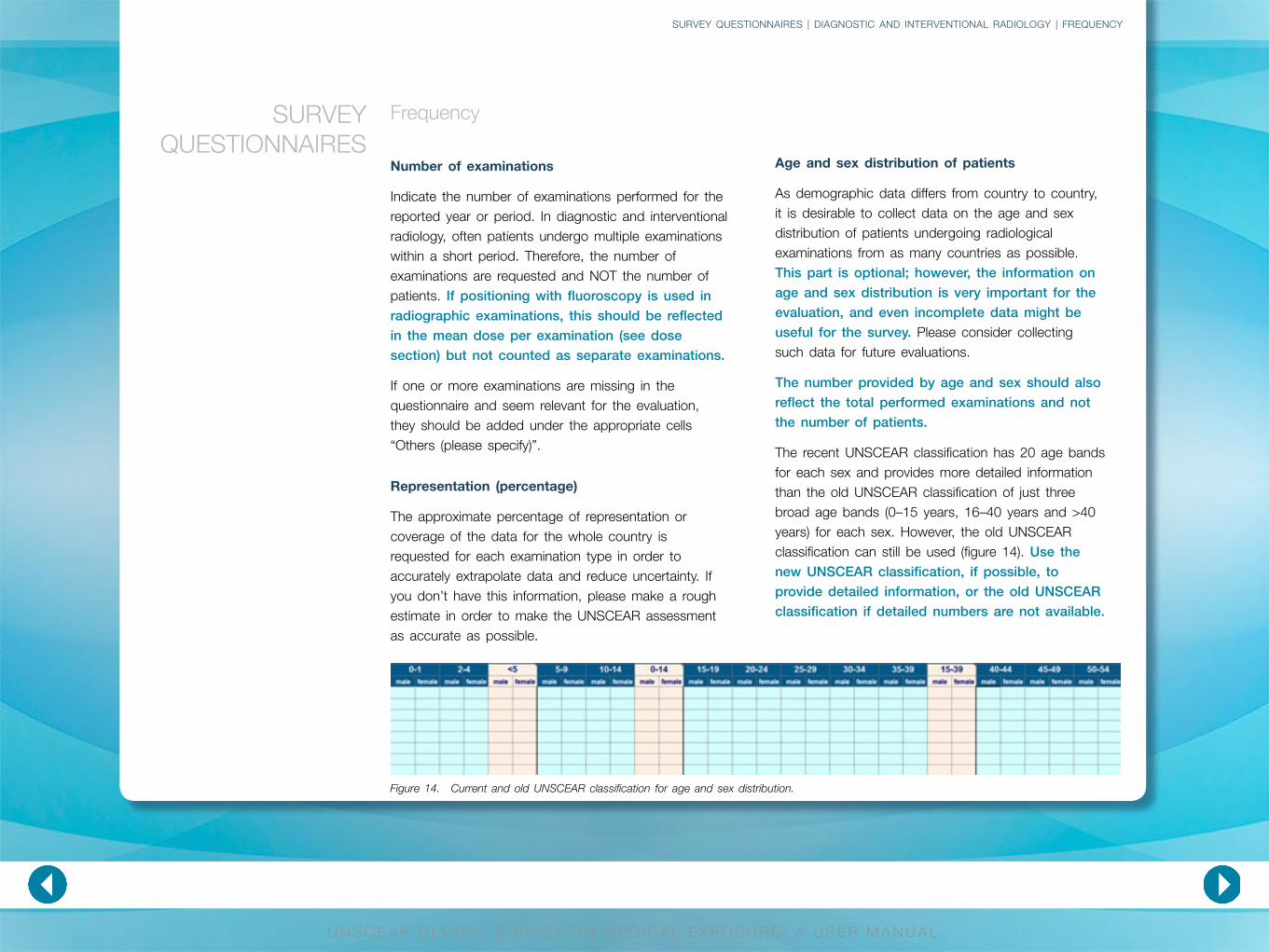
The recent UNSCEAR classification has 20 age bands for each sex and provides more detailed information than the old UNSCEAR classification of just three broad age bands (0–15 years, 16–40 years and >40 years) for each sex. However, the old UNSCEAR classification can still be used (figure 14). Use the new UNSCEAR classification, if possible, to provide detailed information, or the old UNSCEAR classification if detailed numbers are not available.
Figure 14. Current and old UNSCEAR classification for age and sex distribution.

CONTENTS INTRODUCTION GENERAL INFORMATION
SURVEY QUESTIONNAIRES
ONLINE PLATFORM REFERENCES
U N S C E A R G L O B A L S U R V E Y O N M E D I C A L E X P O S U R E : A U S E R M A N U A L
Dose
This section of the questionnaire aims to collect dose related information for the major examinations/procedures in diagnostic and interventional radiology. The classification of examination categories is similar to the one used in the frequency sheet and will not be repeated here. The purpose of the collection of mean doses (including mean effective doses) is for the estimation of the global collective effective dose. UNSCEAR is not looking for diagnostic reference levels (DRLs) per examination as these would overestimate the population dose. Depending on the modality, different dose quantities are requested. This section provides information about effective dose calculation and about the physical dose quantities used in this survey.
SURVEY QUESTIONNAIRES
SURVEY QUESTIONNAIRES | DIAGNOSTIC AND INTERVENTIONAL RADIOLOGY | DOSE
Dose quantities for diagnostic radiography examinations and interventional procedures
The dose area product (DAP), also named kerma-area product (KAP) represents the product of the air kerma (usually in mGy) at the centre of a certain plane of the X-ray beam (e.g. surface of the patient) multiplied by the area of the X-ray field at that plane (usually in cm2). Generally, the DAP/KAP is expressed in Gy∙cm2.
The DAP/KAP is the most widely used dose metric in projection radiography and fluoroscopy. The usefulness of the DAP/KAP is that it can be directly multiplied by examination dependent factors to provide an estimation of effective dose. These factors will be presented in the section dealing with effective dose calculation. In case DAP/KAP values are not available, other dose quantities need to be provided with the corresponding units and variation in the respective fields (figure 15).
Figure 15. Screenshot of dosimetry quantities for diagnostic and interventional radiography.

CONTENTS INTRODUCTION GENERAL INFORMATION
SURVEY QUESTIONNAIRES
ONLINE PLATFORM REFERENCES
U N S C E A R G L O B A L S U R V E Y O N M E D I C A L E X P O S U R E : A U S E R M A N U A L
Dose
Computed tomography (CT) is based on different physical principles from those applying to projection radiography and fluoroscopy. The main dose quantities used for CT dosimetry are the volume computed tomography dose index (CTDIvol) and the dose length product (DLP). The CTDIvol provides an assessment of the average dose to the scanned volume of the standard acrylic CT phantom independently of the specific pitch value. This quantity is important because, multiplied by the scan length, it yields the DLP which in turn leads to the calculation of effective dose by use of examination specific conversion factors. These factors will also be presented in the section dealing with effective dose calculation. CT examinations which comprise more than one phase/scan should reflect this in the total DLP to determine the appropriate effective dose per examination.
SURVEY QUESTIONNAIRES
SURVEY QUESTIONNAIRES | DIAGNOSTIC AND INTERVENTIONAL RADIOLOGY | DOSE
Some cone beam computed tomography (CBCT) machines use DAP/KAP for dose reporting. These CBCTs may additionally provide these values along with CTDIvol and DLP. All values available for CBCT (CTDIvol, DLP or DAP/KAP) should be provided in the relevant section of the questionnaire (figure 16).
Information on radiation dose quantities required in the dosimetry section of the RD questionnaire are summarized in table 3.
Figure 16. Screenshot of dosimetry quantities for CT and CBCT.

U N S C E A R G L O B A L S U R V E Y O N M E D I C A L E X P O S U R E : A U S E R M A N U A L
CONTENTS INTRODUCTION GENERAL INFORMATION
SURVEY QUESTIONNAIRES
ONLINE PLATFORM REFERENCES
PAGE 1 of 3
Radiation dose quantity Description/definition Information and remarksEffective dose Effective dose E (Unit: sievert), is defined as the
weighted sum of the mean radiation doses to a number of radiosensitive tissues or organs in the body [I5, I8].
Mean effective dose values per examination/procedure.
The variation in terms of standard deviation.
Provide further information on the method used to determine the effective dose (e.g. ICRP weighting factors).
Please leave the field empty if no effective doses were calculated for the national survey.
Quantities used in projection radiography, fluoroscopy and interventional procedures
Dose area product (DAP)/kerma area product (KAP)
Dose area product (DAP), also named kerma-area product (KAP), represents the product of the air kerma (usually in mGy) at the centre of a certain plane of the X-ray beam (e.g. the surface of the patient) multiplied by the area of the X-ray field at that plane (usually in cm2).
Generally DAP/KAP is expressed in Gy∙cm2. DAP/KAP values can be multiplied by anatomy and procedure specific factors in order to deduce effective dose values.
DAP/KAP values may be displayed on the console of radiography/fluoroscopy machines.
Average DAP/KAP values per examination/procedure.
The variation in terms of standard deviation.
Possible alternative quantities if DAP/KAP is not available
Air kerma free in air The air kerma free in air [Kα] is the dose absorbed to air free in air. This quantity is directly measured with an appropriate calibrated dosimeter at 1 m distance (radiography) or 50 cm distance (fluoroscopy). Unit: mGyIf it is normalized to the (tube current—exposure time product) is then called “dose yield” or “X-ray tube output”.Unit: mGy/mAs
Average value of quantity per examination/procedure.
Units of quantity.
The variation in terms of standard deviation.
Table 3. Information on radiation dose quantities in diagnostic and interventional radiology used in the UNSCEAR Global Survey
SURVEY QUESTIONNAIRES | DIAGNOSTIC AND INTERVENTIONAL RADIOLOGY | DOSE

U N S C E A R G L O B A L S U R V E Y O N M E D I C A L E X P O S U R E : A U S E R M A N U A L
CONTENTS INTRODUCTION GENERAL INFORMATION
SURVEY QUESTIONNAIRES
ONLINE PLATFORM REFERENCES
PAGE 2 of 3Table 3. Information on radiation dose quantities in diagnostic and interventional radiology used in the UNSCEAR Global Survey
SURVEY QUESTIONNAIRES | DIAGNOSTIC AND INTERVENTIONAL RADIOLOGY | DOSE
Radiation dose quantity Description/definition Information and remarks
Incident air kerma [Kα,i]:Incident air kerma is measured free in air on the central axis of the X-ray beam at the focus skin distance, (FSD). It is related to air kerma free in air by the inverse square law:
Kα,i = K(d)α × (d/dFSD)2, where K(d)α is the air kerma free in air at a distance d from the focus.
Unit: mGy
Average value of quantity per examination/procedure.
Units of quantity.
The variation in terms of standard deviation.
Entrance surface dose (ESD) or Entrance surface air kerma (ESAK)
Entrance surface dose (ESD) or entrance surface air kerma (ESAK) [Kα,e]: this quantity equals the air kerma multiplied by a backscatter factor B.
Kα,e = Kα,i × B
Unit: mGy
The ESD is useful as it can be calculated by using the tube output, the distance from the focus of the X-ray beam and the backscatter factor.
Average value of quantity per examination/procedure.
Units of quantity.
The variation in terms of standard deviation.
Mean glandular dose (MGD) Mean glandular dose (MGD) is a quantity used in mammography in conjunction with age/sex-specific risk factors for radiation-induced breast cancer.
MGD is the average dose to the radiosensitive glandular tissue of the breast. It can be calculated from the incident air kerma [Ka,i] by using appropriate Monte Carlo based conversion factors provided for various radiation qualities and breast thicknesses and compositions.
Unit: mGy
MGD is of interest although the effective dose may be calculated without using it.
Average value of quantity per examination/procedure.
Units of quantity.
The variation in terms of standard deviation.

U N S C E A R G L O B A L S U R V E Y O N M E D I C A L E X P O S U R E : A U S E R M A N U A L
CONTENTS INTRODUCTION GENERAL INFORMATION
SURVEY QUESTIONNAIRES
ONLINE PLATFORM REFERENCES
PAGE 3 of 3Table 3. Information on radiation dose quantities in diagnostic and interventional radiology used in the UNSCEAR Global Survey
SURVEY QUESTIONNAIRES | DIAGNOSTIC AND INTERVENTIONAL RADIOLOGY | DOSE
Radiation dose quantity Description/definition Information and remarks
Quantities used in computed tomography and cone beam computed tomography
Volume computed tomography dose index (CTDIvol):
The volume CTDI (CTDIvol) is equal to the value of CTDIw divided by the CT pitch factor. CTDIvol is a corrected-by-the-pitch-factor CTDIw. It provides an assessment of the average dose to the scanned volume of the standard acrylic CT phantom independently from the specific pitch value.
Unit: mGy
Average CTDIvol values per examination/procedure.
The variation in terms of standard deviation.
Dose length product (DLP) DLP equals CTDIvol multiplied by the scan length.
DLP may be used along with computationally derived factors for the assessment of effective dose in CT examinations.
Unit: mGy × cm
Average DLP values per examination/procedure.
The variation in terms of standard deviation.
Dose area product (DAP)/kerma area product (KAP) in CBCT
Dose area product (DAP), also named kerma area product (KAP) represents the product of the air kerma (usually in mGy) at the centre of a certain plane of the X-ray beam (e.g. the surface of the patient) multiplied by the area of the X-ray field at that plane (usually in cm2).
Generally DAP/KAP is expressed in Gy∙cm2.
Average DAP/KAP values per examination/procedure.
The variation in terms of standard deviation.

CONTENTS INTRODUCTION GENERAL INFORMATION
SURVEY QUESTIONNAIRES
ONLINE PLATFORM REFERENCES
U N S C E A R G L O B A L S U R V E Y O N M E D I C A L E X P O S U R E : A U S E R M A N U A L
Dose
Sample size
The sample size (number of measurements) is used for the estimation of the mean dosimetry value per examination or procedure. If information on effective doses is provided, the sample size could reflect the number of measurements that were the base of each calculation.
Variation
All dose values should be provided as average values per examination or procedure or as mean effective dose per examination or procedure. For uncertainty calculation, it is important to know the variation of these mean values in terms of standard deviation. Standard deviations should be provided separately in the appropriate column for each dose quantity.
Effective dose
For the UNSCEAR Global Survey, users are encouraged to calculate the mean effective dose for the different examinations/procedures reported. The dose quantities requested in this section (e.g. DAP/
KAP and DLP) could be used for these calculations by multiplying appropriate conversion factors. As the calculation methods may vary, additional information about the method used should be included in the “comments” field of the dosimetry section (sheet).
Due to the relatively recent update of the ICRP effective dose weighting factors, users are kindly requested to apply the latest factors [I8] whenever possible. If the old ICRP factors are used [I5] please state this clearly in the “comments” field.
DAP/KAP can be directly multiplied by examination/anatomy dependent factors to provide an estimation of effective dose. These factors have been determined by Monte Carlo simulation methods in conjunction with humanoid computational phantoms. Tables 4 and 5 list dose conversion factors for basic radiographic and fluoroscopic examinations and for image-guided interventional procedures.
SURVEY QUESTIONNAIRES | DIAGNOSTIC AND INTERVENTIONAL RADIOLOGY | DOSE
DIAGNOSTIC AND INTERVENTIONAL
RADIOLOGY

U N S C E A R G L O B A L S U R V E Y O N M E D I C A L E X P O S U R E : A U S E R M A N U A L
CONTENTS INTRODUCTION GENERAL INFORMATION
SURVEY QUESTIONNAIRES
ONLINE PLATFORM REFERENCES
SURVEY QUESTIONNAIRES | DIAGNOSTIC AND INTERVENTIONAL RADIOLOGY | DOSE
Examination typeDose conversion coefficient (DCCE)
[mSv∙(Gy∙cm2)–1]
Chest (PA + Lat) high kV 0.18
Chest (PA + Lat) low kV 0.10
Thoracic spine 0.19
Lumbar spine 0.21
Abdomen 0.26
Pelvis and hip 0.29
Barium meal 0.20
Barium enema 0.28
Barium follow 0.22
IVU 0.18
Cardiac angio 0.20
Table 4. Generalized dose conversion coefficients for radiographic/fluoroscopic examinations (adapted from [E1])

U N S C E A R G L O B A L S U R V E Y O N M E D I C A L E X P O S U R E : A U S E R M A N U A L
CONTENTS INTRODUCTION GENERAL INFORMATION
SURVEY QUESTIONNAIRES
ONLINE PLATFORM REFERENCES
PAGE 1 of 2Table 5. Factors for estimation of effective dose from image-guided interventional procedures (adapted from [N1])
Groups/Subgroups ExaminationsDose conversion coefficient
(DCCE) [mSv∙(Gy∙cm2)–1]
Urinary studies Cystometrography, cystography, excretory urography, micturating cysto-urethrography, urethrography
0.18
Endoscopic retrograde
cholangiopancreatography
(ERCP)
0.26
Arthrograms 0.1
Orthopedic procedures 0.01
Vertebroplasty 0.20
Obstetrics and
gynaecology
Pelvimetry, hysterosalpingogram 0.29
Non-cardiac diagnostic procedures
Peripheral vascular Arteriography (all types)
Peripheral phlebography/venography
Carotid and cerebral angiography
0.26
0.10
0.087
Renal Antegrade pyelography, retrograde pyelography
Renal angiogram, abdominal aortography
Thoracic aortography, arch angiogram
0.18
0.26
0.12
Neurological Cervical spine
Thoracic spine
Lumbar spine
Pulmonary angiography, venacavogram
0.13
0.19
0.21
0.12
SURVEY QUESTIONNAIRES | DIAGNOSTIC AND INTERVENTIONAL RADIOLOGY | DOSE

U N S C E A R G L O B A L S U R V E Y O N M E D I C A L E X P O S U R E : A U S E R M A N U A L
CONTENTS INTRODUCTION GENERAL INFORMATION
SURVEY QUESTIONNAIRES
ONLINE PLATFORM REFERENCES
PAGE 2 of 2
SURVEY QUESTIONNAIRES | DIAGNOSTIC AND INTERVENTIONAL RADIOLOGY | DOSE
Groups/Subgroups ExaminationsDose conversion coefficient
(DCCE) [mSv∙(Gy∙cm2)–1]
Non-cardiac interventional vascular procedures
Percutaneous transluminal angioplasty (PTCA)
0.26
Stent placement Renal/visceral PTCA with stent, Iliac PTCA with stent, bile duct, dilation and stenting
Carotid stent
0.26
0.087
Inferior vena cava filters Filter placement only, hepatic 0.26
Embolization Chemoembolization, pelvic arterial embolization, pelvic vein embolization: ovarian vein, other tumour embolization, embolization
Pulmonary angiography with filter, bronchial artery embolization
Thrombolytic therapy
Transjugular intrahepatic portosystemic shunt (TIPS)
0.26
0.12
0.26
0.26
Cardiac procedures
Diagnostic coronary angiography
0.12
Interventional procedures Angioplasty
Percutaneous transluminal coronary angioplasty
Embolization
Cardiac radiofrequency ablation
0.20-0.26
0.18-0.28
0.26
0.10-0.23
If DAP/KAP is not available and other dose quantities are used, the respective dose conversion coefficients need to be indicated in order to be able to reproduce the calculations.
The mean glandular dose (MGD) is the main quantity used in mammography in conjunction
with age/sex-specific risk factors for radiation-induced breast cancer. However, the effective dose may be derived by incident air kerma by the use of a set of factors that connect incident air kerma and effective dose in mammography for typical mammography exposure conditions (table 6).
Table 5. Factors for estimation of effective dose from image-guided interventional procedures (adapted from [N1])
U N S C E A R G L O B A L S U R V E Y O N M E D I C A L E X P O S U R E : A U S E R M A N U A L
CONTENTS INTRODUCTION GENERAL INFORMATION
SURVEY QUESTIONNAIRES
ONLINE PLATFORM REFERENCES
SURVEY QUESTIONNAIRES | DIAGNOSTIC AND INTERVENTIONAL RADIOLOGY | DOSE
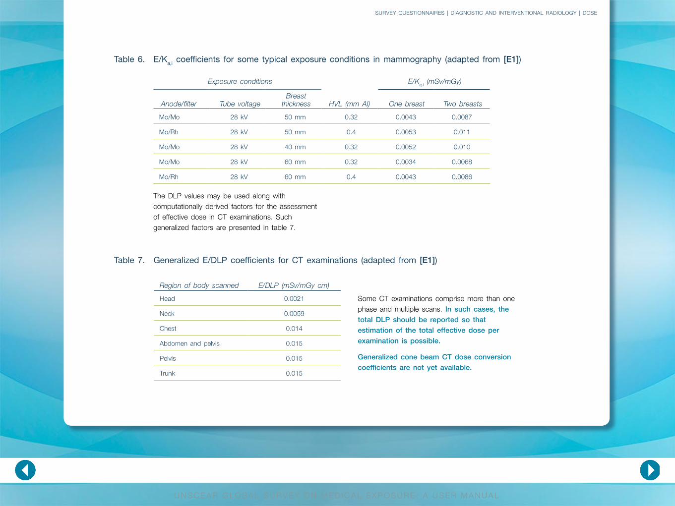
Table 6. E/Ka,i coefficients for some typical exposure conditions in mammography (adapted from [E1])
Table 7. Generalized E/DLP coefficients for CT examinations (adapted from [E1])
Exposure conditions
HVL (mm Al)
E/Ka,i (mSv/mGy)
Anode/filter Tube voltageBreast
thickness One breast Two breasts
Mo/Mo 28 kV 50 mm 0.32 0.0043 0.0087
Mo/Rh 28 kV 50 mm 0.4 0.0053 0.011
Mo/Mo 28 kV 40 mm 0.32 0.0052 0.010
Mo/Mo 28 kV 60 mm 0.32 0.0034 0.0068
Mo/Rh 28 kV 60 mm 0.4 0.0043 0.0086
Region of body scanned E/DLP (mSv/mGy cm)
Head 0.0021
Neck 0.0059
Chest 0.014
Abdomen and pelvis 0.015
Pelvis 0.015
Trunk 0.015
The DLP values may be used along with computationally derived factors for the assessment of effective dose in CT examinations. Such generalized factors are presented in table 7.
Some CT examinations comprise more than one phase and multiple scans. In such cases, the total DLP should be reported so that estimation of the total effective dose per examination is possible.
Generalized cone beam CT dose conversion coefficients are not yet available.

CONTENTS INTRODUCTION GENERAL INFORMATION
SURVEY QUESTIONNAIRES
ONLINE PLATFORM REFERENCES
U N S C E A R G L O B A L S U R V E Y O N M E D I C A L E X P O S U R E : A U S E R M A N U A L
Diagnostic and interventional radiology
From a dosimetry point of view, the major kinds of RD examinations fall into two categories: projectional imaging and cross-sectional imaging. The first category includes radiography, mammography and fluoroscopy thus including interventional procedures. Cross-sectional imaging includes CT and CBCT examinations.
Radiography
In many occasions mean effective dose from projection radiography examinations may be directly calculated. This is possible by multiplying the available mean DAP/KAP value by the anatomy specific, dose conversion coefficients tabulated on table 4. For example an abdomen X-ray with a mean DAP of 7 Gy∙cm2 would yield an effective dose of:
E = 7 Gy∙cm2 × 0.26 mSv∙(Gy∙cm2)–1 = 1.82 mSv (1)
In case a mean DAP/KAP value is not available for an examination, it can be calculated by using more elementary quantities such as the air kerma free in air (Kα). Kα is the dose absorbed to air, free in air (measured in mGy). This quantity is directly measured with an appropriate calibrated dosimeter at 1 m distance (radiography) or 50 cm distance (fluoroscopy). If it is normalized to the (tube current—exposure time product (mAs)), it is then called “dose yield” or “X-ray tube output” measured in mGy/mAs. Assuming reasonable uniformity of X-ray intensity throughout the whole field, it is possible to calculate the cumulative DAP/KAP by multiplying the dose yield with the total mAs and the field size at the distance in question. For example an X-ray
output of 10 mGy/mAs for a 10 × 10 cm2 X-ray field during a 10 mAs exposure will yield a DAP/KAP value as follows:
DAP/KAP = 10 mGy∙mAs-1 × 10 cm × 10 cm × 10 mAs = 10 Gy∙cm2 (2)
The following dose quantities could be available and could possibly be used to calculate the mean effective dose from specific examinations. The definitions and their connections to DAP/KAP and air kerma free in air are described so that the user may track back to the value required for mean effective dose estimation.
Incident air kerma (Kα,i) is measured free in air on the central axis of the X-ray beam at the focus skin distance, (FSD). It is measured in mGy and it is related to air kerma free in air by the inverse square law, as follows:
Kα,i = K(d)α × (d/dFSD)2 (3)
where K(d)α is the air kerma free in air at a distance d from the focus.
Entrance surface dose (ESD) or entrance surface air kerma (ESAK) (Kα,e): This quantity is measured in mGy and equals the air kerma multiplied by a backscatter factor B.
Kα,e = Kα,i × B (4)
The ESD is useful as it can be calculated by using the tube output, the distance from the focus of the X-ray beam and the backscatter factor.
The examples correspond to the two major disciplines for which the
effective dose is applied. Mean values of the dosimetric
quantities should be used in order to calculate mean effective
doses representative of the practice in a country.
APPENDIX: EXAMPLES
OF DOSE CALCULATIONS
APPENDIX | DIAGNOSTIC AND INTERVENTIONAL RADIOLOGY |
CONTENTS INTRODUCTION GENERAL INFORMATION
SURVEY QUESTIONNAIRES
ONLINE PLATFORM REFERENCES
U N S C E A R G L O B A L S U R V E Y O N M E D I C A L E X P O S U R E : A U S E R M A N U A L
Fluoroscopy and interventional procedures
In fluoroscopy and interventional procedures, DAP/KAP is the dose metric of interest. Most of the times it is displayed on the console of the fluoroscopy machine. In examinations/interventional procedures involving a lot of fluoroscopy time, average cumulative DAP/KAP should be used for mean effective dose calculations. Tables 4 and 5 provide anatomy and procedure specific dose conversion coefficients. The calculation of mean effective dose is performed by multiplying the average cumulative DAP/KAP with the appropriate coefficients found in tables 4 and 5. The calculation required is similar to the one presented in equation (1) above.
However, doses in interventional radiology are usually much higher than simple diagnostic radiography and need special attention. For example, a PTCA procedure (e.g for stent placement) with a cumulative mean DAP/KAP of 50 Gy∙cm2 results in 13 mSv by using the appropriate coefficient value for PTCA stent placement from table 5.
E = 50 Gy∙cm2 × 0.26 mSv∙(Gy∙cm2)–1 = 13 mSv (5)
Mammography
In mammography, the mean effective dose may be derived by using the quantity of incident air kerma and a set of factors that connect incident air kerma and effective dose in mammography for typical mammography exposure conditions (table 6). In an exposure of both breasts with the following conditions:
• Breast thickness: 50 mm• 28 kV • Mo/Mo • HVL = 0.32 mm Al • Kα,i = 230 mGy,
would yield the following effective dose:
E = 230 mGy × 0.0087 mSv∙mGy-1 = 2 mSv (6)
Diagnostic and interventional radiologyAPPENDIX: EXAMPLES
OF DOSE CALCULATIONS
APPENDIX | DIAGNOSTIC AND INTERVENTIONAL RADIOLOGY |
CONTENTS INTRODUCTION GENERAL INFORMATION
SURVEY QUESTIONNAIRES
ONLINE PLATFORM REFERENCES
U N S C E A R G L O B A L S U R V E Y O N M E D I C A L E X P O S U R E : A U S E R M A N U A L
Computed tomography
In CT, the quantity of interest is the dose length product (DLP). DLP values are usually displayed on the console of the CT scanner. These values may be multiplied by computationally derived coefficients for the assessment of effective dose in CT examinations. Such generalized coefficients are presented in table 7. As an example, calculation of the effective dose to the patient due to a chest CT scan with an average DLP of 700 mGy∙cm would go as follows:
E = 700 mGy∙cm × 0.014 mSv∙(mGy∙cm)-1 = 9.8 mSv (7)
Sometimes the DLP value may not be available while the average CTDIvol is available. Since DLP is in fact equal to CTDIvol multiplied by the scan length, it is possible to estimate effective dose by multiplying the generalized coefficients with CTDIvol and the total estimated average scan length. The total effective dose arising from a CT scan is calculated by using the total (cumulative) DLP, especially for examinations involving several scans. In case the calculation is done by using CTDIvol, the user should use the total estimated scan length for all phases of the examination.
Diagnostic and interventional radiologyAPPENDIX: EXAMPLES
OF DOSE CALCULATIONS
APPENDIX | DIAGNOSTIC AND INTERVENTIONAL RADIOLOGY |