international journal of pharmaceutical … 667.pdf · of gellan gum (x1) and calcium chloride ......
TRANSCRIPT

Research Article CODEN: IJPRNK IMPACT FACTOR: 1.862 ISSN: 2277-8713 MB Bhatt, IJPRBS, 2014; Volume 3(2): 845-859 IJPRBS
Available Online at www.ijprbs.com 845
FORMULATION AND EVALUATION OF IONOTROPICALLY GELLED NOVEL HYDROGEL BEADS OF VALSARTAN
BHATT M. B., PANCHAL B. P., PATEL N. N., BHIMANI B. V., PATEL G. V., DR. PATEL U. L. Arihant School of Pharmacy & Bio Research Institute, Adalaj, Gandhinagar.
Accepted Date: 19/04/2014; Published Date: 27/04/2014
Abstract: Objectives: To formulate and evaluate the sustained release hydrogel beads of valsartan using different ratio of polymer and counter ion in order to increase the drug bioavailability, therapeutic efficiency, reduce dosing frequency and improvement of patient compliance. Experimental work: Different formulations were prepared by ionotropic gelation method using various release rate controlling polymer i.e. gellan gum and counter ion like, calcium chloride. Drug-excipients compatibility was carried out by FTIR. Different formulations were evaluated for particle size, swelling index, % drug entrapment efficiency and in vitro drug release. Optimized batch was evaluated for scanning electron microscopy. Mathematical analysis of the release kinetics was carried out to determine the mechanism of drug release. In vitro release data was fitted to various models to ascertain the kinetic of drug release. Response surface graph was prepared to examine the effect of independent variable on dependent variable. A 32 factorial was applied to check the effect of varying the concentration of gellan gum (X1) and calcium chloride (X2) on the dependent variable i.e. swelling index and in vitro drug release. Result: It was observed that optimized batch A7 containing gellan gum (2.5%) and calcium chloride (4%) gives 123 % swelling index after 12 hrs and 100.56 % drug release after 24 hrs, which is nearer to theoretical profile. Keywords: Valsartan, gellan gum, in vitro swelling, in vitro drug release
INTERNATIONAL JOURNAL OF
PHARMACEUTICAL RESEARCH AND BIO-SCIENCE
PAPER-QR CODE
Corresponding Author: MS. BHATT M.B.
Access Online On:
www.ijprbs.com
How to Cite This Article:
MB Bhatt, IJPRBS, 2014; Volume 3(2): 845-859

Research Article CODEN: IJPRNK IMPACT FACTOR: 1.862 ISSN: 2277-8713 MB Bhatt, IJPRBS, 2014; Volume 3(2): 845-859 IJPRBS
Available Online at www.ijprbs.com 846
INTRODUCTION
1.1 Introduction to Hydrogel Beads
The basic rationale of controlled release drug delivery system is to optimize the biopharmaceutical, pharmacokinetic and pharmacodynamic properties of a drug administered by the most suitable route to achieve its maximum utility, to control condition within shortest possible time by using smallest quantity of drug. It also provides constant drug level in the blood with reduced dosing frequency and reduced side-effects, thus increasing patient compliance and decreasing adverse drug effects1.
Generally Multiparticulate drug delivery systems are intended for oral, parenteral and topical formulations and approaches include formulations in the form of pellets, granules, beads, gelispheres, microcapsules, microspheres, lipospheres, microparticles and nanoparticles. In these systems, the dosage of the drug substances is divided on a plurality of subunit, typically consisting of thousands of spherical particles with selective diameter range. To deliver the recommended total dose, these subunits are filled into a sachet and encapsulated or compressed into a tablet2.
1.1.1 Advantages
Considerable research efforts have been spent on oral controlled release multiparticulate drug delivery system due to its advantages over monolithic dosage forms as3, 4, 5.
1) Less inter and intra subject variations in gastric transit time.
2) Taste masking.
3) No risk of dose dumping.
4) Less local irritation.
5) Increased solubility or dispersibility, hence quick diffusion, leading to a more rapid drug release and better absorption, as having large surface area.
6) Avoid risk of toxicity since they have ability to spread uniformly throughout gastrointestinal tract.
7) Improves patient compliance by decreasing dosing frequency.

Research Article CODEN: IJPRNK IMPACT FACTOR: 1.862 ISSN: 2277-8713 MB Bhatt, IJPRBS, 2014; Volume 3(2): 845-859 IJPRBS
Available Online at www.ijprbs.com 847
8) Bioavailability enhances despite first pass effect because fluctuations in plasma drug concentration is avoided, a desirable plasma drug concentration is maintained by continuous drug release.
9) Increased therapeutic efficiency.
10) Improved stability.
Hydrophilic polymers derived from plant, animal, microbial, or synthetic sources when added to water, hydrocolloids are formed which disperse evenly as microscopic particles. At sufficiently high concentrations, the polymers become entangled with each other, forming loose networks that change the rheological properties of solutions. Many hydrocolloids, such as gelatine and pectin can also form gels by hydrogen bonding within and between polymers6. Formulations based on hydrocolloids may have some advantages over other sustained release formulations, for instance, different structures can be obtained upon dehydration of the hydrocolloid formulations which can be modified by the drying conditions and formulation composition. Structural characteristics such as porosity may affect the penetration rate of liquid into the formulations and thus modify the release pattern of the drug. Moreover, the stability and physical properties (dimensions, strength, etc.) of various hydrocolloids are affected by factors such as swelling in water, pH value and enzymes, and therefore vary in different parts of the gastrointestinal tract. Changes in the physical properties of the formulations may also lead to different drug-release patterns in different parts of the gastrointestinal tract, thus providing a wide scope to be utilized as carriers for the controlled release of drugs7.
In addition, hydrocolloid formulation preparation procedures are generally quite simple and the cost of such materials is low. Beads formulated from hydrocolloid polymers are called hydrogel beads whose size ranges from 0.2 to 3 mm and are mostly spherical in shape to give them the desired properties regarding eye-appeal, release properties, or technical demands in general. Shape and size are essential parameters for adequately describing hydrocolloid beads. It is easier to specify the size of a regularly shaped bead (i.e., spherical or ellipsoid); for irregularly shaped beads, the term size should be arbitrarily defined8. The incorporation of suitable concentrations of active ingredients for the therapeutic or cosmetic uses in microbeads are generally carried out by two processes9, 10:
a). Incorporation of the active ingredients during the process of preparation of the microbeads.
b). Adsorption of a solution or suspension of the active ingredients in the previously crosslinked microbeads, in case the active ingredients are incompatible with the dehydration solvents.

Research Article CODEN: IJPRNK IMPACT FACTOR: 1.862 ISSN: 2277-8713 MB Bhatt, IJPRBS, 2014; Volume 3(2): 845-859 IJPRBS
Available Online at www.ijprbs.com 848
Factors Affecting Ionotropic Gelation Method10
1) Polymer and crosslinking electrolyte concentration
Polymer and electrolyte concentration have major effect on formulation of beads by ionotropic gelation method. Concentration of both should in the ratio calculated from number of crosslinking units. Percentage entrapment efficiency varies from the type of electrolytes and also the concentration of electrolytes.
2) Temperature
Temperature also plays important role on size of beads formed by ionotropic gelation method and also on the curing time i.e. time required for crosslinking.
3) pH of crosslinking solution
pH of crosslinking solution also considerable factor during the formulation as it shows effect on reaction rate, shape and size of beads.
4) Drug concentration
Drug to be entrapped in the beads should be in the proper ratio with the polymer, as the drug concentration greatly affects the entrapment efficiency, if drug: polymer ratio exceeds the range then bursting effect may observe, density of gelispheres enhances and the size and shape of gelispheres also increases.
5) Gas forming agent concentration
Gas forming agents such as calcium carbonate, sodium bicarbonate added in to the formulation to develop porous gelispheres, which tremendously affect the gelispheres size and shape.
Ionotropic Gelation Method11, 12
Ionotropic gelation involves simply the interaction of an ionic polymer with oppositely charge ion to initiate cross linking.
Methods of ionotropic gelation
There are two methods by which hydrogel beads can be generated using ionotropic gelation technique. These methods differ from each other in the source of the cross linking ion. In one of the methods, the cross linker ion is positioned. Whereas in the other method, the cross linker ion is incorporated within the polymer solution in inactive form. External cross- linking produced thinner films with smoother surface, greater matrix strength, stiffness and

Research Article CODEN: IJPRNK IMPACT FACTOR: 1.862 ISSN: 2277-8713 MB Bhatt, IJPRBS, 2014; Volume 3(2): 845-859 IJPRBS
Available Online at www.ijprbs.com 849
permeability than internally cross-linked films. Externally cross-linked micropellets were also capable of greater drug encapsulation efficiency and slower drug release rate. There are variety of natural and synthetic polymeric systems that have been investigated for the controlled release of drug. Hydrophilic polyionic carbohydrates such as alginate and chitosan have been paid much attention in recent years.
Since the preparation of beads by these materials involves the use of aqueous solvents, environmental problems associated with organic solvents would be minimized. A variety of natural polymers and their derivatised products have been successfully employed in hydrogel system for various pharmaceutical applications. In this review the potential of sodium alginate and chitosan to form the highly cross linked structure and its pharmaceutical applications is discussed. As compared to other natural polymers, sodium alginate and chitosan shows no variations in viscosity and hence produces more uniform gel structure which forms stronger cross linked structure and more loading of entrapped material.
Preparation of ionotropic gelation method
Polyelectrolyte solution
[Sodium Alginate (-)/Gellan gum (-)/CMC (-)/Pectin (-)/ Chitosan (+) + Drug]
↓
Added drop wise under magnetic stirring by needle
↓
Counter ion solution
[Calcium chloride solution (+)/Sodium tripolyphosphate (-)]
↓
Hydrogel Beads

Research Article CODEN: IJPRNK IMPACT FACTOR: 1.862 ISSN: 2277-8713 MB Bhatt, IJPRBS, 2014; Volume 3(2): 845-859 IJPRBS
Available Online at www.ijprbs.com 850
Figure: 1. Schematic representation and diagram of the preparation of hydrogel beads by ionotropic gelation and polyelectrolyte complexation.
Ionotropic gelation is based on the ability of polyelectrolytes to cross link in the presence of counter ions to form hydrogel beads also called as gelispheres. Hydrogel beads are spherical crosslinked hydrophilic polymeric entity capable of extensive gelation and swelling in simulated biological fluids and the release of drug through it controlled by polymer relaxation. The hydrogel beads are produced by dropping a drug-loaded polymeric solution into the aqueous solution of polyvalent cations. The cations diffuses into the drug-loaded polymeric drops, forming a three dimensional lattice of ionically crosslinked moiety. Biomolecules can also be loaded into these hydrogel beads under mild conditions to retain their three dimensional structure13.
MATERIALS AND METHODS
Valsartan was obtained from Cadila Pharmaceuticals Ltd; gellan gum and calcium chloride (CaCl2), methanol, ethanol, sodium hydroxide was obtained from S.D fines chem, Ltd; Mumbai.
Preparation of valsartan loaded hydrogel beads14
The beads were prepared by the ionotropic gelation technique. Gellan gum solution was prepared by dissolving in deionized water and heated at 60°. Concentrations of the drug was dissolved/ dispersed uniformly in 100 ml of gellan solution (1.5, 2.0, 2.5 %) below 40° under continuous stirring. The stirring was continued after complete addition until a uniform dispersion was obtained. The resultant homogeneous bubble free slurry dispersion was dropped through a 25G syringe needle into 100 ml of calcium chloride solution (4 %, 6 %, 10 %)
Research Article CODEN: IJPRNK IMPACT FACTOR: 1.862 ISSN: 2277-8713 MB Bhatt, IJPRBS, 2014; Volume 3(2): 845-859 IJPRBS
Available Online at www.ijprbs.com 851
which was kept under stirring to improve the mechanical strength of the beads and also to prevent aggregation of the formed beads. Immediate formation of small gellan beads took place; after 5 min of curing time, the formed beads were collected by filtration and dried at 40°.
The hydrogel beads were prepared by using different compositions of the polymers as shown in the table: 1.
Table: 1. Composition of Valsartan Hydrogel beads.
Batch code Gellan gum (%w/v) CaCl2 (%w/v) Valsartan
(%w/v)
A1 1.5 4 1
A2 1.5 6 1
A3 1.5 8 1
A4 2.0 4 1
A5 2.0 6 1
A6 2.0 8 1
A7 2.5 4 1
A8 2.5 6 1
A9 2.5 8 1
CHARACTERIZATION OF VALSARTAN LOADED HYDROGEL BEADS 15-18
Drug excipient compitability by fourier transformation infrared (FTIR) spectroscopy
FTIR spectra were recorded on a KBr Press Model SHIMADZU FTIR-5300 Samples were thoroughly grounded with exhaustively dried KBr and pellets were prepared by compression under vacuum and their corresponding FTIR spectra were recorded.
Particle size determination
Particle size of hydrogel beads was determined by micrometer, having an accuracy of 0.001. The average diameter of 50 particles per batch was selected.

Research Article CODEN: IJPRNK IMPACT FACTOR: 1.862 ISSN: 2277-8713 MB Bhatt, IJPRBS, 2014; Volume 3(2): 845-859 IJPRBS
Available Online at www.ijprbs.com 852
Drug entrapment efficiency
Known amount of beads (20 mg) were added to 20 ml USP phosphate buffer of pH 7.4 solution for complete swelling at 37°. The beads were crushed in a glass mortar with pestle the solution was than kept for 24 hrs to extract the drug completely and centrifuged to remove polymeric debris. The clear supernatant solution was analyzed for drug content using UV-visible spectrophotometer at 250 nm (UV/Visible spectrophotometer, Simadzu).
In vitro swelling study
The release of the entrapped drug from hydrogels depends on the swelling behaviour, because swelling is directly proportional to drug release in case of hydrogels. As the hydrogel swells, the pores of network open and release of the entrapped solute occurs. Therefore, the dynamic swelling study of the prepared beads was carried out. The dynamic swelling behaviour of the beads was studied by mass measurement. The 50 mg of beads were incubated into the glass vials, with 25 ml pH 7.4 phosphate buffer. The beads were taken out at different time intervals and blotted carefully without pressing hard to remove the excess surface liquid. The swollen beads were weighed using the electronic microbalance. The weights determined were used for plotting the swelling profile. The swelling index was calculated using the following equation.
Q= [(W2-W1)/W1] ×100
Where, Q= Percentage swelling
W1 is mass of the dry beads
W2 is the mass of swollen beads.
In vitro drug release
In-vitro drug release studies were performed using USP dissolution test apparatus I (basket type). Formulation is put in basket. The dissolution studies were performed in 900 ml dissolution medium 0.1 N HCl (pH 1.2) for 2 hrs and, in phosphate buffer (pH 6.8) for 6 hrs and the same amount of phosphate buffer (pH 7.4) till end of the study at 50 rpm maintained at 37 ± 0.5ºC. At predetermined time intervals an aliquot of 5 ml was withdrawn and replenished with fresh medium. Amount of drug in each aliquot was determined on a UV-Spectrophotometer at 250 nm using a suitable blank. All trials were conducted in triplicate and the average (± SD) reading was noted.

Research Article CODEN: IJPRNK IMPACT FACTOR: 1.862 ISSN: 2277-8713 MB Bhatt, IJPRBS, 2014; Volume 3(2): 845-859 IJPRBS
Available Online at www.ijprbs.com 853
Evaluation of release kinetics
The mechanism of the drug release from the hydrogel beads was investigated by fitting the release data using zero order, first order, Higuchi, Korsemeyer - Peppas, and Erosion models as shown in the Table: 2.
Table: 2. Mathematical modeling for the study of release kinetics Model Equation
Zero order Q = Qo + Kot
Higuchi Q t= Qo + KH t1/2
Korsmeyer-Peppas Mt / M∞ = K × tn
SEM analysis
The surface morphology of the beads was investigated using scanning electron microscope (JEOL, JFC Japan). The hydrogel beads were mounted onto stubs using double sided adhesive tape and sputter coated with platinum using a sputter coater. The SEM analysis of the hydrogel beads was carried out using Jeol JSM 5300, Japan. The hydrogel beads were viewed at an accelerating voltage of 10 kV. The scanning electron microscopy was done for the optimized batch.
RESULTS
Drug-Excipients compatibility study
Figure: 2. Drug-Excipients compatibility study by FTIR
Research Article CODEN: IJPRNK IMPACT FACTOR: 1.862 ISSN: 2277-8713 MB Bhatt, IJPRBS, 2014; Volume 3(2): 845-859 IJPRBS
Available Online at www.ijprbs.com 854
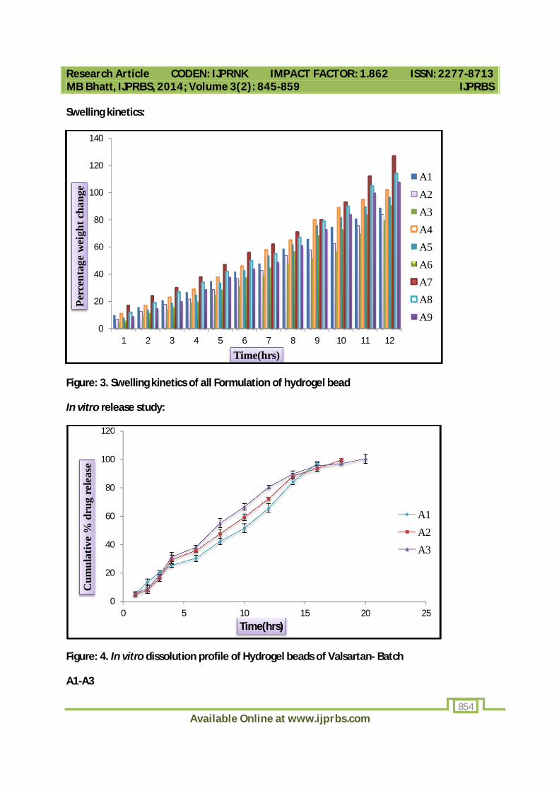
Swelling kinetics:
Figure: 3. Swelling kinetics of all Formulation of hydrogel bead
In vitro release study:
Figure: 4. In vitro dissolution profile of Hydrogel beads of Valsartan- Batch
A1-A3
0
20
40
60
80
100
120
140
1 2 3 4 5 6 7 8 9 10 11 12
Perc
enta
ge w
eigh
t cha
nge
Time(hrs)
A1A2A3A4A5A6A7A8A9
0
20
40
60
80
100
120
0 5 10 15 20 25
Cum
ulat
ive
% d
rug
rele
ase
Time(hrs)
A1A2A3

Research Article CODEN: IJPRNK IMPACT FACTOR: 1.862 ISSN: 2277-8713 MB Bhatt, IJPRBS, 2014; Volume 3(2): 845-859 IJPRBS
Available Online at www.ijprbs.com 855
Figure: 5. In vitro dissolution profile of Hydrogel beads of Valsartan- Batch
A4-A6
Figure: 6. In vitro dissolution profile of Hydrogel beads of Valsartan- Batch
A7-A9
0
20
40
60
80
100
120
0 5 10 15 20 25 30
Cum
ulat
ive
% d
rug
rele
ase
Time(hrs)
A4A5A6
0
20
40
60
80
100
120
0 5 10 15 20 25 30Cum
ulat
ive
% d
rug
rele
ase
Time(hrs)
A7
A8
A9

Research Article CODEN: IJPRNK IMPACT FACTOR: 1.862 ISSN: 2277-8713 MB Bhatt, IJPRBS, 2014; Volume 3(2): 845-859 IJPRBS
Available Online at www.ijprbs.com 856
Scanning Electron Microscopy (SEM):
Figure: 7. SEM of valsartan loaded Hydrogel beads
Table: 4. Model fitting of drug release from Hydrogel beads
Batch Zero order Higuchi First order K-peppas
A1 0.931 0.964 0.841 0.859
A2 0.923 0.979 0.753 0.833
A3 0.942 0.987 0.827 0.862
A4 0.891 0.982 0.753 0.884
A5 0.932 0.963 0.798 0.872
A6 0.947 0.972 0.802 0.863
A7 0.925 0.987 0.792 0.895
A8 0.915 0.965 0.819 0.879
A9 0.895 0.978 0.826 0.846
Research Article CODEN: IJPRNK IMPACT FACTOR: 1.862 ISSN: 2277-8713 MB Bhatt, IJPRBS, 2014; Volume 3(2): 845-859 IJPRBS
Available Online at www.ijprbs.com 857
DISCUSSION
Drug excipient compatibility by FTIR spectra obtained were shown in figure 2. From the graphs it was observed that valsartan and gellan gum showed sharp and strong bands between 3000-2900 cm-1 due to C-H Stretching, 3500-2400 cm-1 due to O-H Stretching, 3500-3300 cm-1due to N-H Stretching, 1725-1705 cm-1 due to –C=O Stretching and 1650-1600 cm-1 due to –COOH Stretching.
The hydrogel formulation confirmed the compatibility between the drug and the polymers. Swelling study showed in figure: 3. It is mainly due to the presence of gellan gum a complex extra cellular polysaccharide. The rate of swelling mainly depends upon the cross linking nature of the hydrogel. The hydrodynamic free volume is high if the gel network is less which in turn lowers the cross linking density. The higher swelling is due to the accommodation of more of the solvent molecules. The swelling rate was found to be different basing on the concentrations of gellan gum and calcium chloride.
The entrapment efficiency of Valsartan in the hydrogel beads was almost similar irrespective of their concentrations and ratios.
The results of the drug release study of the hydrogel were shown in the figure: 4, 5 and 6. The drug release from a hydrogel can be attributed by a number of factors like chemical structure of the polymers, composition of the hydrogel, network structure release condition, release conditions, concentration of the crosslinker etc. In this study the drug release from the hydrogel beads was affected by the concentrations of the polymers and the cross linking agent have been examined.
Formulation A-7 was formulated using 2.5 % gellan gum and 4 % CaCl2. It gave desired results in terms of swelling index and t90 (100.56 %) and it shows 123 % (higher) swelling at 12 hrs as compared to other batches. The cumulative percentage drug release was observed to reduce slowly with the increase in the concentration of cross linking agent.
The size, shape and surface morphology of the dried beads as visualized by SEM as shown in figure: 7 for the best formulation revealed the beads are very spherical with a soft surface.
Kinetics of drug release of optimized batch was studied and it show that Higuchi model was best fit for release of valsartan from dosage form.
CONCLUSION
A controlled release oral drug delivery system of Valsartan was developed in the form of hydrogel beads using various proportions of polymer (gellan gum) and different concentrations

Research Article CODEN: IJPRNK IMPACT FACTOR: 1.862 ISSN: 2277-8713 MB Bhatt, IJPRBS, 2014; Volume 3(2): 845-859 IJPRBS
Available Online at www.ijprbs.com 858
of crosslinking agent. Presence of different concentration of gellan gum and calcium chloride made the hydrogel beads mechanically strenghthen and possibility for the bead formation and maximum swelling rate. Increasing the concentration of crosslinking agent can be used further to control the drug release.
Thus, the hydrogel bead system as an effective drug delivery system was developed which offered a solubilizing cross-linked network matrix for the poorly soluble drug, Valsartan providing the best controlled release of the drug over 24 hrs.
ACKNOWLEDGEMENTS
My sincere thanks to the continuous support given by Mr. Bhavin Bhimani, M. Pharm (Pharmaceutics), Assistant Professor, Arihant School Of Pharmacy & Bio-Research Institute.
REFERENCES
1. Young CR, Koleng JJ, McGinity JW. “Production of spherical pellets by a hot-melt extrusion and spheronization process”. International Journal of Pharmaceutics 2002; 242: 87-92.
2. Gaikwad A, Pathade P, Bidkar S, Pawar A. “Formulation and Evaluation of Multi Particulate Drug Delivery System of Glipizide by Using Crossed Linked Chitosan”. Journal of Pharmacy Research 2011; 4 suppl 1: 300-303.
3. Dey NS, Majumdar S, Rao MEB. “Multiparticulate Drug Delivery Systems for Controlled Release”. Tropical Journal of Pharmaceutical Research 2008; 7(3): 1067-1075.
4. Mennini N, Furlanetto S, Maestrelli F, Pinzauti S, Mura P. “ Response surface methodology in the optimization of chitosan Ca pectinate bead formulations”. European journal of pharmaceutical sciences 2008; 35: 318-325.
5. Kulkarni RV, Boppana, Setty CM, Kalyane NV. “Carboxymethylcellulose-aluminum hydrogel microbeads for prolonged release of simvastatin”. Acta Pharmaceutica Sciencia 2010; 52: 137-143.
6. Laura Cassiday. “Hydrocolloids get personal”. Inform. 2012; 23(6): 349-352.
7. Amos Nussinovich, Avi Gal. “Hydrocolloid carrier beads with inert filler material”. 2010; Patent number: 20100015192.
8. “Biodegradable microbeads for the pharmaceutical and cosmetic uses”. Patent Number: 0563876, 1993.

Research Article CODEN: IJPRNK IMPACT FACTOR: 1.862 ISSN: 2277-8713 MB Bhatt, IJPRBS, 2014; Volume 3(2): 845-859 IJPRBS
Available Online at www.ijprbs.com 859
9. Amos Nussinovitch. “Polymer macro- and micro-gel beads fundamentals and applications, physical properties of beads and their estimation”. 2010; springer science+business media USA.
10. Yoav Basha and Juan-Pablo HernandezLuis. “Alginate microbeads as inoculant carriers for plant growth-promoting bacteria”. Biol Ferti Soils. 2002; 35:359-368.
11. Wayne RG, Siow F W. “Protein release from alginate matrices”. Adv Drug Delivery Rev. 1998;1:267-285.
12. Thanou M et al. “Oral drug absorption enhancement by chitosan and its derivatives”. Adv Drug Delivery Rev. 2001; 52: 117-126.
13. Poncelet D, Babak , DulieuC, Picot Colloids and Surfaces A: “A physico-chemical approach to production of alginate beads by emulsification-internal ionotropic gelation”. Physicochemical and Engineering Aspects, 1999; 155 (2-3), 171-176.
14. Piyakulawat p, Muangsin N. “Preparation and evaluation of Chitosan Beads for Controlled release of Sodium Diclofenac.”AAPS Pharmsci Tech, 2007; 1-11.
15. Halder A, Mukherjee S, Sa B. “Development and evaluation of polyethyleneimine-treated calcium alginate beads for sustained release of diltiazem”. J Microencapsul. 2005 Feb; 22(1):67-80.
16. Archita M, Kishor R, Madhu babu D and Prabhakar reddy V, “Design and in vitro evaluation of sustained release matrix tablets of timolol maleate.” Amer. J. Pharmatech. Res. 2011, 1, 339-354.
17. Prasanth Sai RV, Pujitha I, Perreddy K and Pradeep Chandra K, “Formulation evaluation and characterization of sustained release matrix tablets of timolol maleate using hydrophilic, hydrophobic and plastic polymers.” Int. J. Pharm.Tech. 2011, 3, 2807-2847.
18. Naggar V, Halder A, Sokar M. and Kamel A., “Preparation of ketoprofen floating microparticles”, Int J Pharm, 220, 2001, 13-21.