inter-homolog crossing-over and synapsis in … genetics_ inter...... interhomolog crossingover ......
TRANSCRIPT

Abstract
In this study we have analysed AtASY3, a coiledcoil domain protein that is required for normal meiosis in Arabidopsis. Analysis ofan Atasy31 mutant reveals that loss of the protein compromises chromosome axis formation and results in reduced numbers ofmeiotic crossovers (COs). Although the frequency of DNA doublestrand breaks (DSBs) appears moderately reduced in Atasy31,the main recombination defect is a reduction in the formation of COs. Immunolocalization studies in wildtype meiocytes indicatethat the HORMA protein AtASY1, which is related to Hop1 in budding yeast, forms hyperabundant domains along thechromosomes that are spatially associated with DSBs and early recombination pathway proteins. Loss of AtASY3 disrupts the axialorganization of AtASY1. Furthermore we show that the AtASY3 and AtASY1 homologs BoASY3 and BoASY1, from the closelyrelated species Brassica oleracea, are coimmunoprecipitated from meiocyte extracts and that AtASY3 interacts with AtASY1 viaresidues in its predicted coiledcoil domain. Together our results suggest that AtASY3 is a functional homolog of Red1. Sincestudies in budding yeast indicate that Red1 and Hop1 play a key role in establishing a bias to favor interhomolog recombination(IHR), we propose that AtASY3 and AtASY1 may have a similar role in Arabidopsis. Loss of AtASY3 also disrupts synaptonemalcomplex (SC) formation. In Atasy31 the transverse filament protein AtZYP1 forms small patches rather than a continuous SC. Thefew AtMLH1 foci that remain in Atasy31 are found in association with the AtZYP1 patches. This is sufficient to prevent the ectopicrecombination observed in the absence of AtZYP1, thus emphasizing that in addition to its structural role the protein is important forCO formation.
Author Summary
Homologous recombination (HR) during prophase I of meiosis leads to the formation of physical connections, known as chiasmata,between homologous chromosomes (homologs). Chiasmata are essential for accurate homolog segregation at the first meioticdivision. HR is initiated by the formation of DNA doublestrand breaks (DSBs). As DNA replication prior to meiosis results in theduplication of each homolog to form two identical sister chromatids, a DSB in one sister chromatid could potentially be repairedusing the other as the repair template rather than one of the two nonsister chromatids of the homolog. If this route werepredominant, the formation of chiasmata would be disfavored and chromosome segregation would be compromised. However,during meiosis there is a strong bias towards interhomolog recombination (IHR). In this study we have identified AtASY3, acomponent of the proteinaceous axes that organize the chromosomes during meiosis in Arabidopsis. We find that AtASY3 interactswith AtASY1, a previously identified axis protein that is essential for crossover formation. We show that loss of AtASY3 disrupts theaxisorganization of AtASY1. This results in a substantial reduction in chiasmata, and there is extensive chromosome missegregation. We propose that loss of AtASY3 affects the efficiency of the interhomolog bias.
Citation: Ferdous M, Higgins JD, Osman K, Lambing C, Roitinger E, Mechtler K, et al. (2012) InterHomolog CrossingOverand Synapsis in Arabidopsis Meiosis Are Dependent on the Chromosome Axis Protein AtASY3. PLoS Genet 8(2): e1002507.doi:10.1371/journal.pgen.1002507
Editor: R. Scott Hawley, Stowers Institute for Medical Research, United States of America
Received: July 26, 2011; Accepted: December 11, 2011; Published: February 2, 2012
Copyright: © 2012 Ferdous et al. This is an openaccess article distributed under the terms of the Creative CommonsAttribution License, which permits unrestricted use, distribution, and reproduction in any medium, provided the original authorand source are credited.
Funding: This work was funded by EUFP7/2007–2014 grant KBBE2009222883 (http://cordis.europa.eu/fp7/home_en.html);Biotechnology and Biological Sciences Research Council, United Kingdom, supported JDH and CL. The funders had no rolein study design, data collection and analysis, decision to publish, or preparation of the manuscript.
Competing interests: The authors have declared that no competing interests exist.
Introduction
Meiotic recombination is initiated by the formation of Spo11catalysed DSBs during early prophase I [1]. Each break is resected toproduce 3′ singlestranded DNA tails which then interact with the RecA homologs Rad51 and Dmc1 to form nucleoprotein filaments.The filament on one side of the break then invades the homologous duplex DNA on either of the two nonsister chromatids resultingin strand displacement to form a displacement loop (Dloop). Extension of the invading strand via DNA synthesis increases the sizeof the Dloop, thus enabling the capture of the 3′end of the DNA from the other side of the DSB. Further DNA synthesis and ligationof the DNA ends leads to the formation of two fourway junctions termed a doubleHolliday junction (dHj) that, on resolution, resultsin the formation of a CO (reviewed in [2]).
An important feature of meiotic recombination is its close coordination with the alignment, pairing and synapsis of homologouschromosomes during prophase I (reviewed in [3]). Mutants that are defective in chromosome axis or SC morphogenesis exhibit aprofound effect on recombination and subsequent CO formation. For example, during meiosis in budding yeast IHR predominates
Published: February 2, 2012 DOI: 10.1371/journal.pgen.1002507
Inter-Homolog Crossing-Over and Synapsis in Arabidopsis MeiosisAre Dependent on the Chromosome Axis Protein AtASY3Maheen Ferdous , James D. Higgins , Kim Osman, Christophe Lambing, Elisabeth Roitinger, Karl Mechtler,
Susan J. Armstrong, Ruth Perry, Mónica Pradillo, Nieves Cuñado, F. Chris H. Franklin

over intersister chromatid recombination, this bias being in part dependent on the chromosome axis proteins Hop1 and Red1 [4].The Hop1related proteins AtASY1 and Hormad1/2 are thought to perform the equivalent role in Arabidopsis and mouserespectively [5]–[7]. It is proposed that AtASY1 is essential for AtDMC1dependent IHR. In the absence of AtASY1, the associationof AtDMC1 with the early recombination intermediates appears compromised such that virtually all IHR is aborted [8]. Mutation ofthe budding yeast SC transverse filament gene ZIP1 results in a failure of COdesignated intermediates to progress to form COs[9]. CO formation is also affected, albeit in a distinct manner, in the corresponding Arabidopsis and rice mutants [10], [11]. In thecase of Arabidopsis lacking the Zip1 homolog, AtZYP1, there is a moderate reduction in CO frequency which is accompanied bythe occurrence of recombination between nonhomologous chromosomes. In rice, mutation of the ZEP1 gene leads to an apparentincrease in CO/chiasma frequency. The interrelationship between recombination proteins and meiotic chromosome organization isfurther emphasized in a study in the filamentous fungus Sordaria macrospora [12]. This revealed that, in addition to their previouslydescribed recombination functions, Mer3, Msh4 and Mlh1 have roles in ensuring accurate juxtaposition of the homologouschromosomes. Nevertheless, understanding the functional interrelationship between the recombination machinery and thechromosome axes and SC and the extent to which it is conserved between species has proved challenging. Although the structuralorganization of meiotic chromosomes is conserved, the chromosome axis and SC proteins exhibit a high level of primary sequencedivergence. This limits the efficacy of straightforward homology searches as a route to identification of homologues in differentspecies [13] and raises the question of how functionally related these proteins may be.
As one approach to overcome this problem we have begun to make an inventory of proteins present in meiocytes from Arabidopsisand the closely related species, Brassica oleracea, in order to analyze novel meiotic proteins and identify meiotic protein complexes([14], KO, KM, ER and FCHF unpublished). Used in combination with homology searches this has enabled us to identify anArabidopsis meiosisspecific coiledcoil protein, AtASY3. Analysis of AtASY3 has revealed that it is a component of thechromosome axes during meiotic prophase I. We demonstrate that loss of AtASY3 compromises AtASY1 localization leading to areduced level of CO formation and a defect in chromosome synapsis. Our study provides further insights into the role ofchromosome axisassociated proteins and the SC in the control of CO formation.
Results
Reduced fertility and meiotic defects in plants lacking the coiledcoil protein AtASY3
Analysis of proteins from B. oleracea meiocytes by mass spectrometry (MS) resulted in the detection of 4 peptides with homologyto the predicted product of the Arabidopsis gene, At2g46980 (Figure S1A). We noted that At2g46980 (referred to hereafter asAtASY3) is predicted to encode an 88 kDa protein with a coiledcoil domain in its Cterminus region (Figure S1B). A databasesearch revealed that the predicted coiledcoil protein was most closely related to a rice meiotic gene PAIR3 (25.6% identity) andhad weak homology to Red1 from budding yeast (16.4% identity) [15]–[18]. RTPCR analysis indicated that AtASY3 wastranscribed in reproductive tissues but not in vegetative tissues suggesting a potential role during the reproductive stage ofdevelopment (Figure S1C). Thus we decided to characterize AtASY3 further, to determine whether it encoded a meiotic protein andto establish its role.
We obtained three TDNA insertion lines of AtASY3 from the Nottingham Arabidopsis Stock Centre (NASC) (Figure S1B). Molecularcharacterization of each line confirmed the positions of the TDNA insertions within AtASY3 (Figure S2). Homozygous plants fromeach line showed normal vegetative growth but fertility was reduced by around 75% (Figure S3A).
The reduction in fertility was consistent with a defect in meiosis. To confirm this, DAPIstained chromosome spread preparationsfrom pollen mother cells (PMCs) were examined by fluorescence microscopy. Since the three lines were cytologicallyindistinguishable, the analysis of Atasy31 is presented in Figure 1 and Atasy32 and Atasy33 are shown in Supplementary FigureS3B. Chromosome behavior was apparently normal from G2 through early prophase I (Figure 1A, 1G). However, normal pachytenenuclei were not observed (Figure 1B, 1H). As the chromosomes began to condense during late diplotene/diakinesis it became clearthat a proportion of the homologous chromosome pairs had failed to form chiasmata (Figure 1C, 1I). This was confirmed by thepresence of univalent chromosomes at metaphase I leading to missegregation at both meiotic divisions resulting in the formationof aneuploid tetrads (Figure 1D–1F, 1J–1L).
Figure 1. Meiotic stages.From wildtype (A–F) and Atasy31 (G–L) pollen mother cells. (A,G) Leptotene. (B,H) Pachytene. (C,I) Diakinesis. (D,J)Metaphase I. (E,K) Dyad. (F,L) Tetrad. Early prophase I stages in Atasy31 are similar to that of wildtype, however, atpachytene (H) homologous chromosomes fail to undergo full synapsis in Atasy31. Some univalents are present at diakinesis(I) and metaphase I (J) in Atasy31 which can lead to chromosome missegregation at meiotic divisions and give rise tounbalanced dyad (K) and tetrad (L) nuclei. Bar, 10 µm.doi:10.1371/journal.pgen.1002507.g001

That the meiotic phenotype was due to mutation of AtASY3 was confirmed by an allelism test in which a homozygous Atasy31/Atasy32 double mutant was found to exhibit meiotic defects indistinguishable from the parental lines (Figure S3A–S3C) and acomplementation test in which a full length AtASY3 cDNA cloned under the control of the AtDMC1 promoter in pPF408 [19], wasfound to restore normal meiosis in Atasy31 (Figure S3D–S3I).
Cytological analysis of AtASY3 and AtASY1 localization during meiotic prophase I in wild type
The distribution of AtASY3 was studied by immunolocalization on chromosome spread preparations of wildtype PMCs using antiAtASY3 antibody (Figure 2). AtASY3 foci were first detected on the chromatin at late G2 together with accumulation of the protein inthe nucleolus (Figure 2A). At leptotene the nucleolar signal of AtASY3 disappeared and the protein was detected along thechromosome axes (Figure 2B). This persisted through zygotene and pachytene during which partial colocalization with the SCtransverse filament protein AtZYP1 [10] was observed (Figure 2C–2F).
Figure 2. Immunolocalization of AtASY3 and AtASY1.Immunolocalization of AtASY3 (red) on DAPI stained (blue) wildtype meiocytes during meiotic prophase I (A–F). (A) Late G2,(B) Leptotene, (C) Zygotene, (D) Pachytene, dual localization with AtZYP1 (green) (E) and merged image (F).Immunolocalization of AtASY1 (red) on DAPI stained wildtype meiocytes during prophase I (G–I). (G) Late G2, (H) Leptotene(deconvoluted image), (I) Pachytene (deconvoluted image). Bar, 10 µm. (J) Immunogold labelling of AtASY1 on Crepiscapillaris at meiotic prophase I. Bar 100 nm.doi:10.1371/journal.pgen.1002507.g002
In wildtype meiocytes at the transition from late G2 to leptotene, which is cytologically typified by a prominent centralizednucleolus, numerous chromatinassociated foci and short stretches of AtASY1 staining were observed (Figure 2G). By leptotenethese developed into a linear, yet not entirely uniform, signal along the axes. Analysis using deconvolution software (see Materialsand Methods) revealed that the variation in signal intensity at leptotene arises because AtASY1 is distributed along the axes as aseries of diffuse hyperabundant patches or domains separated by stretches of lower abundance (Figure 2H). This is accentuatedat pachytene when the DAPIstained chromosomes appear as thick, ropelike structures. At this stage the AtASY1 signal isdepleted along the axis and the domains become focilike in appearance (Figure 2I). The foci appear relatively evenly distributedand consistent in number (Mean number of foci per nucleus = 160; n = 10). The tendency for AtASY1 to form domains along the axisis supported by electron microscopy (EM) studies in the plant Crepis capillaris. Immunologold localization of ASY1 in chromosomespread preparations of C. capillaris meiocytes reveals that the gold particles form discrete axisassociated clusters rather than anevenly distributed signal (Figure 2J).
Organization of the chromosome axis proteins and synaptonemal complex in Atasy31
Application of antiAtASY3 antibody to prophase I spread preparations of chromosomes from Atasy31 PMCs did not result in anyAtASY3 signal (Figure 3A). This confirmed the specificity of the antiAtASY3 antibody and supported the RTPCR analysis whichindicated that the AtASY3 transcript was absent in the mutant lines (Figure S1D).

Figure 3. Immunolocalisation of chromosome axis proteins on Arabidopsis wildtype and chromosome axis mutants.At prophase I (A) AtASY3 (red) on Atasy31, (B) AtSMC3 (red) on wildtype and (C) Atasy31, (D) AtSYN1 (red) on wildtypeand (E) Atasy31. (F) AtASY3 (red) on an Atsyn1 mutant, G) AtASY1(red) on Atasy31 at late G2 and (H) at late prophase I,(I) Immunolocalization of AtASY3 (red) on an Atasy1 mutant at leptotene, (J) AtASY1(red) on an Atspo1114/Atasy31 mutantat late prophase I, (K) Immunolocalization of AtZYP1 (red) on wildtype at pachytene and (L) on Atasy31 at late prophase I.DNA is stained with DAPI (blue) for all. Bar, 10 µm.doi:10.1371/journal.pgen.1002507.g003
Since AtASY3 localized to the chromosome axes during prophase I, we investigated the effect of loss of the protein in Atasy31 onother axis components. Immunolocalization of the cohesin proteins AtSMC3 and AtSYN1, the Arabidopsis ortholog of the buddingyeast meiotic cohesin Rec8 [20], [21], on spread preparations of Atasy31 PMCs was indistinguishable from wildtype. Bothproteins were detected as linear chromosome axisassociated signals during prophase I (Figure 3B–3E), suggesting global sisterchromatid cohesion is present. In contrast, localization of AtASY3 was dependent on the cohesin complex, as it was completelydisrupted in an Atsyn1 mutant (Figure 3F).
Localization of AtASY1 in Atasy31 meiocytes at late G2/early leptotene was similar to wildtype, with numerous chromatinassociated foci and short stretches of signal observed (Figure 3G). As prophase I progressed, AtASY1 colocalized with the axes.However, rather than forming a linear signal with the underlying domain organization observed in wildtype, the protein wasdetected as discrete, evenlydistributed foci which persisted until the chromosomes began to condense at the end of prophase I(Figure 3H). These appeared rather heterogeneous in shape and size. The mean number of the AtASY1 foci was 69 per nucleus (n = 30), but there was considerable variation between individual nuclei, with the number ranging from 39–115.
In contrast to the aberrant localization of AtASY1 in Atasy31, that of AtASY3 in an Atasy1 mutant was indistinguishable from wildtype. This suggests that while normal localization of AtASY1 is dependent on AtASY3, this relationship is not reciprocal (Figure 3I).The same situation has been observed in budding yeast where Hop1 loading requires Red1 but not viceversa [22].
Previously we have shown that the association of AtASY1 with the chromosome axes is independent of AtSPO11induced DSBformation [8]. Consistent with this we observed that the axisassociated AtASY1 foci remained in an Atasy31/Atspo1114 doublemutant (Figure 3J). As anticipated, the double mutant failed to form chiasmata, confirming that those detected in Atasy31 are DSBdependent (Figure S4B A,B).
As the initial cytological analysis of Atasy31 indicated a defect in chromosome synapsis we investigated this in more detail. In wildtype meiocytes, the SC transverse filament protein, AtZYP1, polymerized to form the linear central region of the SC, such that atpachytene each pair of homologous chromosomes was fully synapsed (Figure 3K) [10]. In Atasy31 the linearization of AtZYP1 toform a continuous SC did not occur. In most cases the AtZYP1 signals remained as foci or on occasion formed short stretches thatwere often abnormally thick and distorted in appearance. In some instances these structures could represent the accumulation ofpolycomplexes, nucleating at sites where AtZYP1 was unable to polymerize correctly along the lateral elements of the pairedhomologs (Figure 3L).
That SC formation was disrupted in Atasy31 was supported by the analysis of silverstained chromosome spread preparationsusing electron microscopy. In wildtype, fully synapsed homologous chromosomes were observed at pachytene (Figure 4A). Inchromosome spread preparations of the Atasy3 mutants using the same conditions the nuclei were diffuse and the chromosomeaxes could not be readily discerned (Figure 4B). However, by modifying the chromosome spreading conditions (see Materials andMethods) it was possible to detect nuclei where more extensive regions of axis were visible (Figure 4C). In some cases these werealigned, although the spacing often appeared variable. These observations support the immunolocalization studies that indicatedthat SC formation was disrupted. They also suggest that although chromosome axes are formed in Atasy31, there is likely somestructural defect, possibly making them more susceptible to fragmentation by the chromosome spreading procedure. Alternatively,axis formation may be incomplete in the mutant.
Figure 4. Electron micrographs of silver stained meiotic prophase I nuclei in wildtype and Atasy31.(A) wildtype, (B) Atasy31 (C) Atasy31 (modified method) showing aligned stretches of axis (arrow). Bar, 10 µm.

doi:10.1371/journal.pgen.1002507.g004
Chiasma frequency is significantly reduced in Atasy3 mutants
To quantify the reduction in COs in the Atasy3 mutants we analysed the chiasma frequency and distribution in 50 Atasy31 PMCs.This revealed nuclei containing 0–6 chiasmata with an overall mean chiasma frequency of 3.3 (Figure 1J, Figure S4A, Table S1).Similar results were obtained for Atasy32 (3.17 n = 50) and Atasy33 (3.32 n = 50). These observations contrast with wildtype nucleiwhich contain 8–12 chiasmata with an overall mean of 9.76 [23]. Inspection of the chiasma distribution in the Atasy3 mutantsrevealed that 74.8% of the residual chiasmata were localized to the distal regions of the chromosomes. This figure is unchangedfrom that of wildtype (73.8% n = 50) (Table S1).
To further analyze the functional relationship between AtASY3 and AtASY1 we constructed an Atasy31/Atasy1 double mutant andcompared the effect on chiasma formation to that in the Atasy31 and Atasy1 single mutants. This revealed a reduction in the meanchiasma frequency from 3.3 (n = 50) observed in Atasy31 to 1.78 (n = 50; P<10 ) in Atasy31/Atasy1, but no significant differencebetween the double mutant and Atasy1 (1.88 n = 50; P = 0.681) (Figure S4B C,D). Thus AtASY1 is epistatic to AtASY3 with regard toCO formation, whereas the relationship is reversed in terms of protein loading. A similar relationship exists for DSB formation andRed1 and Hop1 loading in budding yeast [22], [24]. Whereas Hop1 loading is greatly reduced in a red1 mutant, but not viceversa,a hop1 mutant exhibits a stronger defect in DSB formation. Our data suggest that the higher CO frequency in Atasy31 ascompared to Atasy31/Atasy1 may be attributable to the AtASY1 foci that remain in the single mutant, but that this is insufficient topromote wildtype levels of COs. However, this interpretation assumes that immunolocalization detects all the axisassociatedAtASY1 that is present in Atasy31.
In budding yeast a group of proteins referred to as ZMM, comprising Zip1, Zip2, Zip3, Zip4, Msh4, Msh5 and Mer3, are crucial forthe formation of interferencesensitive COs [9]. Homologs of the ZMM genes have been identified in Arabidopsis where theirmutation results in a ∼85% reduction in CO formation [reviewed in 25]. The remaining COs (∼15%) exhibit a random numericaldistribution based on a Poisson analysis [26], [27]. Since loss of AtASY3 resulted in a substantial reduction in chiasmata/COs wesurmised that the protein was required for the formation of normal levels of ZMMdependent COs. To investigate this, an Atasy31/Atmsh4 double mutant was constructed and the chiasma frequency determined to establish if loss of AtASY3 resulted in anyreduction in chiasmata over that observed in Atmsh4. In a survey of 30 metaphase I nuclei from the Atasy31/Atmsh4 line nochiasmata were detected, whereas the mean chiasma frequency in the Atmsh4 mutant was 1.1 (n = 30) (Figure S4B E,F). Thisindicates that AtASY3 has a role in the formation of all meiotic COs.
Loss of AtASY3 affects DSB formation and localization of recombination pathway proteins
To address the basis for the reduced chiasma formation in Atasy31, we began by investigating the level of DSB formation in themutant. The meiosisspecific histone H2AX is rapidly phosphorylated in chromatin surrounding the site of a DSB [28], [29], [30].This phosphorylated form, γH2AX, can be detected by immunolocalization as foci in chromosome spread preparations frommeiocytes. In wildtype nuclei a mean of 160.8 (n = 5) γH2AX foci were detected at leptotene (Figure 5A). In Atasy31 thecorresponding number was 114.2 (n = 5) (Figure 5B). This suggested that the formation of DSBs was significantly reduced in theAtasy31 mutant (P<0.01). However, based on these observations we cannot exclude the possibility that the observed difference inthe number of foci in the mutant compared to wildtype was due to an accelerated turnover of DSBs. It also assumes that H2AX isphosphorylated at all DSBs in the mutant.
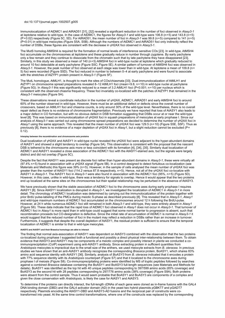
Figure 5. Dual immunolocalisation of AtASY1 and AtZYP1 with recombination pathway proteins in wildtype and Atasy31 meiotic prophase Inuclei.(A) AtASY1 (green) and γH2AX (red) on wildtype and (B) Atasy31 at leptotene, (C) AtASY1 (green) and AtDMC1 (red) onwildtype and (D) on Atasy31 at leptotene. (E) AtZYP1 (green) and AtMSH4 (red) on wildtype and (F) on Atasy31 (AtMSH4arrowed). (G) AtZYP1 (green) and AtMLH1 (red) on wildtype and (H) on Atasy31 (AtMLH1 arrowed). DAPI (blue) for all. Bar,10 µm.
−7
doi:10.1371/journal.pgen.1002507.g005
Immunolocalization of AtDMC1 and AtRAD51 [31], [32] revealed a significant reduction in the number of foci observed in Atasy31at leptotene relative to wildtype. In the case of AtDMC1, the figures for Atasy31 and wildtype were 106.8 (n = 5) and 143.8 (n = 5)(P = 0.02) respectively (Figure 5C, 5D). For AtRAD51, the mean number of foci in Atasy31 was 99.8 (n = 5) compared to 141 (n = 5)(P<0.01) in wildtype meiocytes (Figure S5A, S5B). Although the numbers of AtDMC1 and AtRAD51 foci only indirectly reflect thenumber of DSBs, these figures are consistent with the decrease in γH2AX foci observed in Atasy31.
The MutS homolog AtMSH4 is required for the formation of normal levels of interference sensitive COs [23]. In wildtype, AtMSH4foci accumulate on the chromosomes at leptotene and these gradually reduce in number through zygotene. By early pachyteneonly a few remain and they continue to dissociate from the chromatin such that by late pachytene they have disappeared [23].Similarly, in this study we observed a mean of 140 (n = 5) AtMSH4 foci in wildtype nuclei at leptotene which gradually reduced toaround 10 foci detectable at early pachytene (Figure S5C; Figure 5E). A similar pattern of turnover of AtMSH4 foci was observed inAtasy31. However, the peak number of foci observed at each stage was lower than in wildtype. At leptotene a mean of 109.8 (n = 5) foci were recorded (Figure S5D). The foci reduced in number to between 0–4 at early pachytene and were found to associatewith the stretches of AtZYP1 protein present in Atasy31 (Figure 5F).
The MutL homologue, AtMLH1, is thought to mark the sites of COs/chiasmata [33]. Dualimmunolocalization of AtMLH1 andAtZYP1 on chromosome spread preparations revealed 8–12 AtMLH1 foci (mean = 9.8, n = 10) in wildtype nuclei at pachytene(Figure 5G). In Atasy31 this was significantly reduced to a mean of 3.2 AtMLH1 foci (P<0.001; n = 10) per nucleus which isconsistent with the observed chiasma frequency. These foci invariably colocalized with the patches of AtZYP1 that remained in theAtasy31 meiocytes (Figure 5H).
These observations reveal a coordinate reduction in the number of γH2AX, AtDMC1, AtRAD51 and early AtMSH4 foci to around60% of the number observed in wildtype. However, there must be an additional defect or defects since the overall number ofcrossovers, based on AtMLH1 foci and chiasma counts, is only around 30% of the wildtype level. Nevertheless, there is no overallrepair defect as there is no evidence of chromosome fragmentation. Previously we have reported that loss of AtASY1 also leads toa major defect in CO formation, but with no obvious defect in DSB formation suggesting that DSBs occur at or near the wildtypelevel [8]. This was based on immunolocalization of γH2AX foci in squash preparations of meiocytes at early prophase I. Since ouranalysis of Atasy31 was carried out using chromosome spread preparations we decided to determine the number of γH2AX foci inAtasy1 using the same approach. This revealed that the mean number of γH2AX foci was 129.5 (n = 10) (Figure S5E, S5F). Thus aspreviously [8], there is no evidence of a major depletion of γH2AX foci in Atasy1, but a slight reduction cannot be excluded (P = 0.12).
Interplay between the recombination and chromosome axis proteins
Dual localization of γH2AX and AtASY1 in wildtype nuclei revealed the γH2AX foci were adjacent to the hyperabundant domainsof AtASY1 and showed a slight tendency to overlap (Figure 5A). This observation is consistent with the proposal that the nascentDSB is tethered to the chromosome axis more or less coincident with its formation [4], [34], [35]. Similarly dual localization ofAtDMC1 and AtASY1 revealed a close association of the AtDMC1 foci with the AtASY1stained axis, but the signals were largelydistinct and did not overlap (Figure 5C).
Despite the fact that AtASY1 was present as discrete foci rather than hyperabundant domains in Atasy31, these were virtually all(97.4% n = 5) found in association with a γH2AX signal (Figure 5B). In a control designed to detect fortuitous colocalization (seeMaterials and Methods) this figure was 39% (n = 5). However, in the sample of cells analysed the mean number of γH2AX foci wasgreater than the number of AtASY1 foci (114.2 versus 87.8 respectively, n = 5). Hence, not all of the γH2AX foci colocalize withAtASY1 in Atasy31. The AtASY1 foci in Atasy31 were also found in association with the AtDMC1 foci (95%, n = 5) (Figure 5D).However, in this case, unlike in wildtype, there was a tendency for signals to overlap. Hence it would appear that the two proteinsend up localized at the same sites on the chromosomes but their spatial relationship may be perturbed in the absence of AtASY3.
We have previously shown that the stable association of AtDMC1 foci to the chromosome axes during early prophase I requiresAtASY1 [8]. Since AtASY1 localization is disrupted in Atasy31, we investigated the localization of AtDMC1 in Atasy31 in moredetail. The chronology of AtDMC1 localization was determined by carrying out the immunolocalization of the protein together withprior BrdU pulselabeling of the PMCs during meiotic Sphase as described previously [8]. This revealed that in Atasy31, Atasy1and wildtype maximum numbers of AtDMC1 foci accumulated on the chromosomes around 12 h following the BrdUpulse.However, at 24 h while numerous AtDMC1 foci still remained in both Atasy31 and wildtype, they were entirely absent in Atasy1(Figure S6). These data indicate that the rapid loss of AtDMC1 foci observed in Atasy1 does not occur in Atasy31. That turnover ofAtDMC1 foci in Atasy1 is more rapid than in wildtype could suggest that some normal barrier to progression is absent, such thatrecombination proceeds but COdesignation is defective. Since the initial rate of accumulation of AtDMC1 is normal in Atasy31 itwould suggest that the reduced number of foci in the mutant may reflect a reduction in DSBs rather than an increase in turnover.Furthermore, it suggests that despite the overall depletion of AtASY1, the residual protein is sufficient to ensure that the temporallocalization of AtDMC1 is similar to that in wildtype meiocytes.
AtASY3 and AtASY1 and their Brassica homologs are able to interact
The finding that normal axisassociation of AtASY1 was dependent on AtASY3 combined with the observation that the two proteinscolocalized during prophase I suggested both a functional and possibly a direct physical interrelationship between them. To obtainevidence that AtASY3 and AtASY1 may be components of a meiotic complex and possibly interact in planta we conducted a coimmunoprecipitation (CoIP) experiment using antiAtASY1 antibody. Since extracting protein in sufficient quantities fromArabidopsis meiocytes is impractical due to the small size of the anthers, we used meiocyte extracts from B. oleracea. In previousstudies we have shown that an antiAtASY1 antibody recognizes the corresponding Brassica protein, BoASY1, which shares 83%amino acid sequence identity with AtASY1 [36]. Similarly, analysis of BoASY3 from B. oleracea indicated that it encodes a proteinwith 77% sequence identity with its Arabidopsis counterpart (Figure S7) and that it localized to the chromosome axes duringprophase I of meiosis (Figure S8). Coimmunoprecipitating proteins were identified by MS of tryptic peptides followed by searchesagainst a combined Brassica database containing the BoASY1 and BoASY3 fulllength sequences (see Materials and Methods fordetails). BoASY1 was identified as the top hit with 40 unique peptides corresponding to 397/599 amino acids (65% coverage) andBoASY3 as the second hit with 28 peptides corresponding to 297/776 amino acids (38% coverage) (Figure S9A). Both proteinswere absent from the control sample. Thus it would seem probable that BoASY1 and BoASY3 are components of a complex andgiven the close conservation with Arabidopsis, is likely the case for AtASY1 and AtASY3.
To determine if the proteins can directly interact, the fulllength cDNAs of each gene were cloned as inframe fusions with the GAL4DNA binding domain (DBD) and the GAL4 activator domain (AD) in the yeast two hybrid plasmids pGBKT7 and pGADT7respectively. Plasmids encoding the AtASY1DBD and AtASY3AD fusions and the reciprocal pair of constructs were cotransformed into yeast. At the same time control transformations, where one of the constructs was replaced by the corresponding

empty vector, were also carried out. All plasmid combinations enabled the transformed yeast to grow on synthetic dropout medium(Leu/Trp) that selected the auxotrophic markers on the cloning vectors. When the plasmid combinations were tested on lowstringency selective plates (Leu/Trp/His) the yeast cells containing AtASY1 and AtASY3 as reciprocal DBD/AD fusions enabledsignificant growth at all dilutions. Some slight growth was also detected in controls containing the AtASY1DBD and AtASY3ADplasmids. However, under higher stringency selection (Leu/Trp/His/Ade) growth was entirely restricted to the combinationscarrying both genes (Figure 6). Further experiments with a series of plasmids in which truncated regions of AtASY3 were fused tothe GAL4 activator domain indicated that the interaction of AtASY3 with AtASY1 was dependent on amino acid residues 623–793 ofAtASY3 which correspond to the predicted coiledcoil region (Figure 6). These data suggested that AtASY1 and AtASY3 caninteract and likely do so in planta.
Figure 6. Yeast twohybrid analysis of AtASY3 and AtASY1 with schematic illustration of predicted domains and relative positions of aminoacid residues.Plasmid constructs were cotransformed into Y2HGold yeast cells before plating on SD Leu/Trp (LT), SD Leu/Trp/His (LTH) and SD Leu/Trp/His/Ade (LTHA). Growth on –LTH and –LTHA confirms a proteinprotein interaction between AtASY1and AtASY3 that is dependent on AtASY3 residues 623–793.doi:10.1371/journal.pgen.1002507.g006
Discussion
AtASY3 is a component of the meiotic chromosome axes in Arabidopsis
We have identified AtASY3, a chromosome axis protein that is required for normal levels of COs and SC formation in Arabidopsis.AtASY3 is predicted to contain a coiledcoil domain towards the Cterminus. This structural feature is found in other meioticproteins, such as Red1 in budding yeast, SCP3/Cor1 in mammals and OsPAIR3 in rice, that are components of the axial regions ofthe SC, yet on the basis of amino acid sequence homology are reported to have no close homologs in other species [15], [16], [18],[37], [38]. AtASY3 appears similar in this respect. Although it shares 77% sequence identity with BoASY3 from the closely relatedB. oleracea, the level of sequence identity with Red1 and OsPAIR3 is limited, at 16.4% and 25.6% respectively. This sequencedivergence appears to be a recurrent feature of proteins that are components of the chromosome axes.
DSBs appear reduced and crossover formation is compromised in Atasy31
In Arabidopsis the level of DSBs may be inferred from the number of γH2AX and/or AtDMC1 or AtRAD51 foci detected in meiocytesat early prophase I [8]. Our data suggested a consistent reduction in DSBs from 150–160 in wildtype meiocytes to about 100 inAtasy31, an overall reduction of around 33%. However, as DSB detection is indirect, we cannot formally rule out the possibility thatnot all DSBs are detected in the mutant. Nevertheless, as the number of γH2AX foci is very similar to those of the strandexchangeproteins this seems a reasonable conclusion.
The physical association of the recombination machinery and the chromosome axes has been known for many years [3], [34], [39].Recent studies in budding yeast indicate that the DSB machinery becomes tethered to the chromosome axes prior to breakformation and that the axis components Red1 and Hop1 are required for this [4], [35]. Analysis of red1 mutants has revealed adefect in DSB formation [40]–[42]. It is conceivable that AtASY3 may play a similar role in axis organization that is crucial to enablenormal levels of DSB formation. The reduction in foci corresponding to γH2AX and the strandexchange proteins is consistent witha scenario whereby DSB formation occurs in the context of the axis, rather than the recombination complex associating with theaxis following break formation, as in the latter instance, a reduction in γH2AX foci would not be anticipated. However, it remainspossible that depletion of AtASY3 may induce a change in chromatin structure that in turn results in a reduction in DSBs.
Although DSB formation appears to be significantly reduced in Atasy31, any reduction due to the loss of AtASY1 appears moremarginal. This is in broad agreement with our earlier study [8]. It is worth noting that this difference mirrors chromosome axisformation in the two mutants. Whereas loss of AtASY3 disrupts the axes, the Atasy1 mutant has clearly defined axes, albeit withsome minor discontinuities, seemingly indicating the importance of the chromosome axes for efficient DSB formation [43]. However,it contrasts with observations in budding yeast and mouse, where mutants lacking Hop1 and HORMAD respectively exhibit stronglyreduced numbers of DSBs [44], [45]. At present the basis and significance of this difference remains unknown. It may reflect anunderlying difference in the control of DSB formation, although the formation of DSBs in hop1 and red1 mutants in different buddingyeast strains also shows some variation [40], [46].
A reduction in the mean chiasma/CO frequency to around 30% of the wildtype level was observed in all Atasy3 lines. Analysis ofthe Atmsh4/Atasy31 double mutant indicates that loss of AtASY3 compromises the formation of COs which are subject to COinterference, and also noninterfering COs. This would suggest that AtASY3 plays a crucial role at an early stage in therecombination pathway. A reduction in CO formation is also a characteristic of mutants in the axisassociated proteins Red1 andOsPAIR3 [18], [24], [40], [41]. A recent study in budding yeast has indicated that loss of Red1 results in a conversion from interhomolog bias to intersister bias for DSB repair. It is proposed that this is due to a loss of constraint on Rec8 loading at therecombination site, thus favoring intersister recombination [4]. It is conceivable that a similar situation arises through the loss ofAtASY3. If so, this could explain why there is a proportionally greater reduction in CO formation in Atasy31 than might be expectedfrom the apparent reduction in DSBs. Simple extrapolation would suggest that if the ratio of COs to nonCOs was maintained in the

mutant, the mean CO/chiasma frequency would be 6–7 rather than the 3.5 observed. This implies that there is a loss of interhomolog bias and that a greater proportion of the DSBs are repaired via another route such as inter sisterchromatid exchange.Alternatively, loss of AtASY3 may result in preferential processing of some or all of the recombination intermediates to favor nonCOs rather than COs. At present the data does not enable us to distinguish between these possibilities.
The observation that 75.7% of the chiasmata that remain in Atasy31 are subtelomeric/distal is consistent with previous studies inArabidopsis showing that the telomeres cluster on the nucleolus during early prophase I and as a result the subtelomeric regionsof the homologous chromosomes are placed in proximity [47]. A similar situation was observed in Atasy1 where virtually all theresidual chiasmata are distal [48].
Axisassociation of AtASY1 is AtASY3dependent and occurs in the absence of DSB formation
Deconvolution of the linear AtASY1 signal in wildtype meiocytes at leptotene through early pachytene revealed that it comprisesevenlyspaced axisassociated domains of hyperabundance interspersed with more lightly staining regions. This organization wassupported by immunogold studies in meiocytes from C. capillaris. The number of AtASY1 domains appeared quite consistent, ataround 160 per nucleus. The organization of AtASY1 is highly reminiscent of that of Hop1 which has been observed to formdomains of alternating hyperabundance and lowerabundance in budding yeast at early pachytene [49], [50]. Loss of AtASY3resulted in a significant effect on the distribution of AtASY1, such that axisassociated foci were observed during prophase I. Thesewere fewer in number than the AtASY1 domains observed in wildtype and this varied from cell to cell ranging from 39–115 in thesample examined. Nevertheless, duallocalization studies with γH2AX and AtDMC1 indicated that these AtASY1 foci coincided withsites of recombination. However, further investigation will be required to determine whether or not localization of AtASY1 at the axialregion still occurs with normal spatial specificity in Atasy31. Studies in budding yeast initially reported that localization of theAtASY1 homolog, Hop1, was dependent on the chromosome axis protein Red1 [22]. However, more recently it was suggested thatwhile normal levels of Hop1 loading require Red1, some Hop1 is loaded at the sites of DSBs independently [46]. Our observationssuggest a similar relationship between AtASY1 and AtASY3.
In wildtype, the AtASY1abundant regions along the axes at prophase I correlated both spatially and numerically with the γH2AXand AtDMC1 foci, suggesting that the DSBs are positioned within AtASY1enriched domains. This is consistent with the proposedrole of AtASY1 in promoting IHR [8]. Interestingly, whereas the γH2AX and AtASY1 signals overlapped, the AtDMC1 signal wasadjacent to, but did not merge with the AtASY1 signal. Further study will be required to explore the significance of this observation.It appears that a similar spatial relationship between DSBs and AtASY1 is maintained in Atasy31 as virtually all the AtASY1 foci inthe mutant colocalize with γH2AX. Thus, it suggests that recruitment of AtASY1 and the recombination machinery to the axialregion is spatially coordinated. However, these may not be interdependent events since localization of AtASY1 in the Atspo1114null mutant appears, based on immunofluorescence, normal [8]. Similarly in this study, examination of AtASY1 localization in anAtasy31/Atspo1114 double mutant suggests that the AtASY1 foci observed in Atasy31 are still formed and associated with theaxial region. Nevertheless, further studies will be required to establish if the AtASY1 domains observed in Atspo1114 and Atasy31/Atspo1114 are identical to those in the wildtype. If so, then it suggests that AtASY1 is initially recruited to predeterminedchromosomal regions that also encompass DSB hotspots, possibly establishing a spatial relationship favoring IHR. Alternatively, itis conceivable that the AtASY1 domain formation observed in wildtype may be guided or influenced by DSB formation or the preDNA break recombinosome complex. Studies in budding yeast have also led to the proposal that the interhomolog bias isestablished before DSB formation with enforcement of the bias occurring at the transition from the nascent DSB to a joint moleculerecombination intermediate [41].
Although the majority of the AtASY1 foci in Atasy31 were associated with γH2AX foci at early prophase I, there were additionalγH2AX signals that did not colocalize. If loss of DSBs and the destabilization of AtASY1 localization observed in Atasy31 occursstochastically, then, given that formation of AtASY1 foci and DSBs does not appear to be codependent, one would expect to see asimilar proportion of γH2AX and AtASY1 foci that were not associated with one another. As this does not appear to be the case, itseems possible that a subset of DSBs occur in regions that are not associated with AtASY1. It may simply be that although theAtASY1 foci that persist in Atasy31 correspond to the position of the domains observed in wildtype they are substantially smaller.Hence a proportion of the γH2AX foci no longer colocalize despite the normal spatial recruitment of the recombination complexesto the chromatin. Alternatively, some DSBs may occur in regions of lower AtASY1 abundance. Previously, it has been proposed inbudding yeast that some DSBs are formed at random sites that are not associated with DSB hotspots [41]. Hence, if the AtASY1domains are coincident with hotspots, which would seem logical, this may also be the case in Arabidopsis.
Evidence suggests that AtASY3 and Red1 share functional similarities
Despite amino acid sequence variation it has been suggested that Red1 and SCP3/Cor1 may be structural analogs [15].Nevertheless, to date a functional ortholog of Red1 in a multicellular organism has not been reported. However, the studiesdescribed here suggest that AtASY3 has at least some functional similarity to Red1. The most compelling evidence arises from theclose functional interrelationships that both proteins share with their corresponding HORMA domain proteins. Loss of Red1 andAtASY3 proteins results in a disruption of Hop1 and AtASY1 localization respectively, during prophase I [22]. In budding yeast Red1has been shown to interact with Hop1 in coimmunoprecipitation experiments [46]. Yeast twohybrid studies have shown that the290 amino acids at the Cterminus of Red1 are essential for the interaction with Hop1. This part of the protein is predicted to form acoiledcoil domain. In this study MS analysis of proteins that were coimmunoprecipitated from Brassica meiocytes using an antiAtASY1 antibody revealed a likely interaction between BoASY1 and BoASY3, the homologs of AtASY1 and AtASY3 respectively.That AtASY3 and AtASY1 can directly interact was confirmed by yeast twohybrid analysis. Moreover further study revealed that theCterminal of AtASY3 that contains a predicted coiledcoil region is essential for the interaction between the two proteins. Studiesindicate that Red1 is required for normal levels of DSB formation. This also appears to be the case for AtASY3, although this effectdoes not appear as pronounced in Atasy31 as that in a red1 mutant where a reduction in DSB formation to ∼25% of wildtype hasbeen reported [42].
It seems likely that PAIR3 in rice may also be a functional homolog of Red1. While a direct interaction with the rice HORMA domainprotein OsPAIR2 has not yet been demonstrated, localization of OsPAIR2 is OsPAIR3 dependent and an Ospair3 mutant has asimilar phenotype to that of Atasy31 [17], [18].
SC nucleation is sufficient to prevent aberrant ectopic recombination
In addition to its structural role within the SC, the budding yeast protein Zip1 has been shown to play a key role in meioticrecombination [51], [52]. Zip1 together with other members of the ZMM group of proteins, Zip2, Zip3, Zip4, Msh4, Msh5 and Mer3,is crucial for the formation of interferencedependent COs [51]. In Arabidopsis loss of AtZYP1 only results in a modest reduction inchiasma frequency to about 80% of the wildtype level. However, many of the remaining chiasmata occur between ectopicchromosome regions, possibly between duplicated sequences that amount to around 60% of the Arabidopsis genome [10]. Thestudies described here revealed that loss of AtASY3 had a profound effect on the formation of the SC. In most nuclei it appearedthat alignment of the chromosome axes was extensively disrupted and immunolocalization studies with the transverse filamentprotein AtZYP1 indicated little evidence of normal SC assembly. In general, nuclei contained a mixture of AtZYP1 foci or occasional

short stretches which appeared abnormally thickened and deformed. Although Atasy31 is essentially asynaptic, there is noevidence of the nonallelic recombination observed in plants lacking AtZYP1. The bivalents that remained in the Atasy31 meiocytesat metaphase I comprised homologous chromosomes and there was no evidence of multivalent formation. Immunolocalizationstudies revealed a reduction in AtMLH1 foci that reflected the reduction in chiasmata in Atasy31. These AtMLH1 foci, which arethought to localize to CO sites [33], were invariably associated with the residual AtZYP1 present in the mutant. Hence, it wouldappear that the presence of AtZYP1 at the site of recombination is important for the prevention of nonallelic recombination, butextensive SC polymerization is not required. This finding provides further evidence that in addition to SC formation, AtZYP1 playsan important role in the formation of COs in Arabidopsis.
In summary, these studies provide further insight into meiotic CO formation. Moreover they emphasize that despite the lack ofsequence homology between the chromosome axes components from different species, it seems likely that a close functionalrelationship remains.
Materials and Methods
Plant material and nucleic acid extraction
A. thaliana ecotype Columbia (0) was used for wildtype analysis. TDNA insertion lines SALK_143676, SALK_050971 andSAIL_423_H01 were obtained from NASC for mutant analysis [53]. Plants were grown, material harvested and nucleic acidextractions were performed as previously described by Higgins et al. [23].
TDNA insertion site mapping
The TDNA insertion site of the mutant lines was confirmed as previously described [23] (Figure S2). Details of the primers used arepresented in Table S2.
Complementation of Atasy31
Primers ASY3CMF1 and ASY3CMR1 (Table S2) were used to amplify the entire AtASY3 coding sequence with flanking 5′ and 3′UTR regions from cDNA clone pda 19140 (Riken, Japan). The PCR product was cloned into the binary vector pPF408 [19] usingSpeI sites incorporated into the primers. The construct was confirmed by sequencing. The binary plasmid construct was introducedinto Agrobacterium tumefaciens LBA 4404 and plants transformed as previously described [23].
RNA extraction and RT–PCR
RNA extraction and RTPCR was carried out as previously described [54]. Details of the primers are given in Table S2.
Nucleic acid sequencing
Nucleotide sequencing was carried out by the Genomics and Proteomics Unit, School of Biosciences, University of Birmingham,UK.
Antibody production
Primers ASY3ABF1 and ASY3ABR1 (Table S2) were used to amplify a 702bp fragment comprising amino acid residues 560 to793 of AtASY3 from cDNA clone pda 19140 (Riken, Japan). The PCR product was cloned into the expression vector pET21b(Novagen) using NdeI and Xhol sites incorporated into ASY3ABF1 and ASY3ABR1 respectively. Recombinant Histaggedprotein was isolated from E. coli BL21 (Novagen) under native conditions using Niagarose following the manufacturer's protocol(QIAGEN). Polyclonal antiserum against the recombinant protein was raised in rabbit (BioGenes GmbH, Germany).
Cytological procedures
Cytological studies were carried out as previously described [23]. The following antibodies were used: antiAtASY3 (rabbit, 1/200dilution) antiAtASY1 (rabbit/rat, 1/1000 dilution), antiAtMSH4 (rabbit, 1/500 dilution), antiAtZYP1 (rabbit/rat, 1/500 dilution), antiAtSMC3 (rabbit 1/500 dilution), antiAtSYN1 (rabbit 1/500 dilution), antiAtDMC1 (rabbit 1/500 dilution), antiAtRAD51 (rabbit, 1/500dilution), antiAtMLH1 (rabbit/rat, 1/200 dilution) and antiγH2AX (ser 139, catalog no. 07164 Upstate Biotechnology; rabbit, 1/100dilution) [10], [23], [55]. Microscopy was carried out using a Nikon 90i Fluorescence Microscope (Tokyo, Japan). Image capture,image analysis and processing were conducted using NISElementsF software (Nikon, Tokyo, Japan). Image deconvolution wascarried out using the function “Mexican hat”. This allows better discrimination of the signals. This function performs filtration on theintensity component (or on every selected component when working with multichannel images) of an image using convolution with5×5 kernel. Mexican Hat kernel is defined as a combination of Laplacian kernel and Gaussian kernel it marks edges and alsoreduces noise. In doublestaining experiments the level of chance overlap of foci was assessed using the misorientation methodwhereby one of the two images is rotated through180 degrees following which colocalizing foci are counted as previously described[56].
Electron microscopy and immunogold labeling was performed as previously described [36], [57] except for the modified Atasy3chromosome spread protocol where the detergent was Triton X100 0.1%+Lipsol 0.05% and the digestion time was increased to 7min.
Chiasma counts were carried out as previously described [58]. Chromosome spread preparations from PMCs at metaphase I wereexamined by light microscopy after fluorescence in situ hybridization (FISH) using 45S and 5S rDNA probes. The use of FISHenabled the identification of individual chromosomes. The overall shape of individual bivalents allowed the number and position ofindividual chiasmata to be determined and this was also informed by the position of the FISH signals.
Statistical procedures
The statistical procedures were carried out as described previously [23].
Proteomic analysis of Brassica meiocytes
AtASY3 peptides were initially identified in meiocyte extracts prepared from Brassica oleracaea var. alboglabra A12DHd aspreviously described [14]. CoIP experiments were conducted on meiocytes extracted from the same material. Protein extractionwas under nondenaturing conditions at 4°C. Briefly, tissue was powdered by grinding in liquid nitrogen, resuspended in IP Buffer(20 mM TrisHCl pH 7.5, 150 mM NaCl, 10% glycerol, 2 mM EDTA, 0.1% NP40, protease inhibitor cocktail (Roche, #11836170001),phosphatase inhibitor (Thermo Scientific #78420)) and cell debris removed by centrifugation. Protein extracts were usedimmediately. Antibodies were crosslinked to AffiPrep Protein A beads (BioRad, #1560006) using DMP (Sigma #D8388) and preeluted with glycine to remove any noncrosslinked antibody. Meiocyte extracts were precleared by incubation with nonspecificIgG. Parallel CoIPs were carried out using affinity purified antiAtASY1 antibody and an unrelated antibody as a control. Followingwashing to remove nonspecific proteins, bound proteins were glycineeluted, trypsindigested and analysed on an LTQOrbitrapVelos mass spectrometer (Thermo Fisher Scientific). Since a complete sequence of the Brassica oleracea genome is currently

unavailable, protein identification was carried out against a combined Brassica rapa/napus/oleracea database, downloaded fromNCBI (http://www.ncbi.nlm.nih.gov) in 2010, into which the BoASY1 and BoASY3 fulllength sequences had been manuallyinserted. For details of the MS analysis see Figure S9B.
Yeast 2hybrid analysis
Yeast twohybrid screens were performed according to the Yeastmaker Yeast transformation System 2 manual (Clontech, USA).Briefly, Y2HGold yeast cells were cotransformed with pGADT7 and pGBKT7 using the polyethylene glycol/lithium acetate method.The cotransformed yeasts were grown in SD Leu/Trp, SD Leu/Trp/His and SD Leu/Trp/His/Ade for testing proteinproteininteraction through the activation of the two reporter genes His3 and Ade2. The strength of the interaction was assayed by drop testusing serial dilutions of midexponentialphase cultures. 3 µl drops of undiluted, 10 and 100fold diluted culture were spotted on theselective agar medium and incubated at 30°C for 2 days. Details of the primers used for plasmid construction are shown in TableS2. Plasmid constructs are as shown in Figure 6.
Supporting Information
Figure S1.
A. Peptides from Brassica oleracea meiocytes with homology to gene At2g46980 (AtASY3) identified by mass spectrometry.Peptides were identified in two independent experiments using the procedure described in SanchezMoran et al., 2005 Cytogenetand Genome Res. 109:181–189 [14]. The Arabidopsis TAIR database was used for peptide identification. B. (i) Diagrammaticrepresentation of the 793 aa, 88 kDa AtASY3 protein indicating the relative position of the putative coiledcoil domain (black box).(ii) Map of the ∼3.5 kb At2g46980 locus showing the exon/intron organization of AtASY3. The exons are represented by numberedblack boxes. The triangles indicate the TDNA insertion sites in Atasy31, Atasy32 and Atasy33. C. Expression analysis of AtASY3using semiquantitative RTPCR indicates that in wildtype (WT) expression is highest in bud tissue (B) with a low level present inopen flowers (F). Expression is not detected in stem (S) or leaf (L). AtASY3 expression is absent in the Atasy31, Atasy32 andAtasy33 mutants. AtGAPD was used as an expression control.doi:10.1371/journal.pgen.1002507.s001(PDF)
Figure S2.
Nucleotide sequencing of the TDNA insertion sites in Atasy31, Atasy32 and Atasy33.doi:10.1371/journal.pgen.1002507.s002(PDF)
Figure S3.
A. Fertility in Atasy31 is reduced compared to wildtype Col 0. (A) wildtype, (B) Atasy31, (C) wildtype silique, (D) Atasy31silique. Bar 10 mm. Atasy31 exhibited a reduction in mean silique length of 37% (n = 50) and a reduction in seedset of 73% (n = 50). B. Representative meiotic stages of Atasy32 (A–F) and Atasy33 (G–L). Leptotene (A,G); pachytene (B,H); diakinesis (C,I);metaphase (D,J); dyad (E,K) tetrad (F,L). Bar, 10 µm. C. An allelism test was carried out by reciprocally crossing heterozygousAtasy31 and Atasy32. Cytological analysis of Atasy31/Atasy32 reveals asynapsis at pachytene (A) and univalents in metaphaseI (B). This leads to missegregation at meiotic divisions resulting in the subsequent formation of unbalanced tetrads (C). Bar, 10 µm.Fertility in an Atasy31 complementation line (F) was restored to the normal level observed in wildtype (D) in contrast to that ofAtasy31 (E). Bar, 10 mm. Cytological analysis confirmed that normal meiosis was restored in the Atasy31 complementation line.Homologous chromosomes underwent normal synapsis in pachytene (G). A full complement of five bivalents was observed inmetaphase I (H). These underwent normal segregation leading to the formation of balanced tetrads (I). Bar, 10 µm.doi:10.1371/journal.pgen.1002507.s003(PDF)
Figure S4.
A. Chromosome spread preparations from PMCs at metaphase I were examined by light microscopy after fluorescence in situhybridization (FISH) using 45S (green) and 5S (red) rDNA probes. The use of FISH enabled the identification of individualchromosomes. The overall shape of individual bivalents allowed the number and position of individual chiasmata to be determinedand this was also informed by the position of the FISH signals. For full details of the chiasma scoring procedure see: SanchezMoran et al. (2002) Genetics 162: 1415–1422 [58]. Analyses of metaphase I nuclei of wildtype (A) ab. Rod bivalents, singleinterstitial chiasma in the long arm Chr. 2 and Chr. 4 respectively; ce. Ringbivalents, 2 chiasmata Chr. 1, Chr 5 and Chr.3respectively. Atasy31 (B) a. Chr. 5 rod bivalent distal chiasma; b. Chr. 1 rod bivalent distal chiasma, c. Chr. 4 rod bivalent singleshort arm chiasma. Analysis indicated that mean chiasma frequency in Atasy31 was significantly reduced to 3.40 in contrast towildtype, which had an overall mean chiasma frequency of 9.84. Bar, 10 µm. B. Cytological analyses of metaphase I chromosomespreads indicated the presence of univalents in Atasy31/Atspo1114 (A). No chiasmata were observed in this double mutant incontrast to wildtype (B), where five bivalents were observed in all of the metaphase I cells analysed. This confirms that thechiasmata in Atasy31 are DSBdependent. Comparison of the mean chiasma frequency of Atasy31/Atasy1 (C) and Atasy1 (D)revealed no significant difference between the double mutant and the latter suggesting a close functional relationship betweenAtASY3 and AtASY1. Analysis of 30 metaphase I nuclei from Atasy31/Atmsh4 (E) shows that the double mutant fails to formchiasmata in contrast to Atmsh4 (F), in which a mean chiasma frequency of 1.1 (n = 30) was observed. Bar, 10 µm.doi:10.1371/journal.pgen.1002507.s004(PDF)
Figure S5.
Immunolocalization of recombination proteins in Atasy31 and γH2AX in Atasy1. Immunolocalization of AtRAD51 (red) on DAPIstained (blue) wildtype (A) and Atasy31(B) meiocytes at early prophase I. (C) and (D) show corresponding images for AtMSH4. Inboth cases there is a reduction in foci in the Atasy31 mutant. Immunolocalization of γH2AX (red) on DAPI stained (blue) meiocytesfrom wildtype (E) and Atasy1 (F). (see main text for details). Bar, 10 µm.doi:10.1371/journal.pgen.1002507.s005(PDF)
Figure S6.
Timecourse analysis of AtDMC1 (red) localization in wildtype (WT), Atasy1 and Atasy31. The study revealed that AtDMC1 foci inAtasy31 are stabilized and persist at least up to 24 h post BrdU (green) pulselabeling at S phase before gradually decreasing tovery low numbers by 30 h. This observation was similar to that in WT but contrast with that of Atasy1, where AtDMC1 foci are

1.
View Article PubMed/NCBI Google Scholar
2.View Article PubMed/NCBI Google Scholar
3.View Article PubMed/NCBI Google Scholar
4.
View Article PubMed/NCBI Google Scholar
5.
View Article PubMed/NCBI Google Scholar
6.
View Article PubMed/NCBI Google Scholar
7.
destabilized soon after loading and their numbers decrease rapidly at ∼18 h. Almost all of AtDMC1 foci in Atasy1 were lost by 24 hpost BrdU pulse labeling as previously reported by SanchezMoran et al. 2007 [8].doi:10.1371/journal.pgen.1002507.s006(PDF)
Figure S7.
Sequence alignment of AtASY3 and BoASY3. The proteins exhibit 77% sequence identity.doi:10.1371/journal.pgen.1002507.s007(PDF)
Figure S8.
Immunolocalization of BoASY3 protein using antiAtASY3 antibody to wildtype Brassica oleracea chromosome spreadpreparations from meiocytes at leptotene, zygotene and pachytene. The BoASY3 protein localises to meiotic chromosomes asnumerous foci in leptotene and gradually polymerizes to form a continuous linear signal by pachytene. The localisation of BoASY3is indistinguishable to that of AtASY3 in Arabidopsis. BoASY3 could not be detected using preimmune antiAtASY1 antiserum. Bar,10 µm.doi:10.1371/journal.pgen.1002507.s008(PDF)
Figure S9.
A. BoASY3 sequence showing protein coverage following coimmunoprecipitation with BoASY1 and MS analysis (highlightedyellow). B. Mass spectrometry conditions.doi:10.1371/journal.pgen.1002507.s009(PDF)
Table S1.
Chiasma counts for Atasy31 and wildtype. A. Atasy31 mean chiasma frequency 3.3 (n = 50). Proportion of distal chiasma = 74.8%.B. Arabidopsis (Col0) wildtype mean chiasma frequency 9.76 (n = 50). Proportion of distal chiasma = 73.8%. The proportion ofdistal chiasmata is not significantly different in the mutant.doi:10.1371/journal.pgen.1002507.s010(PDF)
Table S2.
Primer sequences used during the study. Primers 1–8 were used for confirming TDNA insertions sites; primers 9–12 were used forthe analysis of AtASY3 expression; Primers 13 and 14 were used for complementation studies; primers 15 and 16 were used forthe production of recombinant AtASY3 for antibody production. Primers 17–22 were for yeast twohybrid studies. The regionsamplified are indicated; F/R denotes forward and reverse.doi:10.1371/journal.pgen.1002507.s011(PDF)
Acknowledgments
We are grateful to Gareth Jones and Juan Santos for useful discussions. We thank Nancy Kleckner and the anonymous reviewersfor their helpful comments. We thank Steve Price and Karen Staples for technical support.
Author Contributions
Conceived and designed the experiments: FCHF MF JDH KO. Performed the experiments: MF JDH KO CL ER SJA RP MP NC.Analyzed the data: FCHF JDH KO ER. Contributed reagents/materials/analysis tools: KM. Wrote the paper: FCHF KO JDH.
References
Keeney S, Giroux CN, Kleckner N (1997) Meiosisspecific DNA doublestrand breaks are catalyzed by Spo11, a member of a widely conserved proteinfamily. Cell 88: 375–384.
Neale MJ, Keeney S (2006) Clarifying the mechanics of DNA strand exchange in meiotic recombination. Nature 442: 153–158.
Kleckner N (2006) Chiasma formation: chromatin/axis interplay and the role(s) of the synaptonemal complex. Chromosoma 115: 175–194.
Kim KP, Weiner BM, Zhang LR, Jordan A, Dekker J, et al. (2010) Sister Cohesion and Structural Axis Components Mediate Homolog Bias of MeioticRecombination. Cell 143: 924–937.
Caryl AP, Armstrong SJ, Jones GH, Franklin FCH (2000) A homologue of the yeast HOP1 gene is inactivated in the Arabidopsis meiotic mutant asy1.Chromosoma 109: 62–71.
Shin YH, Choi Y, Erdin SU, Yatsenko SA, Kloc M, et al. (2010) Hormad1 Mutation Disrupts Synaptonemal Complex Formation, Recombination, andChromosome Segregation in Mammalian Meiosis. Plos Genetics 6:
Wojtasz L, Daniel K, Roig I, BolcunFilas E, Xu HL, et al. (2009) Mouse HORMAD1 and HORMAD2, Two Conserved Meiotic Chromosomal Proteins, AreDepleted from Synapsed Chromosome Axes with the Help of TRIP13 AAAATPase. Plos Genetics 5:

View Article PubMed/NCBI Google Scholar
8.
View Article PubMed/NCBI Google Scholar
9.
View Article PubMed/NCBI Google Scholar
10.
View Article PubMed/NCBI Google Scholar
11.
View Article PubMed/NCBI Google Scholar
12.
View Article PubMed/NCBI Google Scholar
13.
View Article PubMed/NCBI Google Scholar
14.
View Article PubMed/NCBI Google Scholar
15.
View Article PubMed/NCBI Google Scholar
16.
View Article PubMed/NCBI Google Scholar
17.
View Article PubMed/NCBI Google Scholar
18.
View Article PubMed/NCBI Google Scholar
19.
View Article PubMed/NCBI Google Scholar
20.
View Article PubMed/NCBI Google Scholar
21.
View Article PubMed/NCBI Google Scholar
22.View Article PubMed/NCBI Google Scholar
23.
View Article PubMed/NCBI Google Scholar
24.View Article PubMed/NCBI Google Scholar
25.
View Article PubMed/NCBI Google Scholar
SanchezMoran E, Santos JL, Jones GH, Franklin FCH (2007) ASY1 mediates AtDMC1dependent interhomolog recombination during meiosis inArabidopsis. Genes & Development 21: 2220–2233.
Borner GV, Kleckner N, Hunter N (2004) Crossover/noncrossover differentiation, synaptonemal complex formation, and regulatory surveillance at theleptotene/zygotene transition of meiosis. Cell 117: 29–45.
Higgins JD, SanchezMoran E, Armstrong SJ, Jones GH, Franklin FCH (2005) The Arabidopsis synaptonemal complex protein ZYP1 is required forchromosome synapsis and normal fidelity of crossing over. Genes Dev 19: 2488–2500.
Wang M, Wang KJ, Tang D, Wei CX, Li M, et al. (2010) The Central Element Protein ZEP1 of the Synaptonemal Complex Regulates the Number ofCrossovers during Meiosis in Rice. Plant Cell 22: 417–430.
Storlazzi A, Gargano S, RuprichRobert G, Falque M, David M, et al. (2010) Recombination Proteins Mediate Meiotic Spatial Chromosome Organizationand Pairing. Cell 141: 94–106.
Page SL, Hawley RS (2004) The genetics and molecular biology of the synaptonemal complex. Annual Review of Cell and Developmental Biology 20:525–558.
SanchezMoran E, Mercier R, Higgins JD, Armstrong SJ, Jones GH, et al. (2005) A strategy to investigate the plant meiotic proteome. CytogenetGenome Res 109: 181–189.
de los Santos T, Hollingsworth NM (1999) Red1p, a MEK1dependent phosphoprotein that physically interacts with Hop1p during meiosis in yeast.Journal of Biological Chemistry 274: 1783–1790.
Thompson EA, Roeder GS (1989) Expression and DNA sequence of RED1, a gene required for meiosis I chromosome segregation in yeast. Molecular &General Genetics 218: 293–301.
Wang KJ, Wang M, Tang D, Shen Y, Qin BX, et al. (2011) PAIR3, an axisassociated protein, is essential for the recruitment of recombination elementsonto meiotic chromosomes in rice. Molecular Biology of the Cell 22: 12–19.
Yuan WY, Li XW, Chang YX, Wen RY, Chen GX, et al. (2009) Mutation of the rice gene PAIR3 results in lack of bivalent formation in meiosis. PlantJournal 59: 303–315.
Siaud N, Dray E, Gy I, Gerard E, Takvorian N, et al. (2004) Brca2 is involved in meiosis in Arabidopsis thaliana as suggested by its interaction withDmc1. Embo Journal 23: 1392–1401.
Bai XF, Peirson BN, Dong FG, Xue C, Makaroff CA (1999) Isolation and characterization of SYN1, a RAD21like gene essential for meiosis inArabidopsis. Plant Cell 11: 417–430.
Lam WS, Yang XH, Makaroff CA (2005) Characterization of Arabidopsis thaliana SMC1 and SMC3: evidence that AtSMC3 may function beyondchromosome cohesion. Journal of Cell Science 118: 3037–3048.
Smith AV, Roeder GS (1997) The yeast Red1 protein localizes to the cores of meiotic chromosomes. Journal of Cell Biology 136: 957–967.
Higgins JD, Armstrong SJ, Franklin FCH, Jones GH (2004) The Arabidopsis MutS homolog AtMSH4 functions at an early step in recombination:evidence for two classes of recombination in Arabidopsis. Genes & Development 18: 2557–2570.
Rockmill B, Roeder GS (1990) Meiosis in asynaptic yeast. Genetics 126: 563–574.
Osman K, Higgins JD, SanchezMoran E, Armstrong SJ, Franklin FCH (2011) Pathways to meiotic recombination in Arabidopsis thaliana. NewPhytologist 190: 523–544.

26.View Article PubMed/NCBI Google Scholar
27.
View Article PubMed/NCBI Google Scholar
28.
View Article PubMed/NCBI Google Scholar
29.
View Article PubMed/NCBI Google Scholar
30.
View Article PubMed/NCBI Google Scholar
31.
View Article PubMed/NCBI Google Scholar
32.
View Article PubMed/NCBI Google Scholar
33.
View Article PubMed/NCBI Google Scholar
34.
View Article PubMed/NCBI Google Scholar
35.
View Article PubMed/NCBI Google Scholar
36.
View Article PubMed/NCBI Google Scholar
37.
View Article PubMed/NCBI Google Scholar
38.
View Article PubMed/NCBI Google Scholar
39.
View Article PubMed/NCBI Google Scholar
40.
View Article PubMed/NCBI Google Scholar
41.
View Article PubMed/NCBI Google Scholar
42.View Article PubMed/NCBI Google Scholar
43.
View Article PubMed/NCBI Google Scholar
Copenhaver GP, Housworth EA, Stahl FW (2002) Crossover interference in Arabidopsis. Genetics 160: 1631–1639.
Higgins JD, Buckling EF, Franklin FCH, Jones GH (2008) Expression and functional analysis of AtMUS81 in Arabidopsis meiosis reveals a role in thesecond pathway of crossingover. Plant Journal 54: 152–162.
Paull TT, Rogakou EP, Yamazaki V, Kirchgessner CU, Gellert M, et al. (2000) A critical role for histone H2AX in recruitment of repair factors to nuclearfoci after DNA damage. Current Biology 10: 886–895.
Rogakou E, Pilch D, Orr A, Ivanova U, Bonner W (1998) DNA doublestranded breaks induce histone H2AX phophorylation on serine 139. J Biol Chem273: 5858–5868.
Shroff R, ArbelEden A, Pilch D, Ira G, Bonner WM, et al. (2004) Distribution and dynamics of chromatin modification induced by a defined DNA doublestrand break. Current Biology 14: 1703–1711.
Klimyuk VI, Jones JDG (1997) AtDMC1, the Arabidopsis homologue of the yeast DMC1 gene: Characterization, transposoninduced allelic variation andmeiosisassociated expression. Plant Journal 11: 1–14.
Li WX, Chen CB, MarkmannMulisch U, Timofejeva L, Schmelzer E, et al. (2004) The Arabidopsis AtRAD51 gene is dispensable for vegetativedevelopment but required for meiosis. Proceedings of the National Academy of Sciences of the United States of America 101: 10596–10601.
Jackson N, SanchezMoran E, Buckling E, Armstrong SJ, Jones GH, et al. (2006) Reduced meiotic crossovers and delayed prophase I progression inAtMLH3deficient Arabidopsis. Embo Journal 25: 1315–1323.
Blat Y, Protacio RU, Hunter N, Kleckner N (2002) Physical and Functional Interactions among Basic Chromosome Organizational Features Govern EarlySteps of Meiotic Chiasma Formation. Cell 111: 791–802.
Panizza S, Mendoza MA, Berlinger M, Huang L, Nicolas A, et al. (2011) Spo11Accessory Proteins Link DoubleStrand Break Sites to the ChromosomeAxis in Early Meiotic Recombination. Cell 146: 372–383.
Armstrong SJ, Caryl AP, Jones GH, Franklin FC (2002) Asy1, a protein required for meiotic chromosome synapsis, localizes to axisassociatedchromatin in Arabidopsis and Brassica. J Cell Sci 115: 3645–3655.
Dobson MJ, Pearlman RE, Karaiskakis A, Spyropoulos B, Moens PB (1994) Synaptonemal complex proteins: occurrence, epitope mapping andchromosome disjunction. Journal of Cell Science 107: 2749–2760.
Lammers JHM, Offenberg HH, Vanaalderen M, Vink ACG, Dietrich AJJ, et al. (1994) The gene encoding a major component of the lateral elements ofsynaptonemal complexes of the rat is related to Xlinked lymphocyteregulated genes. Molecular and Cellular Biology 14: 1137–1146.
Carpenter ATC (1975) Electronmicroscopy of meiosis in Drosophila melanogaster females: II: The recombination nodule a recombinationassociatedstructure at pachytene? Proceedings of the National Academy of Sciences of the United States of America 72: 3186–3189.
MaoDraayer Y, Galbraith AM, Pittman DL, Cool M, Malone RE (1996) Analysis of meiotic recombination pathways in the yeast Saccharomycescerevisiae. Genetics 144: 71–86.
Schwacha A, Kleckner N (1997) Interhomolog bias during meiotic recombination: Meiotic functions promote a highly differentiated interhomologonlypathway. Cell 90: 1123–1135.
Xu LH, Weiner BM, Kleckner N (1997) Meiotic cells monitor the status of the interhomolog recombination complex. Genes & Development 11: 106–118.
Pradillo M, Lopez E, Romero C, SanchezMoran E, Cunado N, et al. (2007) An analysis of univalent segregation in meiotic mutants of Arabidopsisthaliana: A possible role for synaptonemal complex. Genetics 175: 505–511.

44.
View Article PubMed/NCBI Google Scholar
45.
View Article PubMed/NCBI Google Scholar
46.
View Article PubMed/NCBI Google Scholar
47.View Article PubMed/NCBI Google Scholar
48.
View Article PubMed/NCBI Google Scholar
49.
View Article PubMed/NCBI Google Scholar
50.
View Article PubMed/NCBI Google Scholar
51.
View Article PubMed/NCBI Google Scholar
52.
View Article PubMed/NCBI Google Scholar
53.View Article PubMed/NCBI Google Scholar
54.
View Article PubMed/NCBI Google Scholar
55.
View Article PubMed/NCBI Google Scholar
56.
View Article PubMed/NCBI Google Scholar
57.
View Article PubMed/NCBI Google Scholar
58.
View Article PubMed/NCBI Google Scholar
Schwacha A, Kleckner N (1994) Identification of joint molecules that form frequently between hmologs but rarely between sister chromatids during yeastmeiosis. Cell 76: 51–63.
Daniel K, Lange J, Hached K, Fu J, Anastassiadis K, et al. Meiotic homologue alignment and its quality surveillance are controlled by mouseHORMAD1. Nature Cell Biology 13: 599–U232.
Woltering D, Baumgartner B, Bagchi S, Larkin B, Loidl J, et al. (2000) Meiotic segregation, synapsis, and recombination checkpoint functions requirephysical interaction between the chromosomal proteins Red1p and Hop1p. Molecular and Cellular Biology 20: 6646–6658.
Armstrong SJ, Jones GH (2001) Female meiosis in wildtype Arabidopsis thaliana and in two meiotic mutants. Sexual Plant Reproduction 13: 177–183.
Ross KJ, Fransz P, Armstrong SJ, Vizir I, Mulligan B, et al. (1997) Cytological characterization of four meiotic mutants of Arabidopsis isolated from TDNA_transformed lines. Chromosome res 5: 551–559.
Boerner GV, Barot A, Kleckner N (2008) Yeast Pch2 promotes domainal axis organization, timely recombination progression, and arrest of defectiverecombinosomes during meiosis. Proceedings of the National Academy of Sciences of the United States of America 105: 3327–3332.
Joshi N, Barot A, Jamison C, Boerner GV (2009) Pch2 Links Chromosome Axis Remodeling at Future Crossover Sites and Crossover Distribution duringYeast Meiosis. Plos Genetics 5:
Boerner GV, Kleckner N, Hunter N (2004) Crossover/noncrossover differentiation, synaptonemal complex formation, and regulatory surveillance at theleptotene/zygotene transition of meiosis. Cell 117: 29–45.
Storlazzi A, Xu LZ, Schwacha A, Kleckner N (1996) Synaptonemal complex (SC) component Zip1 plays a role in meiotic recombination independent ofSC polymerization along the chromosomes. Proceedings of the National Academy of Sciences of the United States of America 93: 9043–9048.
Alonso JM (2003) Genomewide insertional mutagenesis of Arabidopsis thaliana (vol 301, pg 653, 2003). Science 301: 1849–1849.
Chen Z, Higgins JD, Hui JTL, Li J, Franklin FCH, et al. (2011) Retinoblastoma protein is essential for early meiotic events in Arabidopsis. Embo Journal30: 744–755.
Mercier R, Armstrong SJ, Horlow C, Jackson NP, Makaroff CA, et al. (2003) The meiotic protein SWI1 is required for axial element formation andrecombination initiation in Arabidopsis. Development 130: 3309–3318.
Gasior SL, Wong AK, Kora Y, Shinohara A, Bishop DK (1998) Rad52 associates with RPA and functions with Rad55 and Rad57 to assemble meioticrecombination complexes. Genes & Development 12: 2208–2221.
Lopez E, Pradillo M, Romero C, Santos JL, Cunado N (2008) Pairing and synapsis in wild type Arabidopsis thaliana. Chromosome Research 16: 701–708.
SanchezMoran E, Armstrong SJ, Santos JL, Franklin FCH, Jones GH (2002) Variation in chiasma frequency among eight accessions of Arabidopsisthaliana. Genetics 162: 1415–1422.