integumentary system a.k.a.: skin chapter 6. integumentary overall functions 1. protect underlying...
TRANSCRIPT

Integumentary SystemA.K.A.: SkinChapter 6


Integumentary Overall Functions
1. Protect Underlying tissues & organs
2. Excrete salts, water, & organic wastes (excretory organ)
3. Maintain normal body temperature
4. Synthesize vitamin D in epidermis
5. Sensation of touch, pressure, pain, & temp.
Integumentary Overview Main Structure:
1. Cutaneous Membrane
1. Epidermis
2. Dermis
2. Subcutaneous layer
Accessory StructuresHair FolliclesExocrine Glands
SebaceousSweat
NailsNerve Endings

Skin

Epidermal Layers:Stratified Squamous1. Stratum Germinativum – Stratum Basale
Mitosis2. Stratum Spinosum
Some cells still divide3. Stratum Granulosum
Keratin (protein): Water resistant, durable (poor envmt for
bacteria) also nails, hooves, horns, quills No more mitosis
4. Stratum Lucidum: palms & soles only5. Stratum Corneum (superficial)
Flattened, dead, keratinized cells
Day 1
Day 14
Shed after 2 wks

Epidermal Layers
Eyelid Palm & Sole

1. Vitamin D synthesis Stratum spinosum & germinativum
UV light + steroid precursor Vitamin D Vitamin D Purpose:
Needed for bones to absorb calcium Ricketts: bone disease
Epidemic in inner city
2. Protection/ Waterproof
3. Protective Senses
Epidermal Functions

Skin Color: 2 Determinants1. Hemoglobin: Good O2 reddish tint
• More red, inflamed• Less pale• Cyanosis: sustained decr. O2 blue
• Caused by extreme cold or cardiopulm. dz.
2. Skin Pigments1. Carotene: (carrots)2. Melanin:
• Melanocytes (germinativum & dermis), Melanocyte stimulating hormone (MSH)- pituitary gland & Melatonin- pineal gland• Albino: can’t produce melanin

Cyanotic Foot

Melanocytes

Nerve Endings of Epidermis1. Free Nerve Endings:
Pain & Temp. (epidermis & dermis)
2. Merkel’s discs: static L.T. (epidermis)
3. Meissner’s Corpuscles: dynamic L.T. (dermis)
4. Pacinian Corpuscles: deep pressure & vibration (deeper tissue- subcutaneous)

Dermis: 2 layers1. Papillary Layer
Areolar connective tissue Rich in capillaries & nerves Dermal papillae: extensions
into epidermis Epidermal ridge (fingerprints)
2. Reticular Layer Dense irregular connective
tissue Support, attachment, & allows
flexibility

Other Dermal Components & Functions
Accessory Organs: hair, sweat & sebaceous glands
Blood vessels Lymphatics: assists in tissue repair Nerve fibers:
Sensory receptors & nerve fibersControls blood flow & gland secretion rates

Dermis: Wrinkles & Stretch Marks
UV radiation & over stretched skin (pregnancy)Stretch elastin beyond limitsWrinkles & stretch marks
Retin-A: vitamin A derivativeAcne treatment
incr. blood flow to incr. dermal repair & decr. wrinkles

Subcutaneous Layer: Hypodermis
Structure: loose connective tissue w/ abundant adipocytes In babies: more adipose WHY??Adults: fat located differently in male, female
Function: stabilizes skin, store energy, insulateVery elasticGood for med injection
Slow, steady infusion No vital organs & limited # capillaries
Accessory Structure: Hair Follicles Hairs found everywhere except palms, soles,
lips, sides of fingers & toes Hair Structure:
1. Shaft: size, shape, and color of hair 2. Root: encloses matrix3. Hair Follicle:
Papilla: areolar tissue w/ capillaries Matrix:
Medulla- division (inner layer) Cortex- begin keratinizing Cuticle- fully keratinized (outer layer)
4. Arrector pili muscle: smooth muscles5. Nerve Root Plexus

Hair Follicle

Hair Types & ColorsTypes:1. Vellus: fine, “peach fuzz” (most of body)
2. Terminal: heavy, colored (head, eyebrows)
3. Intermediate: in-between (legs & arms)
Colors:1. Determined by melanocytes at papilla
2. Genetic
3. Pigment production slows w/ age graying

Exocrine: Sebaceous Glands1. Sebaceous glands
Holocrine glands Associated with hair follicle: secrete sebum Sebum: prevents drying & inhibits bacterial growth Shampoo: removes natural oilstiff, dry hair
2. Sebaceous Follicle Secretes directly to epidermis
face, back, chest Acne: (caused by hormone changes)
Glands blocked, secretions accumulate inflammation bacterial infection
3. Ceruminous Gland: cerumen (earwax)to protect inner ear

Sebaceous Follicle & Gland
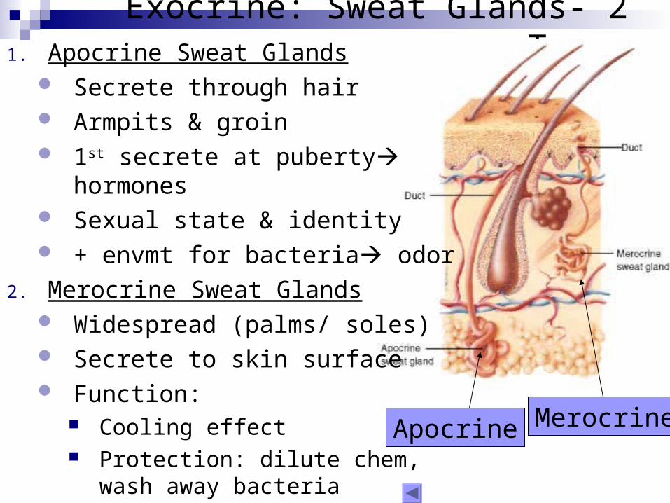
Exocrine: Sweat Glands- 2 Types
MerocrineApocrine
1. Apocrine Sweat Glands Secrete through hair Armpits & groin 1st secrete at puberty hormones Sexual state & identity + envmt for bacteria odor
2. Merocrine Sweat Glands Widespread (palms/ soles) Secrete to skin surface Function:
Cooling effect Protection: dilute chem, wash
away bacteria

Skin Local Control : Inflammation Epidermis= great protection
Thick, keratinized cellsTight junctions b/w cellsSebaceous & sweat glandsAvascular
Papillary layer= bacterial heavenOnce bacteria enters papillary
layer inflamm response Mast cells initiate response

Skin Local Control: Regeneration1. Clot Formation:
1. Platelets & fibrin (scab) Isolates area Creates network for new
cell migration
2. Division, Migration, & Phagocytosis:
1. Stratum germinativum Divide & Migrate Granulation tissue: fibrin
clot, fibroblasts, capillary network
2. Macrophages Phagocytosis

Regeneration (con’t)3. Scar tissue (lots of
collagen, little blood)1. Fibroblasts
Damaged hair follicles, sweat & sebaceous glands replaced w/ scar tissue
4. Contraction: 1. Platelets & epithelial cells
Pull wound together
5. Clot dissolves

Aging Integumentary System

Temperature Control
1. Sweat Glands Cooling effect
2. Muscles Mostly skeletal & arrector pili
3. Blood Flow Superficial: heat transfer Deeper: maintain heat

Application: Skin Cancer Basal cell carcinoma: Most common
Malignant Germinativum (basale) layer Common Cause: UV radiation Metastasis rare
Squamous Cell Carcinoma: 2nd most common Malignant Common cause: UV radiation Metastasis rare
Melanoma: most dangerous & rare Malignant Cancerous melanocytes Common cause: UV radiation High Metastasis rate
Diagnosis: ABCD Assymetry, Border, Color, Diameter

Application: Burns & Skin Grafts Depth of Burn1. First degree
Superficial epidermis Erythema, sunburn red, blisters, painful Heals in few days
2. Second degree Down to germinativum & dermis red, moist,
blisters, blanch with pressure Painful Accessory structures unaffected Minor if >15% body burned 3 weeks to heal
3. Third degree Down to subcutaneous white, brown, black, or
whit, dry Less painful WHY? Hard to heal if extensive through body skin graft
may be needed
PartialThickness
FullThickness
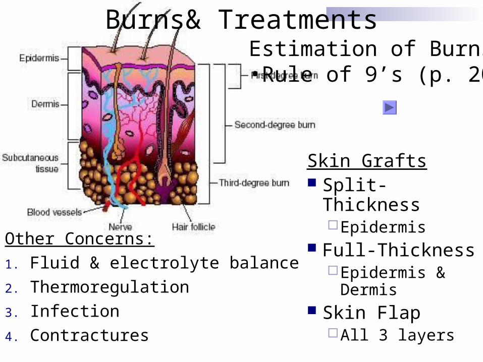
Burns& TreatmentsEstimation of Burns•Rule of 9’s (p. 204)
Skin Grafts Split- Thickness
Epidermis Full-Thickness
Epidermis & Dermis
Skin FlapAll 3 layers
Other Concerns:
1. Fluid & electrolyte balance
2. Thermoregulation
3. Infection
4. Contractures

Rule of 9’s

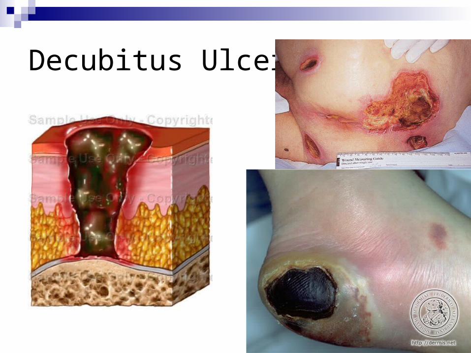
Application: Decubitus Ulcer Decubitus Ulcer= Pressure Sore
Most common: over bony areas (i.e. sacrum, heel) Cut off blood supply decrease nutrients & O2 to tissues Population most affected:
Bed ridden Decreased sensation: diabetics (feet & hands), SCI Wheelchair bound
Stages: I: reddening II: blister III: all 3 skin layers affected IV: all 3 layers of skin & underlying tissues (muscle/
tendons/ bones) Prevention: positioning every 2 hours, weight shifts in
wheelchair, cushioning/ supports
Risk ofInfection
Decubitus Ulcers