integument review are you ready? don’t forget plates also!
TRANSCRIPT

Integument Review
Are you ready?
Don’t forget plates also!

Apocrine VS Eccrine
1. Eccrine
duct to pore on surface
2. Apocrine
Ducts empty into hair follicles
Associated with pheromones in underarm & pubic areas

Cause of a suntan
• Melanocytes make melanin in response to UV exposure

Characteristics of papillary layer
Top part of dermis
• Projections called dermal papillae
• Pain receptors
• Touch receptors
• Blood vessels

Complications of 3rd degree burn
• Associated dangers- Dehydration
- Electrolyte imbalance
- Circulatory shock

Function of sebum
• Oil made by sebaceous glands– - Lubricant for skin– - Kills bacteria
• Most with ducts that empty into hair follicles

Functions of skin
1.Protects deeper tissues
2. prevents desiccation
3. helps in heat regulation
4.Makes vit D
5. Excretion of waste

How are fingernails made?
Scale-like modifications of epidermis
- Heavily keratinized
Stratum basale under nail bed
- Responsible for growth

How does body cool off

Layers of hair & how produced
Cuticle – outside; transparent; heavily keratinized; scaly; gives elasticity & resiliency
Cortex – middle; rope-like proteins; if exposed – moisture loss, unravel, split ends
Medulla – inside; support structure

Male pattern baldness results from what body chemicals?
Scientific name for baldness – Alopecia

Order of epidermis strata layers

Parts of a fingernail
Free edge
Body
Root of nail
Eponychium – proximal nail fold that projects onto the nail body

Rules of Nines
• Way to determine the extent of burns
• Body is divided into 11 areas for quick estimate
• Each area represents ~ 9%

Medical terms: bedsores, cancer, bruise, redness
• Decubitus ulcer
• Melanoma
• Hematoma
• Erythema

Skin problems caused by
• Virus
• Fungus
• Bacteria
• Allergies
• Inflammation of sebaceous glands
• Inflammation of hair follicle
Cold sores
Athlete’s foot
Boil or Impetigo
Contact Dermatitis
Acne
Boil

4 types of membranes & examples• Cutaneous membrane -
skin
• Mucous membrane – inside mouth
• Serous membrane – covering organs
• Synovial membrane – certain joints

3 pigments of skin colorMelanin
Yellow, brown or black
Carotene
Orange-yellow from vegetables
Hemoglobin
Red from blood cells in dermis
Oxygen content determines redness

Tissue type of external skin layer
Epidermis – •outer layer
•Stratified squamous epithelium•Often keratinized

Visceral vs parietal
• Words to describe each layer of serous membranes
• V – on organs
• P – lining outer wall
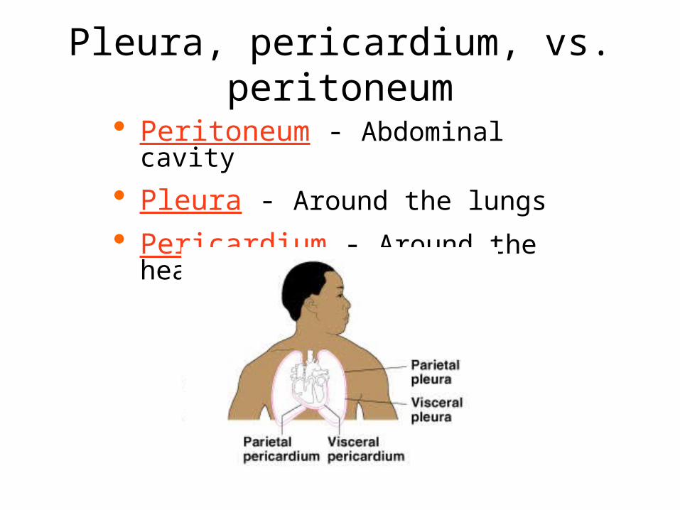
Pleura, pericardium, vs. peritoneum
Peritoneum - Abdominal cavity
Pleura - Around the lungs
Pericardium - Around the heart

What are dermal papillae?
• Projections called dermal papillae in Papillary layer of dermis
• Fingerprints

Melanoma mole looks likeA = Asymmetry
– Two sides of pigmented mole do not match
B = Border irregularity – Borders of mole are not
smooth
C = Color – Different colors in pigmented
area
D = Diameter – Spot is larger then 6 mm in
diameter

What is composition of sweat
• Composition–Mostly water–Some metabolic waste–Fatty acids and proteins (apocrine only)

What’s happening in each stratum?1. Stratum basale
Cells undergoing mitosis
Lies next to dermis
2. Stratum spinosum
connections to adjacent cells
3. Stratum granulosum
granules of keratin
4. Stratum lucidum
Occurs only in thick skin
5. Stratum corneum Shingle-like dead cells

What is keratin, where is it found
• Waterproof protein
• Added by Stratum granulosum
• Found in hair, nails, skin

What is melanin, where is it found
• Pigment made by melanocytes in stratum basale
• Found in skin and hair
• Protects nucleus from UV

Mucous membrane location
• Body cavities that open to the outside

Where are the three kinds of dermal receptors found?
Free nerve endings
• In dermal papillae
Meissner’s corpuscles
• In dermal papillae.
Pacinian corpuscles
• In reticular layer

Which kind of membrane is all connective tissue
SynovialSynovial
MembraneMembrane

Which layer(s) has no blood supply

Which layer(s) insulate(s)

Which receptors are for pain (temp), touch, pressure?
Free nerve endings
• pain
Meissner’s corpuscles
• touch
Pacinian corpuscles
• pressure