integument cell gelatinisation—the fate of the integumentary cells · pdf...
TRANSCRIPT

ORIGINAL ARTICLE
Integument cell gelatinisation—the fate of the integumentarycells in Hieracium and Pilosella (Asteraceae)
Bartosz J. Płachno1 & Piotr Świątek2& Małgorzata Kozieradzka-Kiszkurno3 &
Zbigniew Szeląg4 & Piotr Stolarczyk5
Received: 12 February 2017 /Accepted: 2 May 2017 /Published online: 15 May 2017# The Author(s) 2017. This article is an open access publication
Abstract Members of the generaHieracium and Pilosella aremodel plants that are used to study the mechanisms of apo-mixis. In order to have a proper understanding of apomixis,knowledge about the relationship between the maternal tissueand the gametophyte is needed. In the genus Pilosella, previ-ous authors have described the specific process of the Blique-faction^ of the integument cells that surround the embryo sac.However, these observations were based on data only at thelight microscopy level. The main aim of our paper was toinvestigate the changes in the integument cells at the ultra-structural level in Pilosella officinarum and Hieraciumalpinum. We found that the integument peri-endothelial zonein both species consisted of mucilage cells. The mucilage was
deposited as a thick layer between the plasma membrane andthe cell wall. The mucilage pushed the protoplast to the centreof the cell, and cytoplasmic bridges connected the protoplastto the plasmodesmata through the mucilage layers. Moreover,an elongation of the plasmodesmata was observed in the mu-cilage cells. The protoplasts had an irregular shape and werefinally degenerated. After the cell wall breakdown of the mu-cilage cells, lysigenous cavities that were filled with mucilagewere formed.
Keywords Apomixis . Asteraceae . Integument . Lysigenouscavities .Mucilage cells . Ovule . Plasmodesmata .
Ultrastructure . Idioblasts
Introduction
Members of the genera Hieracium L. and Pilosella Vaill. areimportant model plants for understanding the mechanisms ofapomixis in angiosperms (e.g. Koltunow et al. 2011a,b;Tucker et al. 2012; Okada et al. 2013; Hand and Koltunow2014; Hand et al. 2015; Shirasawa et al. 2015; Rabiger et al.2016; Rotreklová and Krahulcová 2016).
According to Koltunow et al. (1998), the development ofthe embryo and endosperm in Hieracium aurantiacum L. [=Pilosella aurantiaca (L.) F. W. Schultz & Sch. Bip.],H. pilosella L. [= P. officinarum Vaill.] and P. piloselloides(Vill.) Soják [= H. piloselloides Vill.] coincide with the inten-sive Bliquefaction^ of the integument cells that surround theembryo sac. This process was observed in both sexual andapomictic plants and relied on changes in integument cell wallfollowed by integument cell liquefaction near the endotheliumand finally the accumulation of carbohydrate-rich material.According to Koltunow et al. (1998), this material may servea nutritive role and moreover, they suggested that the
Handling Editor: Heiti Paves
Electronic supplementary material The online version of this article(doi:10.1007/s00709-017-1120-1) contains supplementary material,which is available to authorized users.
* Bartosz J. Pł[email protected]
1 Department of Plant Cytology and Embryology, JagiellonianUniversity in Kraków, 9 Gronostajowa St., 30-387 Kraków, Poland
2 Department of Animal Histology and Embryology, University ofSilesia in Katowice, 9 Bankowa St., 40-007 Katowice, Poland
3 Department of Plant Cytology and Embryology, University ofGdańsk, 59 Wita Stwosza St., 80-308 Gdańsk, Poland
4 Department of Botany, Pedagogical University of Kraków, 3Podchorążych St., 30-084 Kraków, Poland
5 Unit of Botany and Plant Physiology, Institute of Plant Biology andBiotechnology, Faculty of Biotechnology and Horticulture,University of Agriculture in Kraków, 29 Listopada 54 Street,31-425 Kraków, Poland
Protoplasma (2017) 254:2287–2294DOI 10.1007/s00709-017-1120-1

accumulation of a large pool of nutrients around the embryosac might have helped the evolution of the apomictic traitwithin the genus. This suggestion about a nutritionalfunction of these specific integumentary cells was acceptedand repeated by other authors, e.g. Van Baarlen et al. (1999)wrote that the ovules of Hieracium and Taraxacum contain aprotein-rich storage tissue, which nourishes the embryo andreduces the importance of the endosperm function. It was alsosuggested by these authors that the presence of this tissuemight explain the evolution of autonomous embryo develop-ment in most of the Asteraceae apomicts. A similar suggestionwas repeated in the case of Taraxacum and Chondrilla byMusiał et al. (2013) and later by Musiał and Kościńska-Pająk (2013). These integumentary cells were called integu-mentary Bnutritive tissue^ and its presence and ultrastructurein different members of Asteraceae was discussed by Kolczyket al. (2014). Although data about the ultrastructure of theintegument in both Hieracium and Pilosella are still lacking,progress has been made in the case of another apomictic ge-nus, Taraxacum, which belongs to the same subfamily.Płachno et al. (2016) showed that the Bnutritive tissue^ (=peri-endothelial tissue) in the Taraxacum ovule consists ofspecialised mucilage cells. During the differentiation of thesecells and the deposition of mucilage, the plasmodesmata be-come elongated and are associated with structures calledBcytoplasmic bridges.^
It is well known that the plasmodesmata are plant cell com-munication channels that are crucial for controlling the inter-cellular transport of macromolecules such as mRNA, signalsincluding proteins and transcriptional factors (e.g. Oparka2004; Gursanscky et al. 2011; Hyun et al. 2011).Symplasmic isolation/communication between the ovularsporophytic tissues and the megagametophyte and later theembryo is necessary for successful development (e.g.Ingram 2010; Bencivenga et al. 2011; Marzec andKurczynska 2008, 2014; Wróbel-Marek et al. 2017 and liter-ature therein). Sporophytic ovule tissues also have an influ-ence on apomixis, e.g. Tucker et al. (2012) showed that inPilosella ovules, sporophytic information is potentiated bythe growth of the funiculus and also that polar auxin transportinfluences ovule development, the initiation of apomixis andthe progression of embryo sac. According to Okada et al.(2013), signalling molecules such as the kinases from the spo-rophytic ovule cells have an influence on the aposporous em-bryo sac formation in the apomictic Hieracium species. Thus,in order to properly understand the symplasmic isolation/communication in Pilosella and Hieracium ovules, basicknowledge about the ultrastructure of the sporophyte tissuesis needed.
It should be stressed that the selection of our research ma-terial is not accidental. There are amphimictic diploids andalso apomictic polyploids (mitotic diplospory) among the ge-nus Hieracium. However, in the genus Pilosella, both
amphimictic taxa (diploids, sometimes tetra- and hexaploids)and poliploidal facultative apomicts (apospory) are known.Thus, we would like to compare if any differences occur inthe integument structure of these genera.
Aims
The main aim of our paper was to investigate the changes inthe integument cells that surround the embryo sac inHieracium and Pilosella.
Another question is what happens to the plasmodesmata inthese cells. Are the plasmodesmata in the ovule integumentarycells of Hieracium and Pilosella associated with the cytoplas-mic bridges (the thin strands of cytoplasm) like in Taraxacumovules?
We also wanted to investigate whether there is an accumu-lation of a large pool of nutrients (protein and lipid storage) inthe peri-endothelial integument cells that surround the embryosac.
Material and methods
Plant material
The plants of an amphimictic P. officinarum Vaill. clone forthe present study were collected by BJP in their natural habitatin Kokotek near the town of Lubliniec, Poland (tetraploidclone x = 9; Sak et al. 2016). Another plant of P. officinarum[hexaploid clone x = 9, Ilnicki and Szeląg 2011] was collectedby ZS in the Mt. Treskovac, Banat, Romania. Plants ofH. alpinum L. [diploid cytotype x = 9, Ilnicki and Szeląg2011] were collected by ZS in the Retezat Mountains,Southern Carpathians, Romania. In both species, the flowersthat were used in this study were harvested before and duringanthesis. They contained ovules with mature embryo sacs ofthe Polygonum type (Figs. 1a, b and 2a, b). Transmissionelectron microscopy (TEM) analysis was performed on atleast three different samples from each species. About 200TEM pictures were taken and analysed.
Light and electron microscopy studies
The preparation of the samples for TEM followed the proce-dure used by Płachno and Świątek (2011) and Kozieradzka-Kiszkurno and Płachno (2012). Semithin sections werestained using aqueous methylene blue with azure II for generalhistology (Humphrey and Pittman, 1974) for 1–2 min (MB/AII) and examined using an Olympus BX60 microscope. Thecytochemical tests included Aniline Blue Black (Jensen,1962) for proteins and Sudan Black B for lipids (Bronner,1975). The periodic acid-Schiff (PAS) reaction was used to
2288 B. J. Płachno et al.

visualise the total carbohydrates of insoluble polysaccharides(Wędzony 1996).
Ultrathin sections were cut on a Leica Ultracut UCT ultra-microtome. After contrasting with uranyl acetate and lead cit-rate, the sections were examined using a Hitachi H500 elec-tron microscope at 75 kV in the Faculty of Biology andEnvironmental Protection, University of Silesia in Katowiceand a Jeol JEM 100 SX; JEOL, Tokyo, Japan, at 80 kV in theDepartment of Cell Biology and Imaging, Institute ofZoology, Jagiellonian University in Kraków.
Results
In both species, at the mature female gametophyte stage(Figs. 1a, b and 2a, b), the ovule had a considerably thick,multilayer integument, which had a heterogeneous structure(outer epidermis, highly vacuolated parenchyma,periendothelial tissue, integumentary tapetum; Figs. 1 and2). The female gametophyte was surrounded by peri-endothelial tissue (Figs. 1 and 2), which was very well devel-oped especially at the chalazal pole of the ovule (Figs. 1a, c
and 2a, d). The peri-endothelial tissue consisted of mucilagecells (Figs. 3 and 4). The protoplasts of these cells had anirregular shape (Figs. 3a, b and 4a, b). Mucilage was amor-phous, electron translucent with electron-dense reticulatecomponents. It was formed by hypertrophied dictyosomes(Figs. 3b, c and 4c, d) and deposited in the extraplasmaticspace between the cell wall and the plasmalemma (Figs. 3b,c and 4a, b). The cytoplasm contained a large nucleus (Fig. 3a,b), many vesicles with mucilage (from the dictyosomes), arough endoplasmic reticulum and ribosomes (Figs. 3c and4c, d). Although the mucilage pushed the protoplast to thecentre of the cell, the mucilage cells were still symplasmicallyconnected (Figs. 3c and 4a, b).
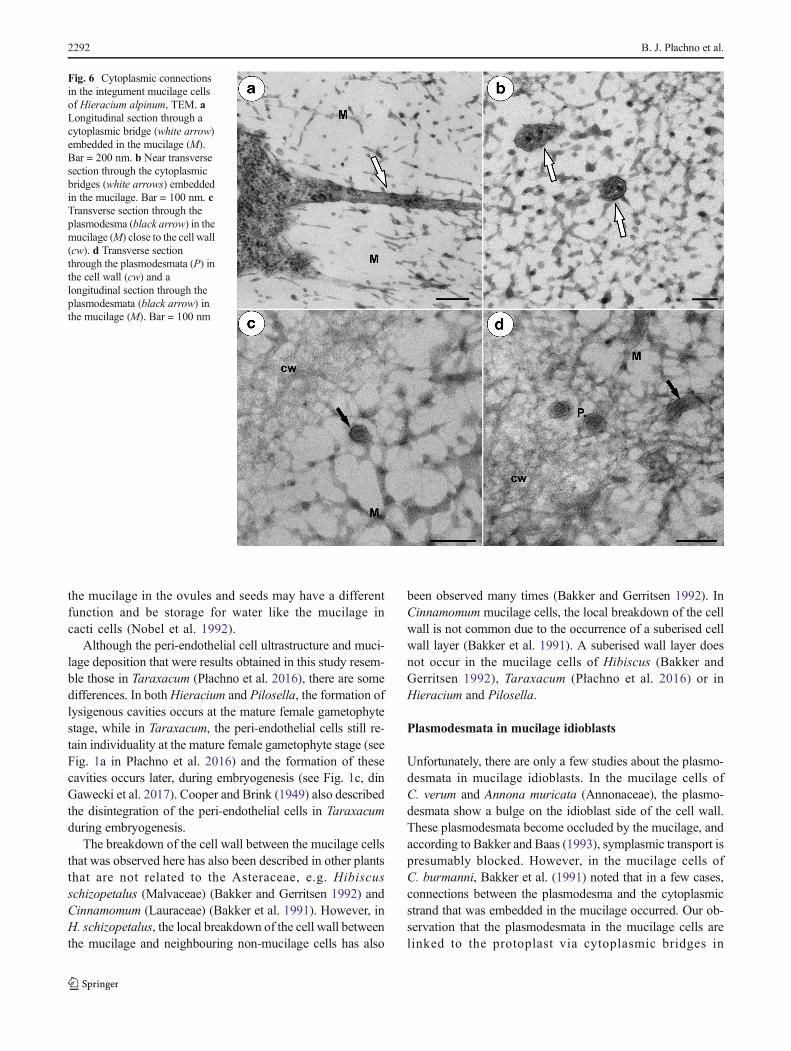
In both species, there were thin strands of cytoplasm (cy-toplasmic bridges) that connected the protoplast with the plas-modesmata (Figs. 3c, 5a–d, and 6a). In the cytoplasmic brid-ges, microtubules and ribosomes were visible (Figs. 3c, 5b–d,and 6a, b). In the mucilage close to the primary wall, cytoplas-mic bridges were connected with the plasmodesmata, whichhad passed the plasmodesmata in the primary cell wall(Fig. 5a–d). On the transverse and longitudinal sections ofthe plasmodesmata, the plasmalemma and the desmotubule
Fig. 1 Pilosella officinarum, Light microscopy. a Semithin sectionthrough an ovary (ov), ovule with an embryo sac (es). Peri-endothelialtissue–mucilage cells (Mu), micropyle (M), chalaza (Ch). Bar = 50 μm. bSemithin section through an ovule with an embryo sac showing thestructure of the periendothelial zone cells (Mu) and mucilage cavities(L). Egg cell (eg), central cell (cc), synergids (s) synergid filiformapparatus (arrow), integumental tapetum (It). Bar = 20 μm. c Chalazalperiendothelial tissue note irregular shape of protoplasts of mucilagecells; mucilage cavities (L), bar = 20 μm
Fig. 2 Hieracium alpinum, light microscopy. a Semithin section throughan ovule with an embryo sac showing the structure of the peri-endothelialzone cells–mucilage cells (Mu). Embryo sac (es), micropyle (M), chalaza(Ch). Bar = 50 μm. b Higher magnification of the embryo sac andperiendothelial tissue. Egg cell (eg), central cell (cc), mucilage cavities(L), integumental tapetum (It). Bar = 20 μm. c, d Peri-endothelialmucilage cells in d at the chalazal pole. Bar = 20 μm
The fate of integumentary cells in Asteraceae 2289

Fig. 4 Ultrastructure of the peri-endothelial zone cells inHieracium alpinum, TEM. a, bGeneral ultrastructure of themucilage cells. Notecytoplasmic–plasmodesmataconnections between themucilage cells (black arrows),mucilage (M), cell wall (cw).Bar = 0.8 μm and bar = 0.85 μm.c, d Hypertrophied dictyosomes(D) with numerous vesiclescontaining mucilage. Nucleus(N), mitochondria, roughendoplasmic reticulum (Er). Bothbars = 500 nm
Fig. 3 Ultrastructure of the peri-endothelial zone cells in Pilosellaofficinarum, TEM. a General ultrastructure of the mucilage cells.Bar = 3.5 μm. b Ultrastructure of the mucilage cells: dictyosomes withnumerous vesicles (arrow), mitochondrion (m), nucleus (N), mucilage(M). Bar = 1 μm. c Plasmodesmata connected with the cytoplasmic
bridge (white arrow); note that the plasmodesma (black arrow) ispartially embedded in the mucilage (M), cell wall (cw), dictyosome (D).In insert, there is a magnified part of cytoplasmic bridge to showmicrotubule. Bar = 0.15 μm
2290 B. J. Płachno et al.

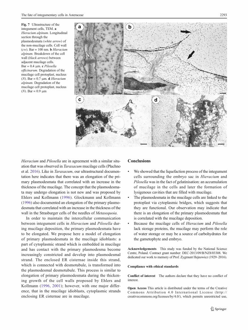
were clearly visible in the mucilage (Fig. 6c, d). The structureof these plasmodesmata was similar to the typical plasmodes-mata in the cell walls of non-mucilage parenchyma cells(Fig. 7a).
A breakdown of the cell wall between the adjacent muci-lage cells occurred (Fig. 7b), after which lysigenous cavitiesthat were filled with mucilage were formed (Figs. 1b, c and2b). The protoplast of mucilage cells was finally degraded(Fig. 7c, d).
The cytochemical tests for the storage lipids and proteingave negative results in the case of the mucilage cells (notshown). However, we observed small lipid droplets in thecytoplasm of the mucilage cells using TEM (Supplementarymaterial 1). There was positive staining after periodic-acid-Schiff reaction. The total carbohydrates of insoluble polysac-charides (including mucilage carbohydrates) stain pink to pur-plish red (Supplementary material 2).
Discussion
We showed that the intensive Bliquefaction^ of the integ-ument cells surrounding the embryo sac, which was
previously described by Koltunow et al. (1998), was inthe fact gelatinisation: an accumulation of the mucilage inthe cells and later the formation of lysigenous cavities thatwere filled with mucilage. Koltunow et al. (1998) wrotethat the material that was accumulated during the inten-sive changes of the integument cells surrounding the em-bryo sac was carbohydrate-rich (positive staining afterperiodic-acid-Schiff reaction). This also agreed with ourobservation that this material is mucilage. The ultrastruc-ture of the mucilage in Hieracium and Pilosella is similarto the previously observed mucilage in the integumentmucilage cells of other Asteraceae genera—Taraxacum,Onopordum, Solidago, Chondrilla and Bellis (Płachnoet al. 2016; Kolczyk et al. 2014, 2016 and literature there-in). However, our results disagree with Van Baarlen et al.(1999) who wrote that the ovules of Hieracium contain aprotein-rich storage tissue, because we did not find pro-te in bodies or pro te in s torage vacuoles in theperiendothelial tissue. However, during the gelatinisationof the periendothelial cells and the degeneration of theirprotoplasts, some nutrients might be released andtransported to the female gametophyte. But, such a hy-pothesis requires experimental confirmation. Moreover,
Fig. 5 Cytoplasmic connectionsin the integument mucilage cellsofHieracium alpinum, TEM. a–dPlasmodesmata connected withthe cytoplasmic bridges (whitearrow). Note that theplasmodesmata (black arrow) arepartially embedded in themucilage (M), Btypical^plasmodesmata in cell wall (P). Inthe cytoplasmic bridge,ribosomes are visible. a, cBar = 200 nm. b, d Bar = 100 nm
The fate of integumentary cells in Asteraceae 2291
the mucilage in the ovules and seeds may have a differentfunction and be storage for water like the mucilage incacti cells (Nobel et al. 1992).
Although the peri-endothelial cell ultrastructure and muci-lage deposition that were results obtained in this study resem-ble those in Taraxacum (Płachno et al. 2016), there are somedifferences. In bothHieracium and Pilosella, the formation oflysigenous cavities occurs at the mature female gametophytestage, while in Taraxacum, the peri-endothelial cells still re-tain individuality at the mature female gametophyte stage (seeFig. 1a in Płachno et al. 2016) and the formation of thesecavities occurs later, during embryogenesis (see Fig. 1c, dinGawecki et al. 2017). Cooper and Brink (1949) also describedthe disintegration of the peri-endothelial cells in Taraxacumduring embryogenesis.
The breakdown of the cell wall between the mucilage cellsthat was observed here has also been described in other plantsthat are not related to the Asteraceae, e.g. Hibiscusschizopetalus (Malvaceae) (Bakker and Gerritsen 1992) andCinnamomum (Lauraceae) (Bakker et al. 1991). However, inH. schizopetalus, the local breakdown of the cell wall betweenthe mucilage and neighbouring non-mucilage cells has also
been observed many times (Bakker and Gerritsen 1992). InCinnamomum mucilage cells, the local breakdown of the cellwall is not common due to the occurrence of a suberised cellwall layer (Bakker et al. 1991). A suberised wall layer doesnot occur in the mucilage cells of Hibiscus (Bakker andGerritsen 1992), Taraxacum (Płachno et al. 2016) or inHieracium and Pilosella.
Plasmodesmata in mucilage idioblasts
Unfortunately, there are only a few studies about the plasmo-desmata in mucilage idioblasts. In the mucilage cells ofC. verum and Annona muricata (Annonaceae), the plasmo-desmata show a bulge on the idioblast side of the cell wall.These plasmodesmata become occluded by the mucilage, andaccording to Bakker and Baas (1993), symplasmic transport ispresumably blocked. However, in the mucilage cells ofC. burmanni, Bakker et al. (1991) noted that in a few cases,connections between the plasmodesma and the cytoplasmicstrand that was embedded in the mucilage occurred. Our ob-servation that the plasmodesmata in the mucilage cells arelinked to the protoplast via cytoplasmic bridges in
Fig. 6 Cytoplasmic connectionsin the integument mucilage cellsof Hieracium alpinum, TEM. aLongitudinal section through acytoplasmic bridge (white arrow)embedded in the mucilage (M).Bar = 200 nm. b Near transversesection through the cytoplasmicbridges (white arrows) embeddedin the mucilage. Bar = 100 nm. cTransverse section through theplasmodesma (black arrow) in themucilage (M) close to the cell wall(cw). d Transverse sectionthrough the plasmodesmata (P) inthe cell wall (cw) and alongitudinal section through theplasmodesmata (black arrow) inthe mucilage (M). Bar = 100 nm
2292 B. J. Płachno et al.
Hieracium and Pilosella are in agreement with a similar situ-ation that was observed in Taraxacummucilage cells (Płachnoet al. 2016). Like in Taraxacum, our ultrastructural documen-tation here indicates that there was an elongation of the pri-mary plasmodesmata that correlated with an increase in thethickness of the mucilage. The concept that the plasmodesma-ta may undergo elongation is not new and was proposed byEhlers and Kollmann (1996). Glockmann and Kollmann(1996) also documented an elongation of the primary plasmo-desmata that correlated with an increase in the thickness of thewall in the Strasburger cells of the needles of Metasequoia.
In order to maintain the intercellular communicationbetween integument cells in Hieracium and Pilosella dur-ing mucilage deposition, the primary plasmodesmata haveto be elongated. We propose here a model of elongationof primary plasmodesmata in the mucilage idioblasts: apart of cytoplasmic strand which is embedded in mucilageand has contact with the primary plasmodesma becomeincreasingly constricted and develop into plasmodesmalstrand. The enclosed ER cisternae inside this strand,which is connected with desmotubule, is transformed intothe plasmodesmal desmotubule. This process is similar toelongation of primary plasmodesmata during the thicken-ing growth of the cell walls proposed by Ehlers andKollmann (1996, 2001); however, with one major differ-ence, that in the mucilage idioblasts, cytoplasmic strandsenclosing ER cisternae are in mucilage.
Conclusions
& We showed that the liquefaction process of the integumentcells surrounding the embryo sac in Hieracium andPilosellawas in the fact of gelatinisation: an accumulationof mucilage in the cells and later the formation oflysigenous cavities that are filled with mucilage.
& The plasmodesmata in the mucilage cells are linked to theprotoplast via cytoplasmic bridges, which suggests thatthey are functional. Our observation may indicate thatthere is an elongation of the primary plasmodesmata thatis correlated with the mucilage deposition.
& Because the mucilage cells of Hieracium and Pilosellalack storage proteins, the mucilage may perform the roleof water storage or may be a source of carbohydrates forthe gametophyte and embryo.
Acknowledgements This study was funded by the National ScienceCentre, Poland. Contract grant number: DEC-2013/09/B/NZ8/03308. Wededicated our work to memory of Prof. Zygmunt Hejnowicz (1929–2016).
Compliance with ethical standards
Conflict of interest The authors declare that they have no conflict ofinterest.
Open Access This article is distributed under the terms of the CreativeCommons At t r ibut ion 4 .0 In te rna t ional License (h t tp : / /creativecommons.org/licenses/by/4.0/), which permits unrestricted use,
Fig. 7 Ultrastructure of theintegument cells, TEM. aHieracium alpinum. Longitudinalsection through theplasmodesmata (white arrow) ofthe non-mucilage cells. Cell wall(cw). Bar = 100 nm. b Hieraciumalpinum. Breakdown of the cellwall (black arrows) betweenadjacent mucilage cells.Bar = 0.4 μm. c Pilosellaofficinarum. Degradation of themucilage cell protoplast, nucleus(N). Bar = 0.7 μm. d Hieraciumalpinum. Degradation of themucilage cell protoplast, nucleus(N). Bar = 0.9 μm
The fate of integumentary cells in Asteraceae 2293

distribution, and reproduction in any medium, provided you giveappropriate credit to the original author(s) and the source, provide a linkto the Creative Commons license, and indicate if changes were made.
References
Bakker ME, Baas P (1993) Cell walls in oil and mucilage cells. Acta BotNeerl 42(2):133–139
Bakker ME, Gerritsen AF (1992) The development of mucilage cells inHibiscus schizopetalus. Acta Bot Neerl 41:31–42
Bakker ME, Gerritsen AF, van der Schaaf PJ (1991) Development of oiland mucilage cells in Cinnamomum burmanni. An ultrastructuralstudy. Acta Bot Neerl 40:339–356
Bencivenga S, Colombo L, Masiero S (2011) Cross talk between thesporophyte and the megagametophyte during ovule development.Sex Plant Reprod 24:113–121
Bronner R (1975) Simultaneous demonstration of lipid and starch in planttissues. Stain Technol 50:1–4
Cooper DC, Brink RA (1949) The endosperm-embryo relationship in theautonomous apomict, Taraxacum officinale. Bot Gaz 111:139–152
Ehlers K, Kollmann R (1996) Formation of branched plasmodesmata inregenerating Solanum nigrum—protoplasts. Planta 199:126. doi:10.1007/BF00196889
Ehlers K, Kollmann R (2001) Primary and secondary plasmodesmata:structure, origin, and functioning. Protoplasma 216:1–30
Gawecki R, Sala K, Kurczyńska EU, Świątek P, Płachno BJ (2017)Immunodetection of some pectic, arabinogalactan proteins andhemicelluloses epitopes in the micropylar transmitting tissue of apo-mictic dandelions (Taraxacum, Asteraceae, Lactuceae). Protoplasma254:657–668. doi:10.1007/s00709-016-0980-0
Glockmann C, Kollmann R (1996) Structure and development of cellconnections in the phloem ofMetasequoia glyptostroboides needles.I. Ultrastructural aspects of modified primary plasmodesmata inStrasburger cells. Protoplasma 193:191–203
Gursanscky NR, Searle IR, Carroll BJ (2011) Mobile microRNAs hit thetarget. Traffic 12:1475–1482
Hand ML, Koltunow AM (2014) The genetic control of apomixis: asex-ual seed formation. Genetics 197:441–450
Hand ML, Vít P, Krahulcová A, Johnson SD, Oelkers K, Siddons H et al(2015) Evolution of apomixis loci in Pilosella and Hieracium(Asteraceae) inferred from the conservation of apomixis-linkedmarkers in natural and experimental populations. Hered 114:17–26
Humphrey CD, Pittman FE (1974) A simple methylene blue-azure IIbasic fuchsin stain for epoxy-embedded tissue sections. StainTechnol 49:9–14
Hyun TK, Uddin MN, Rim Y, Kim J-Y (2011) Cell-to-cell trafficking ofRNA andRNA silencing through plasmodesmata. Protoplasma 248:101–116
Ilnicki T, Szeląg Z (2011) Chromosome numbers in Hieracium andPilosella (Asteraceae) from central and southeastern Europe. ActaBiol Cracov Ser Bot 53(1):102–110. doi:10.2478/v10182-011-0014-3
Ingram GC (2010) Family life at close quarters: communication and con-straint in angiosperm seed development. Protoplasma 247:195–214
JensenWA (1962) Botanical histochemistry. W. H. Freeman and Co, SanFrancisco
Kolczyk J, Stolarczyk P, Płachno BJ (2014) Comparative anatomy ofovules in Galinsoga, Solidago and Ratibida (Asteraceae). ActaBiol Cracov Ser Bot 56(2):115–125. doi:10.2478/abcsb-2014-0024
Kolczyk J, Stolarczyk P, Płachno BJ (2016) Ovule structure of scotchthistle Onopordum acanthium L. (Cynareae, Asteraceae). ActaBiol Cracov Ser 58:19–28. doi:10.1515/abcsb-2016-0001
Koltunow AM, Johnson SD, Bicknell RA (1998) Sexual and apomicticdevelopment in Hieracium. Sex Plant Reprod 11:213–230
Koltunow AM, Johnson SD, Okada T (2011a) Apomixis in hawkweed:Mendel’s experimental nemesis. J Exp Bot 62:1699–1707
Koltunow AM, Johnson SD, Rodrigues JC, Okada T, Hu Y, Tsuchiya Tet al (2011b) Sexual reproduction is the default mode in apomicticHieracium subgenus Pilosella, in which two dominant loci functionto enable apomixis. Plant J 66:890–902
Kozieradzka-Kiszkurno M, Płachno BJ (2012) Are there symplastic con-nections between the endosperm and embryo in some angio-sperms?—A lesson from the Crassulaceae family. Protoplasma249:1081–1089. doi:10.1007/s00709-011-0352-8
MarzecM, Kurczynska EU (2008) Symplasmic communication/isolationand plant cell differentiation (in Polish). Postepy Biol Komorki 35:369–390
Marzec M, Kurczynska E (2014) Importance of symplasmic communi-cation in cell differentiation. Plant Sig & Beh 9:e27931. doi:10.4161/psb.27931tt
Musiał K, Kościńska-Pająk M (2013) Ovules anatomy of selected apo-mictic taxa from Asteraceae family. Mod Phytomorphol 3:35–38
Musiał K, Płachno BJ, Świątek P, Marciniuk J (2013) Anatomy of ovaryand ovule in dandelions (Taraxacum, Asteraceae). Protoplasma 250:715–722. doi:10.1007/s00709-012-0455-x
Nobel PS, Cavelier J, Andrade JL (1992) Mucilage in cacti: its apoplasticcapacitance, associated solutes and influence on tissue water rela-tions. J Exp Bot 43:641–648
Okada T, Hu Y, Tucker MR, Taylor JM, Johnson SD, Spriggs A et al(2013) Enlarging cells initiating apomixis in Hieracium praealtumtransition to an embryo sac program prior to entering mitosis. PlantPhysiol 163:216–231
Oparka KJ (2004) Getting the message across: how do plant cells ex-change macromolecular complexes? Trends Plant Sci 9:33–41
Płachno BJ, Świątek P (2011) Syncytia in plants: cell fusion in endosperm-placental syncytium formation in Utricularia (Lentibulariaceae).Protoplasma 248:425–435. doi:10.1007/s00709-010-0173-1
Płachno BJ, Kurczyńska E, Świątek P (2016) Integument cell differenti-ation in dandelions (Taraxacum, Asteraceae, Lactuceae) with specialattention paid to plasmodesmata. Protoplasma 253:1365–1372. doi:10.1007/s00709-015-0894-2
Rabiger DS, Taylor JM, Spriggs A et al (2016) Generation of an integrat-ed Hieracium genomic and transcriptomic resource enables explo-ration of small RNA pathways during apomixis initiation. BMCBiol 14:86. doi:10.1186/s12915-016-0311-0
Rotreklová O, Krahulcová A (2016) Estimating paternal efficiency in anagamic polyploid complex: pollen stainability and variation in pol-len size related to reproduction mode, ploidy level andhybridogenous origin in Pilosella (Asteraceae). Folia Geobot 51:175. doi:10.1007/s12224-016-9240-5
Sak D, Janas A,MusiałK, Płachno BJ (2016) Sexual reproductive traits intetraploid Pilosella officinarum (Asteraceae, Cichorioideae): DICmi-croscope study of cleared whole-mount tissue. XXXII Conference onEmbryology Plants Animals Humans, May 18–21, 2016,Wojsławice, Poland. Acta Biol Cracov Ser Bot 58(suppl. 1):90
ShirasawaK, HandML, Henderson ST, Okada T, Johnson SD, Taylor JMet al (2015) A reference genetic linkage map of apomicticHieracium species based on expressed markers derived from devel-oping ovule transcripts. Ann Bot 115:567–580
Tucker MR, Okada T, Johnson SD, Takaiwa F, Koltunov AMG (2012)Sporophytic ovule tissues modulate the initiation and progression ofapomixis in Hieracium. J Exp Bot 63:3229–3241
Van Baarlen P, Verduijn M, Van Dijk PJ (1999) What can we learn fromnatural apomicts? Trends Plant Sci 4:43–44
Wędzony M (1996) Fluorescence microscopy for botanists (in Polish).Dept. Plant Physiology Monographs 5, Kraków, p 128
Wróbel-Marek J, Kurczyńska E, Płachno BJ, Kozieradzka-Kiszkurno M(2017) Distribution of symplasmic transport fluorochromes withinthe embryo and seed of Sedum acre L. (Crassulaceae). Planta245(3):491–505 doi:10.1007/s00425-016-2619-y
2294 B. J. Płachno et al.