insight into the pathogenesis of lyme disease · insight into the pathogenesis of lyme disease ......
TRANSCRIPT

Journal of Bacteriology and Virology 2014. Vol. 44, No. 1 p.10 – 22 http://dx.doi.org/10.4167/jbv.2014.44.1.10
Insight into the Pathogenesis of Lyme Disease
Ok Sarah Shin1,2*
1Department of Biomedical Sciences, College of Medicine, Korea University, Gurodong, Gurogu, Seoul; 2Department of Microbiology, College of Medicine, Korea University, Seoul Korea
Lyme disease is the most common vector-borne disease in the United States and Europe, caused by a tick-borne spirochete, Borrelia burgdorferi. Life cycle alternation between arthropod and mammals enhanced B. burgdorferi to adapt to two diverse niches. Although B. burgdorferi infection in these reservoir hosts appears asymptomatic, infection in human can typically cause inflammation in the skin, nervous system, musculoskeletal system and heart. In this review, we discuss the basic molecular characteristics and cell biology of B. burgdorferi and provide an overview of spirochete-induced activation of innate and adaptive immunity, resulting in particular immunopathology. Advancing understanding of the immune evasion mechanisms of B. burgdorferi provides important implications for ongoing research and clinical practice of Lyme disease.
Key Words: Lyme disease, B. burgdorferi, Pathogenesis
Introduction
Lyme disease was first described in Lyme, Connecticut,
after an outbreak of what was thought to be "juvenile
rheumatoid arthritis" (1). Since juvenile rheumatoid arthritis
does not occur in outbreaks, researchers studied these
patients, which led to the identification of Lyme arthritis.
Later, it was founded that Lyme disease affects different
organs of Lyme disease patients during different stages of
the infection (2). Despite improvements in diagnostic tests
and public awareness of Lyme disease, there remain still
approximately 30,000 cases of Lyme disease patients per
year in the United States (3).
Lyme disease is diagnosed clinically based on symptoms,
objective physical findings (such as erythema migrans, facial
palsy, or arthritis), a history of possible exposure to infected
ticks, as well as serological tests. The clinical manifestations
of Lyme disease are complex and can be divided into three
different stages: acute-localized, acute-disseminated and
chronic. When a B. burgdorferi-infected tick feeds on a
human, it inoculates the spirochetes into the skin. During
an acute localized stage, the spirochetes are localized in the
skin, where they cause an inflammatory rash, known as
erythema migrans (EM). In the weeks or months after
infection, the spirochetes disseminate through blood and
lymph, reaching other organs, such as the heart, joints and
nervous system. Chronic infection reflects the establishment
in tissues, where the spirochetes persist even in the face of
the specific host immune response. When left untreated
with antibiotics, infection with B. burgdorferi can result in
chronic arthritis that may progress to a severe, erosive
Received: January 6, 2014/ Revised: January 14, 2014/ Accepted: January 28, 2014
*Corresponding author: Ok Sarah Shin, Ph.D. Department of Biomedical Sciences, College of Medicine, Korea University, Gurodong, Gurogu, Seoul, 152-703, Korea. Phone: +82-2-2626-3280, Fax: +82-2-2626-1962, e-mail: [email protected]
**This work was supported by Korea University Grant (Korea University, Korea). ○CC This is an Open Access article distributed under the terms of the Creative Commons Attribution Non-Commercial License (http://creativecommons.org/license/by-nc/3.0/).
10
Review Article
B. burgdorferi and Lyme Disease Pathogenesis 11
arthritis with histopathologic similarities to rheumatoid
arthritis. In most people, treatment with antibiotics is very
effective in eliminating symptoms, preventing progression to
later manifestations of the disease, and curing the infection.
Some symptoms improve rapidly with this treatment,
whereas other symptoms gradually improve over weeks to
months.
Epidemiology of B. burgdorferi
The epidemiology of human Lyme disease is determined
by the geographic distribution and life cycle of its tick
vector, described in Fig. 1. B. burgdorferi is transmitted by
the bite of infected Ixodes ticks (I. scapularis in the northern
U.S., I. pacificus in the western U.S. and Canada, I. ricinus
and I. persulcatus in Europe and I. persulcatus in Asia).
Ixodes ticks have three life stages that require blood: larvae,
nymphs, and adults, feeding only once during every active
stage. At any stage, ticks can be infected with B. burgdorferi.
Male ticks rarely feed and never engorge but female ticks
can feed on host animals. After feeding for a few days
(about 3 days for larvae, 5 for nymphs, and 7 days for adult
females), the ticks drop off their host and locate on or near
the soil surface and take several months to develop into their
next developmental stage, or, in the case of adult females,
lay about 2000 eggs. The length of a tick's life cycle varies
between 2 years and 6 years, depending on climate, or host
availability.
B. burgdorferi sensu stricto (the sole genospecies
transmitted in the eastern U.S.) is not vertically transmitted
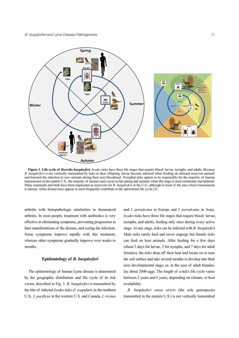
Figure 1. Life cycle of Borrelia burgdorferi. Ixodes ticks have three life stages that require blood: larvae, nymphs, and adults. Because B. burgdorferi is not vertically transmitted by ticks to their offspring, larvae become infected when feeding on infected reservoir animals and transmit the infection to new animals during their next bloodmeal. Nymphal ticks appear to be responsible for the majority of human transmission in the eastern U.S.; the majority of human cases occur in late spring and summer when this stage is most commonly encountered. Many mammals and birds have been implicated as reservoirs for B. burgdorferi in the U.S., although in most of the sites where transmissionis intense, white-footed mice appear to most frequently contribute to the spirochetal life cycle (3).
Erythemamigrans
Larva
Eggs
Adult
Spring
Summer
Autumn
Winter
Nymph
Eggs
12 O Sarah Shin
by ticks to their offspring, larvae become infected when
feeding on infected reservoir animals and transmit the
infection to new animals during their next blood meal.
Nymphal ticks appear to be responsible for the majority of
human transmission in the eastern U.S.; the majority of
human cases occur in late spring and summer when this
stage is most commonly encountered. Many mammals and
birds have been implicated as reservoirs for B. burgdorferi
in the U.S., although in most of the sites where transmission
is intense, white-footed mice appear to most frequently
contribute to the spirochetal life cycle. Other rodents such
as chipmunks and Norway rats may locally contribute to
transmission.
Microbiology of B. burgdorferi
B. burgdorferi was first identified as a causative organism
for Lyme disease by Dr. Willy Burgdorfer in 1982 (4). B.
burgdorferi belongs to a species of bacteria of the class
spirochetes, which include Treponema Pallidum, and Lepto-
spira. B. burgdorferi exhibit a characteristic morphology,
with inner and outer membranes surrounding periplasmic
flagella and a flexible cell wall. The outer membrane is rich
in lipoproteins, including the highly immunogenic outer-
surface proteins (Osp), but does not contain lipopoly-
saccharide (LPS). The unique style of spirochete motility
results from endoflagella contained within the periplasmic
space between a semi rigid peptidoglycan helix and a
multi-layer, flexible outer membrane sheath. When the
filaments rotate within this space, spirochetes move in a
cork-screw fashion (3).
B. burgdorferi was the first spirochete for which the
complete genome was sequenced and the genome of B.
burgdorferi strain B31 was published in 1997 (5, 6). There
are several unique aspects to its composition. For example,
the B. burgdorferi genome is composed of a small linear
chromosome of approximately 900 kb and >20 different
plasmids ranging in size from 5 to 56 kb. Some plasmids
are unstable during in vitro propagation, but are required
for infectivity in vivo (7). The most remarkable aspect of
the B. burgdorferi genome is the large number of sequences
encoding predicted or known lipoproteins.
B. burgdorferi genome encodes for over 160 lipoproteins
that become expressed during different stages of its life
cycle. Because B. burgdorferi has unusually large numbers
of lipoproteins, many scientists focused on investigating
the function of these lipoproteins. It is believed that B.
burgdorferi changes surface protein expression to enable a
successful infection and survive in two different environ-
ments, the tick and the mammalian host. Among many outer
surface lipoproteins, well-characterized paradigm in the
protein expression shift is that of OspA and OspC. OspA is
highly up-regulated by B. burgdorferi within the midgut of
unfed ticks but is down-regulated in B. burgdorferi as ticks
take a blood meal and are transmitted to the mammalian
host (8). In contrast, in unfed ticks, OspC is not expressed by
B. burgdorferi (8, 9), however, OspC is quickly up-regulated
upon entry to the mammalian host (10, 11).
Role of innate immunity
Both innate and adaptive immunity are important for
controlling the pathogenesis of B. burgdorferi. Before the
discovery of toll-like receptors (TLRs), the main focus of
immunological research in Lyme disease was on the role of
adaptive immunity. However, with the discovery of TLRs,
many studies pointed to the importance of innate immune
cells, such as neutrophils and macrophages, in initiating an
inflammatory response and controlling the clearance of the
organisms.
B. burgdorferi lipoproteins are potent stimulants that can
activate pro-inflammatory products in many different cell
types, including macrophages, endothelial cells, neutrophils
and B lymphocytes (12~17). It has been shown that
recognition of B. burgdorferi lipoprotein OspA is mediated
through activation of TLR1/2 heterodimers, leading to
nuclear translocation of the transcription factor NFκB, which
is crucial for initiation of inflammatory responses (18).
Ligation of TLR2 by B. burgdorferi lipoproteins has been
shown in vitro to result in activation of peripheral blood
mononuclear cells (PBMCs) and release of pro-inflammatory
cytokines and chemokines (18). Although it was believed

B. burgdorferi and Lyme Disease Pathogenesis 13
that inflammatory manifestations of B. burgdorferi infection
were mediated by TLR signaling pathways, mice deficient
in CD14, TLR2, or MyD88 still developed arthritis and
harbored a higher organism burden than their wild-type
counterparts (19~23). Specifically, MyD88 deficiency in
mice resulted in 100 fold higher replication of spirochetes
in the joints, heart and ears, whereas TLR2 deficiency in
mice increased pathogen burden by 10 fold, compared with
wild type mice at 2, 4, and 8 weeks post infection (19, 22~
24). Joint inflammation in B. burgdorferi-infected MyD88-
deficient mice has been variously reported by three different
groups as either unchanged from wild-type or increased
(19, 22, 23). It was expected the absence of TLRs would
result in the reduced induction of major inflammatory
signaling pathways, however surprisingly, these mice
showed increased inflammation and increased cytokine/
chemokine production in arthritic joints. This increased
inflammation is ineffective at clearing the organisms and
the bacterial loads in the joints are 1~2 logs higher than in
wild-type mice (19, 22~24). Carditis, which is the other
major manifestation of B. burgdorferi infection in mice,
was unchanged from wild-type levels in MyD88-deficient
mice (23), but myositis was greatly increased (22). In
conclusion, there might be additional receptors that are
required for TLR-independent activation of inflammatory
symptoms in mice deficient in TLR2 or MyD88.
Recently, it was discovered that in addition to TLR2, other
TLRs, such as TLR5, TLR7, TLR8 and TLR9, cooperate
with TLR2-TLR1 to induce pro-inflammatory molecules,
including type I interferons (IFNs) (25~27). Besides TLRs,
NOD-like receptors (NLRs) also play an important role in
sensing B. burgdorferi on dendritic cells and macrophages
within the dermis (Fig. 2) (28~30). NLRs sense the
presence of intracellular muropeptides derived from bacterial
peptidoglycans and NOD1 and NOD2 are mainly expressed
by epithelial cells and antigen-presenting cells (APCs) such
as macrophages and dendritic cells (31). It was shown that
B. burgdorferi up-regulates NOD2 on astrocytes after
exposure to several TLR-ligands (32). In addition, NOD2
is involved in the release of several different inflammatory
cytokines induced by B. burgdorferi infection, such as IL-6
(33, 34). Association of inflammasome in sensing B.
burgdorferi was also reported that B. burgdorferi-induced
production of IL-1 require activation of the inflammasome
components ASC and caspase-1. Furthermore, it was found
that B. burgdorferi induces inflammasome-mediated caspase-
1 activation, although the inflammasome activation does
not appear to be mediated via NLRP3 receptor (28, 35, 36).
However, more details of how TLRs and NLRs interact and
sense B. burgdorferi in the pathogenesis of Lyme disease
remains unknown.
In addition to TLRs and NLRs, integrins are also key
signaling molecules that can activate TLR-independent
signaling pathways for matrix metalloproteinase (MMP)
production (37). Previous work showed that B. burgdorferi
binds to integrins αII2β3, αvβ3, α5β1 and, α3β1 (38~40).
Furthermore, at least two different integrins, αMβ2 integrin
(also known as CR or CD18-CD11b) and α3β1 integrin
mediate internalization of spirochetes in the absence of
antibodies (41, 42).
Although persistently elevated numbers of spirochetes in
tissues from these mice were thought to be due to defective
development of antibody response, surprisingly, the antibody
response appeared normal and functional, suggesting TLR-
independent activation of B lymphocytes could function
efficiently in the absence of TLRs (19). Although Liu et al
reported that deletion of MyD88 in B. burgdorferi-infected
mice led to a shift in a humoral response to Th2-associated
antibody isotypes with an increase in the production of Th2
associated antibody isotypes, IgG1, in MyD88 deficient
mice (23), B. burgdorferi-specific antibody production in
TLR2 or MyD88 deficient mice does not lead to efficient
clearance of spirochetes, suggesting that inability to control
numbers of spirochetes in the absence of MyD88 can be
due to a defect in effector cells involved in innate immune
response (19, 24). In summary, the recognition of B.
burgdorferi components by TLR2 or MyD88 is not essential
for the development of the early adaptive immune response.
Role of adaptive immunity
Previous studies suggested that the development of

14 O Sarah Shin
specific humoral immunity plays a major role in the control
of spirochete burden. B cells have been shown to be very
important in the resolution of arthritis and control of
spirochete burden, whereas T cells appear to be necessary
for inflammation, but are questionably important in control
of spirochete burdens. Studies of B. burgdorferi infection
of severe combined immunodeficiency (SCID) and RAG-
deficient mice, which lack both T and B cells, or of mice
deficient in B cells alone, resulted in higher pathogen
burdens but severity of arthritis was similar to wild type
mice (43~47).
In humans, clinical symptoms during early stage of Lyme
Figure 2. Innate and adaptive immune activation in response to B. burgdorferi infection. B. burgdorferi are initially recognized by innate immune effector cells such as dendritic cells (DCs), neutrophils and macrophages; activation of these cells increases following internalization and degradation of spirochetes within phagolysosomes. DCs that have taken up spirochetes migrate to the lymph nodes, where they present processed B. burgdorferi antigens to T cells and B cells. While T cells enter the circulation and are recruited to the site of infection, B cells can be differentiated into plasma cells that can secrete specific antibodies that can kill B. burgdorferi via complement-dependent and -independent pathways. Production of pro-inflammatory cytokines by activated macrophages results in the recruitment of additional neutrophils, T cells, macrophages and DCs to the bite site, and eventually the development of erythema migrans (3).
B. burgdorferi and Lyme Disease Pathogenesis 15
disease, specifically, biopsies of erythema migrans lesions
show infiltration by T cells (CD8+ cells as well as CD4+
cells), macrophages, plasmacytoid and monocytoid dendritic
cells, and neutrophils. T cells also appear to be directly
involved in the development of inflammatory symptoms
such as carditis and arthritis, but are not critical for the
resolution of the disease (48). It has been shown that Th1
cells dominate the immune response in the synovial fluid
of patients with Lyme disease and that the severity of
arthritis correlates with the ratio of Th1 cells to Th2 cells in
the synovial fluids (49). C3H/HeJ mice whose predominant
CD4+ T cell response to B. burgdorferi is Th1 type show
more severe arthritis than mice whose response is Th2 type
(BALB/C mice) (50~52). However, it is likely that other
genetic factors also play a role and recent data have pointed
away from a key role for Th1/Th2 responses (53).
Recent studies have indicated potential roles of other T
cell types, including natural killer T cells (NKT) and Th17
cells. Glycolipids of B. burgdorferi can be recognized by
NKT cells and antigen-specific activation of NKT cells
prevents persistent joint inflammation and promotes a
spirochetal clearance (54, 55). Furthermore, studies of the
role of a newly discovered member of T cell subsets, Th17,
suggested that neutrophils can drive the differentiation of
Th17 cells in response to B. burgdorferi (56).
Unlike T cell's role in inflammation control, B cell-
mediated response is important for clearing the pathogen
(57). IgM, T cell independent antibodies, is crucial for the
initial reduction of spirochetal burdens, whereas T cell-
dependent production of IgG by B cells is typically
detectable by the second week of infection (58, 59).
Mouse models of Lyme arthritis
Animal models have been very important in the under-
standing of the immune response to B. burgdorferi.
Although both dogs and monkeys develop arthritis in
response to B. burgdorferi infection, the vast majority of
studies have focused on mice due to availability and the
potential for genetic manipulations. Murine Lyme arthritis
is characterized by early onset of arthritis (typically < 2
weeks) followed by spontaneous resolution of the ankle
swelling/arthritis with the development of a specific immune
response (typically ~8 weeks). However, low levels of
spirochetes appear to persist for prolonged periods of time.
Histologically, during acute infection, there is evidence of
inflammation of joints, tendons, ligaments and peri-articular
connective tissue (60). This is characterized by neutrophilic
infiltration and synovial hyperplasia. Arthritis appears to
correlate with spirochete burden in the joint in certain strains
of mice (BALB/C) but not others (C57BL/6 or C3H). It is
important to note that all immuno-competent mice resolve
their arthritis spontaneously regardless of antibiotic therapy,
although mild recurrent arthritis may occur in a small
percentage.
There are significant differences in the response to B.
burgdorferi among strains of mice. Numerous studies have
indicated that genetic regulation of inflammatory responses
control the severity of Lyme arthritis. Among the inbred
strains, C3H mice develop the most significant levels of
arthritis while C57BL/6 mice are relatively resistant (43,
61). BALB/C mice develop variable arthritis depending
upon the numbers of spirochetes present. Studies have
focused on identifying unique pathways associated with
the differential severity of Lyme arthritis (62~64). Although
this difference is not dependent on major histocompatibility
complex (MHC) alleles, it has been linked to quantitative
trait loci (QTL) on chromosomes 1, 4, 5, 11, and 12 (62).
Another factor that influences disease severity in mice is
the levels of pro- and anti-inflammatory immunomodulators
produced by host cells. C57BL/6 mice are better able to
regulate inflammation in response to B. burgdorferi in-
fection than C3H mice, because C57BL/6 macrophages
produce larger amounts of the anti-inflammatory cytokine,
interleukin-10 (IL-10), than did C3H macrophages, whereas
there are increased production of tumor necrosis factor-α
(TNF-α), interleukin-6 (IL-6) and interferon-γ (IFN-γ) in
macrophages from C3H mice (65). Consequently, the
deficiency of IL-10 results in more severe Lyme arthritis in
mice, suggesting an essential role of IL-10 in the regulation
of severity of Lyme arthritis (65).
16 O Sarah Shin
Inflammatory signaling pathways in Lyme disease
B. burgdorferi stimulates a robust inflammatory response
at sites of localization. Infection with B. burgdorferi results
in the activation of inflammatory signaling pathways that
lead to the release of cytokines and chemokines and an
influx of inflammatory cells that contribute to many of the
clinical manifestations of Lyme disease. Therefore, it is
important to understand receptors and signaling pathways
involved in the generation of inflammation and identify
host signaling pathways, which modulate the inflammatory
pathology that is characteristic of Lyme disease.
The pathology at sites such as the skin, heart, central
nervous system and joints occurs as a result of a host
inflammatory response to the organism that results in the
induction and release of cytokines, chemokines and pro-
teases such as matrix metalloproteinases (MMPs) that
destroy tissue (66~72). Specifically, Lyme arthritis appears
to be caused by the host inflammatory system in response
to invasion of joints by the spirochete and is characterized
by edema, synovial thickening, tendonitis, and a leukocytic
infiltration consisting mainly of neutrophils and mono-
nuclear cells. B. burgdorferi produces many outer membrane
lipoproteins which possess potent inflammatory potential
and are believed to be responsible for the inflammatory
properties attributed to this spirochete (16). These lipo-
proteins (such as OspA) are capable of activating a wide
variety of cell types, including macrophages, neutrophils,
and endothelial cells, which results in the production of a
broad spectrum of pro- and anti-inflammatory mediators
that have been linked to inflammatory response by B.
burgdorferi (12~17).
The induction of pro-inflammatory molecules is mediated
through the activation of specific signaling pathways. The
signaling pathways involved are typically specific to the
stimulus and to the cell type. Several laboratories have
studied the role of major signaling pathways in the
development of Lyme arthritis. Among them are mitogen
activated protein kinase (MAPK), and janus kinase/signal
transducer and activator of transcription (JAK/STAT). Behera
et al. have shown that the addition of B. burgdorferi to
cultures of human chondrocytes results in the activation of
at least two arms of the MAPK pathway, p38 and JNK, and
components of the JAK/STAT pathway including STAT-3
and STAT-6, but not STAT-1 (69). Inhibition of each of these
signaling pathways results in a diminution of the expression
of specific groups of cytokines, chemokines and MMPs
(69). Anguita et al. have demonstrated the importance of
the MAPK pathways in vivo using mice that are deficient
in MAP kinase kinase-3 (MKK-3) which is an upstream
activator of p38 (73, 74). These mice showed significantly
reduced production of pro-inflammatory cytokines and a
reduction in arthritis compared to matched controls.
Consistent with in vitro studies of chondrocyte stimulation
with B. burgdorferi showing lack of STAT-1 activation (69),
Brown et al. have recently shown using STAT-1 deficient
mice that STAT-1 is involved in carditis in response to B.
burgdorferi but not arthritis (75).
Phagocytosis of B. burgdorferi
The presence of phagocytes, such as neutrophils and
macrophages, at the sites of B. burgdorferi infection in-
dicates that the phagocytosis may play an important role in
efficient clearance of the organisms. It has previously been
shown that B. burgdorferi are efficiently phagocytosed by
macrophages, neutrophils and dendritic cells (23, 76~78)
and recruited to a lysosome within 20 min post B.
burgdorferi infection. Specifically, macrophages in vitro
rapidly ingest and kill B. burgdorferi in large numbers, with
or without opsonization and participation of Fc receptors
(79). The role of TLR signaling for phagocytosis of B.
burgdorferi was studied using bone marrow-derived macro-
phages and peritoneal macrophages. MyD88-mediated effect
on uptake of B. burgdorferi is thought to be through TLR
signaling-mediated activation of phosphoinositide 3 kinase
(PI3K) and recruitment of actin-related protein complexes
(Arp2/3), which result in actin polymerization to initiate
phagocytosis (80, 81). Phagocytosis is not only required
for efficient clearance of the organisms, it is also found to
be important for generating signals for host inflammatory

B. burgdorferi and Lyme Disease Pathogenesis 17
response. The phagocytosis of B. burgdorferi and sub-
sequent degradation within phagolysosomes further were
shown to amplify the release of inflammatory cytokines
through activation of TLR signaling within phagolysosome
(26).
Immune evasions by B. burgdorferi
As discussed above, a robust humoral and cellular
immune response occurs upon B. burgdorferi infection,
however, infection with B. burgdorferi can persist. While
the exact mechanism by which B. burgdorferi evades the
immune system is not known, several mechanisms have
been hypothesized. One possibility is that B. burgdorferi
evades immune response by binding to various components
of the extracellular matrix and migrating rapidly intra-
cellular compartments and the extracellular matrix. After B.
burgdorferi reaches the dermis, it expresses binding proteins
called adhesins to facilitate its dissemination. In particular,
decorin-binding adhesins (DbpA and DbpB) seem to play an
important role by binding to decorin, a collagen-associated
proteoglycan (82).
Another possibility is that B. burgdorferi frequently
changes its surface antigens to evade immune detection.
Once B. burgdorferi enter the skin, the spirochetes are
thought to downregulate lipoproteins, such as OspC, which
are no longer required to establish or maintain infection
(83). Expression of OspC plays an essential part in the
establishment of infection in a mammalian host, although
the mechanism by which OspC promotes B. burgdorferi
infectivity is unknown. B. burgdorferi also uses a system
of antigenic variation to evade antibodies. VlsE is a 35 kDa
lipoprotein that undergoes antigenic variation through the
recombination of sequences from silent cassettes into the
expressed vlsE locus (9, 83, 84). Lastly, B. burgdorferi may
induce autoimmunity in the host following the initial
infection, which could produce chronic infection. This
hypothesis was suggested by the findings of the structural
homology between OspA and human gene called leukocyte
function-associated antigen (LFA-1) (85, 86). These and
other findings indicate that the B. burgdorferi alter antigen
expression during infection in order to evade B cell-
mediated antibody response and do not elicit effective
memory responses to protective antigens. Thus, it is
challenging to identify target antigens that can induce
protective immunity and this needs further investigation.
Conclusion
B. burgdorferi infections and the diseases that they cause
are a growing public health problem, and there is currently
no available vaccine in use. In 2012, a first patient diagnosed
with Lyme disease was for the first time officially confirmed
by the national surveillance system in Korea (87). Because
of wide distribution of ixodes ticks among mountains in
Korea, which carry B. burgdorferi, it is necessary to perform
continuous surveillance system for Lyme disease and alert
the public about how to protect themselves from getting
tick bites.
Despite continued advances in our understanding of the
pathogenesis of Lyme disease, there remains much to learn
about how host immune defenses react to the organism.
How host immune system modulates sensing of the
pathogens and activates various signaling pathways requires
more attention. In addition, host genes responsible for
Lyme disease susceptibility in humans is largely unknown
and require more studies because we need to answer the
question of why some infected patients have only subclinical
disease, whereas others develop overt manifestations. These
and other advances will hopefully lead to a better under-
standing of the determinants of vector and host specificity
for B. burgdorferi, and to the manipulation of these
determinants to interrupt the cycle of transmission.
Lastly, it will be essential to develop a new vaccine for
Lyme disease. An OspA-based human vaccine to protect
against B. burgdorferi infection was developed and approved
by the US Food and Drug Administration (FDA). However,
due to low demand, the vaccine was removed from the
market in early 2002 by the manufacturer, GlaxoSmith-
Kline (GSK). Limitations and failed public acceptance of a
human vaccine led to its demise, yet current research
involving new paradigms for future vaccine design that

18 O Sarah Shin
would include elements of both the vector and the pathogen
will be interesting to follow.
REFERENCES
1) Steere AC, Hardin JA, Malawista SE. Erythema
chronicum migrans and Lyme arthritis: cryoimmuno-
globulins and clinical activity of skin and joints. Science
1977;196:1121-2.
2) Hu LT. In the clinic. Lyme disease. Ann Intern Med
2012;157:ITC2-2 - ITC2-16.
3) Radolf JD, Caimano MJ, Stevenson B, Hu LT. Of ticks,
mice and men: understanding the dual-host lifestyle of
Lyme disease spirochaetes. Nat Rev Microbiol 2012;
10:87-99.
4) Burgdorfer W, Barbour AG, Hayes SF, Benach JL,
Grunwaldt E, Davis JP. Lyme disease-a tick-borne
spirochetosis? Science 1982;216:1317-9.
5) Fraser CM, Casjens S, Huang WM, Sutton GG, Clayton
R, Lathigra R, et al. Genomic sequence of a Lyme
disease spirochaete, Borrelia burgdorferi. Nature 1997;
390:580-6.
6) Casjens S, Palmer N, van Vugt R, Huang WM,
Stevenson B, Rosa P, et al. A bacterial genome in flux:
the twelve linear and nine circular extrachromosomal
DNAs in an infectious isolate of the Lyme disease
spirochete Borrelia burgdorferi. Mol Microbiol 2000;
35:490-516.
7) Rosa PA, Tilly K, Stewart PE. The burgeoning
molecular genetics of the Lyme disease spirochaete.
Nat Rev Microbiol 2005;3:129-43.
8) Schwan TG, Piesman J, Golde WT, Dolan MC, Rosa
PA. Induction of an outer surface protein on Borrelia
burgdorferi during tick feeding. Proc Natl Acad Sci U
S A 1995;92:2909-13.
9) Ohnishi J, Piesman J, de Silva AM. Antigenic and
genetic heterogeneity of Borrelia burgdorferi popu-
lations transmitted by ticks. Proc Natl Acad Sci U S A
2001;98:670-5.
10) Ramamoorthi N, Narasimhan S, Pal U, Bao F, Yang
XF, Fish D, et al. The Lyme disease agent exploits a
tick protein to infect the mammalian host. Nature 2005;
436:573-7.
11) Grimm D, Tilly K, Byram R, Stewart PE, Krum JG,
Bueschel DM, et al. Outer-surface protein C of the
Lyme disease spirochete: a protein induced in ticks for
infection of mammals. Proc Natl Acad Sci U S A 2004;
101:3142-7.
12) Norgard MV, Arndt LL, Akins DR, Curetty LL, Harrich
DA, Radolf JD. Activation of human monocytic cells
by Treponema pallidum and Borrelia burgdorferi
lipoproteins and synthetic lipopeptides proceeds via a
pathway distinct from that of lipopolysaccharide but
involves the transcriptional activator NF-kappa B. Infect
Immun 1996;64:3845-52.
13) Norgard MV, Riley BS, Richardson JA, Radolf JD.
Dermal inflammation elicited by synthetic analogs of
Treponema pallidum and Borrelia burgdorferi lipo-
proteins. Infect Immun 1995;63:1507-15.
14) Morrison TB, Weis JH, Weis JJ. Borrelia burgdorferi
outer surface protein A (OspA) activates and primes
human neutrophils. J Immunol 1997;158:4838-45.
15) Ma Y, Seiler KP, Tai KF, Yang L, Woods M, Weis JJ.
Outer surface lipoproteins of Borrelia burgdorferi
stimulate nitric oxide production by the cytokine-
inducible pathway. Infect Immun 1994;62:3663-71.
16) Ma Y, Weis JJ. Borrelia burgdorferi outer surface
lipoproteins OspA and OspB possess B-cell mitogenic
and cytokine-stimulatory properties. Infect Immun 1993;
61:3843-53.
17) Wooten RM, Modur VR, McIntyre TM, Weis JJ.
Borrelia burgdorferi outer membrane protein A induces
nuclear translocation of nuclear factor-kappa B and
inflammatory activation in human endothelial cells. J
Immunol 1996;157:4584-90.
18) Hirschfeld M, Kirschning CJ, Schwandner R, Wesche
H, Weis JH, Wooten RM, et al. Cutting edge: inflam-
matory signaling by Borrelia burgdorferi lipoproteins
is mediated by toll-like receptor 2. J Immunol 1999;
163:2382-6.
19) Bolz DD, Sundsbak RS, Ma Y, Akira S, Kirschning CJ,
Zachary JF, et al. MyD88 plays a unique role in host
defense but not arthritis development in Lyme disease.
J Immunol 2004;173:2003-10.
20) Wang G, Ma Y, Buyuk A, McClain S, Weis JJ, Schwartz
I. Impaired host defense to infection and Toll-like
receptor 2-independent killing of Borrelia burgdorferi
B. burgdorferi and Lyme Disease Pathogenesis 19
clinical isolates in TLR2-deficient C3H/HeJ mice.
FEMS Microbiol Lett 2004;231:219-25.
21) Wang X, Ma Y, Weis JH, Zachary JF, Kirschning CJ,
Weis JJ. Relative contributions of innate and acquired
host responses to bacterial control and arthritis
development in Lyme disease. Infect Immun 2005;73:
657-60.
22) Behera AK, Hildebrand E, Bronson RT, Perides G,
Uematsu S, Akira S, et al. MyD88 deficiency results in
tissue-specific changes in cytokine induction and in-
flammation in interleukin-18-independent mice infected
with Borrelia burgdorferi. Infect Immun 2006;74:1462
-70.
23) Liu N, Montgomery RR, Barthold SW, Bockenstedt LK.
Myeloid differentiation antigen 88 deficiency impairs
pathogen clearance but does not alter inflammation in
Borrelia burgdorferi-infected mice. Infect Immun 2004;
72:3195-203.
24) Wooten RM, Ma Y, Yoder RA, Brown JP, Weis JH,
Zachary JF, et al. Toll-like receptor 2 is required for
innate, but not acquired, host defense to Borrelia
burgdorferi. J Immunol 2002;168:348-55.
25) Cervantes JL, La Vake CJ, Weinerman B, Luu S,
O'Connell C, Verardi PH, et al. Human TLR8 is
activated upon recognition of Borrelia burgdorferi RNA
in the phagosome of human monocytes. J Leukoc Biol
2013;94:1231-41.
26) Cervantes JL, Dunham-Ems SM, La Vake CJ, Petzke
MM, Sahay B, Sellati TJ, et al. Phagosomal signaling
by Borrelia burgdorferi in human monocytes involves
Toll-like receptor (TLR) 2 and TLR8 cooperativity and
TLR8-mediated induction of IFN-beta. Proc Natl Acad
Sci U S A 2011;108:3683-8.
27) Petzke MM, Brooks A, Krupna MA, Mordue D,
Schwartz I. Recognition of Borrelia burgdorferi, the
Lyme disease spirochete, by TLR7 and TLR9 induces
a type I IFN response by human immune cells. J
Immunol 2009;183:5279-92.
28) Oosting M, Buffen K, Malireddi SR, Sturm P,
Verschueren I, Koenders MI, et al. Murine Borrelia
arthritis is highly dependent on ASC and caspase-1, but
independent of NLRP3. Arthritis Res Ther 2012;14:
R247.
29) Berende A, Oosting M, Kullberg BJ, Netea MG, Joosten
LA. Activation of innate host defense mechanisms by
Borrelia. Eur Cytokine Netw 2010;21:7-18.
30) Sterka D Jr, Marriott I. Characterization of nucleotide-
binding oligomerization domain (NOD) protein expres-
sion in primary murine microglia. J Neuroimmunol
2006;179:65-75.
31) Kumar H, Kawai T, Akira S. Pathogen recognition by
the innate immune system. Int Rev Immunol 2011;30:
16-34.
32) Sterka D Jr, Rati DM, Marriott I. Functional expression
of NOD2, a novel pattern recognition receptor for
bacterial motifs, in primary murine astrocytes. Glia
2006;53:322-30.
33) Petnicki-Ocwieja T, DeFrancesco AS, Chung E, Darcy
CT, Bronson RT, Kobayashi KS, et al. Nod2 suppresses
Borrelia burgdorferi mediated murine Lyme arthritis
and carditis through the induction of tolerance. PloS
One 2011;6:e17414.
34) Oosting M, Berende A, Sturm P, Ter Hofstede HJ, de
Jong DJ, Kanneganti TD, et al. Recognition of Borrelia
burgdorferi by NOD2 is central for the induction of an
inflammatory reaction. J Infect Dis 2010;201:1849-58.
35) Oosting M, van de Veerdonk FL, Kanneganti TD, Sturm
P, Verschueren I, Berende A, et al. Borrelia species
induce inflammasome activation and IL-17 production
through a caspase-1-dependent mechanism. Eur J
Immunol 2011;41:172-81.
36) Liu N, Belperron AA, Booth CJ, Bockenstedt LK. The
caspase 1 inflammasome is not required for control of
murine Lyme borreliosis. Infect Immun 2009;77:3320
-7.
37) Behera AK, Hildebrand E, Uematsu S, Akira S, Coburn
J, Hu LT. Identification of a TLR-independent pathway
for Borrelia burgdorferi-induced expression of matrix
metalloproteinases and inflammatory mediators through
binding to integrin alpha 3 beta 1. J Immunol 2006;177:
657-64.
38) Coburn J, Magoun L, Bodary SC, Leong JM. Integrins
alpha(v)beta3 and alpha5beta1 mediate attachment of
lyme disease spirochetes to human cells. Infect Immun
1998;66:1946-52.
39) Coburn J, Leong JM, Erban JK. Integrin alpha IIb beta
3 mediates binding of the Lyme disease agent Borrelia
burgdorferi to human platelets. Proc Natl Acad Sci U S
20 O Sarah Shin
A 1993;90:7059-63.
40) Coburn J, Barthold SW, Leong JM. Diverse Lyme
disease spirochetes bind integrin alpha IIb beta 3 on
human platelets. Infect Immun 1994;62:5559-67.
41) Coburn J, Leong JM, Erban JK. Integrin alpha IIb beta
3 mediates binding of the Lyme disease agent Borrelia
burgdorferi to human platelets. Proc Natl Acad Sci U S
A 1993;90:7059-63.
42) Marre ML, Petnicki-Ocwieja T, DeFrancesco AS, Darcy
CT, Hu LT. Human integrin alpha(3)beta(1) regulates
TLR2 recognition of lipopeptides from endosomal
compartments. PLoS One 2010;5:e12871.
43) Barthold SW, Sidman CL, Smith AL. Lyme borreliosis
in genetically resistant and susceptible mice with severe
combined immunodeficiency. Am J Trop Med Hyg
1992;47:605-13.
44) Bockenstedt LK, Kang I, Chang C, Persing D, Hayday
A, Barthold SW. CD4+ T helper 1 cells facilitate
regression of murine Lyme carditis. Infect Immun 2001;
69:5264-9.
45) McKisic MD, Redmond WL, Barthold SW. Cutting
edge: T cell-mediated pathology in murine Lyme
borreliosis. J Immunol 2000;164:6096-9.
46) Ruderman EM, Kerr JS, Telford SR 3rd, Spielman A,
Glimcher LH, Gravallese EM. Early murine Lyme
carditis has a macrophage predominance and is in-
dependent of major histocompatibility complex class
II-CD4+ T cell interactions. J Infect Dis 1995;171:362
-70.
47) Schaible UE, Gay S, Museteanu C, Kramer MD,
Zimmer G, Eichmann K, et al. Lyme borreliosis in the
severe combined immunodeficiency (scid) mouse
manifests predominantly in the joints, heart, and liver.
Am J Pathol 1990;137:811-20.
48) McKisic MD, Redmond WL, Barthold SW. Cutting
edge: T cell-medicated pathology in murine Lyme
borreliosis. J Immunol 2000;164:6096-9.
49) Gross DM, Steere AC, Huber BT. T helper 1 response
is dominant and localized to the synovial fluid in
patients with Lyme arthritis. J Immunol 1998;160:1022
-8.
50) Keane-Myers A, Nickell SP. Role of IL-4 and IFN-
gamma in modulation of immunity to Borrelia
burgdorferi in mice. J Immunol 1995;155:2020-8.
51) Matyniak JE, Reiner SL. T helper phenotype and genetic
susceptibility in experimental Lyme disease. J Exp Med
1995;181:1251-4.
52) Yssel H, Shanafelt MC, Soderberg C, Schneider PV,
Anzola J, Peltz G. Borrelia burgdorferi activates a T
helper type 1-like T cell subset in Lyme arthritis. J Exp
Med 1991;174:593-601.
53) Potter MR, Noben-Trauth N, Weis JH, Teuscher C,
Weis JJ. Interleukin-4 (IL-4) and IL-13 signaling
pathways do not regulate Borrelia burgdorferi-induced
arthritis in mice: IgG1 is not required for host control
of tissue spirochetes. Infect Immun 2000;68:5603-9.
54) Kinjo Y, Tupin E, Wu D, Fujio M, Garcia-Navarro R,
Benhnia MR, et al. Natural killer T cells recognize
diacylglycerol antigens from pathogenic bacteria. Nat
Immunol 2006;7:978-86.
55) Tupin E, Benhnia MR, Kinjo Y, Patsey R, Lena CJ,
Haller MC, et al. NKT cells prevent chronic joint
inflammation after infection with Borrelia burgdorferi.
Proc Natl Acad Sci U S A 2008;105:19863-8.
56) Codolo G, Amedei A, Steere AC, Papinutto E, Cappon
A, Polenghi A, et al. Borrelia burgdorferi NapA-driven
Th17 cell inflammation in lyme arthritis. Arthritis
Rheum 2008;58:3609-17.
57) Connolly SE, Benach JL. The versatile roles of
antibodies in Borrelia infections. Nat Rev Microbiol
2005;3:411-20.
58) Belperron AA, Dailey CM, Booth CJ, Bockenstedt LK.
Marginal zone B-cell depletion impairs murine host
defense against Borrelia burgdorferi infection. Infect
Immun 2007;75:3354-60.
59) Tunev SS, Hastey CJ, Hodzic E, Feng S, Barthold SW,
Baumgarth N. Lymphoadenopathy during lyme
borreliosis is caused by spirochete migration-induced
specific B cell activation. PLoS Pathog 2011;7:e1002066.
60) Moody KD, Barthold SW. Lyme borreliosis in
laboratory mice. Lab Anim Sci 1998;48:168-71.
61) Barthold SW, Beck DS, Hansen GM, Terwilliger GA,
Moody KD. Lyme borreliosis in selected strains and
ages of laboratory mice. J Infect Dis 1990;162:133-8.
62) Weis JJ, McCracken BA, Ma Y, Fairbairn D, Roper RJ,
Morrison TB, et al. Identification of quantitative trait
loci governing arthritis severity and humoral responses
in the murine model of Lyme disease. J Immunol 1999;
B. burgdorferi and Lyme Disease Pathogenesis 21
162:948-56.
63) Crandall H, Dunn DM, Ma Y, Wooten RM, Zachary JF,
Weis JH, et al. Gene expression profiling reveals unique
pathways associated with differential severity of Lyme
arthritis. J Immunol 2006;177:7930-42.
64) Miller JC, Ma Y, Crandall H, Wang X, Weis JJ. Gene
expression profiling provides insights into the pathways
involved in inflammatory arthritis development: murine
model of Lyme disease. Exp Mol Pathol 2008;85:20-7.
65) Brown JP, Zachary JF, Teuscher C, Weis JJ, Wooten RM.
Dual role of interleukin-10 in murine Lyme disease:
regulation of arthritis severity and host defense. Infect
Immun 1999;67:5142-50.
66) Sellati TJ, Bouis DA, Caimano MJ, Feulner JA, Ayers
C, Lien E, et al. Activation of human monocytic cells
by Borrelia burgdorferi and Treponema pallidum is
facilitated by CD14 and correlates with surface exposure
of spirochetal lipoproteins. J Immunol 1999;163:2049
-56.
67) Radolf JD, Arndt LL, Akins DR, Curetty LL, Levi ME,
Shen Y, et al. Treponema pallidum and Borrelia
burgdorferi lipoproteins and synthetic lipopeptides
activate monocytes/macrophages. J Immunol 1995;154:
2866-77.
68) Tatro JB, Romero LI, Beasley D, Steere AC, Reichlin S.
Borrelia burgdorferi and Escherichia coli lipopoly-
saccharides induce nitric oxide and interleukin-6 pro-
duction in cultured rat brain cells. J Infect Dis 1994;
169:1014-22.
69) Behera AK, Thorpe CM, Kidder JM, Smith W,
Hildebrand E, Hu LT. Borrelia burgdorferi-induced
expression of matrix metalloproteinases from human
chondrocytes requires mitogen-activated protein kinase
and Janus kinase/signal transducer and activator of
transcription signaling pathways. Infect Immun 2004;
72:2864-71.
70) Brown CR, Blaho VA, Loiacono CM. Susceptibility to
experimental Lyme arthritis correlates with KC and
monocyte chemoattractant protein-1 production in joints
and requires neutrophil recruitment via CXCR2. J
Immunol 2003;171:893-901.
71) Gebbia JA, Coleman JL, Benach JL. Selective induction
of matrix metalloproteinases by Borrelia burgdorferi
via toll-like receptor 2 in monocytes. J Infect Dis 2004;
189:113-9.
72) Gebbia JA, Coleman JL, Benach JL. Borrelia spiro-
chetes upregulate release and activation of matrix
metalloproteinase gelatinase B (MMP-9) and collage-
nase 1 (MMP-1) in human cells. Infect Immun 2001;
69:456-62.
73) Anguita J, Barthold SW, Persinski R, Hedrick MN, Huy
CA, Davis RJ, et al. Murine Lyme arthritis development
mediated by p38 mitogen-activated protein kinase
activity. J Immunol 2002;168:6352-7.
74) Hedrick MN, Olson CM Jr, Conze DB, Bates TC,
Rincón M, Anguita J. Control of Borrelia burgdorferi-
specific CD4+-T-cell effector function by interleukin-
12- and T-cell receptor-induced p38 mitogen-activated
protein kinase activity. Infect Immun 2006;74:5713-7.
75) Brown CR, Blaho VA, Fritsche KL, Loiacono CM.
Stat1 deficiency exacerbates carditis but not arthritis
during experimental lyme borreliosis. J Interferon
Cytokine Res 2006;26:390-9.
76) Montgomery RR, Nathanson MH, Malawista SE. The
fate of Borrelia burgdorferi, the agent for Lyme disease,
in mouse macrophages. Destruction, survival, recovery.
J Immunol 1993;150:909-15.
77) Montgomery RR, Malawista SE. Entry of Borrelia
burgdorferi into macrophages is end-on and leads to
degradation in lysosomes. Infect Immun 1996;64:2867
-72.
78) Montgomery RR, Lusitani D, de Boisfleury Chevance
A, Malawista SE. Human phagocytic cells in the early
innate immune response to Borrelia burgdorferi. J
Infect Dis 2002;185:1773-9.
79) Montgomery RR, Nathanson MH, Malawista SE. Fc-
and non-Fc-mediated phagocytosis of Borrelia
burgdorferi by macrophages. J Infect Dis 1994;170:
890-3.
80) Shin OS, Miller LS, Modlin RL, Akira S, Uematsu S,
Hu LT. Downstream signals for MyD88-mediated
phagocytosis of Borrelia burgdorferi can be initiated
by TRIF and are dependent on PI3K. J Immunol 2009;
183:491-8.
81) Shin OS, Isberg RR, Akira S, Uematsu S, Behera AK,
Hu LT. Distinct roles for MyD88 and Toll-like receptors
2, 5, and 9 in phagocytosis of Borrelia burgdorferi and
cytokine induction. Infect Immun 2008;76:2341-51.
22 O Sarah Shin
82) Brown EL, Wooten RM, Johnson BJ, Iozzo RV, Smith
A, Dolan MC, et al. Resistance to Lyme disease in
decorin-deficient mice. J Clin Invest 2001;107:845-52.
83) Liang FT, Yan J, Mbow ML, Sviat SL, Gilmore RD,
Mamula M, et al. Borrelia burgdorferi changes its
surface antigenic expression in response to host immune
responses. Infect Immun 2004;72:5759-67.
84) McDowell JV, Sung SY, Hu LT, Marconi RT. Evidence
that the variable regions of the central domain of VlsE
are antigenic during infection with lyme disease
spirochetes. Infect Immun 2002;70:4196-203.
85) Gross DM, Forsthuber T, Tary-Lehmann M, Etling C,
Ito K, Nagy ZA, et al. Identification of LFA-1 as a
candidate autoantigen in treatment-resistant Lyme
arthritis. Science 1998;281:703-6.
86) Trollmo C, Meyer AL, Steere AC, Hafler DA, Huber
BT. Molecular mimicry in Lyme arthritis demonstrated
at the single cell level: LFA-1 alpha L is a partial
agonist for outer surface protein A-reactive T cells. J
Immunol 2001;166:5286-91.
87) Moon S, Gwack J, Hwang KJ, Kwon D, Kim S, Noh Y,
et al. Autochthonous lyme borreliosis in humans and
ticks in Korea. Osong Public Health Res Perspect 2013;
4:52-6.