in vitro evaluation of tio2 nanotubes as cefuroxime
TRANSCRIPT
HAL Id: hal-02386062https://hal.archives-ouvertes.fr/hal-02386062
Submitted on 29 Nov 2019
HAL is a multi-disciplinary open accessarchive for the deposit and dissemination of sci-entific research documents, whether they are pub-lished or not. The documents may come fromteaching and research institutions in France orabroad, or from public or private research centers.
L’archive ouverte pluridisciplinaire HAL, estdestinée au dépôt et à la diffusion de documentsscientifiques de niveau recherche, publiés ou non,émanant des établissements d’enseignement et derecherche français ou étrangers, des laboratoirespublics ou privés.
In vitro evaluation of TiO2 nanotubes as cefuroximecarriers on orthopaedic implants for the prevention of
periprosthetic joint infectionsP. Chennell, E. Feschet-Chassot, T. Devers, K.O. Awitor, S. Descamps, V.
Sautou
To cite this version:P. Chennell, E. Feschet-Chassot, T. Devers, K.O. Awitor, S. Descamps, et al.. In vitro evaluationof TiO2 nanotubes as cefuroxime carriers on orthopaedic implants for the prevention of peripros-thetic joint infections. International Journal of Pharmaceutics, Elsevier, 2013, 455 (1-2), pp.298-305.�10.1016/j.ijpharm.2013.07.014�. �hal-02386062�
1
In-vitro evaluation of TiO2 nanotubes as cefuroxime carriers on 1
orthopedic implants for the prevention of periprosthetic joint 2
infections 3
4
Structured Abstract: 5
Context: The prevention of periprosthetic joint infections requires an antibiotic 6
prophylactic therapy, which could be delivered locally using titanium dioxide 7
nanotubes as novel reservoirs created directly on the orthopaedic implant titanium 8
surface. 9
Objective: In this study, the influence of several parameters that could impact the use 10
of titanium dioxide nanotubes as cefuroxime carriers was investigated. 11
Method: Cefuroxime loading and release was studied for 90 minutes with three nano-12
topography conditions (nano-smooth, nano-rugged and nano-tubular), two 13
cefuroxime loading solution concentrations (150 mg/mL and 25 mg/mL) and two 14
nano-tubular crystalline structures. 15
Results: In all tested conditions, maximum amount of cefuroxime was obtained within 16
two minutes. For both cefuroxime loading solution concentrations, nano-smooth 17
samples released the least cefuroxime, and the nano-tubular samples released the 18
most, and a six-fold increase in the concentration of the cefuroxime loading increased 19
the amount of cefuroxime quantified by more than seven times, for all tested nano-20
topographies. However, the nano-tubes’ crystalline structure did not have any 21
influence on the amount of cefuroxime quantified. 22
Conclusion: The results demonstrated that the surface nano-topography and loading 23
solution concentration influence the efficiency of titanium dioxide nanotubes as 24
cefuroxime carriers and need to be optimized for use as novel reservoirs for local 25
delivery of cefuroxime to prevent periprosthetic infections. 26
27
28

2
29
30
3
In-vitro evaluation of TiO2 nanotubes as cefuroxime carriers on 31
orthopedic implants for the prevention of periprosthetic joint 32
infections 33
34
P. Chennell1,2, E. Fechet-Chassot1, T. Devers3, K.O. Awitor1, S. Descamps1,4, V. 35
Sautou1,2 36
1 EA 4676 C-BIOSENSS, Clermont Université, Université d’Auvergne, BP 10448, F-37
63000 Clermont-Ferrand, France 38
2 Service Pharmacie, CHU Clermont-Ferrand, F-63003 Clermont-Ferrand, France 39
3 FRE 3520, CNRS - Université d’Orléans, 1b rue de la férollerie, 45071 Orléans 40
Cedex 2, France 41
4 Service de Chirurgie Orthopédique, CHU Clermont-Ferand, F-63003 Clermont-42
Ferrand, France 43
44
Corresponding author : 45
Philip Chennell 46
C-BIOSENSS, 4th R2, Faculty of Medicine and Pharmacy, Place Henri Dunant, 48
63000 Clermont-Ferrand cedex, France 49
Mobile : +33(0)6.38.13.79.11 50
51
52
Keywords: 53
Periprosthetic joint infections, Drug delivery systems, Titanium dioxide, Nanotubes, 54
Cefuroxime 55
56
Introduction 57
Total knee and hip arthroplasties (TKA and THA) are frequent orthopaedic 58
procedures, with over 4,400,000 TKA and 2,100,000 THA having been performed 59
4
between 1998 and 2008 in the United States (Memtsoudis et al., 2012). 60
Periprosthetic joint infection incidences vary notably with patient comorbidities, but 61
are estimated to range between 0.86% to 1.1% for TKA and between 0.3% to 1.63% 62
for THA (Dale et al., 2011; Jämsen et al., 2010; Ong et al., 2009; Phillips, 2006; 63
Pulido et al., 2008). With the number of TKA and THA predicted to grow by nearly 64
600% and more than 100% respectively between 2005 and 2030 in the United States 65
(S. Kurtz et al., 2007; Iorio et al., 2008), the absolute number of periprosthetic joint 66
infections is set to increase. The most prevalent etiological agents of orthopaedic 67
infections are gram positive opportunistic cocci, namely Staphylococcus aureus and 68
Staphylococcus epidermidis, responsible for more than two thirds of all infections 69
(Campoccia et al., 2006; Montanaro et al., 2011). If surgical debridement combined 70
with antibiotic therapy and implant retention is still possible in the early stages of 71
infections, in the other cases a surgical revision with implant ablation and several 72
weeks of intravenous antibiotic therapy is necessary to treat the infection completely 73
(Cuckler, 2005; Senthi et al., 2011). 74
A prophylactic systemic antibiotic treatment is recommended when using 75
implantable medical devices such as articular implants (AlBuhairan et al., 2008; 76
Meehan et al., 2009; Kuong et al., 2009; Gillespie and Walenkamp, 1996; Société de 77
Pathologie Infectieuse de Langue Française (SPILF), 2009). Regrettably, due to the 78
disturbed bone structure and local vascularity, caused by the trauma of the surgery, 79
as well as probable initial bad distribution of antibiotics into the bone (Smilack et al., 80
1976), it is thought that the achieved in situ concentrations might not be enough to 81
exceed minimal inhibition concentrations (Schmidmaier et al., 2006; Zilberman and 82
Elsner, 2008), leading to a suboptimal efficiency. 83
Recently, the use of self-assembled titanium dioxide nanotubes (TiO2 NT) as 84
reservoirs for localized antibiotic prophylactic therapy has been described (Ayon et 85
al., 2006; Gulati et al., 2011; Kang et al., 2007; Losic and Simovic, 2009; Macak et 86
al., 2007; Popat et al., 2007). Created directly on the titanium surface, TiO2 NT, in 87
certain conditions, have been shown to improve osteoblast adhesion, reduce 88
bacterial colonization and serve as a reservoir for drugs (Losic and Simovic, 2009; 89
Gulati et al., 2011; Ghicov and Schmuki, 2009; Roy et al., 2011), possibly allowing an 90
in situ release of antibiotics after the prosthesis implantation. 91
5
In this work, Cefuroxime, a second generation cephalosporin, was chosen as 92
a model drug as its use is recommended by the American Academy of Orthopaedic 93
surgeons (Meehan et al., 2009) and by the French Society of Anaesthesia and 94
Intensive care (Société française d’anesthésie et de réanimation, 2011) for antibiotic 95
prophylactic treatment in orthopaedic surgery. Moreover, to our knowledge, there is 96
no data in the literature concerning the use of nanotubes as cefuroxime carriers. 97
We studied the influence of several parameters that could have an impact on 98
the use of TiO2 NT as a cefuroxime carrier: the surface nano-topography (smooth 99
versus nano-rugged versus nanostructured titanium samples), the loading solution 100
concentration (by comparing the use of a saturated solution of cefuroxime (150 101
mg/mL) to a much lesser concentrated solution (25 mg/mL), for each nano-102
topography condition) and the nanotube’s crystalline structure (annealed nanotubes 103
versus non-annealed nanotubes). 104
105
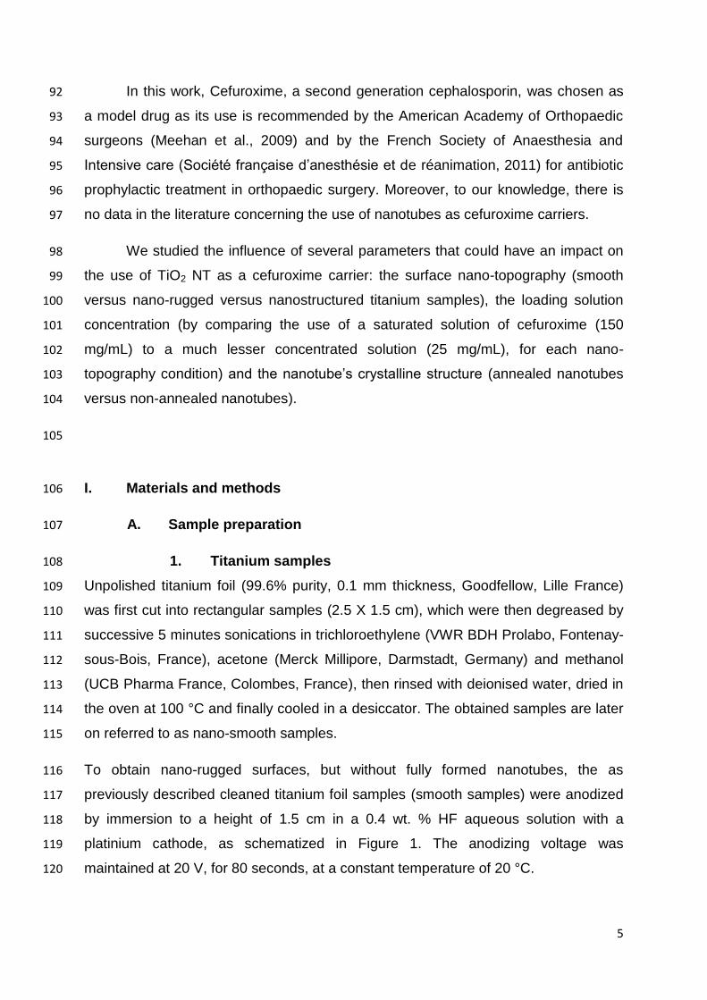
I. Materials and methods 106
A. Sample preparation 107
1. Titanium samples 108
Unpolished titanium foil (99.6% purity, 0.1 mm thickness, Goodfellow, Lille France) 109
was first cut into rectangular samples (2.5 X 1.5 cm), which were then degreased by 110
successive 5 minutes sonications in trichloroethylene (VWR BDH Prolabo, Fontenay-111
sous-Bois, France), acetone (Merck Millipore, Darmstadt, Germany) and methanol 112
(UCB Pharma France, Colombes, France), then rinsed with deionised water, dried in 113
the oven at 100 °C and finally cooled in a desiccator. The obtained samples are later 114
on referred to as nano-smooth samples. 115
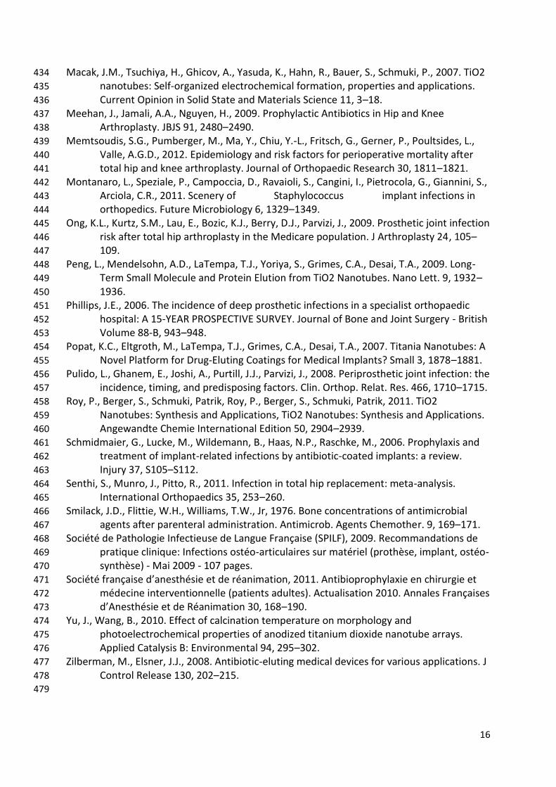
To obtain nano-rugged surfaces, but without fully formed nanotubes, the as 116
previously described cleaned titanium foil samples (smooth samples) were anodized 117
by immersion to a height of 1.5 cm in a 0.4 wt. % HF aqueous solution with a 118
platinium cathode, as schematized in Figure 1. The anodizing voltage was 119
maintained at 20 V, for 80 seconds, at a constant temperature of 20 °C. 120
6
To obtain TiO2 nanotubes (nano-tubular samples), the anodization was carried out in 121
the same conditions as described above but for 20 minutes. 122
All experiments were conducted on the obtained nano-smooth, nano-rugged or nano-123
tubular titanium foil. 124
To convert the amorphous nanotubes into the mixed crystalline phases of anatase 125
and rutile, some samples were annealed at 500°C for 2 hours under oxygen, with a 126
heating and cooling rate of 5 °C min-1. 127
128
2. Cefuroxime loading solutions 129
A 150 mg/mL cefuroxime solution was prepared by dissolving 1.5 g of cefuroxime 130
(Panpharma, Fougeres, France, batch No 104179) with sterile deionised water 131
(VERSYLENE®, Fresenius Kabi, Louviers, France) to a total volume of 10 mL. 132
133
3. Loading method and storage 134
To load the samples with cefuroxime, an adapted soaking technique was used (Ayon 135
et al., 2006; Kim et al., 2008). Each sample was immersed to a height of 1.5 cm, in 136
either a 150 mg/ml or a 25 mg/mL solution of cefuroxime, for 30 minutes, in ambient 137
daylight, at room temperature (22-26°C). At the end of the loading time, the samples 138
were removed from the immersion solution and were immediately air blown to 139
remove excess solution on the surface and to dry them. 140
The samples were stored prior to loading and after loading, until cefuroxime 141
quantification, in a climate chamber (BINDER GmbH, Tuttlingen, Germany), in the 142
dark, at 25°C. 143
144
B. Sample characterizations 145
1. Structural characterizations 146
The crystalline structure and phase of the TiO2 nanotube layers of a smooth, a non-147
annealed and an annealed nano-tubular sample were determined by X-ray diffraction 148

7
(XRD) using a Scintag XRD X’TRA diffractometer with CuKα (λ = 1.54° radiation). 149
The CuKβ radiation was filtered through a nickel filter. The diffraction pattern was 150
achieved between 20 and 80◦ with a step angle of 0.05° and a scanning speed of 151
0.01° per second. 152
For the study of the surface nano-topography, structural characterization of a smooth, 153
a nano-rugged and a nano-tubular sample was performed before and after drug 154
loading with two different concentration cefuroxime solutions using a field emission 155
scanning electron microscope (SEM). It was performed using a Supra 55 VP SEM 156
(Carl Zeiss SMT, Nanterre, France) with secondary emission and in lens detector. 157
The accelerating voltage and the working distance were respectively 3 kV and either 158
5 or 6 mm (image dependent). 159
Images were acquired at different scan sizes from the top surface. 160
For the four different tested conditions of titanium foil nano-topography and crystalline 161
structure (smooth, nano-rugged, non-annealed nano-tubular and annealed nano-162
tubular), surface wettability was investigated using a drop shape analysis system 163
(EasyDrop, Kruss, Hamburg, Germany). The contact angle was measured with a 164
deionized water sensile droplet of 3 μL in ambient conditions. The measurement was 165
taken 5 seconds after the deposition of the water droplet on the substrate. After 166
measurement, the samples were cleaned, dried in nitrogen and stored in a 167
desiccator. 168
169
2. Cefuroxime quantification 170
Each sample was placed into a known volume of sterile deionised water (5 or 10 mL 171
depending on the estimated loaded quantity of cefuroxime), for 90 minutes. At 172
determined times (1; 2; 5; 10; 15; 30; 60 and 90 minutes) a determined volume (1000 173
µL) of release solution was collected and was replaced with the same volume of 174
fresh sterile deionised water. 175
The cefuroxime present in the collected sample was then quantified by HPLC 176
composed of a PU-2080 Plus pump, and an AS-2055 Plus auto-sampler coupled with 177
an UV/VIS spectrophotometer (UV-2075 Plus detector), from Jasco France 178
(Bouguenais, France) 179
8
The HPLC separation column used was a 5 µm Lichrospher 100 RP 18 endcapped 180
column (125 × 4.6 mm ID) (Macherey-Nagel EURL, Hoerd, France) 181
The HPLC mobile phase was composed of 85/15 phosphate buffer/acetonitrile (v/v) 182
mixture. The phosphate buffer used was a 0.1 mol/L solution of H2KPO4 (VWR 183
International Pessac, France). The flow rate through the column for the analysis was 184
set at 1 mL/min, with the column thermo regulated to a temperature of 35°C. The 185
injection volume was of 20 µL. The detection wavelength was set up at 273 nm. 186
Cefuroxime presents a retention time of 3.8 minutes. This chromatographic method is 187
linear for concentrations ranging from 0.25 µg/mL to 20 µg/mL. The mean linear 188
regression equation obtained is y = 35022x + 3014.7 (r2 = 0.9998), where x is the 189
cefuroxime concentration and y the surface area of the corresponding peak. This 190
method has acceptable accuracy and precision as the intra-assay and inter-assay 191
coefficients of variation are below 5%. The limit of quantification of this method is of 192
0.25 mg/mL. 193
Cefuroxime release results were expressed in µg of cefuroxime over the loaded 194
surface of anodized TiO2 (µg/cm2). 195
196
C. Tested parameters 197
In this work, several parameters that could have an influence on the use of 198
TiO2 NT as cefuroxime reservoirs were investigated. 199
1. Influence of the surface nano-topography and loading 200
solution concentration 201
10 anodized (nano-tubular) non annealed Ti samples, 10 nano-rugged Ti samples, 202
and 10 smooth Ti samples were prepared. 5 samples of each surface nano-203
topography (15 samples in total) were loaded by immersion into a 150 mg/mL 204
cefuroxime solution, as previously described, whilst the other 5 of each surface nano-205
topography were loaded with a 25 mg/mL cefuroxime solution. 206
2. Influence of the nanotube’s crystalline structure 207
To see if annealing (heat treating) the nanotubes could impact their use as 208
cefuroxime carriers as it modifies the nanotubes crystalline structure, annealed 209
nanotubes were compared to non-annealed nanotubes. 210
9
7 non-annealed anodized samples and 7 annealed anodized samples were loaded 211
by immersion in a 150 mg/mL cefuroxime solution, as previously described. 212
213
D. Statistical considerations 214
Statistical analysis was performed using a non-parametric Man-Whitney test. The 215
difference was considered significant for a p-value ≤ 0.05. 216
217
II. Results 218
A. Structural characterization and surface wettability 219
Figure 1 shows the current density time curve for Ti anodization obtained in our 220
operating conditions, and illustrates the nanotube growth. SEM pictures of obtained 221
nano-rugged and nano-tubular samples before loading are presented in Figure 2. 222
Anodization occurred on both sides of the anodized samples. 223
224
Nano-smooth non anodized samples typically are not microscopically smooth, but do 225
not present any nano-scale ruggedness. Nano-rugged samples present nano-scale 226
modifications with the formation of what seems to be shallow nanopores, whilst 227
retaining their micro-scale topography. For the 20 minutes anodized samples, the 228
formed nanotubes have dimensions of between 300 to 400 nm high and 70 to 90 nm 229
in diameter. 230
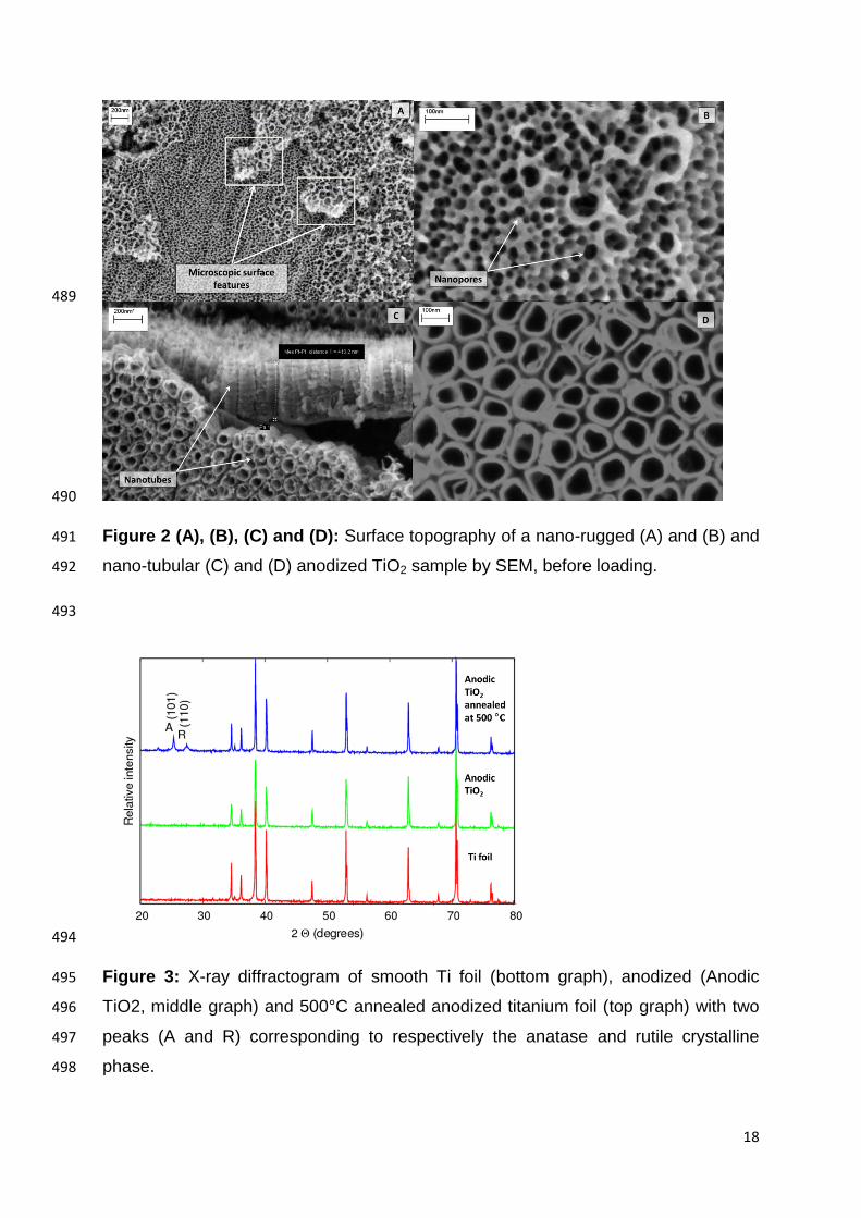
The XRD results are depicted in Figure 3. The peaks characteristic of anatase and 231
rutile crystalline structure only appear after annealing at 500°C, and are not present 232
in Ti foil or as-anodized Ti . 233
234
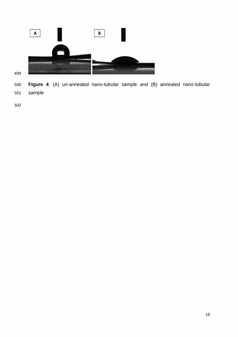
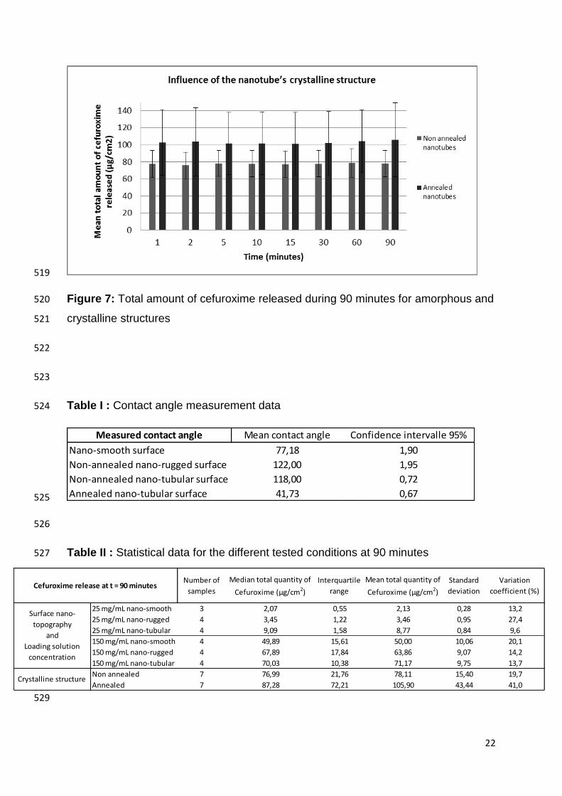
Concerning the contact angle measurements (data summarised in Table I), 235
anodization times of 80 seconds and 1200 seconds greatly increase the contact 236
angle compared to smooth un-anodized titanium; however the contact angle 237

10
measured for an annealed nanotubular surface was lower than un-annealed 238
anodised surface (Figure 4), and even lower than for a nano-smooth surface. 239
240
241
B. Cefuroxime quantification 242
SEM pictures of loaded samples are shown in Figure 5, with light grey/white 243
colouring showing the titanium sample, and dark grey patches being cefuroxime 244
deposits. In Figure 5(A), a relatively large deposit of cefuroxime can clearly be seen, 245
with Figure 5(B) being a closer view of the phenomenon. Figure 5(C) and Figure 5(D) 246
show how cefuroxime infiltrates and sometimes covers the nano-rugged surface 247
nanopores, and in Figure 5(E) and Figure 5(F) the same can be said of the nano-248
tubular surface. 249
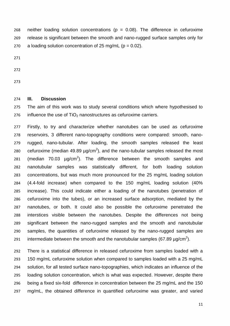
250
251
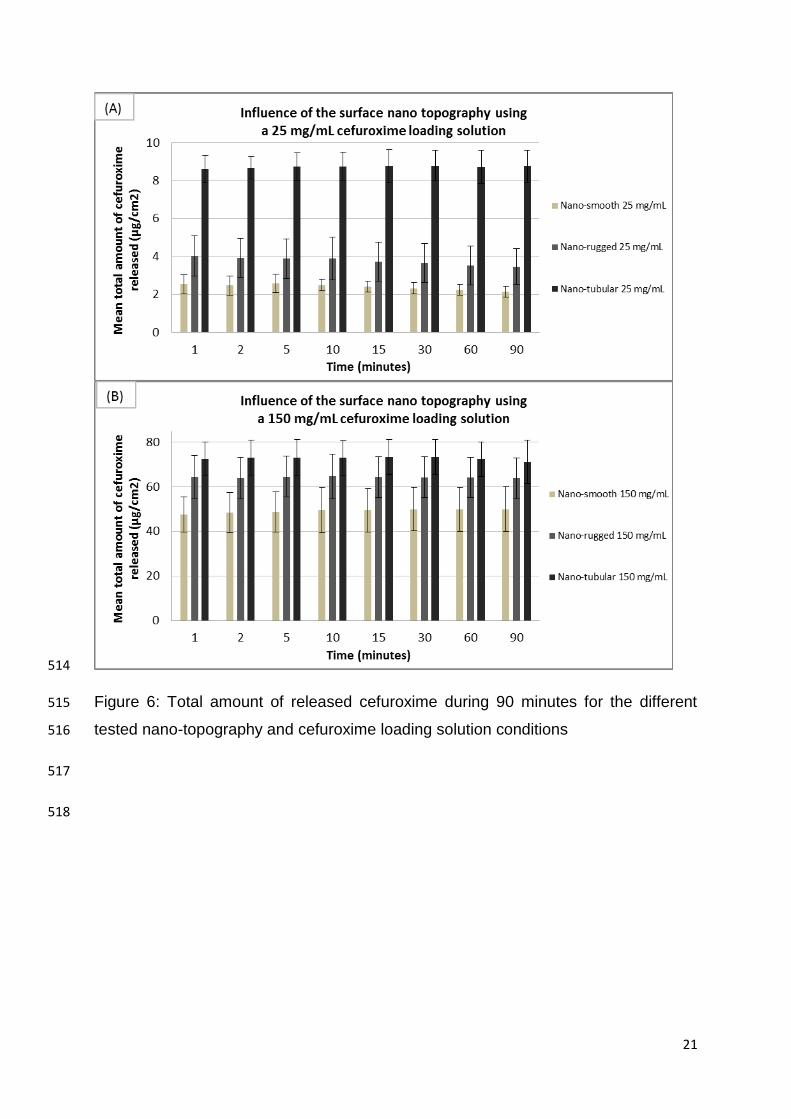
Figure 6 (A) and (B) shows the total amount of cefuroxime released over a 90 252
minutes period from the different tested nano-surfaces with a 25 mg/mL and 150 253
mg/mL cefuroxime loading solution, and Figure 7 shows the total amount of 254
cefuroxime released over a 90 minutes period for annealed and un-annealed nano-255
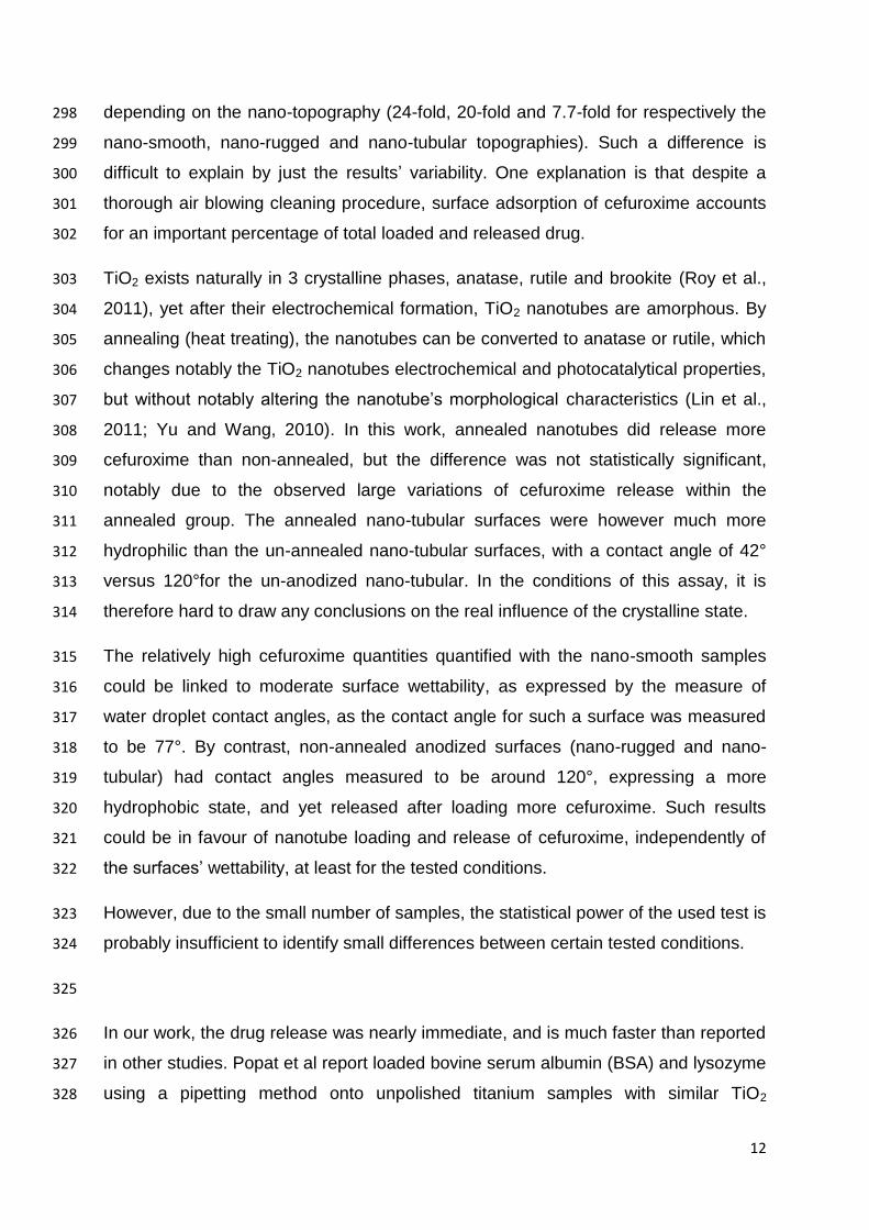
tubular surfaces. Table II summarise the statistical data of the different tested 256
conditions at 90 minutes. 257
Maximum cefuroxime release for the studied conditions was obtained within the first 258
one or two minutes, and no additional cefuroxime release was detected for the rest of 259
the release study period. 260
For the nanotube length tested, there is no statistical influence of the nanotube’s 261
crystalline structure on the amount of cefuroxime quantified (p = 0.28). Cefuroxime 262
loading solution concentration does have a significant impact on cefuroxime 263
quantities released (p = 0.001) as does the surface nano-topography, but only 264
between the smooth samples and the nanotube samples for both tested loading 265
solution concentrations (p = 0.04). The difference between the nano-rugged surface 266
samples and the nanotubular surface samples is not statistically significant, for 267
11
neither loading solution concentrations (p = 0.08). The difference in cefuroxime 268
release is significant between the smooth and nano-rugged surface samples only for 269
a loading solution concentration of 25 mg/mL (p = 0.02). 270
271
272
273
III. Discussion 274
The aim of this work was to study several conditions which where hypothesised to 275
influence the use of TiO2 nanostructures as cefuroxime carriers. 276
Firstly, to try and characterize whether nanotubes can be used as cefuroxime 277
reservoirs, 3 different nano-topography conditions were compared: smooth, nano-278
rugged, nano-tubular. After loading, the smooth samples released the least 279
cefuroxime (median 49.89 µg/cm2), and the nano-tubular samples released the most 280
(median 70.03 µg/cm2). The difference between the smooth samples and 281
nanotubular samples was statistically different, for both loading solution 282
concentrations, but was much more pronounced for the 25 mg/mL loading solution 283
(4.4-fold increase) when compared to the 150 mg/mL loading solution (40% 284
increase). This could indicate either a loading of the nanotubes (penetration of 285
cefuroxime into the tubes), or an increased surface adsorption, mediated by the 286
nanotubes, or both. It could also be possible the cefuroxime penetrated the 287
interstices visible between the nanotubes. Despite the differences not being 288
significant between the nano-rugged samples and the smooth and nanotubular 289
samples, the quantities of cefuroxime released by the nano-rugged samples are 290
intermediate between the smooth and the nanotubular samples (67.89 µg/cm2). 291
There is a statistical difference in released cefuroxime from samples loaded with a 292
150 mg/mL cefuroxime solution when compared to samples loaded with a 25 mg/mL 293
solution, for all tested surface nano-topographies, which indicates an influence of the 294
loading solution concentration, which is what was expected. However, despite there 295
being a fixed six-fold difference in concentration between the 25 mg/mL and the 150 296
mg/mL, the obtained difference in quantified cefuroxime was greater, and varied 297
12
depending on the nano-topography (24-fold, 20-fold and 7.7-fold for respectively the 298
nano-smooth, nano-rugged and nano-tubular topographies). Such a difference is 299
difficult to explain by just the results’ variability. One explanation is that despite a 300
thorough air blowing cleaning procedure, surface adsorption of cefuroxime accounts 301
for an important percentage of total loaded and released drug. 302
TiO2 exists naturally in 3 crystalline phases, anatase, rutile and brookite (Roy et al., 303
2011), yet after their electrochemical formation, TiO2 nanotubes are amorphous. By 304
annealing (heat treating), the nanotubes can be converted to anatase or rutile, which 305
changes notably the TiO2 nanotubes electrochemical and photocatalytical properties, 306
but without notably altering the nanotube’s morphological characteristics (Lin et al., 307
2011; Yu and Wang, 2010). In this work, annealed nanotubes did release more 308
cefuroxime than non-annealed, but the difference was not statistically significant, 309
notably due to the observed large variations of cefuroxime release within the 310
annealed group. The annealed nano-tubular surfaces were however much more 311
hydrophilic than the un-annealed nano-tubular surfaces, with a contact angle of 42° 312
versus 120°for the un-anodized nano-tubular. In the conditions of this assay, it is 313
therefore hard to draw any conclusions on the real influence of the crystalline state. 314
The relatively high cefuroxime quantities quantified with the nano-smooth samples 315
could be linked to moderate surface wettability, as expressed by the measure of 316
water droplet contact angles, as the contact angle for such a surface was measured 317
to be 77°. By contrast, non-annealed anodized surfaces (nano-rugged and nano-318
tubular) had contact angles measured to be around 120°, expressing a more 319
hydrophobic state, and yet released after loading more cefuroxime. Such results 320
could be in favour of nanotube loading and release of cefuroxime, independently of 321
the surfaces’ wettability, at least for the tested conditions. 322
However, due to the small number of samples, the statistical power of the used test is 323
probably insufficient to identify small differences between certain tested conditions. 324
325
In our work, the drug release was nearly immediate, and is much faster than reported 326
in other studies. Popat et al report loaded bovine serum albumin (BSA) and lysozyme 327
using a pipetting method onto unpolished titanium samples with similar TiO2 328

13
nanotubes and obtained maximum release times that varied between 25 and 110 329
minutes (Popat et al., 2007). Using a similar method, Aninwene et al loaded 330
penicillin/streptomycin or dexamethasone and obtained drug elution for 3 days 331
(Aninwene et al., 2008). The very fast release obtained here could be explained by 332
high Cefuroxime water solubility (150 mg/mL), or by a surface layer of Cefuroxime, or 333
possibly by both. Despite our cleaning method, SEM images showed significant 334
Cefuroxime deposit on the surface of the TiO2 layer, on samples with different nano-335
topography, possibly linked to the surface micro-topography. However, since nano-336
tubular samples released significantly more Cefuroxime than smooth samples, it 337
seems quite plausible that Cefuroxime penetrated at least partially into the 338
nanotubes. 339
Longer release times have been reported. Peng et al, also using unpolished TiO2 340
nanotube surfaces and a consistent cleaning technique, found that elution kinetics of 341
paclitaxel and BSA were influences by nanotube height and pore diameter (Peng et 342
al., 2009), with nanotubes of 5 µm height and 100 nm of diameter releasing the most 343
drug for up to 3 weeks. 344
Therefore, in this study, the thickness of the nanotube layer could impact the 345
maximum loading capacity of the tubes, as the longer the tube, the more volume it 346
could contain. It could be possible that 400 nm of length is not enough to allow a 347
significant amount of drug into the nanotubes, with regard to the potential surface 348
adsorption. Therefore, any conclusions about the impact of the nanotubes’ crystalline 349
structure might be premature, as the 400 nm high nanotubes could have been loaded 350
at maximum capacity regardless of the surface wettability. 351
In this work, the titanium foil used for the experiments was unpolished, and wasn’t 352
microscopically smooth. This could have an impact on cefuroxime adsorption, as 353
SEM structural characterization of samples with different nano-topographies, loaded 354
with cefuroxime, showed an inhomogeneous cefuroxime spread. The microscopic 355
surface features could lead to the formation of “beds” of cefuroxime, protected from 356
the surface cleaning procedure. It has also already been hypothesized that in 357
previous studies at least a significant amount of drug stayed on the surface and did 358
penetrate into the nanotubes (Peng et al., 2009). These microscopic features could 359
also account for the high variability between samples of the same series. Also, 360
14
surface adsorption could account for the instant burst-like release that was observed. 361
Therefore, it would seem that in order to more accurately measure the exact quantity 362
of drug actually loaded and then released by the nanotubes, microscopically smooth 363
samples are needed, as well as an adequate and validated surface cleaning method. 364
365
Several improvements could be made to this study. To achieve a microscopically 366
smooth surface, a polishing method (electro-chemical polishing or physical polishing) 367
could have been used. The effects of micro-scale ruggedness would therefore be 368
reduced. Also, higher nanotubes could offer improved loading volume and drug 369
storage capacity, and could also improve loading and elution kinetics. 370
371
The work presented here seems to indicate that some parameters, like nanotube 372
height and the loading solution concentration, have more influence on cefuroxime 373
release from TiO2 nanotubes, whereas the crystalline structure of the nanotubes 374
didn’t influence the amount of cefuroxime released. The nano-tubular samples 375
released more cefuroxime than nano-smooth or nano-rugged samples, for both 150 376
mg/mL and 25 mg/mL cefuroxime loading concentrations. However, cefuroxime 377
release kinetics were too fast for lasting local drug delivery, and need to be extended. 378
Longer nanotubes could increase the amount of cefuroxime loaded and release 379
times, but might also increase overall fragility, and thus need to be tested. Also, the 380
antibacterial efficacy of such a delivery method using cefuroxime still needs to be 381
investigated. 382
383
IV. Acknowledgements 384
The authors thank F. Feschet and B. Pereira for their help with the statistical analysis 385
of the data, and C. Massard and V. Raspal for their insights. 386
387
15
V. Bibliography 388
389
AlBuhairan, B., Hind, D., Hutchinson, A., 2008. Antibiotic prophylaxis for wound infections in 390
total joint arthroplasty: A SYSTEMATIC REVIEW. J Bone Joint Surg Br 90-B, 915–919. 391
Aninwene, G.E., Yao, C., Webster, T.J., 2008. Enhanced osteoblast adhesion to drug-coated 392
anodized nanotubular titanium surfaces. Int J Nanomedicine 3, 257–264. 393
Ayon, A.A., Cantu, M., Chava, K., Agrawal, C.M., Feldman, M.D., Johnson, D., Patel, D., 394
Marton, D., Shi, E., 2006. Drug loading of nanoporous TiO2 films. Biomed Mater 1, 395
L11–15. 396
Campoccia, D., Montanaro, L., Arciola, C.R., 2006. The significance of infection related to 397
orthopedic devices and issues of antibiotic resistance. Biomaterials 27, 2331–2339. 398
Cuckler, J.M., 2005. The Infected Total Knee: Management Options. The Journal of 399
Arthroplasty 20, Supplement 2, 33–36. 400
Dale, H., Skråmm, I., Løwer, H.L., Eriksen, H.M., Espehaug, B., Furnes, O., Skjeldestad, F.E., 401
Havelin, L.I., Engesaeter, L.B., 2011. Infection after primary hip arthroplasty: a 402
comparison of 3 Norwegian health registers. Acta Orthop 82, 646–654. 403
Ghicov, Andrei, Schmuki, Patrik, 2009. Self-ordering electrochemistry: a review on growth 404
and functionality of TiO2 nanotubes and other self-aligned MOx structures. Chem. 405
Commun. 2791–2808. 406
Gillespie, W.J., Walenkamp, G.H., 1996. Antibiotic prophylaxis for surgery for proximal 407
femoral and other closed long bone fractures, in: Cochrane Database of Systematic 408
Reviews. John Wiley & Sons, Ltd. 409
Gulati, K., Aw, M.S., Losic, D., 2011. Drug-eluting Ti wires with titania nanotube arrays for 410
bone fixation and reduced bone infection. Nanoscale Research Letters 6, 571. 411
Iorio, R., Robb, W.J., Healy, W.L., Berry, D.J., Hozack, W.J., Kyle, R.F., Lewallen, D.G., 412
Trousdale, R.T., Jiranek, W.A., Stamos, V.P., Parsley, B.S., 2008. Orthopaedic Surgeon 413
Workforce and Volume Assessment for Total Hip and Knee Replacement in the 414
United States: Preparing for an Epidemic. J Bone Joint Surg Am 90, 1598–1605. 415
Jämsen, E., Varonen, M., Huhtala, H., Lehto, M.U.K., Lumio, J., Konttinen, Y.T., Moilanen, T., 416
2010. Incidence of prosthetic joint infections after primary knee arthroplasty. J 417
Arthroplasty 25, 87–92. 418
Kang, H.-J., Kim, D.J., Park, S.-J., Yoo, J.-B., Ryu, Y.S., 2007. Controlled drug release using 419
nanoporous anodic aluminum oxide on stent. Thin Solid Films 515, 5184–5187. 420
Kim, D., Macak, Jan M, Schimidt-Stein, F., Schmuki, Patrik, 2008. Capillary effects, wetting 421
behavior and photo-induced tube filling of TiO2 nanotube layers. Nanotechnology 19, 422
305710. 423
Kuong, E.E., Ng, F.Y., Yan, C.H., Fang, C.X.S., Chiu, P.K.Y., 2009. Antibiotic prophylaxis after 424
total joint replacements. Hong Kong Med J 15, 458–462. 425
Kurtz, S., Ong, K., Lau, E., Mowat, F., Halpern, M., 2007. Projections of primary and revision 426
hip and knee arthroplasty in the United States from 2005 to 2030. J Bone Joint Surg 427
Am 89, 780–785. 428
Lin, J.Y., Chou, Y.T., Shen, J.L., Yang, M.D., Wu, C.H., Chi, G.C., Chou, W.C., Ko, C.H., 2011. 429
Effects of rapid thermal annealing on the structural properties of TiO2 nanotubes. 430
Applied Surface Science 258, 530–534. 431
Losic, D., Simovic, S., 2009. Self-ordered nanopore and nanotube platforms for drug delivery 432
applications. Expert Opinion on Drug Delivery 6, 1363–1381. 433
16
Macak, J.M., Tsuchiya, H., Ghicov, A., Yasuda, K., Hahn, R., Bauer, S., Schmuki, P., 2007. TiO2 434
nanotubes: Self-organized electrochemical formation, properties and applications. 435
Current Opinion in Solid State and Materials Science 11, 3–18. 436
Meehan, J., Jamali, A.A., Nguyen, H., 2009. Prophylactic Antibiotics in Hip and Knee 437
Arthroplasty. JBJS 91, 2480–2490. 438
Memtsoudis, S.G., Pumberger, M., Ma, Y., Chiu, Y.-L., Fritsch, G., Gerner, P., Poultsides, L., 439
Valle, A.G.D., 2012. Epidemiology and risk factors for perioperative mortality after 440
total hip and knee arthroplasty. Journal of Orthopaedic Research 30, 1811–1821. 441
Montanaro, L., Speziale, P., Campoccia, D., Ravaioli, S., Cangini, I., Pietrocola, G., Giannini, S., 442
Arciola, C.R., 2011. Scenery of Staphylococcus implant infections in 443
orthopedics. Future Microbiology 6, 1329–1349. 444
Ong, K.L., Kurtz, S.M., Lau, E., Bozic, K.J., Berry, D.J., Parvizi, J., 2009. Prosthetic joint infection 445
risk after total hip arthroplasty in the Medicare population. J Arthroplasty 24, 105–446
109. 447
Peng, L., Mendelsohn, A.D., LaTempa, T.J., Yoriya, S., Grimes, C.A., Desai, T.A., 2009. Long-448
Term Small Molecule and Protein Elution from TiO2 Nanotubes. Nano Lett. 9, 1932–449
1936. 450
Phillips, J.E., 2006. The incidence of deep prosthetic infections in a specialist orthopaedic 451
hospital: A 15-YEAR PROSPECTIVE SURVEY. Journal of Bone and Joint Surgery - British 452
Volume 88-B, 943–948. 453
Popat, K.C., Eltgroth, M., LaTempa, T.J., Grimes, C.A., Desai, T.A., 2007. Titania Nanotubes: A 454
Novel Platform for Drug‐Eluting Coatings for Medical Implants? Small 3, 1878–1881. 455
Pulido, L., Ghanem, E., Joshi, A., Purtill, J.J., Parvizi, J., 2008. Periprosthetic joint infection: the 456
incidence, timing, and predisposing factors. Clin. Orthop. Relat. Res. 466, 1710–1715. 457
Roy, P., Berger, S., Schmuki, Patrik, Roy, P., Berger, S., Schmuki, Patrik, 2011. TiO2 458
Nanotubes: Synthesis and Applications, TiO2 Nanotubes: Synthesis and Applications. 459
Angewandte Chemie International Edition 50, 2904–2939. 460
Schmidmaier, G., Lucke, M., Wildemann, B., Haas, N.P., Raschke, M., 2006. Prophylaxis and 461
treatment of implant-related infections by antibiotic-coated implants: a review. 462
Injury 37, S105–S112. 463
Senthi, S., Munro, J., Pitto, R., 2011. Infection in total hip replacement: meta-analysis. 464
International Orthopaedics 35, 253–260. 465
Smilack, J.D., Flittie, W.H., Williams, T.W., Jr, 1976. Bone concentrations of antimicrobial 466
agents after parenteral administration. Antimicrob. Agents Chemother. 9, 169–171. 467
Société de Pathologie Infectieuse de Langue Française (SPILF), 2009. Recommandations de 468
pratique clinique: Infections ostéo-articulaires sur matériel (prothèse, implant, ostéo-469
synthèse) - Mai 2009 - 107 pages. 470
Société française d’anesthésie et de réanimation, 2011. Antibioprophylaxie en chirurgie et 471
médecine interventionnelle (patients adultes). Actualisation 2010. Annales Françaises 472
d’Anesthésie et de Réanimation 30, 168–190. 473
Yu, J., Wang, B., 2010. Effect of calcination temperature on morphology and 474
photoelectrochemical properties of anodized titanium dioxide nanotube arrays. 475
Applied Catalysis B: Environmental 94, 295–302. 476
Zilberman, M., Elsner, J.J., 2008. Antibiotic-eluting medical devices for various applications. J 477
Control Release 130, 202–215. 478
479
17
480
481
482
Figure 1: Current density time curve with experimental setup. Nanotubes appear to 483 be fully formed as of 20 minutes (1200 seconds) of anodizing time (d). The nano-484
rugged surface was obtained with an anodizing time of 80 seconds, which 485
corresponds to the low point of current density curve, between stage (a) and (b). 486
487
488
18
489
490
Figure 2 (A), (B), (C) and (D): Surface topography of a nano-rugged (A) and (B) and 491
nano-tubular (C) and (D) anodized TiO2 sample by SEM, before loading. 492
493
494
Figure 3: X-ray diffractogram of smooth Ti foil (bottom graph), anodized (Anodic 495
TiO2, middle graph) and 500°C annealed anodized titanium foil (top graph) with two 496
peaks (A and R) corresponding to respectively the anatase and rutile crystalline 497
phase. 498
19
499
Figure 4: (A) un-annealed nano-tubular sample and (B) annealed nano-tubular 500
sample 501
502

20
503
504
505
506
Figure 5: (A) and (B): smooth titanium sample after cefuroxime loading, with (B) 507
being a close up view of (A). (C) and (D): nano-rugged titanium sample with 508
cefuroxime deposit and infiltration in the surface nanopores, with (D) being a close up 509
view of (C). (E) and (F): nano-tubular titanium sample with cefuroxime deposit and 510
infiltration in the surface nanopores. 511
512
513
21
514
Figure 6: Total amount of released cefuroxime during 90 minutes for the different 515
tested nano-topography and cefuroxime loading solution conditions 516
517
518
22
519
Figure 7: Total amount of cefuroxime released during 90 minutes for amorphous and 520
crystalline structures 521
522
523
Table I : Contact angle measurement data 524
525
526
Table II : Statistical data for the different tested conditions at 90 minutes 527
528 529
Measured contact angle Confidence intervalle 95%
1,90
0,67
Mean contact angle
77,18
122,00
118,00
41,73Annealed nano-tubular surface
Non-annealed nano-rugged surface 1,95
0,72
Nano-smooth surface
Non-annealed nano-tubular surface
Number of
samples
Median total quantity of
Cefuroxime (µg/cm2)
Interquartile
range
Mean total quantity of
Cefuroxime (µg/cm2)
Standard
deviation
Variation
coefficient (%)
25 mg/mL nano-smooth 3 2,07 0,55 2,13 0,28 13,2
25 mg/mL nano-rugged 4 3,45 1,22 3,46 0,95 27,4
25 mg/mL nano-tubular 4 9,09 1,58 8,77 0,84 9,6
150 mg/mL nano-smooth 4 49,89 15,61 50,00 10,06 20,1
150 mg/mL nano-rugged 4 67,89 17,84 63,86 9,07 14,2
150 mg/mL nano-tubular 4 70,03 10,38 71,17 9,75 13,7
Non annealed 7 76,99 21,76 78,11 15,40 19,7
Annealed 7 87,28 72,21 105,90 43,44 41,0
Surface nano-
topography
and
Loading solution
concentration
Crystalline structure
Cefuroxime release at t = 90 minutes

23
530