immunoglobulins structure and function arpad lanyi bsc public health 5th week, 2015
TRANSCRIPT
IMMUNOGLOBULINSSTRUCTURE AND FUNCTION
Arpad Lanyi
BSc Public Health5th week, 2015
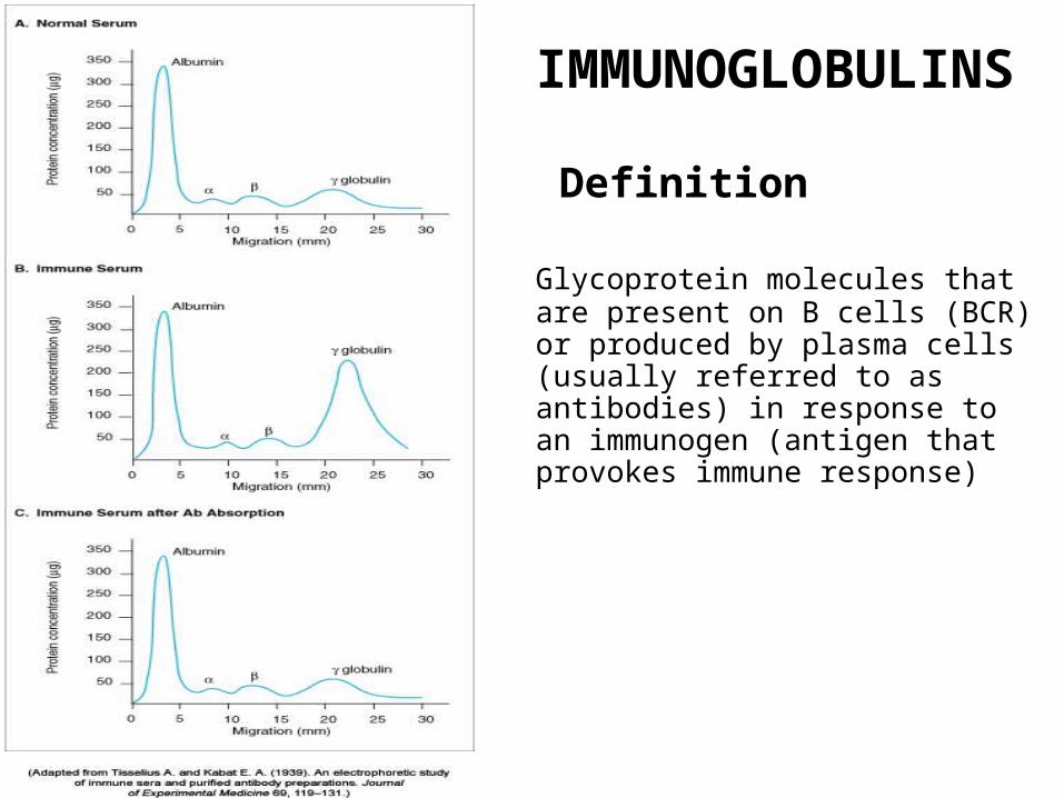
IMMUNOGLOBULINS
Definition
Glycoprotein molecules that are present on B cells (BCR) or produced by plasma cells (usually referred to as antibodies) in response to an immunogen (antigen that provokes immune response)
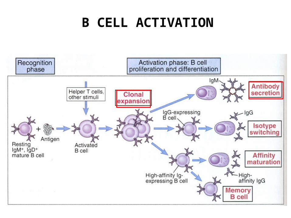
B CELL ACTIVATION
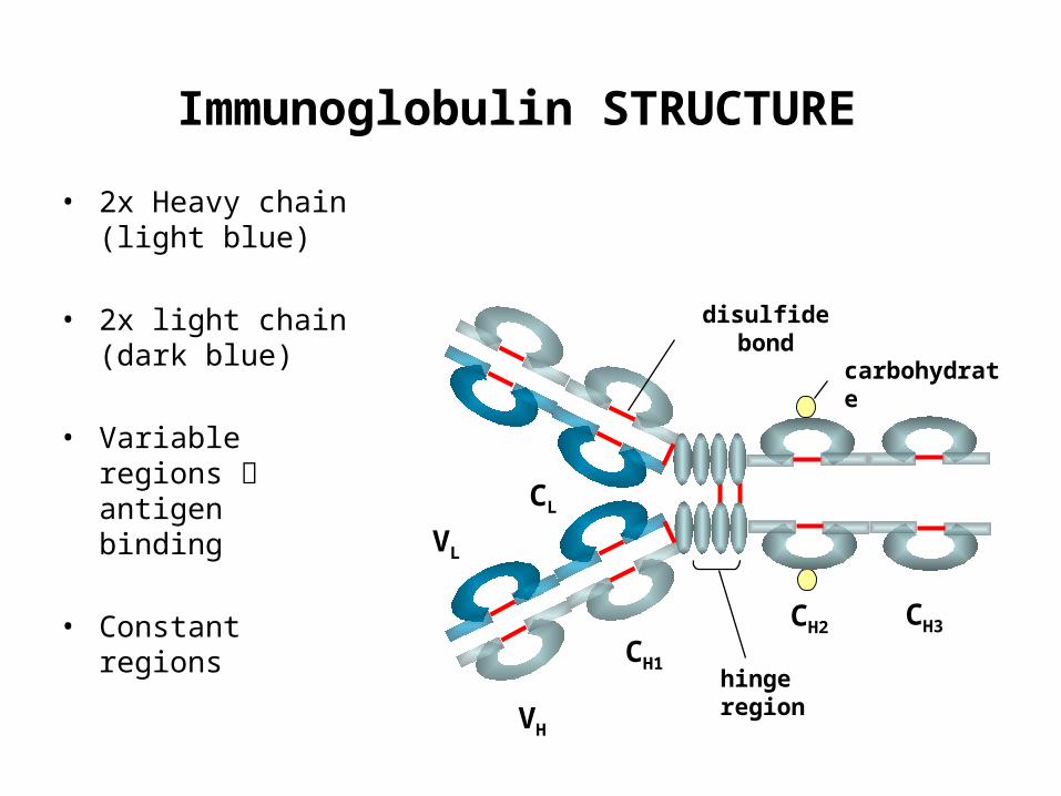
Immunoglobulin STRUCTURE
• 2x Heavy chain (light blue)
• 2x light chain (dark blue)
• Variable regions antigen binding
• Constant regions
hinge region
carbohydrate
disulfide bond
CH1
VL
CL
VH
CH2 CH3

FLEXIBILITY OF ANTIBODIES

AntibodyBCR (B cell receptor)
MEMBRANE BOUND!
Associated chains for signaling
Transmembrane domain
Cytoplasmic domain
Antigen recognition B cell activation
SOLUBLE (freely circulating)
Antigen binding effector functionsProduced by plasma cells
mIg sIg
s
s
s
s
s
s
s
s
s ss s
CH2
CH3s
s
s
s
s
s
s
s
ss
VL
VH
CL
CH1 ss
ss
s
s
s
s
ss
effektor funkciók
konstans domének
antigénkötés
variábilis domének
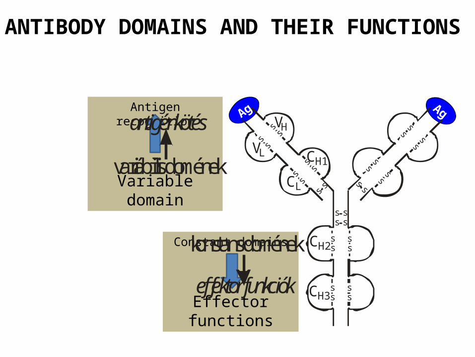
ANTIBODY DOMAINS AND THEIR FUNCTIONS
Ag
Constant domains
Effector functions
Antigen recognition
Variable domain
Ag

B cell
B CELL ACTIVATION
BCR oligomerization results in B cell activation, proliferation and differentiation

ANTIGEN BINDING
Antigen Binding Fragment (Fab)
Complement binding site
Placental transferConstant fragment (Fc)
Binding to Fc receptors on phagocytic cells
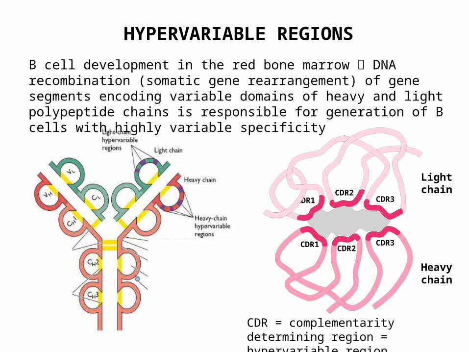
HYPERVARIABLE REGIONS
B cell development in the red bone marrow DNA recombination (somatic gene rearrangement) of gene segments encoding variable domains of heavy and light polypeptide chains is responsible for generation of B cells with highly variable specificity
Epitope
CDR1 CDR2CDR3
CDR1CDR2
CDR3
Light chain
Heavy chain
CDR = complementarity determining region = hypervariable region
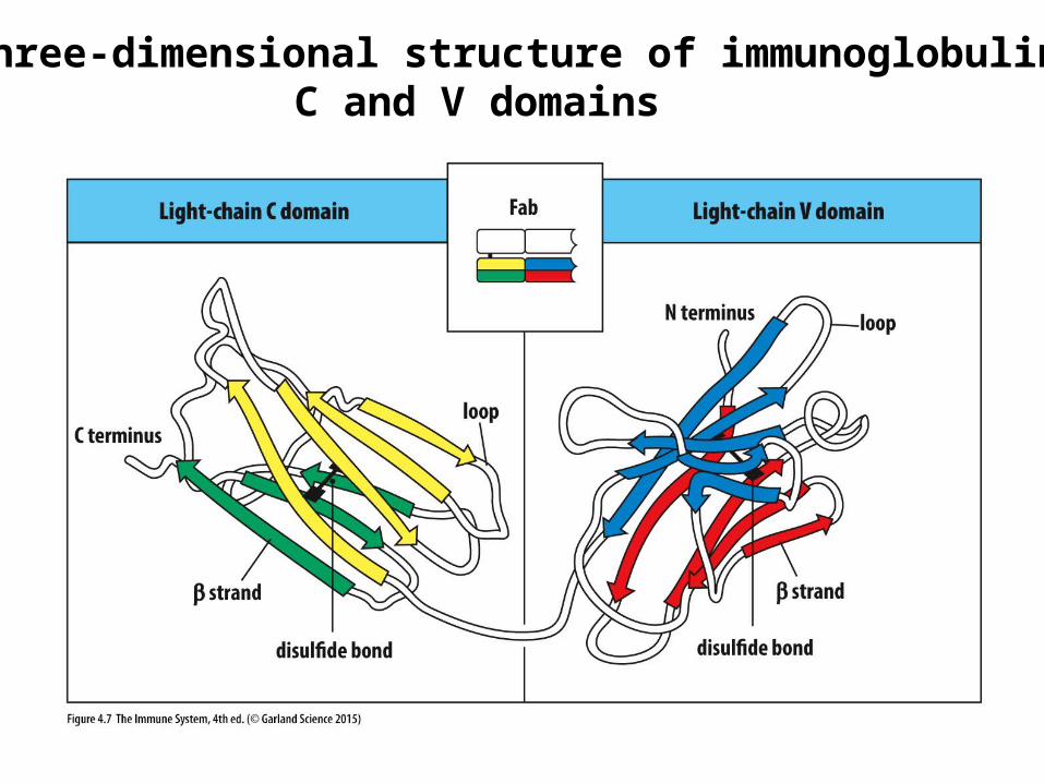
The three-dimensional structure of immunoglobulin C and V domains
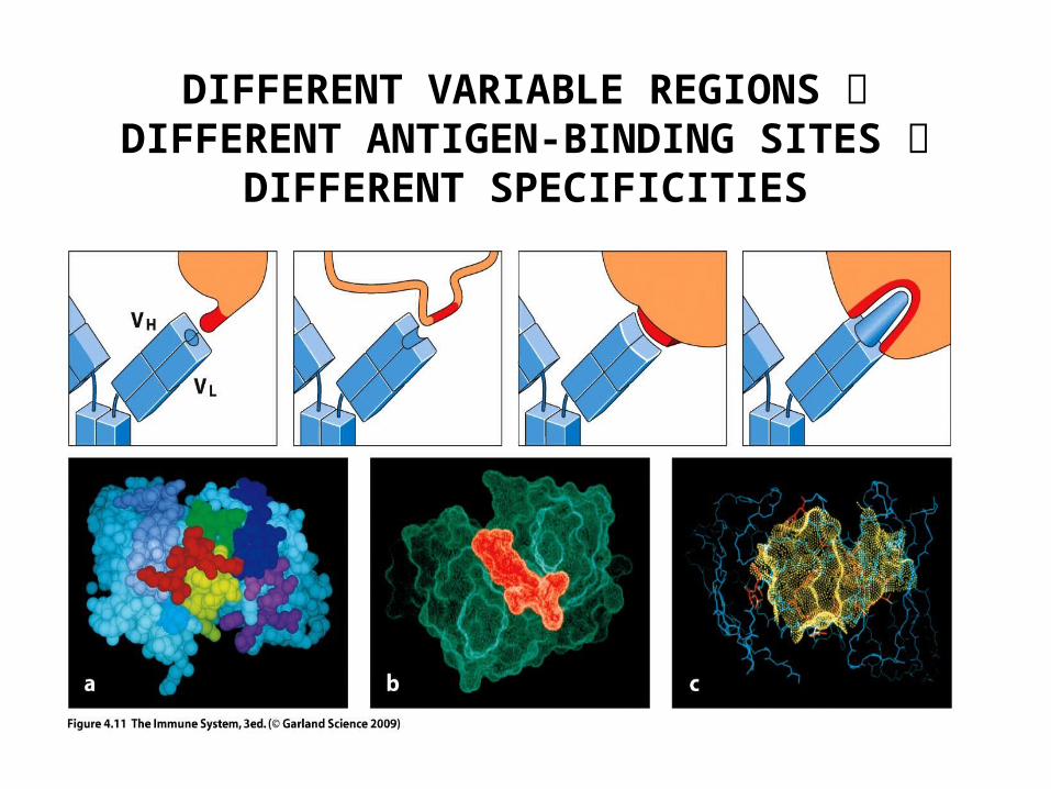
DIFFERENT VARIABLE REGIONS DIFFERENT ANTIGEN-BINDING SITES DIFFERENT
SPECIFICITIES
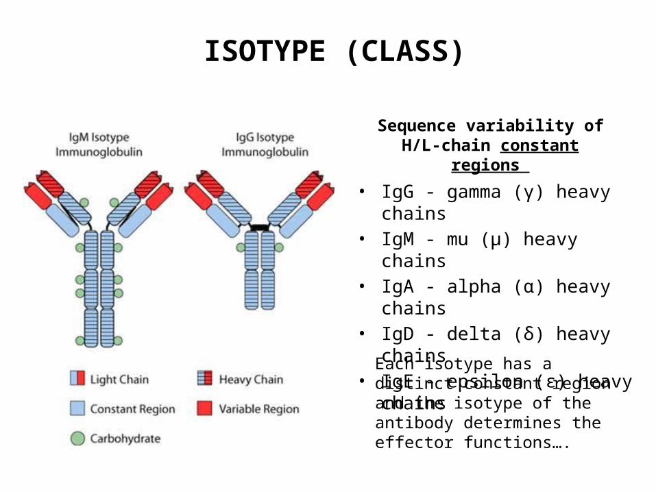
ISOTYPE (CLASS)
Sequence variability of H/L-chain constant regions
• IgG - gamma (γ) heavy chains• IgM - mu (μ) heavy chains• IgA - alpha (α) heavy chains• IgD - delta (δ) heavy chains• IgE - epsilon (ε) heavy chains
Each isotype has a distinct constant region and the isotype of the antibody determines the effector functions….

PHASES OF B CELL RESPONSE
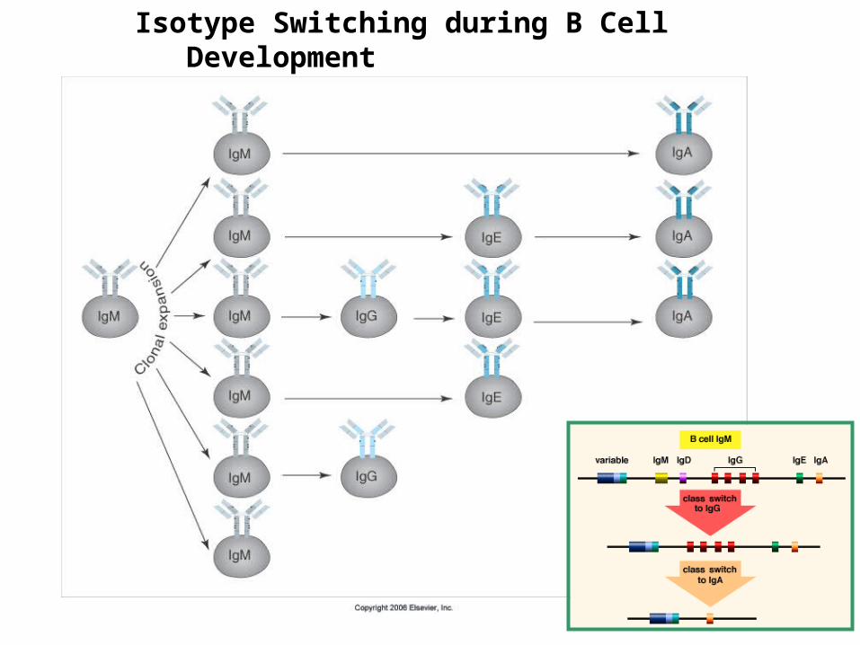
YIsotype Switching during B Cell Development PE SWITCHING
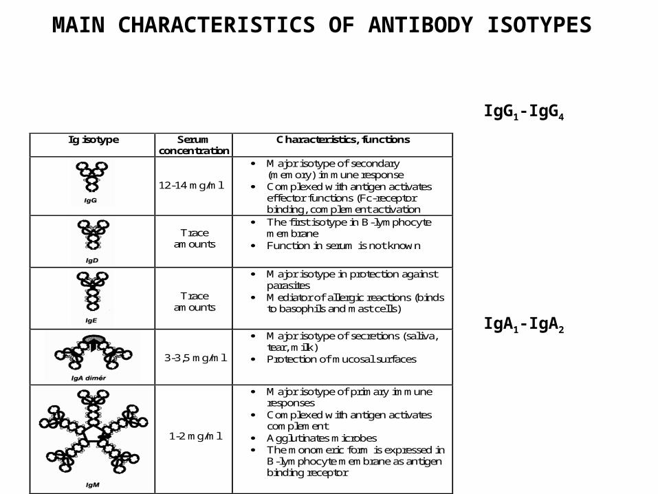
Ig isotype Serum concentration
Characteristics, functions
12-14 mg/ml
Major isotype of secondary (memory) immune response
Complexed with antigen activates effector functions (Fc-receptor binding, complement activation
Trace
amounts
The first isotype in B-lymphocyte membrane
Function in serum is not known
Trace amounts
Major isotype in protection against parasites
Mediator of allergic reactions (binds to basophils and mast cells)
3-3,5 mg/ml
Major isotype of secretions (saliva, tear, milk)
Protection of mucosal surfaces
1-2 mg/ml
Major isotype of primary immune responses
Complexed with antigen activates complement
Agglutinates microbes The monomeric form is expressed in
B-lymphocyte membrane as antigen binding receptor
MAIN CHARACTERISTICS OF ANTIBODY ISOTYPES
IgG1-IgG4
IgA1-IgA2
Ig . C onc entra tion
na p o k
p rim er response
„A” a ntig né
IgM
IgGIgAIgE
Szekund er ’la syec ond a ry response
„A” és a ntig én
„B”
5 10 15 20 25 30
IgM
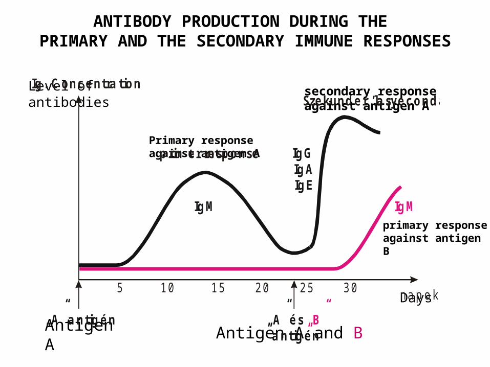
secondary response against antigen A
Primary response against antigen A
Level of antibodies
napok
primary response against antigen B
Antigen A
Days
Antigen A and B
ANTIBODY PRODUCTION DURING THE PRIMARY AND THE SECONDARY IMMUNE RESPONSES

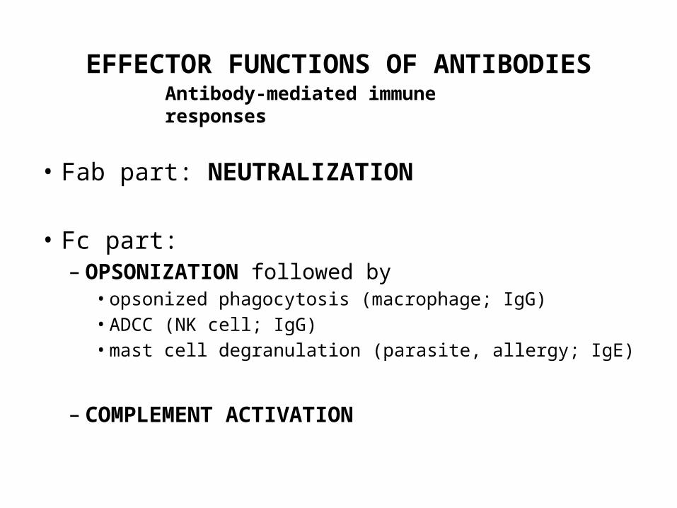
Antibody-mediated immune responses
EFFECTOR FUNCTIONS OF ANTIBODIES
• Fab part: NEUTRALIZATION
• Fc part:– OPSONIZATION followed by
• opsonized phagocytosis (macrophage; IgG)• ADCC (NK cell; IgG)• mast cell degranulation (parasite, allergy; IgE)
– COMPLEMENT ACTIVATION
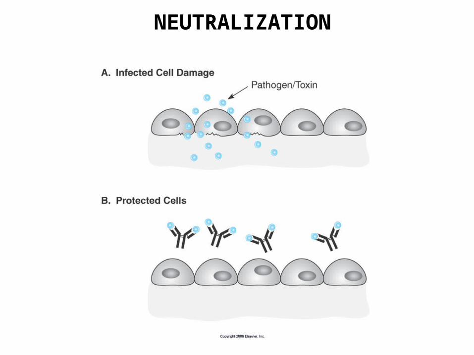
NEUTRALIZATION

Antigen binding
Complement binding site
Placental transfer
Binding to Fc receptors
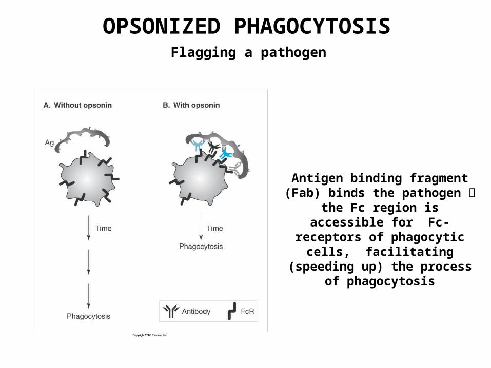
OPSONIZED PHAGOCYTOSISFlagging a pathogen
Antigen binding fragment (Fab) binds the pathogen the Fc region is accessible for Fc-
receptors of phagocytic cells, facilitating (speeding up) the
process of phagocytosis
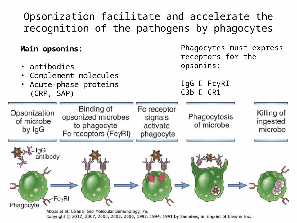
Main opsonins:
• antibodies• Complement molecules• Acute-phase proteins (CRP, SAP)
Opsonization facilitate and accelerate the recognition of the pathogens by phagocytes
Phagocytes must express receptors for the opsonins:
IgG FcγRIC3b CR1
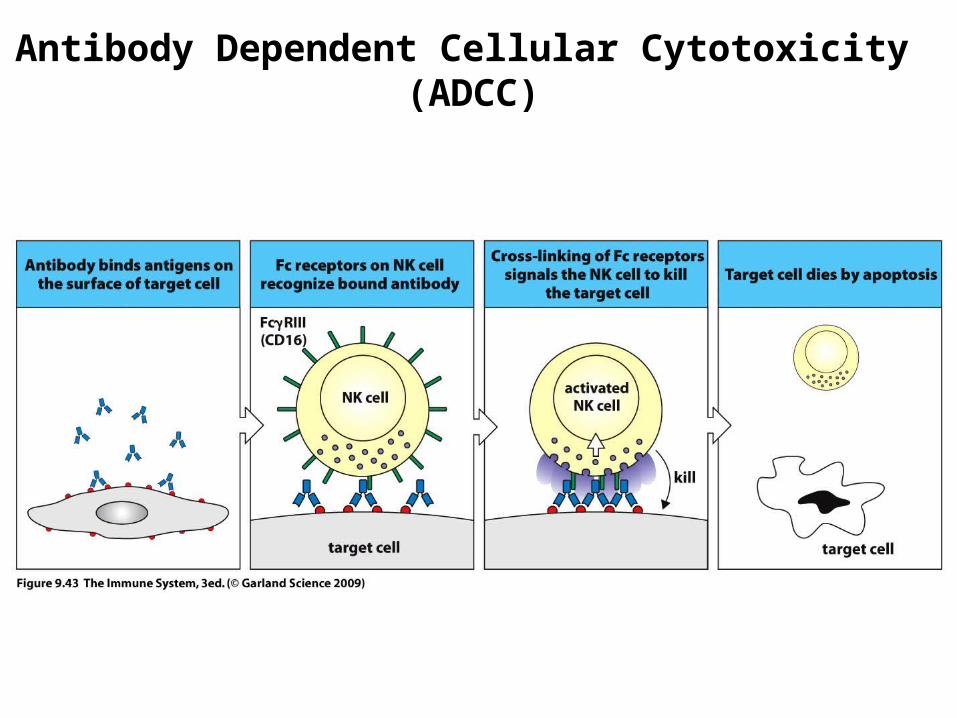
Antibody Dependent Cellular Cytotoxicity (ADCC)
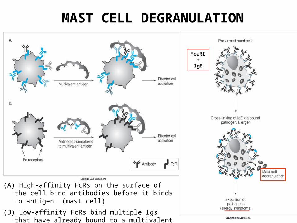
(A) High-affinity FcRs on the surface of the cell bind antibodies before it binds to antigen. (mast cell)
(B) Low-affinity FcRs bind multiple Igs that have already bound to a multivalent antigen. (macrophage, NK cell)
MAST CELL DEGRANULATION
FcεRI+
IgE
The complement system
• The complement system is a set of plasma proteins that act in a cascade to attack and kill extracellular pathogens.
• Approximately 30 components: – activating molecules– complement receptors– regulator factors– membrane proteins wich inhibit the lysis of host cells
• Most of the complement proteins and glycoproteins are produced in the liver in an inactive form (zymogen). Activation is induced by proteolitic cleavage.
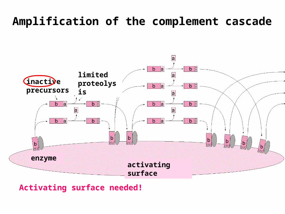
Amplification of the complement cascade
inactive precursors
limitedproteolysis
activating surfaceenzyme
Activating surface needed!
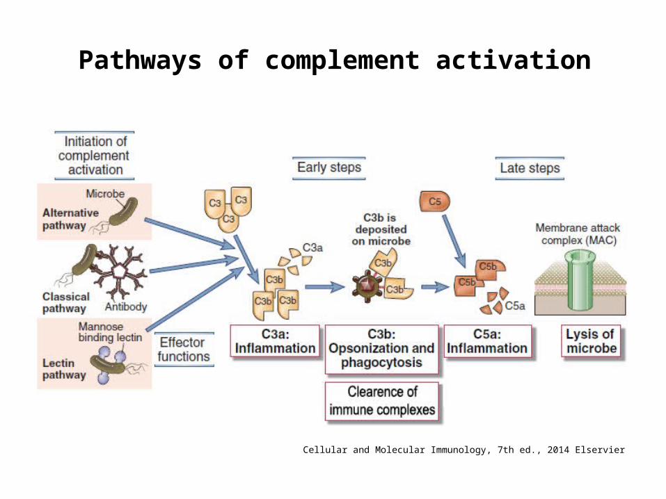
Pathways of complement activation
Cellular and Molecular Immunology, 7th ed., 2014 Elservier


Antigen binding
Complement binding site
Placental transferBinding to Fc
receptors
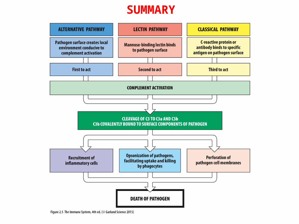
SUMMARY
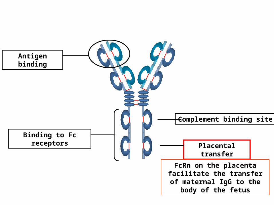
Antigen binding
Complement binding site
Placental transfer
Binding to Fc receptors
FcRn on the placenta facilitate the transfer of maternal IgG to
the body of the fetus
IgG
IgM
IgA
A F T E R B IR T H
breas t milkIgA
0
1 0 0 %( a d u l t )
3 3y e a r
2 546 a d u l t9 1m o n t h
maternal IgG
B E F O R E B IR T H
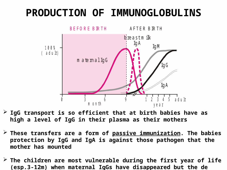
PRODUCTION OF IMMUNOGLOBULINS
IgG transport is so efficient that at birth babies have as high a level of IgG in their plasma as their mothers
These transfers are a form of passive immunization. The babies protection by IgG and IgA is against those pathogen that the mother has mounted
The children are most vulnerable during the first year of life (esp.3-12m) when maternal IgGs have disappeared but the de novo synthesis is at low level
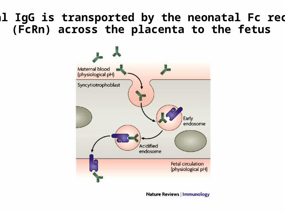
Maternal IgG is transported by the neonatal Fc receptor (FcRn) across the placenta to the fetus
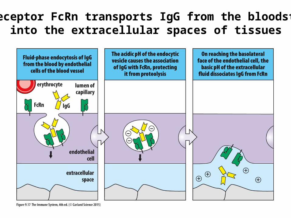
The receptor FcRn transports IgG from the bloodstream into the extracellular spaces of tissues
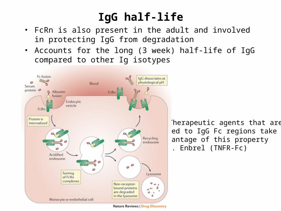
IgG half-life• FcRn is also present in the adult and involved in protecting IgG from
degradation• Accounts for the long (3 week) half-life of IgG compared to other Ig
isotypes
• Therapeutic agents that are fused to IgG Fc regions take advantage of this property e.g. Enbrel (TNFR-Fc)
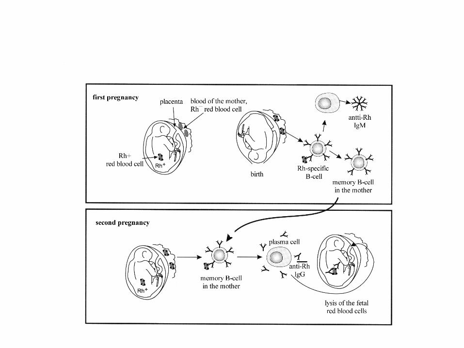
Pathological consequences of placental transport of IgG
(hemolytic disease of the newborn)
Passive anti-D IgG
anti-RhIgM
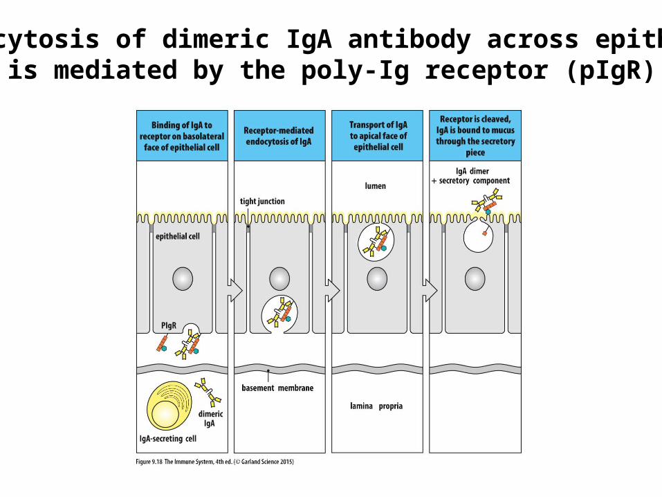
Transcytosis of dimeric IgA antibody across epithelia is mediated by the poly-Ig receptor (pIgR)