immune complex transfer enzyme immunoassay for antibody igm to hiv-1 p17 antigen
TRANSCRIPT
© 1998 Wiley-Liss, Inc.
Journal of Clinical Laboratory Analysis 12:329–336 (1998)
Immune Complex Transfer Enzyme Immunoassay for AntibodyIgM to HIV-1 p17 Antigen
Seiichi Hashida, 1 Setsuko Ishikawa, 1 Ichiro Nishikata, 1 Kazuya Hashinaka, 1
Shinichi Oka, 2 and Eiji Ishikawa 1*1Department of Biochemistry, Miyazaki Medical College, Kiyotake, Miyazaki, Japan
2AIDS Clinical Center, International Medical Center of Japan, Toyama, Shinjuku, Tokyo, Japan
The immune complex transfer enzyme im-munoassay for antibody IgM to HIV-1 p17antigen is described. Serum samples con-taining antibody IgM to HIV-1 p17 antigenwere incubated simultaneously with 2,4-dinitrophenyl-bovine serum albumin-re-combinant p17 (rp17) conjugate and rp17-β-D-galactosidase conjugate, and the immunecomplex formed comprising the three com-ponents was trapped onto colored polysty-rene beads coated with affinity-purified (anti-2,4-dinitrophenyl group) IgG. Subsequently,the immune complex was transferred to whitepolystyrene beads coated with monoclonalmouse (antihuman IgM) IgG in the presenceof excess of eN-2,4-dinitrophenyl-L-lysine. Thesignal for antibody IgM to p17 antigen wasthe fluorescence intensity by fluorometric as-say of β-D-galactosidase activity bound to thewhite polystyrene beads. The periods of timerequired for the formation, trapping, and trans-ferring of the immune complex comprising the
three components were more than 4 hr, 2 hr,and 3 hr, respectively. The immunoassay de-veloped was shown to be specific by inhibi-tion of transferring the immune complex in thepresence of excess of nonspecific IgM butnot IgG. Signals for antibody IgM to p17antigen with serum samples of HIV-1seroconversion serum panels,—that is, withserum samples in early stages of the infec-tion—tended to be higher than those withserum samples from HIV-1 asymptomatic car-riers probably long after the infection and pa-tients with ARC and AIDS. In contrast, sig-nals for antibody IgG to p17 antigen withserum samples of HIV-1 seroconversion se-rum panels tended to be higher than signalsfor antibody IgM to p17 antigen but were muchlower than signals for antibody IgG to p17antigen with serum samples from HIV-1 as-ymptomatic carriers and patients with ARCand AIDS. J. Clin. Lab. Anal. 12:329–336,1998. © 1998 Wiley-Liss, Inc.
Key words: antibody; human immunodeficiency virus type 1; p17 antigen; β-D-galactosidase
INTRODUCTION
Ultrasensitive enzyme immunoassays (immune complextransfer enzyme immunoassays) for antibody IgGs to p17,p24, and reverse transcriptase (RT) of HIV-1 have been de-veloped using recombinant p17, p24, and RT (rp17, rp24, andrRT) as antigens (1–7). The immune complexes, comprising2,4-dinitrophenyl-recombinant protein conjugates, antibodyIgGs to HIV-1 and recombinant protein-β-D-galactosidaseconjugates, were formed by 3-hr incubation and were trappedonto polystyrene beads coated with affinity-purified (anti-2,4-dinitrophenyl group) IgG by overnight incubation. After wash-ing, the immune complexes were eluted from the polystyrenebeads with eN-2,4-dinitrophenyl-L-lysine and were transferredto polystyrene beads coated with affinity-purified (antihumanIgG γ-chain) IgG by 3-hr incubation. Bound β-D-galactosi-dase activity was assayed by fluorometry for 2.5 hr. The vol-ume of serum samples used was 10 µL. These immunoassayshave made possible diagnosis of HIV-1 infection with urine
(1–4) and whole saliva samples (5,6) and have been shown tobe more useful as a confirmatory test with higher sensitivi-ties and specificities than Western blotting (7). Notably, anti-body IgG to p17 of HIV-1 could be detected as early as oreven earlier than antibodies to HIV-1 by conventional meth-ods (7,8). In addition, an ultrasensitive enzyme immunoas-say based on a similar principle for p24 antigen of HIV-1 hasalso been developed (9,10). On the basis of these results, thewindow period after HIV-1 infection (during which diagno-sis of HIV-1 infection is not possible due to the absence ofdetectable antibodies to HIV-1) has been shortened by simul-taneous detection of both p24 antigen and antibody IgGs top17 and RT (8). Recently, it has been made possible to per-
*Correspondence to: Eiji Ishikawa, M.D., Ph.D., Professor of Biochemistry,Department of Biochemistry, Miyazaki Medical College, Kiyotake, Miyazaki889-1692, Japan.
Received 7 April 1998; Accepted 13 April 1998
330 Hashida et al.
form more sensitive immune complex transfer enzyme im-munoassays for antibody IgG to p17 antigen within shorterperiods of time (15 to 60 min each for the formation, trap-ping, and transferring of the immune complex) by incuba-tions for the immunoreactions with shaking using a largervolume of serum samples (100 µL) and solid phases withlarger surface areas (11).
This report describes the immune complex transfer enzymeimmunoassay for antibody IgM to HIV-1, which has beenreported to be detected in early stages of HIV-1 infection(12,13), in comparison with that for antibody IgG to HIV-1.
MATERIALS AND METHODS
Buffers
The regularly used buffer was 10 mmol/L sodium phos-phate buffer, pH 7.0, containing 1.0 g/L bovine serum albu-min (fraction V, Intergen Company, Purchase, NY), 0.1 mol/L NaCl, 1.0 mmol/L MgCl2 and 1.0 g/L NaN3 (buffer A). Inother buffers, the concentration of bovine serum albumin was0.1 g/L (buffer B) and the concentration of NaCl was 0.4 mol/L (buffer C).
Antibodies
Rabbit (antihuman IgG γ-chain) IgG and rabbit (antihu-man IgM µ-chain) IgG were obtained from Medical and Bio-logical Laboratories Co., Ltd., Nagoya, Japan. Monoclonalmouse (antihuman IgM) IgG1 (Product No. 7408) was ob-tained from Oy Medix Biochemica Ab, Kauniainen, Finland.
Polystyrene Beads Coated With Affinity-Purified(Anti-2,4-Dinitrophenyl Group) IgG, Affinity-Purified (Antihuman IgG g-Chain) IgG andMonoclonal Mouse (Antihuman IgM) IgG 1
Colored polystyrene beads (3.2 mm in diameter, ImmunoChemical Inc., Okayama, Japan) were coated by physical ad-sorption with affinity-purified (anti-2,4-dinitrophenyl-bovineserum albumin) IgG (0.05 g/L) (14). White polystyrene beads(3.2 mm in diameter, Immuno Chemical Inc.) were coated byphysical adsorption with affinity-purified rabbit (antihumanIgG γ-chain) IgG (3 to 100 mg/L) and monoclonal mouse (an-tihuman IgM) IgG1 (3 to 30 mg/L), respectively (14).
Rabbit (Antihuman IgG g-Chain) Fab ¢- and Rabbit(Antihuman IgM m-Chain) Fab ¢-b-D-GalactosidaseConjugates
Rabbit (antihuman IgG γ-chain) F(ab′)2 and rabbit (antihu-man IgM µ-chain) F(ab′)2 were prepared by digestion of IgGwith pepsin, and Fab′ was prepared by reduction of F(ab′)2
with 2-mercaptoethylamine (15). Fab′ was conjugated to β-D-galactosidase (EC 3.2.1.23) from Escherichia coli usingN,N¢-o-phenylenedimaleimide (15).
Two-Site Enzyme Immunoassays for HumanIgG and IgM
In two-site enzyme immunoassay for IgG, a sample mixedwith buffer A to 150 µL was incubated with one polystyrenebead coated with affinity-purified rabbit (antihuman IgG γ-chain) IgG (100 µg/ml) for 1 hr. The polystyrene bead waswashed twice with 2 mL of buffer B and was incubated with150 µL of buffer A containing rabbit (antihuman IgG γ-chain)Fab′-β-D-galactosidase conjugate (100 fmol) and nonspecificrabbit F(ab′)2 (0.1 mg) for 1 hr. The incubations were per-formed at room temperature with shaking. The polystyrenebead was washed twice with 2 mL of buffer B, and bound β-D-galactosidase activity was assayed at 30°C for 1 hr by fluo-rometry using 4-methylumbelliferyl-β-D-galactoside assubstrate (16).
In two-site enzyme immunoassay for IgM, polystyrenebeads coated with monoclonal mouse (antihuman IgM) IgG1
(10 µg/ml) and rabbit (antihuman IgM µ-chain) Fab′-β-D-galactosidase conjugate were substituted for polystyrenebeads coated with affinity-purified rabbit (antihuman IgG γ-chain) IgG and rabbit (antihuman IgG γ-chain) Fab′-β-D-ga-lactosidase conjugate, respectively.
2,4-Dinitrophenyl-Bovine Serum Albumin-Recombinant HIV-1 p17 (rp17) Conjugate
Thiol groups introduced into rp17 molecules were reactedwith 6-maleimidohexanoyl-2,4-dinitrophenyl-bovine serumalbumin (1).
HIV-1 rp17-b-D-Galactosidase Conjugate
Thiol groups introduced into rp17 molecules were reactedwith maleimide-β-D-galactosidase (EC 3.2.1.23) (1).
Immune Complex Transfer EnzymeImmunoassays for Antibody IgG to HIV-1p17 Antigen
Antibody IgG to HIV-1 p17 antigen was measured essen-tially in the same way as described previously (7,17). An ali-quot (10 µL) of serum was mixed with 90 µL of buffer C andwas incubated for up to 20 hr with 50 µL of buffer C contain-ing inactive β-D-galactosidase (50 µg) (Mutein, BoehringerMannheim, Mannheim, Germany) and 100 fmol each of 2,4-dinitrophenyl-bovine serum albumin-rp17 conjugate andrp17-β-D-galactosidase conjugate. Subsequently, the reactionmixture was incubated with two colored polystyrene beadscoated with affinity-purified (anti-2,4-dinitrophenyl group)IgG for up to 2 hr. The colored polystyrene beads were washedand incubated for up to 4 hr with two white polystyrene beadscoated with affinity-purified (antihuman IgG γ-chain) IgG(100 µg/ml) in 150 µL of buffer A containing 1 mmol/L eN-2,4-dinitrophenyl-L-lysine. The incubations were performedat room temperature with shaking. The white polystyrene
Enzyme Immunoassay for Anti-HIV-1 IgM 331
beads were washed, and β-D-galactosidase activity bound wasassayed at 30°C for up to 20 hr by fluorometry using 4-methylumbelliferyl-β-D-galactoside as substrate (16). Thefluorescence intensity was measured with a spectro-fluorophotometer (F-3010, Hitachi, Ltd., Tokyo, Japan) us-ing 360 nm for excitation and 450 nm for emissionanalysis. The fluorescence intensity of 1 × 10⁻⁸ mol/L 4-methylumbelliferone in 0.1 mol/L glycine-NaOH buffer,pH 10.3 was adjusted to 100.
Immune Complex Transfer EnzymeImmunoassays for Antibody IgM to HIV-1p17 Antigen
White polystyrene beads coated with monoclonal mouse(antihuman IgM) IgG1 (10 µg/ml) were substituted for thosecoated with affinity-purified (antihuman IgG γ-chain) IgGin the immune complex transfer enzyme immunoassay forantibody IgG to HIV-1 p17 antigen described above.
Other Immunological Methods
The gelatin particle agglutination test for antibodies to HIV-1 was performed using a commercial kit with a lysate of HIV-1 as antigen (SERODIA-HIV, Fujirebio Inc., Tokyo, Japan).Western blotting for antibody IgG to HIV-1 was performedusing a commercial kit preblotted with nine proteins of HIV-1 (gp160, gp120, p66, p55, p51, gp41, p31, p24, and p17)(Ortho HIV Western Blot Kit, Ortho Diagnostic Systems Inc.,Raritan, NJ).
Seroconversion Serum Panels
One seroconversion serum panel (Panel E) was obtainedfrom Boston Biomedica, West Bridgewater, MA. Twoseroconversion serum panels (SV-0051 and SV-0241) wereobtained from North American Biologicals, Inc., Miami, FL.
Serum Samples Randomly Collected From HIV-1Seronegative and Seropositive Subjects
Serum samples were randomly collected from 50 HIV-1seronegative subjects (23 males aged 22–59 yr and 27 fe-males aged 27–73 yr) and 15 HIV-1 seropositive subjects (3male asymptomatic carriers (AC) aged 17–22 years, 2 femaleasymptomatic carriers aged 22 and 39 years, 4 male patientsaged 10–52 years with AIDS-related complex (ARC), onefemale patient aged 18 years with AIDS-related complex, 5male patients aged 14–59 years with AIDS) and were storedat –20°C until use. The seronegativity and seropositivity weretested by the gelatin particle agglutination test. The seroposi-tivity was confirmed by Western blotting.
RESULTS AND DISCUSSION
In the immune complex transfer enzyme immunoassaysfor antibody IgG and IgM to HIV-1 p17 antigen, serum
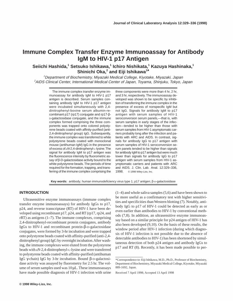
samples were incubated simultaneously with 2,4-dini-trophenyl-bovine serum albumin-recombinant p17 (rp17)conjugate and rp17-β-D-galactosidase conjugate, and the im-mune complexes formed comprising the three componentswere trapped onto colored polystyrene beads coated with af-finity-purified (anti-2,4-dinitrophenyl group) IgG. Subse-quently, the immune complexes were transferred to whitepolystyrene beads coated with affinity-purified (antihumanIgG γ-chain) IgG and monoclonal mouse (antihuman IgM)IgG1 in the presence of excess of eN-2,4-dinitrophenyl-L-lysine (Fig. 1). Incubations for the immunoreactions wereperformed at room temperature with shaking throughout.
In order to develop the immune complex transfer enzymeimmunoassay for antibody IgM to p17 antigen, which is assensitive as that for antibody IgG to p17 antigen, the follow-ing experiments were performed.
Amount of IgG and IgM Released From ColoredPolystyrene Beads Incubated With SerumSamples
As in the immune complex transfer enzyme immunoas-says for antibody IgG and IgM to p17 antigen, colored poly-styrene beads (two beads/assay) coated with affinity-purified(anti-2,4-dinitrophenyl group) IgG were incubated with 10and 100 µL of serum samples in a total volume of 150 µL for
Fig. 1. Immune complex transfer enzyme immunoassay for antibody IgG.DNP: 2,4-dinitrophenyl group. Ag: antigen. Enz: enzyme.
332 Hashida et al.
1 hr and, after washing, with 150 µL of eN-2,4-dinitrophenyl-L-lysine solution for 1 hr. During incubation with serumsamples, IgG and IgM were nonspecifically adsorbed ontothe colored polystyrene beads and were partially releasedduring incubation in the presence of εN-2,4-dinitrophenyl-L-lysine. The amounts of IgG and IgM released into the εN-2,4-dinitrophenyl-L-lysine solution were from 3.7 to 56 fmol/assay and from 0.2 to 11 fmol/assay, respectively, which weremeasured by two-site enzyme immunoassays for IgG and IgM(Table 1). The specificity of the two-site enzyme immunoas-says used was satisfactory for the above experiments. Namely,the detection limits of IgG and IgM by the two-site enzymeimmunoassay for IgG were 0.1 fmol/assay and 20 fmol/as-say, respectively, and those by the two-site enzyme immu-noassay for IgM were >104 fmol/assay and 0.01 fmol/assay,respectively.
Capacity of White Polystyrene Beads Coated WithAffinity-Purified (Antihuman IgG g-Chain) IgG andMonoclonal Mouse (Anti-Human IgM) IgG 1 to TrapIgG and IgM
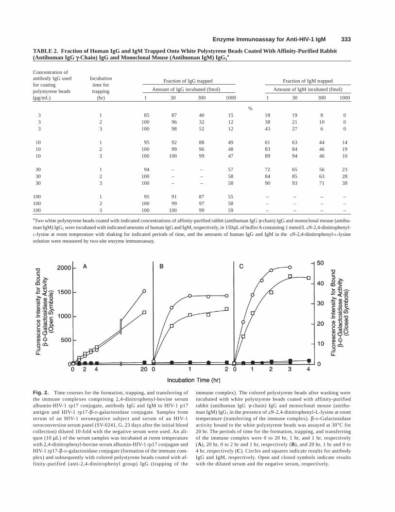
In order to test the capacity of white polystyrene beadscoated with affinity-purified (antihuman IgG γ-chain) IgG andmonoclonal mouse (antihuman IgM) IgG1 to trap IgG andIgM, various amounts (1–1000 fmol) of nonspecific humanIgG and IgM in the εN-2,4-dinitrophenyl-L-lysine solutionwere incubated with two white polystyrene beads coated withincreasing concentrations (from 3 to 100 µg/mL) of the anti-immunoglobulin IgGs for 1, 2, and 3 hr (Table 2). The frac-tions of IgG and IgM trapped were larger when coated with10 to 100 µg/mL than when coated with 3 µg/mL, and thefractions of IgG trapped were larger than those of IgM trapped.The fractions of IgG trapped differed only slightly when thewhite polystyrene beads coated with 10 to 100 µg/mL wereincubated with 1 to 300 fmol of IgG and significantly de-
creased when incubated with 1,000 fmol. The fractions ofIgM trapped differed only slightly when the white polysty-rene beads coated with 10 to 100 µg/mL were incubated with1 to 30 fmol of IgM and significantly decreased when incu-bated with 300 fmol or more.
From these results, transferring of the immune complexescomprising the three components to the white polystyrenebeads was indicated not to be intolerably inhibited by theamounts of nonspecific IgG (3.7–56 fmol) and IgM (0.2–11fmol) released from the colored polystyrene beads incubatedwith 10 to 100 µL of serum samples described above, and theamounts of nonspecific human IgG and IgM to be added fortest of the specificity of the immune complex transfer en-zyme immunoassays for antibody IgG and IgM to p17 anti-gen described below were indicated to be 1,000 fmol (0.15µg) or more of IgG and 300 fmol (0.27 µg) or more of IgM.
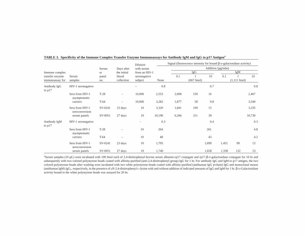
Time Courses of the Immunoreactions Involvedin the Immune Complex Transfer EnzymeImmunoassay
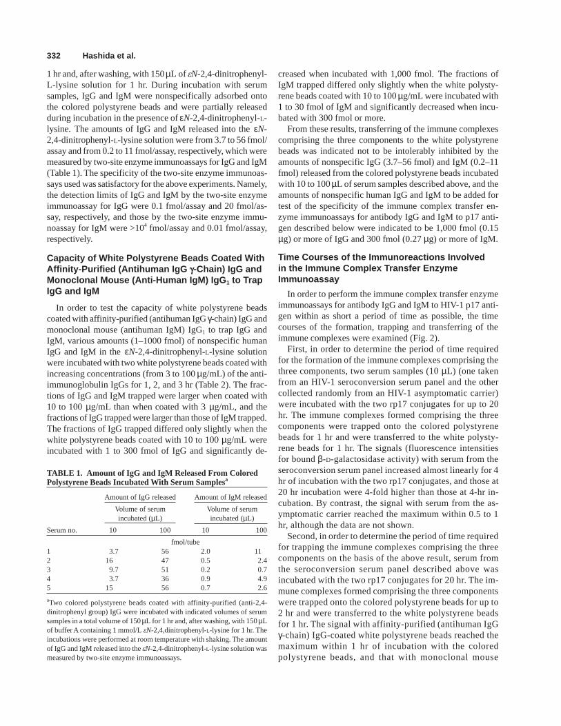
In order to perform the immune complex transfer enzymeimmunoassays for antibody IgG and IgM to HIV-1 p17 anti-gen within as short a period of time as possible, the timecourses of the formation, trapping and transferring of theimmune complexes were examined (Fig. 2).
First, in order to determine the period of time requiredfor the formation of the immune complexes comprising thethree components, two serum samples (10 µL) (one takenfrom an HIV-1 seroconversion serum panel and the othercollected randomly from an HIV-1 asymptomatic carrier)were incubated with the two rp17 conjugates for up to 20hr. The immune complexes formed comprising the threecomponents were trapped onto the colored polystyrenebeads for 1 hr and were transferred to the white polysty-rene beads for 1 hr. The signals (fluorescence intensitiesfor bound β-D-galactosidase activity) with serum from theseroconversion serum panel increased almost linearly for 4hr of incubation with the two rp17 conjugates, and those at20 hr incubation were 4-fold higher than those at 4-hr in-cubation. By contrast, the signal with serum from the as-ymptomatic carrier reached the maximum within 0.5 to 1hr, although the data are not shown.
Second, in order to determine the period of time requiredfor trapping the immune complexes comprising the threecomponents on the basis of the above result, serum fromthe seroconversion serum panel described above wasincubated with the two rp17 conjugates for 20 hr. The im-mune complexes formed comprising the three componentswere trapped onto the colored polystyrene beads for up to2 hr and were transferred to the white polystyrene beadsfor 1 hr. The signal with affinity-purified (antihuman IgGγ-chain) IgG-coated white polystyrene beads reached themaximum within 1 hr of incubation with the coloredpolystyrene beads, and that with monoclonal mouse
TABLE 1. Amount of IgG and IgM Released From ColoredPolystyrene Beads Incubated With Serum Samplesa
Amount of IgG released Amount of IgM released
Volume of serum Volume of serumincubated (µL) incubated (µL)
Serum no. 10 100 10 100
fmol/tube1 3.7 56 2.0 112 16 47 0.5 2.43 9.7 51 0.2 0.74 3.7 36 0.9 4.95 15 56 0.7 2.6
aTwo colored polystyrene beads coated with affinity-purified (anti-2,4-dinitrophenyl group) IgG were incubated with indicated volumes of serumsamples in a total volume of 150 µL for 1 hr and, after washing, with 150 µLof buffer A containing 1 mmol/L eN-2,4,dinitrophenyl-L-lysine for 1 hr. Theincubations were performed at room temperature with shaking. The amountof IgG and IgM released into the eN-2,4-dinitrophenyl-L-lysine solution wasmeasured by two-site enzyme immunoassays.
Enzyme Immunoassay for Anti-HIV-1 IgM 333
TABLE 2. Fraction of Human IgG and IgM Trapped Onto White Polystyrene Beads Coated With Affinity-Purified Rabbit(Antihuman IgG g-Chain) IgG and Monoclonal Mouse (Antihuman IgM) IgG1
a
Concentration ofantibody IgG used Incubationfor coating time for
Fraction of IgG trapped Fraction of IgM trapped
polystyrene beads trapping Amount of IgG incubated (fmol) Amount of IgM incubated (fmol)
(µg/mL) (hr) 1 30 300 1000 1 30 300 1000
%3 1 85 87 40 15 18 19 8 03 2 100 96 32 12 38 21 10 03 3 100 98 52 12 43 27 6 0
10 1 95 92 88 49 61 63 44 1410 2 100 99 96 48 83 84 46 1910 3 100 100 99 47 89 94 46 10
30 1 94 – – 57 72 65 56 2330 2 100 – – 58 84 85 63 2830 3 100 – – 58 90 93 71 39
100 1 95 91 87 55 – – – –100 2 100 99 97 58 – – – –100 3 100 100 99 59 – – – –
aTwo white polystyrene beads coated with indicated concentrations of affinity-purified rabbit (antihuman IgG γ-chain) IgG and monoclonal mouse (antihu-man IgM) IgG1 were incubated with indicated amounts of human IgG and IgM, respectively, in 150 µL of buffer A containing 1 mmol/L eN-2,4-dinitrophenyl-L-lysine at room temperature with shaking for indicated periods of time, and the amounts of human IgG and IgM in the eN-2,4-dinitrophenyl-L-lysinesolution were measured by two-site enzyme immunoassay.
Fig. 2. Time courses for the formation, trapping, and transferring ofthe immune complexes comprising 2,4-dinitrophenyl-bovine serumalbumin-HIV-1 rp17 conjugate, antibody IgG and IgM to HIV-1 p17antigen and HIV-1 rp17-β-D-galactosidase conjugate. Samples fromserum of an HIV-1 seronegative subject and serum of an HIV-1seroconversion serum panel (SV-0241, G, 23 days after the initial bloodcollection) diluted 10-fold with the negative serum were used. An ali-quot (10 µL) of the serum samples was incubated at room temperaturewith 2,4-dinitrophenyl-bovine serum albumin-HIV-1 rp17 conjugate andHIV-1 rp17-β-D-galactosidase conjugate (formation of the immune com-plex) and subsequently with colored polystyrene beads coated with af-finity-purified (anti-2,4-dinitrophenyl group) IgG (trapping of the
immune complex). The colored polystyrene beads after washing wereincubated with white polystyrene beads coated with affinity-purifiedrabbit (antihuman IgG γ-chain) IgG and monoclonal mouse (antihu-man IgM) IgG1 in the presence of eN-2,4-dinitrophenyl-L-lysine at roomtemperature (transferring of the immune complex). β-D-Galactosidaseactivity bound to the white polystyrene beads was assayed at 30°C for20 hr. The periods of time for the formation, trapping, and transferringof the immune complex were 0 to 20 hr, 1 hr, and 1 hr, respectively(A), 20 hr, 0 to 2 hr and 1 hr, respectively (B), and 20 hr, 1 hr and 0 to4 hr, respectively (C). Circles and squares indicate results for antibodyIgG and IgM, respectively. Open and closed symbols indicate resultswith the diluted serum and the negative serum, respectively.
334H
ashida et al.
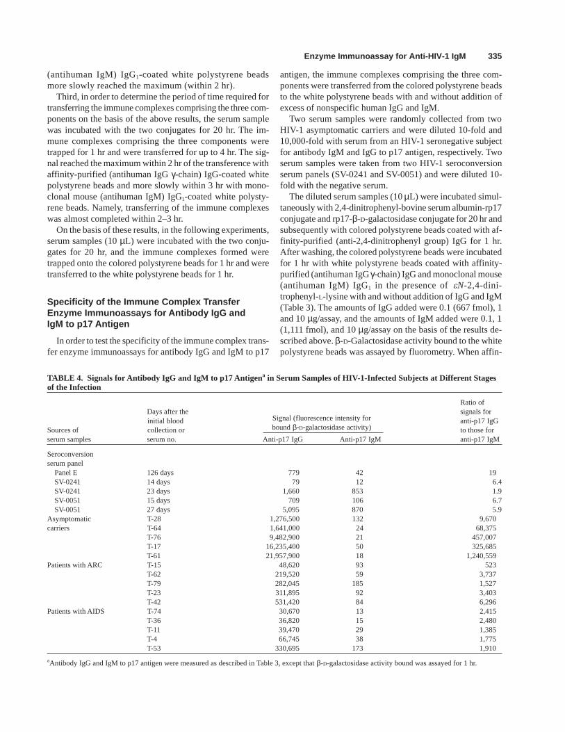
TABLE 3. Specificity of the Immune Complex Transfer Enzyme Immunoassays for Antibody IgM and IgG to p17 Antigena
Dilution Signal (fluorescence intensity for bound β-D-galactosidase activity)
Serum Days after with serum Addition (µg/tube)Immune complex or the initial from an HIV-1 IgG IgMtransfer enzyme Serum panel blood seronegative 0.1 1 10 0.1 1 10immunoassay for samples no. collection subject None (667 fmol) (1,111 fmol)
Antibody IgG HIV-1 seronegative – 0.8 0.7 0.8to p17
Sera from HIV-1 T-28 – 10,000 2,553 2,008 159 16 2,467asymptomaticcarriers T-64 – 10,000 3,282 1,877 58 9.8 3,540
Sera from HIV-1 SV-0241 23 days 10 3,320 1,841 109 15 3,335seroconversionserum panels SV-0051 27 days 10 10,190 6,266 311 39 10,730
Antibody IgM HIV-1 seronegative – 0.3 0.4 0.3to p17
Sera from HIV-1 T-28 – 10 264 261 4.8asymptomaticcarriers T-64 – 10 48 43 4.2
Sera from HIV-1 SV-0241 23 days 10 1,705 1,699 1,451 99 13seroconversionserum panels SV-0051 27 days 10 1,740 1,658 1,338 132 23
aSerum samples (10 µL) were incubated with 100 fmol each of 2,4-dinitrophenyl-bovine serum albumin-rp17 conjugate and rp17-β-D-galactosidase conjugate for 16 hr andsubsequently with two colored polystyrene beads coated wtih affinity-purified (anti-2,4-dinitrophenyl group) IgG for 1 hr. For antibody IgG and IgM to p17 antigen, the twocolored polystyrene beads after washing were incubated with two white polystyrene beads coated with affinity-purified (antihuman IgG γ-chain) IgG and monoclonal mouse(antihuman IgM) IgG1, respectively, in the presence of eN-2,4-dinitrophenyl-L-lysine with and without addition of indicated amounts of IgG and IgM for 1 hr. β-D-Galactosidaseactivity bound to the white polystyrene beads was assayed for 20 hr.
Enzyme Immunoassay for Anti-HIV-1 IgM 335
(antihuman IgM) IgG1-coated white polystyrene beadsmore slowly reached the maximum (within 2 hr).
Third, in order to determine the period of time required fortransferring the immune complexes comprising the three com-ponents on the basis of the above results, the serum samplewas incubated with the two conjugates for 20 hr. The im-mune complexes comprising the three components weretrapped for 1 hr and were transferred for up to 4 hr. The sig-nal reached the maximum within 2 hr of the transference withaffinity-purified (antihuman IgG γ-chain) IgG-coated whitepolystyrene beads and more slowly within 3 hr with mono-clonal mouse (antihuman IgM) IgG1-coated white polysty-rene beads. Namely, transferring of the immune complexeswas almost completed within 2–3 hr.
On the basis of these results, in the following experiments,serum samples (10 µL) were incubated with the two conju-gates for 20 hr, and the immune complexes formed weretrapped onto the colored polystyrene beads for 1 hr and weretransferred to the white polystyrene beads for 1 hr.
Specificity of the Immune Complex TransferEnzyme Immunoassays for Antibody IgG andIgM to p17 Antigen
In order to test the specificity of the immune complex trans-fer enzyme immunoassays for antibody IgG and IgM to p17
antigen, the immune complexes comprising the three com-ponents were transferred from the colored polystyrene beadsto the white polystyrene beads with and without addition ofexcess of nonspecific human IgG and IgM.
Two serum samples were randomly collected from twoHIV-1 asymptomatic carriers and were diluted 10-fold and10,000-fold with serum from an HIV-1 seronegative subjectfor antibody IgM and IgG to p17 antigen, respectively. Twoserum samples were taken from two HIV-1 seroconversionserum panels (SV-0241 and SV-0051) and were diluted 10-fold with the negative serum.
The diluted serum samples (10 µL) were incubated simul-taneously with 2,4-dinitrophenyl-bovine serum albumin-rp17conjugate and rp17-β-D-galactosidase conjugate for 20 hr andsubsequently with colored polystyrene beads coated with af-finity-purified (anti-2,4-dinitrophenyl group) IgG for 1 hr.After washing, the colored polystyrene beads were incubatedfor 1 hr with white polystyrene beads coated with affinity-purified (antihuman IgG γ-chain) IgG and monoclonal mouse(antihuman IgM) IgG1 in the presence of eN-2,4-dini-trophenyl-L-lysine with and without addition of IgG and IgM(Table 3). The amounts of IgG added were 0.1 (667 fmol), 1and 10 µg/assay, and the amounts of IgM added were 0.1, 1(1,111 fmol), and 10 µg/assay on the basis of the results de-scribed above. β-D-Galactosidase activity bound to the whitepolystyrene beads was assayed by fluorometry. When affin-
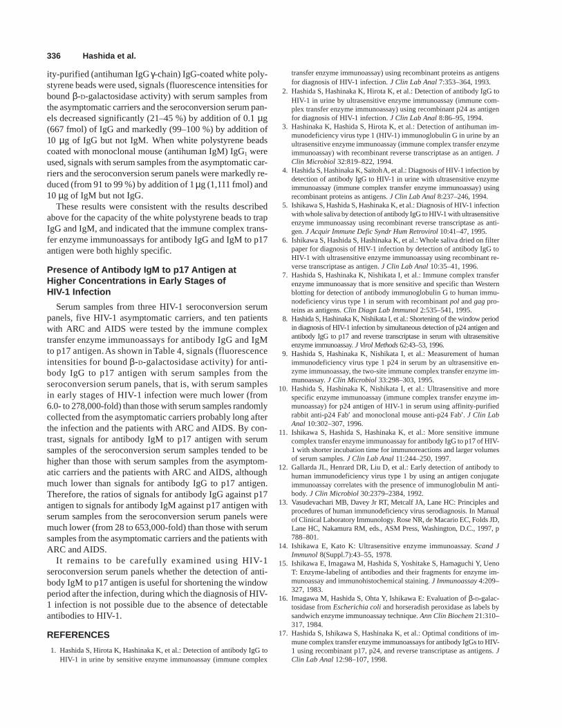
TABLE 4. Signals for Antibody IgG and IgM to p17 Antigena in Serum Samples of HIV-1-Infected Subjects at Different Stagesof the Infection
Ratio ofDays after the signals forinitial blood Signal (fluorescence intensity for anti-p17 IgG
Sources of collection or bound β-D-galactosidase activity) to those forserum samples serum no. Anti-p17 IgG Anti-p17 IgM anti-p17 IgM
Seroconversionserum panel
Panel E 126 days 779 42 19SV-0241 14 days 79 12 6.4SV-0241 23 days 1,660 853 1.9SV-0051 15 days 709 106 6.7SV-0051 27 days 5,095 870 5.9
Asymptomatic T-28 1,276,500 132 9,670carriers T-64 1,641,000 24 68,375
T-76 9,482,900 21 457,007T-17 16,235,400 50 325,685T-61 21,957,900 18 1,240,559
Patients with ARC T-15 48,620 93 523T-62 219,520 59 3,737T-79 282,045 185 1,527T-23 311,895 92 3,403T-42 531,420 84 6,296
Patients with AIDS T-74 30,670 13 2,415T-36 36,820 15 2,480T-11 39,470 29 1,385T-4 66,745 38 1,775T-53 330,695 173 1,910
aAntibody IgG and IgM to p17 antigen were measured as described in Table 3, except that β-D-galactosidase activity bound was assayed for 1 hr.
336 Hashida et al.
ity-purified (antihuman IgG γ-chain) IgG-coated white poly-styrene beads were used, signals (fluorescence intensities forbound β-D-galactosidase activity) with serum samples fromthe asymptomatic carriers and the seroconversion serum pan-els decreased significantly (21–45 %) by addition of 0.1 µg(667 fmol) of IgG and markedly (99–100 %) by addition of10 µg of IgG but not IgM. When white polystyrene beadscoated with monoclonal mouse (antihuman IgM) IgG1 wereused, signals with serum samples from the asymptomatic car-riers and the seroconversion serum panels were markedly re-duced (from 91 to 99 %) by addition of 1 µg (1,111 fmol) and10 µg of IgM but not IgG.
These results were consistent with the results describedabove for the capacity of the white polystyrene beads to trapIgG and IgM, and indicated that the immune complex trans-fer enzyme immunoassays for antibody IgG and IgM to p17antigen were both highly specific.
Presence of Antibody IgM to p17 Antigen atHigher Concentrations in Early Stages ofHIV-1 Infection
Serum samples from three HIV-1 seroconversion serumpanels, five HIV-1 asymptomatic carriers, and ten patientswith ARC and AIDS were tested by the immune complextransfer enzyme immunoassays for antibody IgG and IgMto p17 antigen. As shown in Table 4, signals (fluorescenceintensities for bound β-D-galactosidase activity) for anti-body IgG to p17 antigen with serum samples from theseroconversion serum panels, that is, with serum samplesin early stages of HIV-1 infection were much lower (from6.0- to 278,000-fold) than those with serum samples randomlycollected from the asymptomatic carriers probably long afterthe infection and the patients with ARC and AIDS. By con-trast, signals for antibody IgM to p17 antigen with serumsamples of the seroconversion serum samples tended to behigher than those with serum samples from the asymptom-atic carriers and the patients with ARC and AIDS, althoughmuch lower than signals for antibody IgG to p17 antigen.Therefore, the ratios of signals for antibody IgG against p17antigen to signals for antibody IgM against p17 antigen withserum samples from the seroconversion serum panels weremuch lower (from 28 to 653,000-fold) than those with serumsamples from the asymptomatic carriers and the patients withARC and AIDS.
It remains to be carefully examined using HIV-1seroconversion serum panels whether the detection of anti-body IgM to p17 antigen is useful for shortening the windowperiod after the infection, during which the diagnosis of HIV-1 infection is not possible due to the absence of detectableantibodies to HIV-1.
REFERENCES
1. Hashida S, Hirota K, Hashinaka K, et al.: Detection of antibody IgG toHIV-1 in urine by sensitive enzyme immunoassay (immune complex
transfer enzyme immunoassay) using recombinant proteins as antigensfor diagnosis of HIV-1 infection. J Clin Lab Anal 7:353–364, 1993.
2. Hashida S, Hashinaka K, Hirota K, et al.: Detection of antibody IgG toHIV-1 in urine by ultrasensitive enzyme immunoassay (immune com-plex transfer enzyme immunoassay) using recombinant p24 as antigenfor diagnosis of HIV-1 infection. J Clin Lab Anal 8:86–95, 1994.
3. Hashinaka K, Hashida S, Hirota K, et al.: Detection of antihuman im-munodeficiency virus type 1 (HIV-1) immunoglobulin G in urine by anultrasensitive enzyme immunoassay (immune complex transfer enzymeimmunoassay) with recombinant reverse transcriptase as an antigen. JClin Microbiol 32:819–822, 1994.
4. Hashida S, Hashinaka K, Saitoh A, et al.: Diagnosis of HIV-1 infection bydetection of antibody IgG to HIV-1 in urine with ultrasensitive enzymeimmunoassay (immune complex transfer enzyme immunoassay) usingrecombinant proteins as antigens. J Clin Lab Anal 8:237–246, 1994.
5. Ishikawa S, Hashida S, Hashinaka K, et al.: Diagnosis of HIV-1 infectionwith whole saliva by detection of antibody IgG to HIV-1 with ultrasensitiveenzyme immunoassay using recombinant reverse transcriptase as anti-gen. J Acquir Immune Defic Syndr Hum Retrovirol 10:41–47, 1995.
6. Ishikawa S, Hashida S, Hashinaka K, et al.: Whole saliva dried on filterpaper for diagnosis of HIV-1 infection by detection of antibody IgG toHIV-1 with ultrasensitive enzyme immunoassay using recombinant re-verse transcriptase as antigen. J Clin Lab Anal 10:35–41, 1996.
7. Hashida S, Hashinaka K, Nishikata I, et al.: Immune complex transferenzyme immunoassay that is more sensitive and specific than Westernblotting for detection of antibody immunoglobulin G to human immu-nodeficiency virus type 1 in serum with recombinant pol and gag pro-teins as antigens. Clin Diagn Lab Immunol 2:535–541, 1995.
8. Hashida S, Hashinaka K, Nishikata I, et al.: Shortening of the window periodin diagnosis of HIV-1 infection by simultaneous detection of p24 antigen andantibody IgG to p17 and reverse transcriptase in serum with ultrasensitiveenzyme immunoassay. J Virol Methods 62:43–53, 1996.
9. Hashida S, Hashinaka K, Nishikata I, et al.: Measurement of humanimmunodeficiency virus type 1 p24 in serum by an ultrasensitive en-zyme immunoassay, the two-site immune complex transfer enzyme im-munoassay. J Clin Microbiol 33:298–303, 1995.
10. Hashida S, Hashinaka K, Nishikata I, et al.: Ultrasensitive and morespecific enzyme immunoassay (immune complex transfer enzyme im-munoassay) for p24 antigen of HIV-1 in serum using affinity-purifiedrabbit anti-p24 Fab′ and monoclonal mouse anti-p24 Fab′. J Clin LabAnal 10:302–307, 1996.
11. Ishikawa S, Hashida S, Hashinaka K, et al.: More sensitive immunecomplex transfer enzyme immunoassay for antibody IgG to p17 of HIV-1 with shorter incubation time for immunoreactions and larger volumesof serum samples. J Clin Lab Anal 11:244–250, 1997.
12. Gallarda JL, Henrard DR, Liu D, et al.: Early detection of antibody tohuman immunodeficiency virus type 1 by using an antigen conjugateimmunoassay correlates with the presence of immunoglobulin M anti-body. J Clin Microbiol 30:2379–2384, 1992.
13. Vasudevachari MB, Davey Jr RT, Metcalf JA, Lane HC: Principles andprocedures of human immunodeficiency virus serodiagnosis. In Manualof Clinical Laboratory Immunology. Rose NR, de Macario EC, Folds JD,Lane HC, Nakamura RM, eds., ASM Press, Washington, D.C., 1997, p788–801.
14. Ishikawa E, Kato K: Ultrasensitive enzyme immunoassay. Scand JImmunol 8(Suppl.7):43–55, 1978.
15. Ishikawa E, Imagawa M, Hashida S, Yoshitake S, Hamaguchi Y, UenoT: Enzyme-labeling of antibodies and their fragments for enzyme im-munoassay and immunohistochemical staining. J Immunoassay 4:209–327, 1983.
16. Imagawa M, Hashida S, Ohta Y, Ishikawa E: Evaluation of β-D-galac-tosidase from Escherichia coli and horseradish peroxidase as labels bysandwich enzyme immunoassay technique. Ann Clin Biochem 21:310–317, 1984.
17. Hashida S, Ishikawa S, Hashinaka K, et al.: Optimal conditions of im-mune complex transfer enzyme immunoassays for antibody IgGs to HIV-1 using recombinant p17, p24, and reverse transcriptase as antigens. JClin Lab Anal 12:98–107, 1998.