imaging the skullradfileshare.cvm.ncsu.edu/print/2007/07 skull notes.pdf · 1 imaging the skull...
TRANSCRIPT
1
Imaging theSkull
Reading Assignment
71-8787-89
Skull is Anatomically Complex
Use symmetryCreates problems 3D 2DDV & lateral views are typically inadequate for complete evaluation
2
Ancillary views necessary because of anatomic complexity
Nasal passageMaxillaMandibleFrontal SinusTympanic BullaTemporomandibular Joint
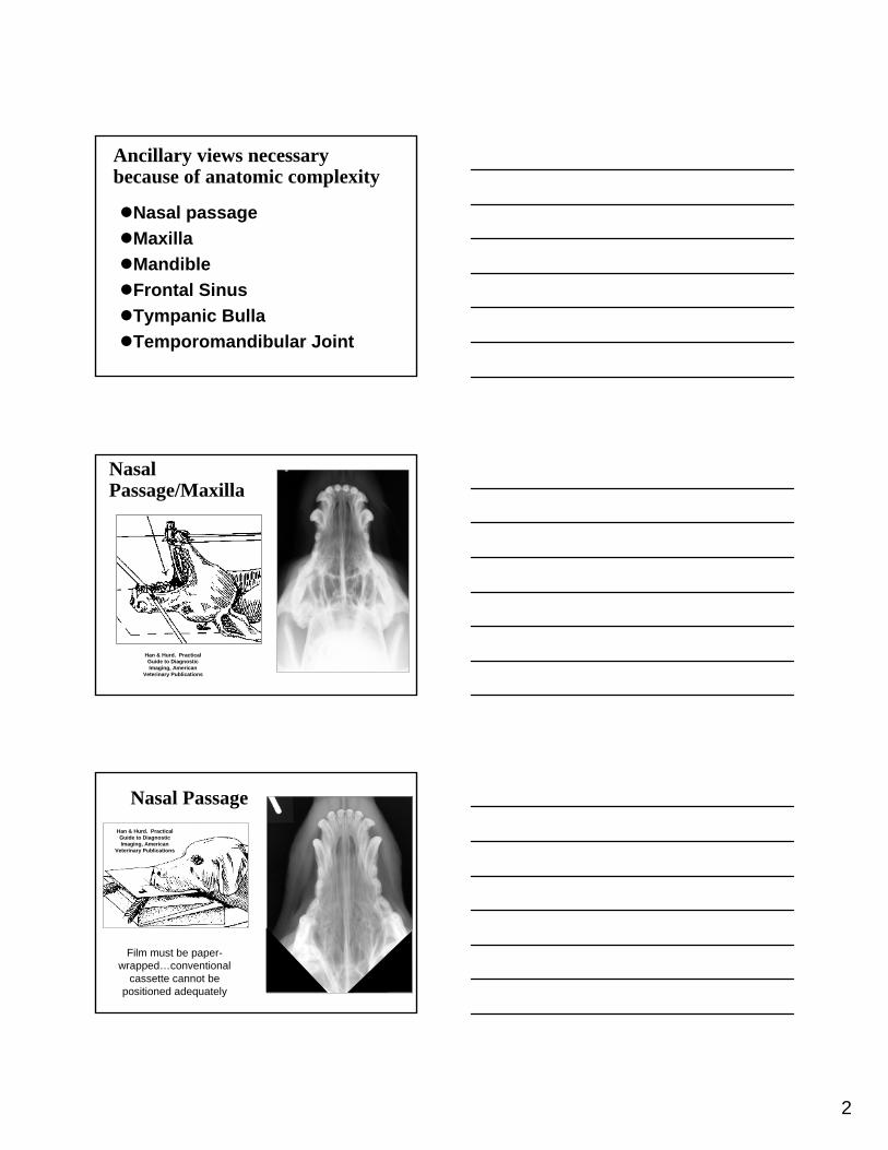
Nasal Passage/Maxilla
Han & Hurd. Practical Guide to Diagnostic Imaging, American
Veterinary Publications
Nasal Passage
Film must be paper-wrapped…conventional
cassette cannot be positioned adequately
Han & Hurd. Practical Guide to Diagnostic Imaging, American
Veterinary Publications
3
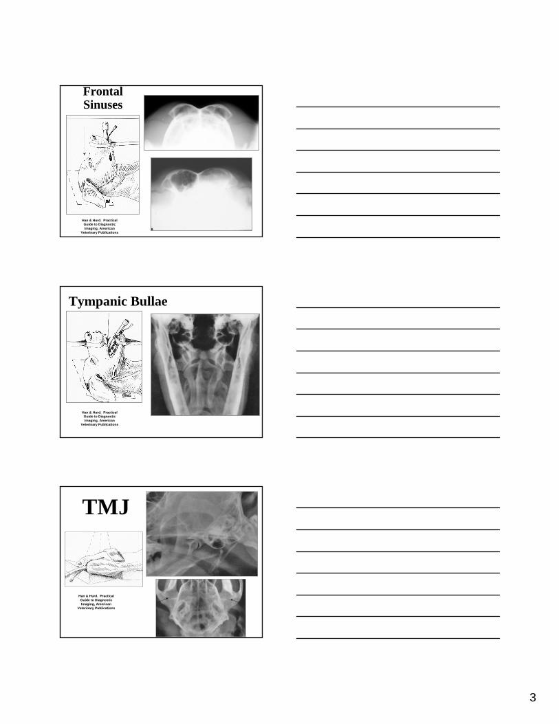
Frontal Sinuses
Han & Hurd. Practical Guide to Diagnostic Imaging, American
Veterinary Publications
Tympanic Bullae
Han & Hurd. Practical Guide to Diagnostic Imaging, American
Veterinary Publications
TMJ
Han & Hurd. Practical Guide to Diagnostic Imaging, American
Veterinary Publications
4
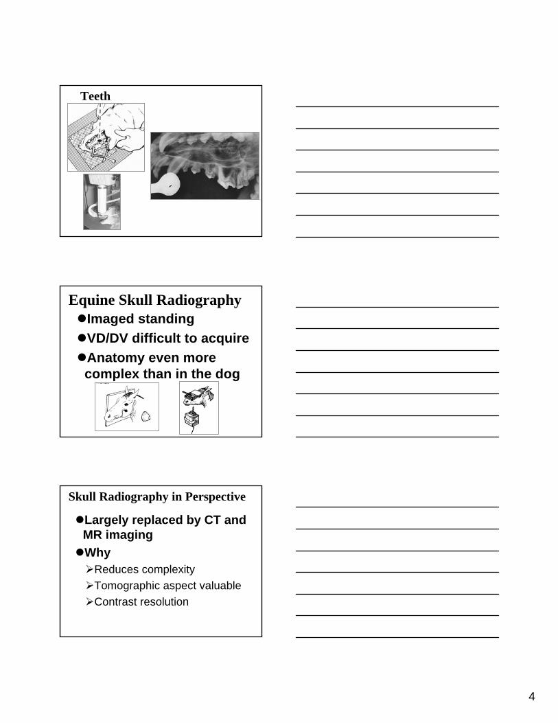
Teeth
Equine Skull RadiographyImaged standingVD/DV difficult to acquireAnatomy even more complex than in the dog
Skull Radiography in Perspective
Largely replaced by CT and MR imagingWhy
Reduces complexityTomographic aspect valuableContrast resolution
5
Uses of RadiographyOtitis mediaMandibular and maxillary tumorsSinusitisNasal cancerRhinitisDental disease
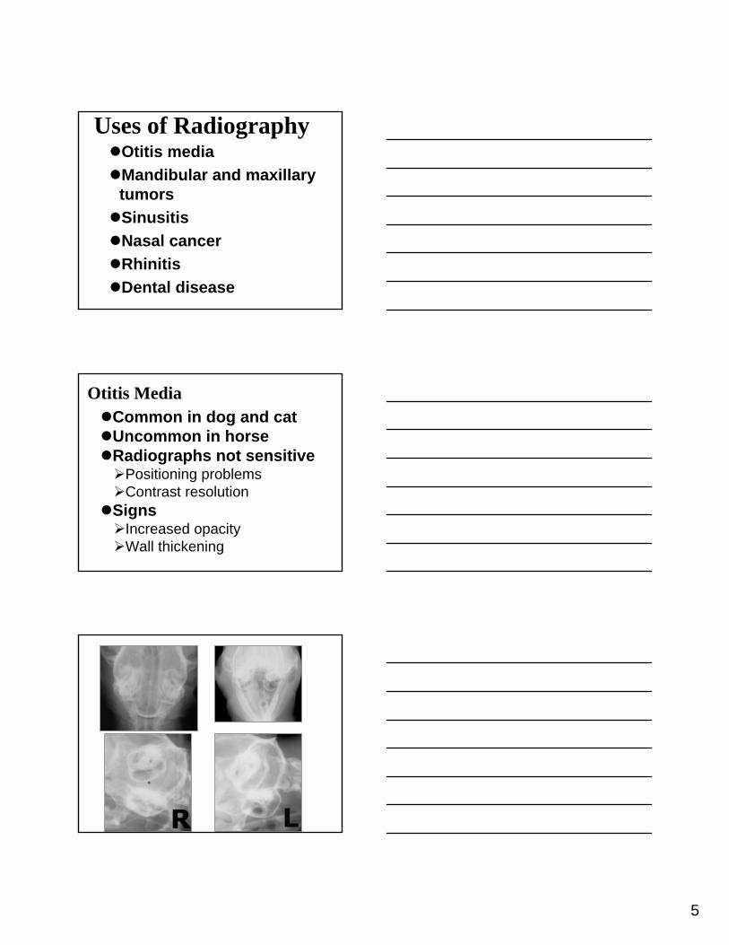
Otitis MediaCommon in dog and catUncommon in horseRadiographs not sensitive
Positioning problemsContrast resolution
SignsIncreased opacityWall thickening
6
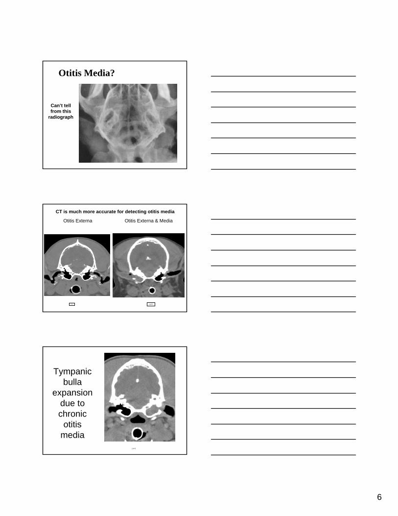
Otitis Media?
Can’t tell from this
radiograph
11457345714
Otitis Externa Otitis Externa & Media
CT is much more accurate for detecting otitis media
110473
Tympanic bulla
expansion due to chronic
otitismedia
7
119499
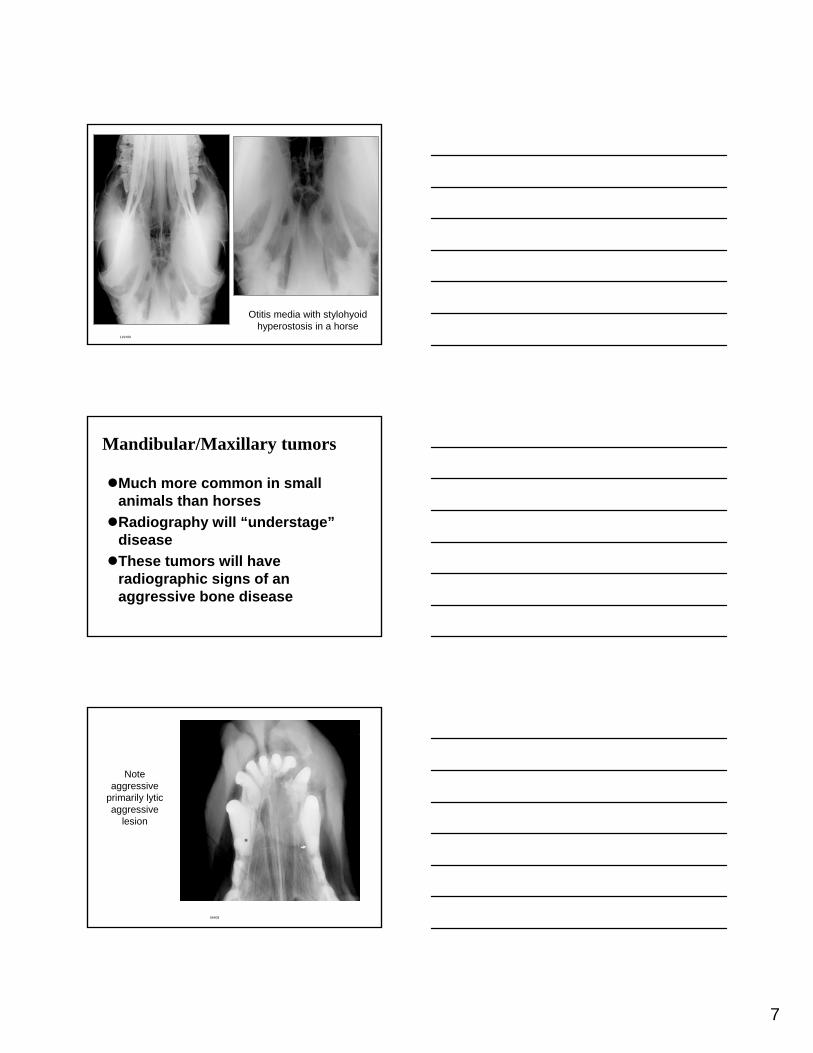
Otitis media with stylohyoidhyperostosis in a horse
Mandibular/Maxillary tumors
Much more common in small animals than horsesRadiography will “understage”diseaseThese tumors will have radiographic signs of an aggressive bone disease
66408
Note aggressive
primarily lytic aggressive
lesion
8
111963
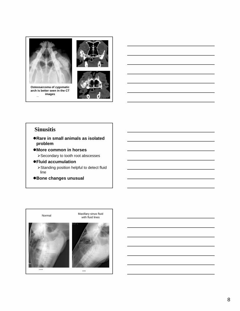
Osteosarcoma of zygomaticarch is better seen in the CT
images
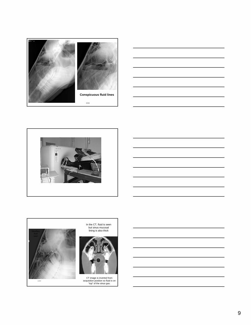
SinusitisRare in small animals as isolated problemMore common in horses
Secondary to tooth root abscessesFluid accumulation
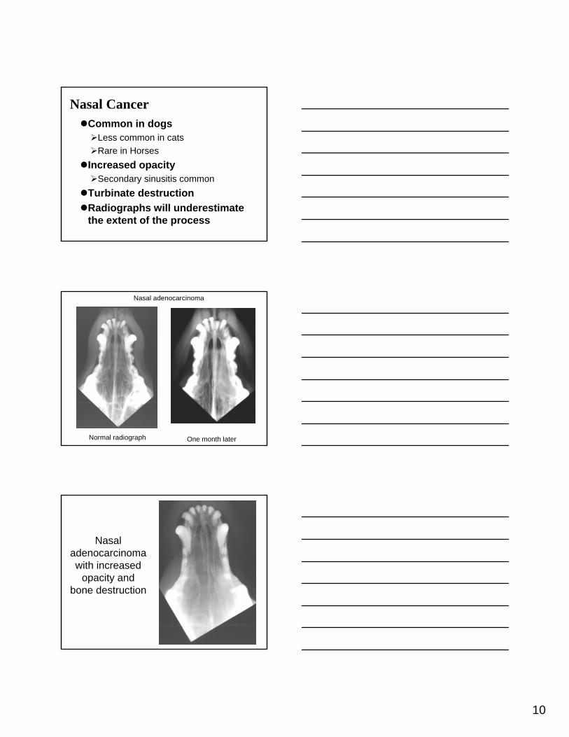
Standing position helpful to detect fluid line
Bone changes unusual
63368114128
Normal Maxillary sinus fluid with fluid lines
9
63368
Conspicuous fluid lines
112029
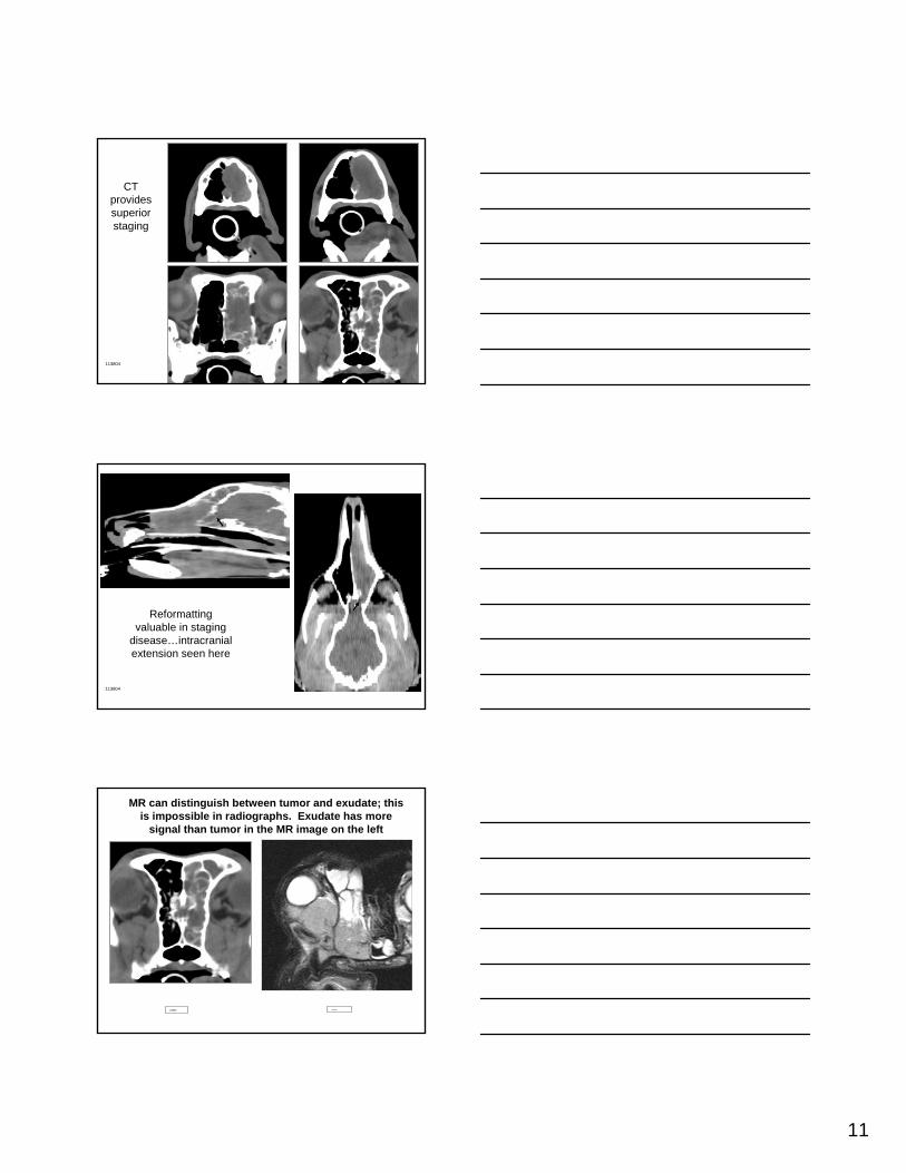
Maxillary fluid seen
In the CT, fluid is seen but sinus mucosal lining is also thick
CT image is inverted from acquisition position so fluid is on
“top” of the sinus gas.
10
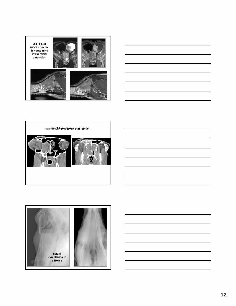
Nasal CancerCommon in dogs
Less common in catsRare in Horses
Increased opacitySecondary sinusitis common
Turbinate destructionRadiographs will underestimate the extent of the process
One month later
Nasal adenocarcinoma
Normal radiograph
Nasal adenocarcinomawith increased
opacity and bone destruction
11
113804
CT provides superior staging
Reformatting valuable in staging
disease…intracranial extension seen here
113804
VA002357113804
MR can distinguish between tumor and exudate; this is impossible in radiographs. Exudate has more
signal than tumor in the MR image on the left
12
T2 T1+C
T1+CT1+C
MR is also more specific for detecting intracranialextension
NC4348
111105
Aggressive nasal tumor in a horseNasal Lymphoma in a Horse
Nasal Lymphoma in
a Horse

13
Nasal Lymphoma in a Horse
2005.06.29Equine nasal lymphoma successfully treated with
radiation
2005.12.15
14
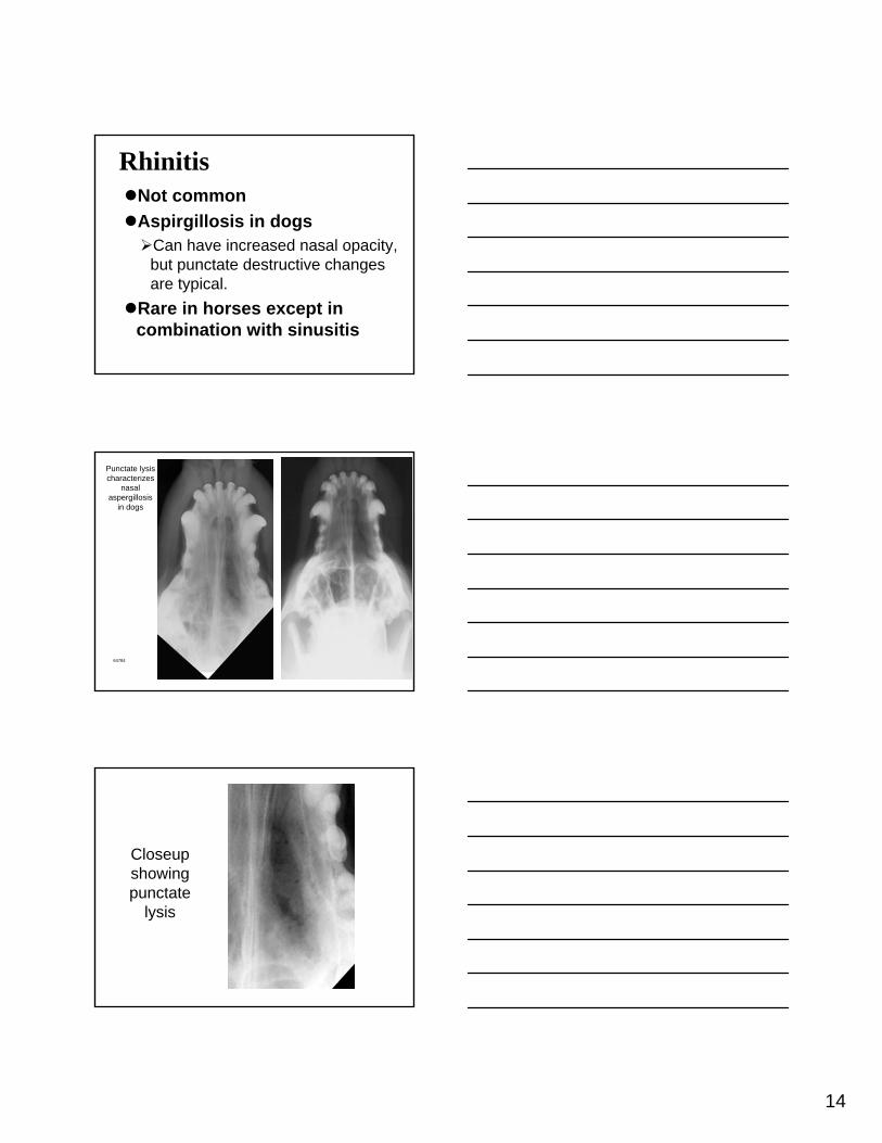
RhinitisNot commonAspirgillosis in dogs
Can have increased nasal opacity, but punctate destructive changes are typical.
Rare in horses except in combination with sinusitis
Punctate lysis characterizes
nasal aspergillosis
in dogs
64784
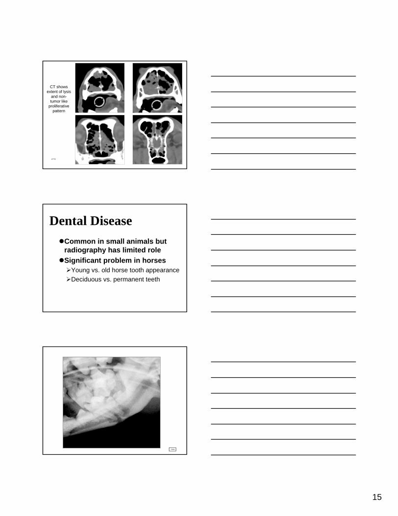
Closeupshowing punctate
lysis
15
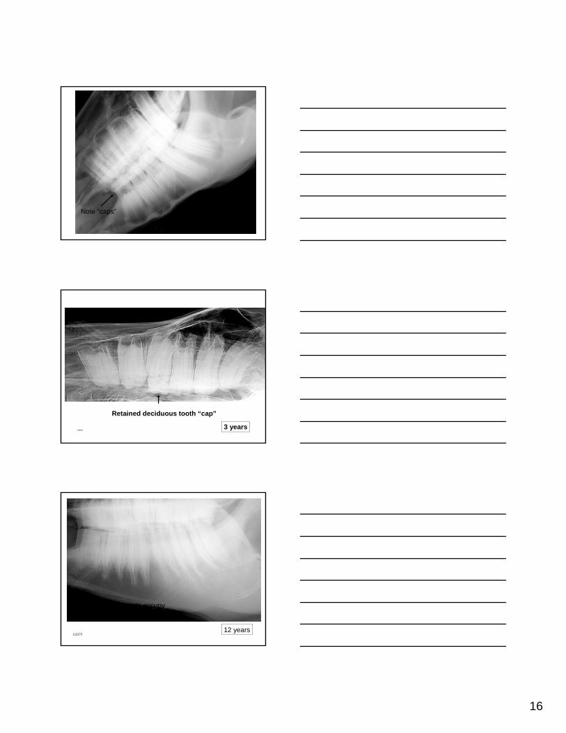
CT shows extent of lysis
and non-tumor like
proliferativepattern
107762
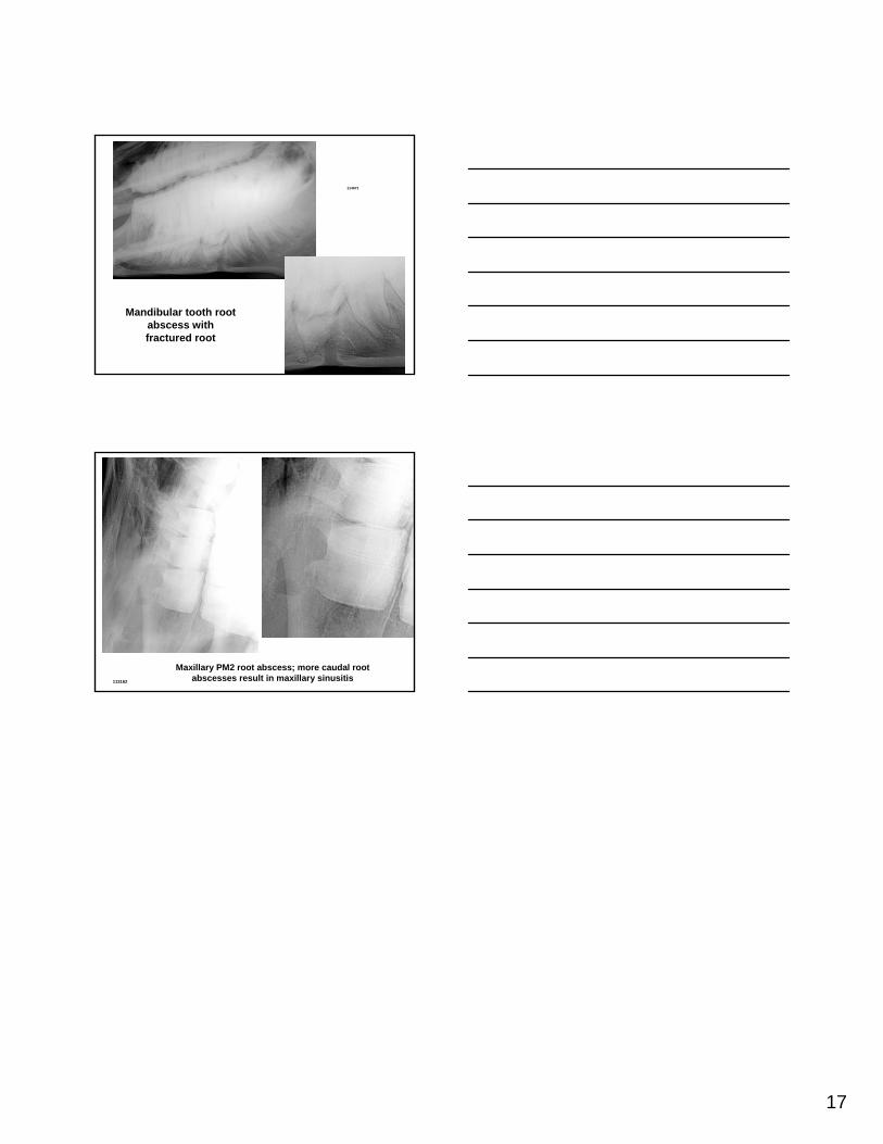
Dental DiseaseCommon in small animals but radiography has limited roleSignificant problem in horses
Young vs. old horse tooth appearanceDeciduous vs. permanent teeth
73944
Canine mandibular tooth root abscess
16
1082062 years
Periapical radiolucent regions normal in
young horsesNote “caps”
1082063 years
Retained deciduous tooth “cap”
11227312 years
With ageing, roots occupy less of mandible
17
114671
Mandibular tooth root abscess with fractured root
113162
Maxillary PM2 root abscess; more caudal root abscesses result in maxillary sinusitis