idf clinical practice recommendations on the diabetic foot ... · diabetic foot is one of the most...
TRANSCRIPT

IDF Clinical Practice Recommendations on the Diabetic Foot – 2017A guide for healthcare professionals

CopyrightAll rights reserved. No part of this publication may be reproduced or transmitted in any form or by any means without the written prior permission of the IDF. Requests to reproduce or translate IDF publications should be addressed to [email protected]
PublisherPublished by the International Diabetes Federation
© International Diabetes Federation, 2017ISBN: 978-2-930229-86-7
Please cite this report as: International Diabetes Federation. Clinical Practice Recommendation on the Diabetic Foot: A guide for health care professionals : International Diabetes Federation, 2017.
CorrespondenceInternational Diabetes Federation, 166 Chaussée de La Hulpe B-1170, Brussels Belgium [email protected]
Duality of interest Authors of the guidelines declared dualities of interest in respect of commercial enterprises, government, and non-governmental organisations. No fees were paid to the authors in connection with the development of this document or the guidelines described herein.
DesignDesigned by Karakas
Main authorsAmmar Ibrahim, MD, FACS, Chair IDF Diabetic Foot Stream, Chair SACA region 2016-2017. General Director ”Instituto Nacional de Diabetes (INDEN), Dominican Republic”. Professor at Iberoamericana University (UNIBE)
Edward Jude, Professor and Consultant Diabetologist, Tameside Hospital NHS Foundation Trust, Ashton under Lyne, UK.
Katia Langton, DC, Certified Pedorthist (C) Island Pedorthic FootCare. Central Vancouver Island Foot and Ulcer Protection Clinic. Canada.
Fermin R. Martinez-De Jesus, MD, Director, San Elian Center for Prevention and Salvage of the Diabetic Foot. Asociación Mexicana de Pie Diabetico. Veracruz, Mexico.
Lawrence B. Harkless, DPM, Founding Dean, College of Podiatric Medicine. Professor of Podiatric Medicine and Surgery. Western University of Health Sciences. Secretary Diabetic Foot Stream Committee. California, USA.
Hanan Gawish, Professor of Diabetes and Endocrinology, Mansoura University. Chair of Egyptian Society of Diabetic Foot (ESDF). Egypt.
Yu-Yao Huang M.D., Ph.D., Director, Department of Medical Nutrition Therapy. Chang Gung Memorial Hospital and Chang Gung University. Taiwan.
Jonathan Labovitz, DPM, FACFAS, CHCQM, Medical Director, Foot and Ankle Center. Professor, College of Podiatric Medicine, Western University of Health Sciences.
Zhangrong XU, MD, Chief physician and Professor of Medicine, Diabetes Center, The 306th Hospital. Beijing, China.
Sharad Pendsey, MD, Consultant in Diabetes. Director, Diabetes Clinic & Research Center, Dhantoli. Nagpur, India.
Fang LIU, MD, PhD, Professor, PhD supervisor,Chief physician, Vice-chief of Dept of Endocrinology & Metabolism. Shanghai Jiao-Tong University Affiliated Sixth People’s Hospital. Shanghai Clinical Center for Diabetes. Shanghai Key Clinical Center of Metabolic Diseases. Shanghai Key Laboratory of Diabetes Mellitus. Shanghai Institute for Diabetes. China.Shaukat Sadikot, MD, President International Diabetes Federation (2016-2017), Diabetes India and Jaslok Hospital, Mumbai, IndiaNam Han Cho, MD, President-Elect International Diabetes Federation (2016-17), Department of Preventive Medicine, Ajou University School of Medicine, Suwon, Korea.
2IDF Clinical Practice Recommendations on the Diabetic Foot 2017
Table of contents
Foreword . . . . . . . . . . . . . . . . . . . . . . . . . . . . . . . . . . . . . . . . . . . . . . . . . . . . . . . . . . . . . . . . . . . . . . . . . . . . . . . . . . . . . . . . . . . . . . . . . . . . 4
Introduction . . . . . . . . . . . . . . . . . . . . . . . . . . . . . . . . . . . . . . . . . . . . . . . . . . . . . . . . . . . . . . . . . . . . . . . . . . . . . . . . . . . . . . . . . . . . . . . . . 8
Diabetic Peripheral Neuropathy . . . . . . . . . . . . . . . . . . . . . . . . . . . . . . . . . . . . . . . . . . . . . . . . . . . . . . . . . . . . . . . . . . . . . . . . . . . . . . 14
Peripheral Arterial Disease . . . . . . . . . . . . . . . . . . . . . . . . . . . . . . . . . . . . . . . . . . . . . . . . . . . . . . . . . . . . . . . . . . . . . . . . . . . . . . . . . . . 34
Ulcers . . . . . . . . . . . . . . . . . . . . . . . . . . . . . . . . . . . . . . . . . . . . . . . . . . . . . . . . . . . . . . . . . . . . . . . . . . . . . . . . . . . . . . . . . . . . . . . . . . . . . . 42
Diabetic Foot Infection . . . . . . . . . . . . . . . . . . . . . . . . . . . . . . . . . . . . . . . . . . . . . . . . . . . . . . . . . . . . . . . . . . . . . . . . . . . . . . . . . . . . . . 51
Charcot Neuro-osteoarthropathy . . . . . . . . . . . . . . . . . . . . . . . . . . . . . . . . . . . . . . . . . . . . . . . . . . . . . . . . . . . . . . . . . . . . . . . . . . . . 62
This icon indicates interactive elements
IDF Clinical Practice Recommendations on the Diabetic Foot
3IDF Clinical Practice Recommendations on the Diabetic Foot 2017

Foreword
4

Foreword from the IDF President
Long-term complications of diabetes develop gradually. The longer you have diabetes — and the less controlled your blood sugar — the higher the risk of complications. With the growing number of people living with diabetes worldwide, healthcare professionals are encouraged to pay attention to the major complications of diabetes in their daily practice. It is therefore IDF’s vision is to develop a series clinical practice recommendations for health care professionals on specific topics, with the aim of creating clinical guidelines in easily digestible and user-friendly format and adaptable to any country, region or health setting.
Diabetic foot is one of the most serious and costly complications of diabetes. These new IDF Clinical Practice Recommendations on the Diabetic Foot are an excellent addition to the knowledge base underlying the delivery of high-quality primary clinical care. We hope that they will promote and improve diabetic foot care within all seven IDF regions.
I would like to thank the IDF Diabetic Foot Committee, headed by Dr. Ammar Ibrahim, for their tireless efforts to produce these guidelines. Using their vast experience in the field, the committee members methodically and critically examined a vast amount of published scientific evidence on the diabetic foot. These clinical practice recommendations are a tribute to the skills of the authors and it is with great pleasure that I pen these words to relate my enthusiasm for their work.
It is my hope and expectation that these clinical practice recommendations will provide an effective learning experience and referenced resource for all health professionals caring for people living with diabetes, resulting in improved patient outcomes. I therefore highly recommend that all primary care health professionals make use of them for an optimal management of diabetic foot complications in their settings.
Dr Shaukat Sadikot IDF President 2016-2017
5IDF Clinical Practice Recommendations on the Diabetic Foot 2017

Foreword from the Chair
The complications of diabetes are far less common and less severe in people who have well-controlled blood glucose. With the correct treatment and recommended lifestyle changes, many people with diabetes are able to prevent or delay the onset of complications, avoiding serious consequences to their health and well-being.
Diabetic foot disease, mainly due to neuropathy, peripheral arterial disease, and/or infection, often leads to ulceration and possible subsequent limb amputation. It is one of the most costly complications of diabetes, and can result in an important economic, social, and public health burden; especially in low-income communities, if there is neither an appropriate educational programme, nor adequate and suitable footwear.
These IDF Clinical Practice Recommendations on the Diabetic Foot are simplified, easy to digest guidelines to prioritize health care practitioner's early intervention of the diabetic foot with a sense of urgency through education. The main goals of these guidelines are to promote early detection and intervention; provide the criteria for time- adequate referral to a second or third level centers and serve as tool to educate people with diabetes about the importance of prevention in this pathology.
They are also designed to provide clinicians with practice recommendations based on published evidence, which have been validated through reviews and field-testing by experienced diabetic foot care clinicians. They are not targeting only specialized diabetic foot health practitioners, but all health professionals, including diabetic educators and nurses, and in some circumstances, people with diabetes and their families.
An abbreviated version of these guidelines, the “Diabetes Foot Screening Pocket Chart, has also been produced and will be distributed to primary care physicians, nurses, registered dietitians and nutritionists, and other health professionals.
Using simple language, appropriate for all segments of the health sector, this clinical manual is a collective work, suitable for daily field practice. First of all, I would like to acknowledge the IDF President, Dr Shaukat Sadikot, for his leadership, vision, and enthusiasm; Katia Langton, Edward Jude and Belma Malanda as secretariats for their support.
In a voluntary and multidisciplinary undertaking of this magnitude, many professionals have contributed to the final product now in your hands. It is impossible to acknowledge them individually here, but to each and every one of them, we extend our sincerest appreciation.
6IDF Clinical Practice Recommendations on the Diabetic Foot 2017

This limitation not-with-standing, a special debt of gratitude is due to the members of the IDF Diabetic Foot Committee: Katia Langton (Canada), Edward Jude (UK), Lawrence B. Harkless (USA), Jonathan Labovitz (USA), Sharad Pendsey (India), Fang Liu (Shanghai), Yu-Yao Huang (Taiwan), Zhangrong Xu (Beijing), Hanan Gawish (Egypt) and Fermin R. Martinez-De Jesus (Mexico). It is their commitment and dedication to the process that has made this document possible.
This is only the beginning of a long journey on this topic. Updated versions, some modifications, local adaptations, improvements and periodic reviews according to the state-of-the art on the topic will be done on a regular basis. With a view to future revisions and to keep the work as close as possible to field realities, the authors would be grateful for suggestions from users of this manual.
It is our hope that these clinical practice recommendations will not only help health care practitioners understand the importance of screening of the diabetic foot but also provide them with the tools to assess and treat their patients more effectively.
Dr Ammar Ibrahim, MD, FACS Chair of the IDF Diabetic Foot Committee
7IDF Clinical Practice Recommendations on the Diabetic Foot 2017

Introduction
8

Diabetes and its complications are rapidly becoming the world’s most significant cause of morbidity and mortality. It is predicted that by 2040 there will be over 642 million people with diabetes in the world.1 With the lifetime incidence of foot ulcers occurring in up to 25% of patients2, we need to pay far more attention to the diabetic foot and shift our focus to preventing ulcers rather than treating them. Diabetes morbidity rates are staggeringly high and the 5-year mortality rate, after a lower extremity amputation, is only second to lung cancer.3
We are in an era where more people are dying globally from non-communicable diseases known as lifestyle related diseases – diabetes, cardiovascular disease, stroke, cancer and chronic lung diseases – than from infectious diseases.4 Non-communicable diseases were responsible for 38 million (68%) of the world’s 56 million deaths in 2012 with the majority of them occurring in low- and middle-income countries.5
So it is a measure of how well we are doing in managing infectious diseases, but also of how lifestyle related diseases fueled by unhealthy diets, insufficient physical activity, and obesity are leading the way for increased deaths.
Keeping people on their feet, walking and mobile is fundamental to preventing the progression of lifestyle related diseases. But people will not walk if they have pain, balance issues or fear they are doing more damage to their feet; and they are unable to walk if they have open ulcers on the plantar surface of their feet. Once these problems arise, people often become increasingly sedentary, and with decreased physical activity, short and long-term blood glucose levels will increase, people put on weight and overall health declines steadily.
Figure 1 Adults who died from diabetes, HIV/AIDS, tuberculosis, and malaria
5.0 millionfrom diabetes
2015 IDF
1.5 millionfrom HIV/AIDS
2013 WHO Global Health Observatory
Data Repository 2013
1.5 millionfrom tuberculosis
2013 WHO Global Health Observatory
Data Repository 2013
0.6 millionfrom malaria
2013 WHO Global Health Observatory
Data Repository 2013
9IDF Clinical Practice Recommendations on the Diabetic Foot 2017

In diabetes, elevated glycaemic levels increase the risk of microvascular and macrovascular complications. These increase the risk of further complications such as retinopathy, cardiovascular disease, and nephropathy, in addition to peripheral neuropathy, which can cause foot ulcerations and may lead to lower limb amputations. Improved blood glucose control, which can be managed with simply walking, will decrease the the impact on macrovascular and microvascular damage. Physical activity remains an important first-line therapeutic approach to improve glycaemic control in individuals who are obese and/or have diabetes.
Numerous studies have shown that blood glucose levels are improved by increasing physical activity. Each 1-hour per day increment of brisk walking was associated with a 34% reduction in risk of developing type 2 diabetes.6
The basic treatment for diabetes should be considered on the basis of individualised and comprehensive treatment targets that include well controlled blood glucose, blood pressure and lipid profile, weight management, smoking cessation, a healthy diet and physical activities such as walking.
Diabetes eventually affects every part of the body, but it frequently involves the feet first. The key to treating this disease is to get ahead of it and treat it earlier in the progression of diabetes. A paradigm shift is urgently needed to treat diabetic foot disease preventatively. As the diabetes pandemic progresses globally; so do foot complications and ulcers, which usually precede the
majority of lower extremity amputations. More than half of all foot ulcers will become infected, requiring hospitalization and 20% of lower extremity infections will result in amputation.7
Foot problems are indeed a global problem and there is no area in the world that does not report the development of foot lesions as a consequence mainly of neuropathy and peripheral vascular disease.8,9,10
The prevalence of active foot ulceration varies from approximately 1% in certain European and North American studies to more than 11% in reports from some African countries. Although there have been many developments in recent years which encourage optimism for future improvement in diabetic foot care, there is still much to be done. Since most advancements focus on new treatments for complications, not preventive measures.
Africa
Europe
North America
1%
11%
Figure 2 Prevalence of active foot ulceration
In some islands of the Caribbean, for example, where the prevalence of diabetes is approaching 20%, foot lesions and gangrene are amongst the most frequent conditions seen on surgical wards.8,9,10
10IDF Clinical Practice Recommendations on the Diabetic Foot 2017

The diabetic foot is a major medical, social and economic problem worldwide. However, the reported frequency of ulceration and amputation varies considerably. This may be due to differences in diagnostic criteria as well as regionally specific social, economic and health-related factors.
In developing countries, foot ulcers and amputations are unfortunately very common. Poverty, a lack of sanitation and hygiene, and barefoot walking often interact to compound the impact of diabetic foot damage. In low-income countries, the lack of access to adequate health care, together with economic and geographical factors, often prevent people with diabetes from seeking medical treatment for foot lesions until these have become severely infected.12
Neuropathy is a frequently encountered complication of diabetes. Diabetic peripheral neuropathy is an impairment of normal activities of the nerves throughout the body and can alter autonomic, motor, and sensory functions. The reported prevalence of diabetic peripheral neuropathy ranges from 16% to as high as 66%.8
Perhaps the most commonly recognized form of neuropathy among people with diabetes is sensory neuropathy, resulting in the loss of sensation beginning in the most distal part of the extremity. Sensory diabetic peripheral neuropathy causes diminished sensory feedback, predisposing patients to become more prone to foot injuries and the above complications.
Due to lack of training, it has been estimated that less than one third of physicians recognize the symptoms of diabetic peripheral neuropathy, even when it is symptomatic, and discuss them with their patients.13 The opportunity is missed to get in front of the progression of diabetes and its complications by treating the diabetic foot in an earlier risk category.
An understanding of the comprehensive management and treatment of the diabetic foot is lacking amongst healthcare providers. Diabetic foot care has been described as ‘fragmented and haphazard’, and dependent on which practitioner the patient happens to be seeing, and which local resources are available.14 Very few clinicians are treating the diabetic foot in a systematic, standardized method with proper risk categorization of foot complications. Our pocket chart for a comprehensive diabetic foot exam will lead practitioners through the full assessment with a thought process on how to treat these patients preventatively.
At the time of diagnosis of diabetes, and at regular diabetes check-ups, warning bells need to go off so the practitioner assesses, triages, and treats the diabetic foot early and preventatively in accordance with the risk category.
All people with diabetes should be screened and placed in the appropriate risk stratification which includes the clinical pathway for prevention and treatment. Members of the team and necessary services such as foot care nursing, diabetes education, pedorthists, skilled wound care team, physicians, podiatrists, prosthetics, home care and counseling are central for good outcomes to improve health-related quality of life.
The goal of these IDF Guidelines is to protect the diabetic foot from breakdown, preventing foot ulceration and lower limb amputations, by taking preventative measures early in the disease process and treating the foot in the early Risk Categories of 1, and 2 and before they become the VERY HIGH Risk Category 3.
In most developed countries, the annual incidence of foot ulceration amongst people with diabetes is about 2% . In these countries, diabetes is the most common cause of non-traumatic amputation; approximately 1% of people with diabetes suffer a lower-limb amputation .11, 12
11IDF Clinical Practice Recommendations on the Diabetic Foot 2017

Since eighty percent of diabetic foot cost are in Risk Category 3, we need to focus on treating these patients earlier and with the aim of preventing ulcers and progression into Risk Category 3. Each country's health care budget will not be able to sustain the demand necessary to treat diabetic foot complications, such as ulcers leading to amputations, as this disease progresses incessantly. Comprehensive diabetic foot assessments and foot care, based on prevention, education and a multi-disciplinary team approach, may reduce foot complications and amputations by up to 85%.15 Globally, we need to front load our resources and shift them into treating diabetic foot disease earlier in the risk categories and away from reactionary ulcer care.
Risk category 0
Risk category 1
Risk category 2
Risk category 3
Normal Plantar Sensation
Loss of Protective Sensation (LOPS)
LOPS with either High Pressure or Poor
Circulation or Structural Foot Deformities or
Onychomycosis
History of Ulceration, Amputation or
Neuropathic Fracture
LOW RISK MODERATE RISK HIGH RISK VERY HIGH RISK
Clinical tip
In the progression of peripheral neuropathy; vibration sense is lost initially. Motor neuropathy and position sense is lost in conjunction with protective sensation. Therefore, even if the patient has full or partial sensation, it is important to check the intrinsic musculature of the feet (small muscles in the feet) progressing to the extrinsic musculature (muscles of the leg) to monitor the progression of neuropathy. This progression limits their ability to walk and maintain mobility. Eventual progression of the neuropathy results in the loss of pain and temperature fibers.
Figure 3 Risk categories
IDF urges all health care practitioners to treat patients earlier in that ‘WINDOW OF PRESENTATION’ between the time a patient presents with neuropathy but before an ulcer develops
12IDF Clinical Practice Recommendations on the Diabetic Foot 2017

References:1. International Diabetes Federation. IDF Diabetes Atlas, 7th edn.
Brussels, Belgium: International Diabetes Federation, 2015. http://www.diabetesatlas.org
2. Singh N, Armstrong DG, Lipsky BA. Preventing foot ulcers in patients with diabetes. Jama. 2005 Jan 12;293(2):217-28.
3. Armstrong DG, Wrobel J, Robbins JM. Guest editorial: are diabetes-related wounds and amputations worse than cancer. Int Wound J. 2007 Dec 1;4(4):286-7.
4. World Health Organization. Assessing national capacity for the prevention and control of noncommunicable diseases. Report of the 2015 global survey. Geneva: WHO. 2015
5. World Health Organization. Global status report on noncommunicable diseases 2014. 2014.
6. Hu FB. Globalization of Diabetes. Diabetes Care. 2011 May 26;34(6):1249.
7. Wu SC, Driver VR, Wrobel JS, Armstrong DG. Foot ulcers in the diabetic patient, prevention and treatment. Vascular health and risk management. 2007 Feb 1;3(1):65.
8. Boulton AJ. The diabetic foot: a global view. Diabetes/Metabolism Research and Reviews. 2000 Sep 1;16(S1):S2-5.
9. Boulton A. The diabetic foot: epidemiology, risk factors and the status of care. Diabetes Voice. 2005 Nov;50(S1):5-7.
10. Boulton AJ, Vileikyte L, Ragnarson-Tennvall G, Apelqvist J. The global burden of diabetic foot disease. The Lancet. 2005 Nov 18;366(9498):1719-24.
11. Bobircã F, Mihalache O, Georgescu D, Pãtraæcu T. The New Prognostic-Therapeutic Index for Diabetic Foot Surgery-Extended Analysis. Chirurgia. 2016;111:151-5.
12. Lazzarini PA, Hurn SE, Fernando ME, Jen SD, Kuys SS, Kamp MC, Reed LF. Prevalence of foot disease and risk factors in general inpatient populations: a systematic review and meta-analysis. BMJ open. 2015 Nov 1;5(11):e008544.
13. Melmed S, Polonsky KS, Larsen PR, Kronenberg HM. Williams Textbook of Endocrinology 13th Edition 2016. Elsevier Inc.
14. Cheung C et al. The diabetic foot: A reconceptualization. Diabetic Foot Canada 2013, Vol1, No1, 11-12.
15. International Diabetes Federation and International Working Group of the Diabetic Foot. Diabetes and Foot Care: Time to Act, Fourth Edition.
13IDF Clinical Practice Recommendations on the Diabetic Foot 2017
Diabetic peripheral neuropathy
1414

Definition In diabetic foot disease; diabetic peripheral neuropathy (DPN) is the primary risk factor for the development of diabetic foot ulcers.1 DPN is one of the most common diabetes complications and it significantly impacts progression to the devastating outcomes of ulcerations that may lead to amputations.
The reported prevalence of diabetic peripheral neuropathy ranges from 16% to as high as 66%2 and its prevelance is believed to increase with the duration of diabetes and poor glucose control. The definition of neuropathy is nerve disease or damage. An internationally recognized definition of DPN is “the presence of symptoms and/or signs of peripheral nerve dysfunction in people with diabetes after exclusion of other causes”.3
PresentationPeripheral neuropathy may manifest as an inability to detect temperature changes, vibration, proprioception, pressure, and, most seriously, pain. Some patients have a form of painful sensory neuropathy that includes symptoms, such as burning and tingling, known as paresthesia.2,4,5
The clinical presentation of DPN can be quite variable.Patients can present with “positive” or “negative” symptoms. Positive symptoms are those that patients complain of (subjective findings), including paresthesia (tingling, hyperesthesia, burning, allodynia or formication). Negative symptoms are usually unveiled by clinical examination (objective findings). They could consist of numbness, dead/asleep feeling, or muscle weakness in the lower limbs.
The majority of patients with neuropathy present with some particular symptom and/or sign of DPN which should be recognized and paid attention to. Up to 50% of patients may experience symptoms, most frequently a burning pain, electrical or stabbing sensations, paresthesia, hyperesthesia, and a deep aching pain.3 Neuropathic pain is typically worse at night and at rest
as it advances, and the symptoms are most commonly experienced in the feet and lower limbs, although in some cases the hands may also be affected. However, as up to half of the patients may be asymptomatic, a diagnosis may only be made on examination or, in some cases, when the patient presents with a painless foot ulcer or foot infection. When neuropathy initially presents, clinicians need to start paying attention and become vigilant in initiating preventative treatment.
EpidemiologyChronic sensorimotor polyneuropathy afflicts sensory, motor and autonomic nerves of the peripheral nervous system. It is the sensory peripheral neuropathy that leads to the loss of the “gift of pain”, this is the feedback from our feet telling us when to rest, stay off our feet, and change our footwear to protect from tissue damage, injury and high peak pressure areas that may lead to tissue breakdown.
The progressive nature of neuropathy, leading to loss of protective sensation in the feet, makes the feet vulnerable to injuries and ulceration. Small afferent nerve fibers conduct the sensations of pain and
1515IDF Clinical Practice Recommendations on the Diabetic Foot 2017

temperature while large nerve fibers conduct touch, vibration and sense of joint position. Affliction of motor nerve fibers leads to the atrophy of small muscles in the feet (intrinsic muscles) leading to foot deformities and reduced motor function. Frequently, this targets the intrinsic musculature of the foot resulting in joint instability. As innervation decreases, muscle wasting is observed. Over time, these imbalances lead to flexible deformities that become progressively more rigid. Rigid deformities are subject to greater pressure and predispose patients to ulcer formation.
Autonomic neuropathy is perhaps the most overlooked in the diabetic limb. Autonomic nerve involvement impairs the impaired vasoregulation and may result in changes to the texture and turgor of the skin, causing the dryness and fissuring.4,5 The dryness predominantly effects the plantar foot. Dysregulation of local perspiration may contribute to increased moisture and increase the risk of fungal infections. With increased stiffness within the skin, areas of friction are less accommodating and hyperkeratotic lesions may develop. Untreated, these lesions may progress with respect to thickness and induration, and exert increased pressure on deep tissues resulting in ulceration.
Table 1 The Progression of Peripheral Neuropathy
A. The first determinant in the escalation of the risk categories and thus leading to an increased risk of complications is the loss of sensation (peripheral neuropathy). This deficit increases the patient from risk category 0 to risk category 1. This is further increased to a Risk Category 2 when found in conjunction with PAD, structural foot deformities or Onychomycosis. The sensory neuropathy is assessed using the 5.07 monofilament (MF) exerting 10 grams of pressure on the foot to test sensation. Other tests might need to be performed if the patient can feel the MF, such as 128 Hz tuning fork (vibration sensation), neuroptip (pain sensation) and temperature sense. Once sensation is lost, it is not just stepping on a piece of broken glass, a thumbtack, other objects, or wearing improper footwear that puts these patients at risk; it is also the repetitive, constant stress of walking that puts the neuropathic foot at risk for ulceration.
B. As motor neuropathy progresses and the small muscles of the foot denervate, we will see weakness, atrophy and imbalance in the intrinsic musculature of the foot causes the high risk ‘Intrinsic Minus Foot’. Flexion of the interphalangeal joints and hyperextension of the metacarpophalangeal (MTP) joints results in clawing of the toes which depresses the metatarsal heads and pushes the metatarsal fat pads distally so they no longer provide cushioning over the bony prominences of the metatarsal heads.6 Muscle imbalances lead to foot deformities which change the biomechanics of the foot subjecting it to the repetitive stress. The repetitive, constant pressure of walking may cause calluses which can ulcerate and then become infected.
C. As neuropathy progresses, the small, unmyelinated nerves that are responsible for pain and temperature will be affected.7 Patients will complain of pain in the feet (burning or lancinating in nature) and either hot or cold feet as the neuropathy advances.
1616IDF Clinical Practice Recommendations on the Diabetic Foot 2017

A. Patient History
Does the patient have any:
Numbness and tingling in the feet?Burning sensation?
Is it worse at night or at rest?Pain in the feet or legs when walking
that is limiting mobility?
Leg or foot symptoms when mobile relieved immediately with sitting or
bending forward?History of foot ulcers? Swelling in the feet or legs?
Are the feet hot or cold?
1717IDF Clinical Practice Recommendations on the Diabetic Foot 2017

B. Diabetic Foot Screening for Peripheral Neuropathy
Using the 10g Monofilament assess the four main areas on the plantar surface of the foot (avoiding areas of callus).8 Place the monofilament on each area of the foot PERPENDICULARLY until the monofilament buckles, and hold for 2 seconds each time with the patient’s eyes closed and answering “yes” each time they feel it. Preferred sites for testing are the plantar surfaces of the 1st, 3rd and 5th metatarsal heads and the plantar surface of the hallux.
The diagosis of neuropathy is determined if the patient does not feel 1 out of 4 areas tested.
1. Touch-pressure sensation
1818IDF Clinical Practice Recommendations on the Diabetic Foot 2017

B. Diabetic Foot Screen for Peripheral Neuropathy
Using a 128-Hz tuning fork.9
1. Ask the patients to close their eyes.
2. Put the patient’s feet on flat surface and tap on the tuning fork.
3. Place the vibrating fork on patient’s distal Hallux (big toe) joint and ask them if they can feel vibration (Show the patient on a bony prominence on their hand first).
4. Have the patient answer yes or no when asked if they can feel the vibration.
5. If they cannot feel vibration on the hallux continue checking bony prominences moving proximally until the patient feels the vibration.
2. Test for vibration loss
1919IDF Clinical Practice Recommendations on the Diabetic Foot 2017

B. Diabetic Foot Screen for Peripheral Neuropathy
Measure VPT using electromechanical instruments such as the Biothesiometer or Vibrameter.8 A VPT value of >25 V in at least one foot has been associated with a higher cumulative risk of neuropathic ulceration. Values between 16 and 24 V indicate intermediate risk, and values <15 V, represent low risk and is considered normal.
3. Measure vibration perception
threshold (VPT)
2020IDF Clinical Practice Recommendations on the Diabetic Foot 2017

B. Diabetic Foot Screen for Peripheral Neuropathy
Test temperature sensation with Tip-Therm or test tubes, one with cold water (5-10°C) and one with warm water (35 to 45°C). Put on the dorsum of the patient’s foot directly on the skin and ask the patient what they feel. Grade the temperature sensation testing as normal, weak or loss of temperature sensation. Please remember that temperature sensation is lost in conjunction with pain sensation (small, unmyelinated nerves) so if the patient has lost temperature sensation then pain is also usually lost.
4. Test temperature sensation
2121IDF Clinical Practice Recommendations on the Diabetic Foot 2017

B. Diabetic Foot Screen for Peripheral Neuropathy
Pain is a common and sometimes severe manifestation in people with diabetes. Most patients with painful diabetic peripheral neuropathy (PDPN) complain of various kinds of painful sensation, such as stinging, burning, lancinating pain, electric like shocks as well as aching pain in the lower extremities.
Evaluation of painThe total symptom score system (TSS) is a recommended diagnostic method.10
Symptom Severity
Frequency no mild moderate severe
occasional 0 1.00 2.00 3.00
often 0 1.33 2.33 3.33
persistant 0 1.66 2.66 3.66
Note: The enrolled symptoms include numbness, cutting, burning and stinging pain. TSS score=summation of 4 feelings, ranged from 0 to 14.64. TSS > 3 is considered positive.
Another simple method to assess pain is the ID pain scale11
Issues Yes No
1. Did the pain feel like pins and needles? +1 0
2. Did the pain feel hot/burning? +1 0
3. Did the pain feel numb? +1 0
4. Did the pain feel like electric shocks? +1 0
5. Is the pain made worse with touch of clothing or bed sheets?
+1 0
6. Is the pain limited to your joints? -1 0
Total score
Criteria of ID pain scale - Results evaluation
Total score -1 0 1 2 3 4 5
JudgementExclude
neuralgia
Not all exclude
neuralgia
Consider neuralgia
Highly consider neuralgia
5. Pain sensation
2222IDF Clinical Practice Recommendations on the Diabetic Foot 2017

B. Diabetic Foot Screen for Peripheral Neuropathy
1. Check the patient’s ankle reflex and patellar reflex on the Achilles tendon or ligamentum patellae with a percussion hammer. This may be weak in the elderly so it is not a specific test.
2. Assess for motor neuropathy by testing splay of the lesser digits, and flexion and extension of the big toe and ankle. As weakness progresses up the leg from intrinsic musculature to extrinsic musculature; ask the patient to walk on their toes and heels to assess extrinsic muscle strength. This important component of neuropathy often goes undetected because practitioners do not look for it.12 Motor neuropathy correlated with intrinsic muscle atrophy plays a role in the weakness of the digit stabilizers progressing to ankle and knee weakness. Overall gait instability will affect the patient’s ability to walk and manage their blood glucose. They may also have an increased risk of falling.
6. Check for ankle reflex
2323IDF Clinical Practice Recommendations on the Diabetic Foot 2017

C. Criteria for Quick diagnosis of DPN13
1. A diabetes history
2. DPN signs with or with out symptoms
3. Abnormal DPN screen (including pain, temperature sensation, touch-pressure sensation, vibration sensation and motor nerve reflex/testing)
4. Before any intervention for managing diabetic polyneuropathy, it is essential to rule out other causes of sensorimotor neuropathies like nutritional deficiency (e.g. vitamin B12 deficiency), alcohol abuse, uremia, hypothyroidism, paraneoplastic neuropathy, drug induced neuropathy (e.g. isoniazid) and spinal cord pathologies such as intermittent neurogenic claudication (lumbar stenosis) or protrusion of lumbar intervertebral disc, etc.
ComplicationsWe have known, since the 1960s, how to diagnose and treat the neuropathic foot and how to prevent long-term loss of ambulation through early preventative off-loading but this still remains a critical challenge. This involves using orthotics and footwear to redistribute plantar pressure over a large surface area which reduces risk of ulceration.
Normally skin is strong and can withstand hundreds of pounds of pressure; however, with sensory neuropathy, motor neuropathy and limited joint mobility, tissue damage causing calluses progressing to ulceration can happen with very low levels of pressure per square inch. It is actually the constant, repetitive stress of walking that can cause the damage in the neuropathic foot, especially if there is limited joint mobility.14
Patients with sensory neuropathy do not alter their stride which causes peak pressures to ensue. When
compounded with motor neuropathy, this weakens the intrinsic musculature of the foot and then progresses up to the extrinsic musculature of the lower extremity. This changes the shape and structure of the foot, creating the ‘Intrinsic Minus Foot’ (loss of intrinsic musculature) and distorts the foot with a heightened arch, prominent metatarsal heads, clawing of the lesser digits, fat pads in the heel and metatarsal heads displaced distally creating the high risk foot more likely to develop significant complications, such as further exposing the foot to ulceration.6
Longstanding hyperglycemia causes a reaction between the glucose and collagen leading to the resultant formation of Advanced Glycation Endproducts (AGEs) .15 The depositions of these AGE’s into the Achilles tendon, capsules and ligaments of the foot, creates collagen toughness and inelasticity causing stiffness and rigidity in the foot. This causes limited joint mobility which results in an inability of the foot to function with its two main goals – to adapt to terrain and to distribute pressure – and there is a relationship between high peak plantar pressures and limited joint mobility.
Patients diagnosed with neuropathy do not alter their stride to absorb shock and distribute pressure and forces throughout the plantar surface of the foot. The areas bearing more of the body weight are heightened due to the structural deformities of the motor neuropathy coupled with the limited joint mobility, which also adds to the high peak pressures.
This high risk foot needs off-loading before an ulcer develops. This is the ‘Window of Presentation’ that we must act upon with urgency. We need to off-load the foot to distribute the pressures that can cause ulceration and we need to treat this patient early in the risk category to prevent small problems from becoming large problems.
IDF urges all health care practitioners to treat patients earlier in that ‘WINDOW OF PRESENTATION’ between when a patient presents with neuropathy but before an ulcer develops
2424IDF Clinical Practice Recommendations on the Diabetic Foot 2017

TreatmentWhat should a comprehensive diabetic foot exam entail to dramatically reduce lower extremity amputations?
See the Pocket chart, Comprehensive Diabetic Foot Assessment with Risk Categorization.
1. Comprehensive Diabetic Foot Assessments with
Risk Categorization
Risk category 0
Risk category 1
Risk category 2
Risk category 3
Normal Plantar Sensation
Loss of Protective Sensation (LOPS)
LOPS with either High Pressure or Poor
Circulation or Structural Foot Deformities or
Onychomycosis
History of Ulceration, Amputation or
Neuropathic Fracture
LOW RISK MODERATE RISK HIGH RISK VERY HIGH RISK
2525IDF Clinical Practice Recommendations on the Diabetic Foot 2017

TreatmentWhat should a comprehensive diabetic foot exam entail to dramatically reduce lower extremity amputations?
• Regular foot care nursing including corn and callus removal and toenail clipping – this prevents little problems from escalating into big problems.
• Treatment of Onychomycosis and Tinea pedis in the person with diabetes
Onychomycosis needs to be attended to seriously in the person with diabetes as it is a progressive infection that increases the risk for secondary systemic bacterial infections and limb amputation. While only a cosmetic nuisance in the general population, in the diabetes population, the likelihood of secondary complications which may lead to amputation is heightened by compromised vascular status and DPN.16 Clinicians should be vigilant in diagnosing and treating this silent infection in the immunocompromised diabetes population and not ignore it.
Onychomycosis is the most ignored infection and is vastly undertreated. It occurs in 2-13% of the general population but in a person with diabetes, this increases to 35% of the population.17 In people living with diabetes, this silent infection escalates the risk of ulceration and gangrene to three fold.18,19
The fungus thickens the nail and as it thickens it pulls away from the nail bed causing erosions in the surrounding nail bed and opens a portal of entry for bacteria and fungus infection. Cross infection into the skin resulting in Tinea Pedis, can create fissuring in the foot which provides a further open portal of entry allowing for secondary bacterial infections. Health practitioners need to understand that Onychomycosis must be identified and treated early and immediately as patients having diabetes in conjunction with Onychomycosis elevates their risk category and risk of complications. It is not just considered a cosmetic nuisance in this immunocompromised population. Early identification allows for treatment with topical medications rather than systemic oral medications necessitating liver testing.
2. Management of foot
problems preventatively
2626IDF Clinical Practice Recommendations on the Diabetic Foot 2017

TreatmentWhat should a comprehensive diabetic foot exam entail to dramatically reduce lower extremity amputations?
Education
Due to a lack of a normal pain response, neuropathic individuals will ignore signs of injury and focus on their task at hand. Lack of pain feedback in the presence of injury creates difficulty with patient adherence and commitment to self-inspection protocols. Intense education and footcare knowledge is necessary to reduce foot complications.
Patient should:
• Check shoes before putting on
• Change shoes daily if able to, as alternate shoes distribute pressures differently
• Not check bathwater with their feet
• Wash feet daily
• Not use perfumed soaps
• Keep feet moisturized with creams but not between the toes
• Never walk barefoot
• Wear shower shoes
Self-Inspection Criteria
• Redness
• Blister
• Callus
• Open sore (ulcer)
• Swelling
• Dryness
• Nail thickness, length or tenderness
3. Patient education and daily self-inspection
2727IDF Clinical Practice Recommendations on the Diabetic Foot 2017

TreatmentWhat should a comprehensive diabetic foot exam entail to dramatically reduce lower extremity amputations?
Risk category 0
Risk category 1
Risk category 2
Risk category 3
The patient has good sensation and can; therefore, protect themselves with intact pain sensation. They must wear sensible footwear on their feet. They can check their own feet regularly and will need to get a comprehensive diabetic foot exam in twelve month’s time to monitor for the progression of neuropathy. Tight glycaemic control is necessary to maintain this risk category.
Use custom foot orthoses casted to the patients foot, to protect the neuropathic foot and accommodate foot deformities. This is the gold standard but if this is unaffordable, then less expensive options exist such as the direct mold diabetic foot inserts that are molded directly to the foot with a heat source. The last option would be off the shelf devices with limited molding but some cushioning. They need a comprehensive diabetic foot exam in six month’s time.
Total contact casted Diabetic custom foot orthoses to be fitted into Diabetic Orthopaedic footwear designed to further aid in increasing the surface area. Diabetic orthopaedic footwear with modifications, if necessary, such as a rigid rocker sole or stabilizer. They need a comprehensive diabetic foot exam in three month’s time.
Offloading with Removable Cast Walker (recommended to be rendered irremovable), Total Contact Cast or Wound shoe to close ulcers quickly and aggressively and to immobilize a Charcot foot. If the RCW is chosen for a Charcot foot; a total contact casted Diabetic foot orthoses can be used in the RCW and later ground down to fit into protective Diabetic Orthopaedic footwear
LOW RISK MODERATE RISK HIGH RISK VERY HIGH RISK
4. Offloading devices for
prevention of incident and recurrent ulcers and to expedite ulcer healing.
2828IDF Clinical Practice Recommendations on the Diabetic Foot 2017

MEDICAL TREATMENT OF DIABETIC PERIPHERAL NEUROPATHY TARGETING ETIOLOGY:
Etiopathogenic treatment:Pathogenesis of diabetic neuropathy is multifactorial. Drug treatment to prevent the occurrence of neuropathy (primary prevention) or to reverse or halt the progress of existing neuropathic damage (secondary prevention) has been extensively studied in both animals and human beings with very little or no success.
Chronic Hyperglycemia: Good glycaemic control in both type 1 and type 2 diabetes has shown promising results. In the Diabetes Control and Complications Trial (DCCT) in type 1 diabetes, it has conclusively shown risk reduction of 69% in the primary prevention of neuropathy in good vs. conventional glycaemic control. Intensive therapy also showed risk reduction by 57% in secondary prevention of neuropathy.21
In type 2 diabetes, the UK Prospective Diabetes Study (UKPDS) showed improved glycaemic control can reduce risk of neuropathy and other microvascular complications. UKPDS was a clinical trial of a programme of intensive control of blood glucose after the diagnosis of type 2 diabetes, which achieved a median hemoglobin A1c (HbA1c) of 7% (53 mmol/mol) compared to 7.9% (63 mmol/mol) in those allocated to conventional treatment over a median 10 years of follow-up. A substantial reduction in the risk of microvascular complications was reported. Each 1% reduction in HbA1c was associated with a 37% decrease in the risk of microvascular complications. The rate of increase of relative risk of microvascular disease with hyperglycemia was greater than that for myocardial infarction, which emphasizes the crucial role of hyperglycemia in the etiology of small vessel disease and may explain the greater rate of microvascular complications seen in populations with less satisfactory control of glycemia.22
Figure 1 Possible etiopathogenesis of diabetic polyneuropathy20
Chronic Hyperglycemia
Advanced glycation end
products
Increased Polyol
Pathway
Glucose oxidation
Oxidative stress
Nerve Ischemia & Nerve cell death
2929IDF Clinical Practice Recommendations on the Diabetic Foot 2017

Aldose reductase inhibitors (ARI): These drugs have been tried in both animals and human beings. Trials with ARI (Epalrestat) in humans have been carried out mostly in Japan. This agent is modestly effective for symptomatic relief and abnormality of vibration sense. It may also delay the progression of the underlying disease process; Epalrestat 50mg three times per day may improve motor and sensory nerve conduction velocity.23,24
Vasodilatory Drugs:Endothelial dysfunction causing occlusion of the vasa nervosum leads to reduced nerve endoneural blood flow resulting in nerve hypoxia. Vasodilator drugs have been tried to improve nerve function. These include calcium channel blockers, angiotensin converting enzyme inhibitors (ACE-I) and nitrates.25
Advanced glycation end products (AGEs):These can result due to the exposure of proteins to chronic hyperglycemia and may play an important role in the pathogenesis of diabetes complications. Aminoguanidine, an inhibitor of nonenzymatic glycation, has shown some beneficial effects in experimental diabetic neuropathy.26,27
Nerve growth factors:Neuronal sprouting and growth are stimulated by nerve growth factors (NGF) and neurotrophic factors. NGF and ACTH analogues are normally present in neuronal membranes and are known to promote neuronal regeneration. Recombinant human nerve growth factor (rhNGF) is also being tried in various clinical trials.28-30
Table 2 Treatment of diabetic neuropathy based on etiopathogenesis3
Mechanism Drugs Aim
Chronic hyperglycemia Pharmacotherapy for Diabetes (Insulin and oral drugs)
To achieve good glycaemic control.
Increased polyol pathway Aldose reductase inhibitor e.g. Sorbinil, Epalrestat
Reduces nerve sorbitol.
Increased Oxidative Stress Alpha Lipoic Acid, Glutathione Reduce oxygen free radicals
Increased Nerve Hypoxia Nitrates, ACE-inhibitors, calcium channel blockers
Increase nerve blood flow
Nerve degeneration Nerve growth factor, ACTH analogue, rhNGF
Increase nerve regeneration
Increased advanced glycosylation end products (AGEs)
Aminoguanidine Decrease AGEs accumulation
* Please note that 1. These drugs have been researched and there is no current evidence that clearly demonstrates efficacy of their use in diabetic peripheral neuropathy; 2. None of these drugs have been approved for the treatment of diabetic peripheral neuropathy by the US Food and Drug Administration (FDA).
3030IDF Clinical Practice Recommendations on the Diabetic Foot 2017
Treatment of painful diabetic neuropathy can be challenging and the treatment pathway should include pharmacological treatment as well as psychosocial intervention. The pharmacological management mainly involved antidepressants and antiepileptics. The antidepressant drugs recommended are the serotonin reuptake inhibitor duloxetine and the tricyclic drugs are amitriptyline and imipramine. Amongst the antiepileptics the treatment of choice are gabapentin and pregabalin. Many patient will require more than one drug for effective pain management. Those who do not respond to the antidepressant and/or antiepileptic treatment may require analgesics as well. Tramadol and morphine are some of the more frequently used analgesics. These analgesics should not be a primary pharmacological treatment and patients must be made aware of the significant side effects of these medications.
Summary: There are several studies both in animals and humans, including randomized controlled trials using different drugs for the pathogenetic treatment of diabetic neuropathy. The evidence is not robust enough to support the use of agents like nerve growth factors, which are essential fatty acids.
There are limited studies showing the benefit of aldose reductase inhibitors, mostly from Japan. Evidence supports the use of alpha-lipoic acid given intravenously, however it is not a universally available agent.
Presently, most patients with painful diabetic peripheral neuropathy will require pharmacological treatment to control the symptoms and improve sleep and overall quality of life at some point. However, the best treatment for primary and secondary control remains achieving good glycemic control. In addition, controlling risk factors such as alcohol abuse and cigarette smoking. Patient education, proper foot care and appropriate footwear coupled with good glycaemic control will go a long way in preventing diabetic foot problems.
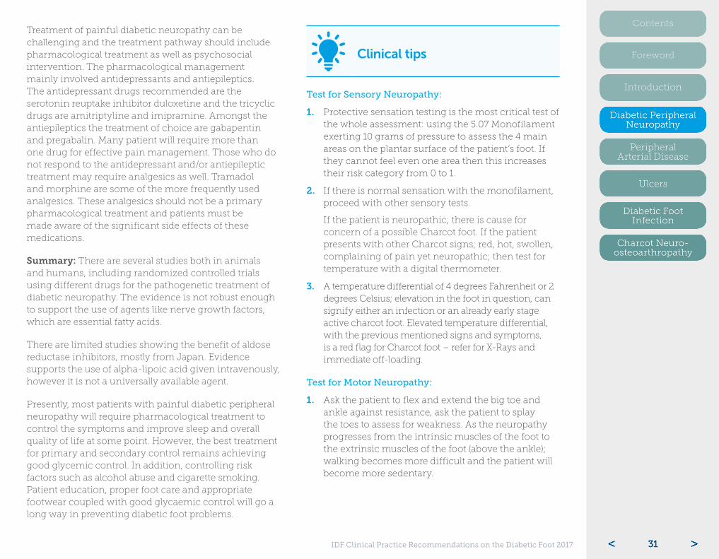
Test for Sensory Neuropathy:
1. Protective sensation testing is the most critical test of the whole assessment: using the 5.07 Monofilament exerting 10 grams of pressure to assess the 4 main areas on the plantar surface of the patient’s foot. If they cannot feel even one area then this increases their risk category from 0 to 1.
2. If there is normal sensation with the monofilament, proceed with other sensory tests.
If the patient is neuropathic; there is cause for concern of a possible Charcot foot. If the patient presents with other Charcot signs; red, hot, swollen, complaining of pain yet neuropathic; then test for temperature with a digital thermometer.
3. A temperature differential of 4 degrees Fahrenheit or 2 degrees Celsius; elevation in the foot in question, can signify either an infection or an already early stage active charcot foot. Elevated temperature differential, with the previous mentioned signs and symptoms, is a red flag for Charcot foot – refer for X-Rays and immediate off-loading.
Test for Motor Neuropathy:
1. Ask the patient to flex and extend the big toe and ankle against resistance, ask the patient to splay the toes to assess for weakness. As the neuropathy progresses from the intrinsic muscles of the foot to the extrinsic muscles of the foot (above the ankle); walking becomes more difficult and the patient will become more sedentary.
Clinical tips
3131IDF Clinical Practice Recommendations on the Diabetic Foot 2017

Test for Vibration Loss:
1. Test for vibration loss with a 128-Hz tuning fork. Test from the distal Hallux initially and if they cannot feel it, move proximally to map out where they are able to feel vibration again. As soon as there is vibration loss proximal to the ankle, it is possible motor neuropathy is progressing proximally. Ask the patient to walk on their heels and toes.
Foot care:
1. Is the patient able to care for their feet and nails?
2. Is the patient cognizant and able to understand the need to assess and care for their feet on a daily basis?
3. Is the patient able to see the bottom of their feet?
4. Is there neuropathy, obesity or retinopathy preventing foot care?
5. Do they understand what diabetic neuropathy and peripheral arterial disease is?
6. Does the patient understand how managing their blood glucose prevents irreversible neuropathy that damages their feet? Do they understand the link between elevated blood glucose, neuropathy, ulcers and amputations leading to death? Do they understand the critical need to keep blood sugars below an HbA1c of seven?
7. Refer for diabetic education and foot care nursing including toenail care and corn and callus removal.
Footwear:
1. What is the structural integrity of the shoe? Is it flexible?
2. Is it appropriate for the insensate foot – is it seamless?
3. Does it have a stable heel counter to control the neuropathic foot? Refer for proper footwear if need be.
4. What is the depth of the removable insert?
5. Is there a thumb width between the end of the longest toe and the end of the shoe?
6. Based on their needs recommended for their Risk Category, do they have diabetic custom orthoses for protection if necessary?
7. Diabetic custom orthoses are flexible, accommodative and usually made of a pink plastazote to show blood if their is any ulcerations.
3232IDF Clinical Practice Recommendations on the Diabetic Foot 2017

References:1. Armstrong DG, Lavery LA. Diabetic foot ulcers: prevention,
diagnosis and classification. American family physician. 1998 Mar;57(6):1325-32.
2. Boulton AJ. The diabetic foot: a global view. Diabetes/Metabolism Research and Reviews. 2000 Sep 1;16(S1):S2-5.
3. Boulton AJ, Vinik AI, Arezzo JC, Bril V, Feldman EL, Freeman R, Malik RA, Maser RE, Sosenko JM, Ziegler D. Diabetic neuropathies. Diabetes care. 2005 Apr 1;28(4):956-62.
4. Boulton A. The diabetic foot: epidemiology, risk factors and the status of care. Diabetes Voice. 2005 Nov;50(S1):5-7.
5. Boulton AJ, Vileikyte L, Ragnarson-Tennvall G, Apelqvist J. The global burden of diabetic foot disease. The Lancet. 2005 Nov 18;366(9498):1719-24.
6. Bernstein RK. Physical signs of the intrinsic minus foot. Diabetes Care. 2003 Jun 1;26(6):1945-6.
7. Reeves A, Swenson R. Chapter 21: Neuromuscular disorders [Internet]. Dartmouth.edu. 2008 [cited 12 May 2017]. Available from: https://www.dartmouth.edu/~dons/part_3/chapter_21.html.
8. Boulton AJ, Armstrong DG, Albert SF, Frykberg RG, Hellman R, Kirkman MS, Lavery LA, LeMaster JW, Mills JL, Mueller MJ, Sheehan P. Comprehensive foot examination and risk assessment. Diabetes care. 2008 Aug 1;31(8):1679-85.
9. O'Brien T, Karem J. An initial evaluation of a proof-of-concept 128-Hz electronic tuning fork in the detection of peripheral neuropathy. Journal of the American Podiatric Medical Association. 2014 Mar;104(2):134-40.
10. Alexander S, Barinov A, Dyck P.J, ...Ziegler D. The Sensory Symptoms of Diabetic Polyneuropathy Are Improved With -Lipoic Acid. Diabetes Care 2003; 26: 770-6.
11. Portenoy R. Development and testing of a neuropathic pain screening questionnaire: ID Pain. Curr Med Res Opin, 2006, 22: 1555–1565.
12. Jacobs AM. A Closer Look at Motor Neuropathy in Patients with Diabetes. Podiatry Today Sept 2008. Volume 21 – Issue 9.
13. Lipsky BA, Aragón-Sánchez J, Diggle M, Embil J, Kono S, Lavery L, Senneville É, Urbančič-Rovan V, Van Asten S, Peters EJ. IWGDF guidance on the diagnosis and management of foot infections in persons with diabetes. Diabetes/metabolism research and reviews. 2016 Jan 1;32(S1):45-74.
14. Brand PW, Yancey P. The Gift Nobody Wants: The Inspiring Story of a Surgeon who Discovers why We Hurt and what We Can Do about it. Zondervan Publ.; 1997.
15. Newton V. Key considerations for assessment and management of limited joint mobility in the diabetic foot. The Diabetic Foot Journal. 2013;16(3):108-14.
16. Chadwick P. Fungal infection of the diabetic foot: the often ignored complication. Diabetic Foot Canada. 2013;1(2):20-4.
17. Pollak R. How to Treat Onychomycosis in Diabetic Patients. Podiatry Today March 2003. Volume 16 – Issue 3.
18. Winston JA, Miller JL. Treatment of onychomycosis in diabetic patients. Clinical Diabetes. 2006 Oct 1;24(4):160-6.
19. Boyko WL, Doyle JJ, Ryu S, Gause DO. PDD5: Onychomycosis and its impact on secondary infection development in the diabetic population. Value in Health. 1999 May 1;2(3):199.
20. Vallianou N, Evangelopoulos A, Koutalas P. Alpha-lipoic acid and diabetic neuropathy. Rev Diabet Stud. 2009 Nov;6(4):230-6.
21. Diabetes Control and Complications Trial Research Group. The effect of intensive treatment of diabetes on the development and progression of long-term complications in insulin-dependent diabetes mellitus. N Engl j Med. 1993 Sep 30;1993(329):977-86.
22. Group UP. Tight blood pressure control and risk of macrovascular and microvascular complications in type 2 diabetes: UKPDS 38. BMJ: British Medical Journal. 1998 Sep 12:703-13.
23. Matsuoka K, Sakamoto N, Akanuma Y, Hotta N, Shichiri M, Toyota T, Oka Y, Kawamori R, Shigeta Y, ADCT Study Group. RETRACTED: A long-term effect of epalrestat on motor conduction velocity of diabetic patients: ARI-Diabetes Complications Trial (ADCT). Diabetes research and clinical practice. 2007 Sep 1;77(3):S263-8.
24. Hotta N, Akanuma Y, Kawamori R, Matsuoka K, Oka Y, Shichiri M, Toyota T, Nakashima M, Yoshimura I, Sakamoto N, Shigeta Y. Long-term clinical effects of epalrestat, an aldose reductase inhibitor, on diabetic peripheral neuropathy. Diabetes care. 2006 Jul 1;29(7):1538-44.
25. Malik RA, Williamson S, Abbott C, Carrington AL, Iqbal J, Schady W, Boulton AJ. Effect of angiotensin-converting-enzyme (ACE) inhibitor trandolapril on human diabetic neuropathy: randomised double-blind controlled trial. The Lancet. 1998 Dec 26;352(9145):1978-81.
26. Brownlee M, Cerami A, Vlassara H. Advanced glycosylation end products in tissue and the biochemical basis of diabetic complications. New England Journal of Medicine. 1988 May 19;318(20):1315-21.
27. Cameron NE, Cotter MA, Dines K, Love A. Effects of aminoguanidine on peripheral nerve function and polyol pathway metabolites in streptozotocin-diabetic rats. Diabetologia. 1992 Oct 1;35(10):946-50.
28. Cameron NE, Cotter MA. Potential therapeutic approaches to the treatment or prevention of diabetic neuropathy: evidence from experimental studies. Diabetic Medicine. 1993 Aug 9;10(7):593-605.
29. Apfel SC, Arezzo JC, Brownlee M, Federoff H, Kessler JA. Nerve growth factor administration protects against experimental diabetic sensory neuropathy. Brain research. 1994 Jan 14;634(1):7-12.
30. Seckel BR. Enhancement of peripheral nerve regeneration. Muscle & nerve. 1990 Sep 1;13(9):785-800.
3333IDF Clinical Practice Recommendations on the Diabetic Foot 2017

Peripheral Arterial Disease (PAD)
34

This chapter aims to provide a reliable and accurate screening process and management specification for health care practitioners and physicians, to decrease the high rates of morbidity and mortality from the misdiagnosis of PAD in diabetic foot disease.
EpidemiologyDisease consequences of the compromised vascular system in diabetes can be among the most devastating complications. Both macrovascular and microvascular diseases are believed to contribute to the consequences of peripheral vascular disease, resulting in the inability of the dysvascular or ischemic limb to heal itself properly. Small injuries may progress to larger wounds because of reduced healing capacity. Delivery of systemic antibiotics can be compromised, leaving infections uncontrolled to the affected foot. Among people with diabetes, all blood vessels regardless of size and function are affected.
The 1999-2000 National Health and Nutrition Examination Survey (NHANES) found that the prevalence of peripheral arterial disease was 4.5% (95% CI 3.4–5.6) in the general population but increased to 9.5% (95% CI 5.5–13.4) in people with diabetes. Other reports have shown higher prevalence of PAD with 12.5% of people with normal glucose tolerance compared to 20.6% of those with diabetes or glucose intolerance.1
In one large population-based study, over half of people with diabetes were found to have absent pedal pulses, a common sign of impaired vascular function. Another study found that in patients with nonpalpable pulses, the relative risk of ulceration was 4.72 (95% CI 3.28, 6.78), compared to a normal exam with all four pulses palpable.1 Ankle-brachial index, despite recognized limitations in the diabetes population, has also been used in diabetes screening. In patients with an ankle-brachial index <0.90, their relative risk has been reported to be 1.25 (95% CI) for developing an ulcer, compared to people with diabetes with a normal ankle-brachial index.2,3
Risk FactorsChanges in lifestyle and an aging population has contributed to diabetes becoming one of the biggest global health challenges. According to the IDF Diabetes Atlas 7th Edition, there were 415 million people living with diabetes in 2015, a total estimated to increase to 642 million by 2040. The Western Pacific region is hit the hardest with 153 million people living with diabetes in 2015, increasing to 215 million by 2040.4
The relationship between abnormal glucose metabolism and lower extremity atherosclerotic lesions (Peripheral Arterial Disease - PAD) is closely related.5,6 Diabetes combined with PAD is not only a risk factor for diabetic foot disease, but also a major cause of amputation. Patients with PAD had much higher rates of cardiovascular events with a prevalence of cardiovascular events as high as 21.14% up to a year after the diagnosis of PAD. This was similar to those without diabetes who had suffered a cardiovascular event.7
Clinical manifestations vary across a wide spectrum from asymptomatic to gangrene of the lower extremity. Most of these patients are unaware that they have PAD and do not seek treatment. Furthermore, some clinicians do not examine and assess their patients with PAD and miss the diagnosis altogether, resulting in high rates of morbidity and mortality.8
35IDF Clinical Practice Recommendations on the Diabetic Foot 2017

ScreeningThese conditions lead to the low rates of assessment, diagnosis, and treatment of patients with PAD.9,10 Early screening and diagnosis would allow appropriate interventions that may delay or even prevent PAD, intermittent claudication, walking impairment and reduce the amputation rate. Additionally, screening for PAD and treating it appropriately can reduce future cardiovascular and cerebrovascular events, including coronary heart disease and stroke, which reduces the mortality rate. Therefore, it is of great clinical significance to strengthen PAD screening and management of people with diabetes and their cardiovascular risks.11
DiagnosisIn order to diagnose PAD, a complete history and physical examination is required. The basic examination must include assessing for skin temperature, discoloration, pedal and posterior tibial artery pulse (which is easy and reliable) and inquiring on the distance the patient is able to walk prior to developing calf pain and/or cramping.
Further examinations are needed for more quantitative, objective and reliable methods of diagnosis. The ABI (ankle-brachial index) is necessary for the diagnosis of PAD. Despite this examination, there are a considerable number of patients who have missed being diagnosed in current clinical practice of endocrinology and metabolism.
High risk populations should be screened annually for PAD
• People with diabetes aged over 50
• People with diabetes with PAD risk factors (such as cardiovascular and cerebral- vascular disease, dyslipidemia, hypertension, cigarette smoking, or duration of diabetes of more than 5 years)
• People with diabetes with a foot ulcer or gangrene should be examined with a comprehensive assessment of arterial disease, regardless of age
36IDF Clinical Practice Recommendations on the Diabetic Foot 2017

Method of Clinical ScreeningPeople with diabetes who complain of leg weakness, thigh or calf muscle pain during walking, or intermittent claudication, should be considered to have PAD until proven otherwise. It is important to remember that Neurogenic Intermittent Claudiation (spinal stenosis) will mimic the symptoms of intermittent claudication due to PAD, but symptoms are usually alleviated after walking in patients with DPN.
Mild to moderate ischemia may present with lower extremity abnormalities, lack of leg hair below the knee, subcutaneous fat atrophy, nail thickening, skin redness (dependent rubor) and diminished pulses.
A patient with severe lower limb ischemia may present with a foot ulcer, severe pain, petechia or ecchymoses, orthostatic edema.12
Patients need a full assessment for chronic occlusive arterial lesions because only 10 to 20% of patients with PAD will have intermittent claudication, and patients with spinal stenosis will also have neurogenic intermittent claudication symptoms. Therefore, if the diagnosis of PAD is only based on the patient’s symptoms or signs, the diagnosis will frequently be missed.
Methods of screening for PAD include:• Intermittent claudication questionnaire score • Comprehensive physical examination of the lower limb
(complete vascular examination, ABI and arterial color Doppler ultrasound examination).
Dorsal pedis artery and posterior tibial artery palpation can provide valuable information for screening for PAD in diabetic patients. Ankle arterial pulse palpation and femoral artery auscultation with a stethoscope are reliable for diagnosing or excluding PAD with very high accuracy (93.8%).13 If the leg and ankle arterial pulses are normal and auscultation reveals no femoral arterial bruit, PAD can be excluded with specificity and negative predictive value as high as 98.3% and 94.9%, respectively.
However, there is still a high misdiagnosis rate despite this. We should therefore emphasize the importance of physical examination in the clinic. If the signs and symptoms of lower limb ischemia are abnormal, normal arterial pulse can exclude PAD.
If PAD is suspected, patients require further investigation, such as ABI and color Doppler ultrasound examination.
Palpation of PulsesPalpation of dorsalis pedis and tibial pulses resulting in a strong arterial pulse (0, non-ischemic), palpable but slightly diminished (1, mild), thready and scarcely palpable (2, moderate) and non-palpable pulses (3, severe).
37IDF Clinical Practice Recommendations on the Diabetic Foot 2017
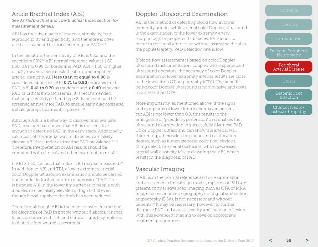
Ankle Brachial Index (ABI) See Ankle/Brachial and Toe/Brachial Index section for
measurement details)
ABI has the advantages of low cost, simplicity, high reproducibility and specificity, and therefore is often used as a standard test for screening for PAD.13,14
In the literature, the sensitivity of ABI is 95%, and the specificity 99%.14 ABI normal reference value is 1.00 - 1.30, 0.91 to 0.99 for borderline PAD. ABI > 1.30 or higher usually means vascular calcification, and impaired arterial elasticity. ABI less than or equal to 0.90 is considered abnormal. ABI 0.71 to 0.90 indicates mild PAD, ABI 0.41 to 0.70 as moderate and ≤ 0.40 as severe PAD or critical limb ischaemia. It is recommended that people with type 1 and type 2 diabetes should be screened annually for PAD, to ensure early diagnosis and initiate prompt treatment, if present.15
Although ABI is a better way to discover and evaluate PAD, research has shown that ABI is not sensitive enough in detecting PAD in the early stage. Additionally, calcinosis of the arterial wall in diabetes, can falsely elevate ABI thus under estimating PAD prevalence.16-19 Therefore, interpretation of ABI results should be combined with clinical and other examination results.
If ABI > 1.30, toe brachial index (TBI) may be measured.19
In addition to ABI and TBI, a lower extremity arterial color Doppler ultrasound examination should be carried out in order to further confirm diagnosis of PAD. This is because ABI in the lower limb arteries of people with diabetes can be falsely elevated or high (> 1.3) even though blood supply to the limb has been reduced.
Therefore, although ABI is the most convenient method for diagnosis of PAD in people without diabetes, it needs to be combined with TBI and clinical signs & symptoms in diabetic foot wound assessment.
Doppler Ultrasound ExaminationABI is the method of detecting blood flow in lower extremity arteries while arterial color Doppler ultrasound is the examination of the lower extremity artery morphology. In people with diabetes, PAD tends to occur in the small arteries, so without assessing distal to the popliteal artery, PAD detection rate is low.
If blood flow assessment is based on color Doppler ultrasound instrumentation, coupled with experienced ultrasound operators, the accuracy of color Doppler examination of lower extremity arteries results are close to the lower limb CT angiography (CTA). The benefit being color Doppler ultrasound is noninvasive and costs much less than CTA.
More importantly, as mentioned above, if the signs and symptoms of lower limb ischemia are present but ABI is not lower than 0.9, this results in the emergence of “pseudo hypertension” and enables the ultrasound examination to successfully diagnose PAD. Color Doppler ultrasound can show the arterial wall, thickening, atherosclerotic plaque and calcification degree, such as lumen stenosis, color flow obvious filling defect, or arterial occlusion, which decreases arterial wall elasticity falsely elevating the ABI, which results in the diagnosis of PAD.
Vascular ImagingIf ABI is in the normal reference and on examination and assessment clinical signs and symptoms of PAD are present, further advanced imaging such as CTA or MRA (magnetic resonance angiography), or digital subtraction angiography (DSA), is not necessary and without benefits.16 It may be necessary, however, to further diagnose PAD and assess severity and location of lesion with this advanced imaging to develop appropriate treatment programmes.
38IDF Clinical Practice Recommendations on the Diabetic Foot 2017

Figure 1 Lower-extremity amputations
0 10 20 30 40 50 60
Mild
Moderate
Severe
Odds ratio
Isc
he
mia
ag
ain
st n
on
isc
hem
icP
atie
nts
(S
core
)
5.7
11.4
53.2
Table 2 Therapeutic interventions by severity grades for ischemia in the diabetic foot syndrome21
Severity Grades
Prevention No ischemia (0) Mild (1) Moderate (2) Severe (3)
Secondary
Early diagnosis,
Prompt intervention
Reducing severity
Limiting damage
Outpatient
Focus in other variables influencing diabetic foot outcomes
Outpatient
Consider vascular assessment.
Endovascular Therapy (ET) or conventional.
Outpatient/Inpatient
Consider ET or by-pass (BP), adjuvants, minor amputations. Prevalence <30% of Lower-extremity amputations
Inpatient/Outpatient
ET or BP is mandatory.prevalence of 70% of Lower-extremity amputations for severe ischemia. Use Jones bandage.
ClassificationResearch data indicates18,20,21 that ischemic classification, graded from mild to severe, is relevant in the prognosis score of the Saint Elian wound classification system (Table 1). Ischemia has the worst prognosis of the ten severity factors for wound healing progress and amputations (Figure 1) in diabetic foot patients.18
Palpation of pedal pulses is an important measurement, and is frequently the only way to assess the arterial perfusion of the feet in many primary care settings. Classification will assist in selecting patients for referral to a vascular or diabetic foot unit or continue their care at the same adequate level. Once the patient is classified, the ischemia grades are useful to provide therapeutic interventions systematically.
39IDF Clinical Practice Recommendations on the Diabetic Foot 2017

The assessment of ischemia in a clinical setting includes patient history and clinical examination in combination with testing such as pedal pulse palpation, the ankle/brachial index (ABI), toe/brachial index (TBI) and waveform analysis.19,22 ABI is a very useful clinical test to assess the arterial blood supply to the foot, but there are limitations to this method when conducted on people with diabetes and TBI is recommended instead.22
Subcategorization of patients by ischemia grades of non-ischaemic patients (scaled as zero), mild (1 point), moderate (2) and severe (3) are categorized after the non-invasive vascular assessment that escalated from pedal pulse palpations to ABI, TBI and waveform pulse analysis.18,20,21
Ankle/Brachial and Toe/Brachial IndexPatients must lay supine for a minimum of 20 minutes and then measure the brachial systolic pressure and the tibialis posterior and dorsalis pedis artery pressures in order to be used for ABI calculation (Hand-held Doppler–8 MHz Doppler probe). Toe pressure is determined by Doppler technique (8 MHz) using a digital cuff on the proximal aspect of the hallux to calculate the TBI. Toe/ brachial index and ABI is determined by dividing the higher systolic pressure of the toe and of the foot or ankle, respectively by the maximum blood pressure of the arms. Ischemia is defined as an ABI < 0.9 and TBI < 0.75.
Toe pressures and the TBI may be used by nurses to diagnose the severity of ischemia in diabetic foot patients.
Clinical tip
Assess pedal pulses
Does the foot feel warm or cold to touch?
Is there hair growing on the toes, feet or legs. This is difficult to assess in women due to shaving.
Can you feel the Dorsalis Pedis pulse. If weak or not present, can you feel the Posterior Tibial pulse?
If weak or not present, can you feel the Popliteal pulse?
Is there Dependent Rubor? This is a fiery to dusky-red coloration visible when the leg is in a dependent position (sitting) but not when it is elevated above the heart. The cause is peripheral arterial disease. To test, elevate the legs from supine to 60 degrees for 1 minute. Pallor within 25 seconds requires an Ankle Brachial Index (ABI) first. If abnormal findings, refer for vascular consultation.
ABI less than 0.90 consistent with Peripheral Arterial Disease – refer for vascular consultation.
40IDF Clinical Practice Recommendations on the Diabetic Foot 2017

References:1. Selvin E, Erlinger TP. Prevalence of and risk factors for peripheral
arterial disease in the United States. Circulation. 2004 Aug 10;110(6):738-43.
2. Boulton AJ, Vileikyte L, Ragnarson-Tennvall G, Apelqvist J. The global burden of diabetic foot disease. The Lancet. 2005 Nov 18;366(9498):1719-24.
3. Bobircã F, Mihalache O, Georgescu D, Pãtraæcu T. The New Prognostic-Therapeutic Index for Diabetic Foot Surgery-Extended Analysis. Chirurgia. 2016;111:151-5.
4. International Diabetes Federation. IDF Diabetes Atlas, 7th edn. Brussels, Belgium: International Diabetes Federation, 2015. http://www.diabetesatlas.org
5. Adler AI, Stevens RJ, Neil A, Stratton IM, Boulton AJ, Holman RR. UKPDS 59: hyperglycemia and other potentially modifiable risk factors for peripheral vascular disease in type 2 diabetes. Diabetes care. 2002 May 1;25(5):894-9.
6. Beks PJ, Mackaay AJ, de Neeling JN, De Vries H, Bouter LM, Heine RJ. Peripheral arterial disease in relation to glycaemic level in an elderly Caucasian population: the Hoorn study. Diabetologia. 1995 Jan 1;38(1):86-96.
7. Steg PG, Bhatt DL, Wilson PW, D’Agostino R, Ohman EM, Röther J, Liau CS, Hirsch AT, Mas JL, Ikeda Y, Pencina MJ. One-year cardiovascular event rates in outpatients with atherothrombosis. Jama. 2007 Mar 21;297(11):1197-206.
8. Hirsch AT, Criqui MH, Treat-Jacobson D, Regensteiner JG, Creager MA, Olin JW, Krook SH, Hunninghake DB, Comerota AJ, Walsh ME, McDermott MM. Peripheral arterial disease detection, awareness, and treatment in primary care. Jama. 2001 Sep 19;286(11):1317-24.
9. Hirsch AT, Murphy TP, Lovell MB, Twillman G, Treat-Jacobson D, Harwood EM, Mohler ER, Creager MA, Hobson RW, Robertson RM, Howard WJ. Gaps in public knowledge of peripheral arterial disease. Circulation. 2007 Oct 30;116(18):2086-94.
10. Lovell M, Harris K, Forbes T, Twillman G, Abramson B, Criqui MH, Schroeder P, Mohler ER, Hirsch AT. Peripheral arterial disease: lack of awareness in Canada. Canadian Journal of Cardiology. 2009 Jan 1;25(1):39-45.
11. Ferket BS, Spronk S, Colkesen EB, Hunink MM. Systematic review of guidelines on peripheral artery disease screening. The American journal of medicine. 2012 Feb 29;125(2):198-208.
12. Halperin JL. Evaluation of patients with peripheral vascular disease. Thrombosis research. 2002 Jun 1;106(6):V303-11.
13. Khan NA, Rahim SA, Anand SS, Simel DL, Panju A. Does the clinical examination predict lower extremity peripheral arterial disease?. Jama. 2006 Feb 1;295(5):536-46.
14. Xu D, Li J, Zou L, Xu Y, Hu D, Pagoto SL, Ma Y. Sensitivity and specificity of the ankle—brachial index to diagnose peripheral artery disease: a structured review. Vascular Medicine. 2010 Oct;15(5):361-9.
15. Norgren L, Hiatt WR, Dormandy JA, Nehler MR, Harris KA, Fowkes FG, Tasc II Working Group. Inter-society consensus for the management of peripheral arterial disease (TASC II). European Journal of Vascular and Endovascular Surgery. 2007 Dec 31;33(1):S1-75.
16. Hoe J, Koh WP, Jin A, Sum CF, Lim SC, Tavintharan S. Predictors of decrease in ankle–brachial index among patients with diabetes mellitus. Diabetic Medicine. 2012 Sep 1;29(9):e304-7.
17. Hirsch AT, Haskal ZJ, Hertzer NR, Bakal CW, Creager MA, Halperin JL, Hiratzka LF, Murphy WR, Olin JW, Puschett JB, Rosenfield KA. ACC/AHA 2005 Practice Guidelines for the management of patients with peripheral arterial disease (lower extremity, renal, mesenteric, and abdominal aortic). Circulation. 2006 Mar 21;113(11):e463-654.
18. Martínez-De Jesús FR. A checklist system to score healing progress of diabetic foot ulcers. The international journal of lower extremity wounds. 2010 Jun;9(2):74-83.
19. Brooks B, Dean R, Patel S, Wu B, Molyneaux L, Yue DK. TBI or not TBI: that is the question. Is it better to measure toe pressure than ankle pressure in diabetic patients?. Diabetic Medicine. 2001 Jul 1;18(7):528-32.
20. Huang Y, Xie T, Cao Y, Wu M, Yu L, Lu S, Xu G, Hu J, Ruan H. Comparison of two classification systems in predicting the outcome of diabetic foot ulcers: the Wagner grade and the saint elian wound score systems. Wound Repair and Regeneration. 2015 May 1;23(3):379-85.
21. Martinez- De Jesus FR. Validation of the Ischemia Severity Scale (ISS) based on non-invasive vascular assessments for outcomes prediction in diabetic foot wounds. (Report in progress)
22. Williams DT, Harding KG, Price P. An evaluation of the efficacy of methods used in screening for lower-limb arterial disease in diabetes. Diabetes care. 2005 Sep 1;28(9):2206-10.
41IDF Clinical Practice Recommendations on the Diabetic Foot 2017

Ulcers
4242

The incidence of diabetic foot ulcers is up to 25% over a patient’s lifetime.1 The onset is variable in patients with type 1 diabetes. Foot ulcers occur in 15-25% of people with diabetes1,2 which equates to slightly more than 2% annually and between 5-7.5% of those patients with neuropathy3,4 Foot ulcers and infections are the most common reason for hospital admission in people with diabetes in the United States. The prevalence of diabetic ulcers is 7-8%.
Since diabetes and obesity are growing at epidemic proportions and with an increasing elderly population with chronic conditions, will make coordinated care more essential and valued. The team approach to ulcer and amputation prevention has been well documented in medical literature, aiming to improve quality of life and decrease cost.
Natural historyThe natural history of a diabetic foot ulcer without medical intervention usually progresses from ulcer to infected ulcer to deep infected ulcer to osteomyelitis (bone infection) and ends in amputation or death. 56% of ulcers become infected and 1 in 5 of these will require some level of amputation. Additionally, it has been estimated that 15% of diabetic foot ulcers result in lower extremity amputations and 85% of diabetic patients who undergo lower extremity amputations had an ulcer prior to amputation.5,6 The 5-year relative mortality after diabetic foot ulcer is 48%.7 This is clearly higher than most cancers (breast, lymphoma, etc.).
EtiologyThe etiology of a diabetic foot ulcer (DFU) is multifaceted. No single risk factor is responsible for a foot ulcer. Several component causes added together create a sufficient cause for ulceration. Peripheral neuropathy (loss of sensation) frequently occurs, 20% at the time of diagnosis and about 8-12 years after developing type 2 diabetes, and is the permissive factor in ulcer development.8
The three main factors that determine the likelihood of ulceration in a neuropathic foot are:9
1. The severity and localization of the sensory loss to the plantar foot
2. The footwear used to disperse the magnitude of the forces on the foot while walking. Since Pressure = Force/Area (P=F/A), with the force being the patient’s body weight, the surface area is indirectly proportional to the plantar peak pressures. Therefore, shoe modifications are designed to increase the surface area, thus reducing peak pressure.
3. The role walking distance plays. The walking distance causes a moderate, repetitive stress that builds up over time. This cumulative effect leads to the point of an inflammatory response, which serves as a warning sign of impending skin breakdown. This inflammation irritates the polymodal nociceptors in the skin, initiating the pathway to ulceration. 9 Calluses will form in areas of structural deformity with limited joint mobility. The hard callus acts like a foreign body and increases the peak plantar pressure. During ambulation, the pressure from the callus causes deep tissue injury with hemorrhage within the calluses and an ulcer then forms. Most ulcers are preceded by a callus.
4343IDF Clinical Practice Recommendations on the Diabetic Foot 2017

Risk factorsReiber et al. described the causal pathways for diabetic foot ulceration.10 They consist of three main factors: peripheral neuropathy, minor trauma and deformity. A minor trauma can be repetitive, low pressure or high pressure over a short duration. A deformity can be visual, like a hammer toe or bunion, or it could be invisible, such as limited joint mobility. When combined together, these three factors were responsible for more than 63% of foot ulcers in a multi-centered retrospective cohort involving 148 patients. Asking three simple clinical questions can stratify patients risk for ulceration:
1. Does the patient have peripheral neuropathy?
Loss of protective sensation (LOPS) is a term that connotes the patient’s Diabetic peripheral neuropathy (DPN) is severe enough to place the foot at risk for ulceration. DPN is the single most important risk factor for the development of diabetic foot ulcers.
DPN affects the three divisions of the peripheral nervous system; sensory, motor and autonomic. Sensory neuropathy ultimately resulting in anaesthesia leaves a patient at risk for unfelt trauma. Repetitive cycles of low to moderate pressure in an insensate foot initially causes inflammation, and progresses to hematoma or bulla (blister) formation, then skin breakdown.9 Motor neuropathy causes intrinsic muscle atrophy, resulting in a high-arched foot with hammer toes (intrinsic minus foot), causing abnormal weight-bearing and increased plantar foot pressure. The autonomic nervous system controls the ability of blood vessels to dilate and constrict. This is nitric oxide(NO) dependent. It has been shown that NO is depleted in people with diabetes.11 Autonomic neuropathy reduces sweating and oil secretion, resulting in dry skin which can cause fissures and this can lead to ulceration.
4444IDF Clinical Practice Recommendations on the Diabetic Foot 2017

The foot should be inspected for bony prominences or any visual deformities. A foot deformity could be hammer toes, bunions, a prominent metatarsal head, or a Charcot foot. Patients with neuropathy and deformity have a more than 12-fold increased risk of ulceration than patients without neuropathy.12 It is important to recognize that foot deformities alone do not cause ulceration. However, when combined with sensory neuropathy, the conditions are favorable for ulcer formation. Some deformities are not visible to inspection. Limited joint mobility (LJM) is just as important predictor for ulceration as a visible deformity.13 LJM causes increased pressure at locations distal to the joint with limited motion. An example is hallux limitus (arthritis in the first metatarsal-phalangeal joint). This helps to explain why the hallux (great toe) is the most common site for diabetic foot ulceration, as the limited metatarsophalangeal joint motion increases pressure at the distal hallux. This is an opportunity where prophylactic surgery can prevent foot ulceration.
2. Does the patient have a deformity?
Risk factorsReiber et al. described the causal pathways for diabetic foot ulceration.6 They consist of three main factors: peripheral neuropathy, minor trauma and deformity. A minor trauma can be repetitive, low pressure or high pressure over a short duration. A deformity can be visual, like a hammer toe or bunion, or it could be invisible, such as limited joint mobility. When combined together, these three factors were responsible for more than 63% of foot ulcers in a multi-centered retrospective cohort involving 148 patients. Asking three simple clinical questions can stratify patients risk for ulceration:
4545IDF Clinical Practice Recommendations on the Diabetic Foot 2017

A history of diabetic foot pathology, such as ulceration, amputation or Charcot foot, is a risk factor for future ulceration. A history of diabetic foot ulcer is 36 times more likely to lead to development of a future ulcer.12 A previous major amputation increases the risk of ulceration and loss of the contralateral leg. A person with diabetes and a lower extremity amputation has a 50% chance of developing a serious lesion on the contralateral limb within two years.14
3. Does the patient have a history of diabetic foot pathology: ulceration, amputation or Charcot foot?
Risk factorsReiber et al. described the causal pathways for diabetic foot ulceration.6 They consist of three main factors: peripheral neuropathy, minor trauma and deformity. A minor trauma can be repetitive, low pressure or high pressure over a short duration. A deformity can be visual, like a hammer toe or bunion, or it could be invisible, such as limited joint mobility. When combined together, these three factors were responsible for more than 63% of foot ulcers in a multi-centered retrospective cohort involving 148 patients. Asking three simple clinical questions can stratify patients risk for ulceration:
4646IDF Clinical Practice Recommendations on the Diabetic Foot 2017

Table 1 Saint Elian Score System for 10 subcategorized wound severity factors and III Grades for prognosis
Score (Severity)
Factors 1 (Mild) 2 (Moderate) 3 (Severe) Score
1. Primary zone (location) Phalanges Metatarsal Tarsal
2. Topographic aspects (location) Dorsal or plantar Lateral or medial Two or more
3. Zone number One Two Three
4. Ischemia
Palpable pulses slightly diminished ABI (0.89-0.7) TBI (0.74-0.60)
Scarcely palpable pulses ABI (0.69-0.5) TBI (0.59-0.30)
Non palpable pulses ABI (<0.5) TBI (<0.30)
5. InfectionErythema < 2 cm Purulent discharge, warmth, tenderness
Erythema > 2 cm Muscles, tendons or bone or joint infection
Systemic Inflammatory Response Syndrome. Secondary hyper or hypoglycemia
6. Edema Periwound One foot or legBilateral secondary to comorbidities
7. Neuropathy
Protective Sensation diminished (128 HZ tuning fork, SWM, Vibratip, Ipswich)
Protective Sensation absent (128 HZ tuning fork, SWM, Vibratip, Ipswich)
Diabetic Neuro-osteoarthropathy (DNOA)-Charcot
8. Area Small: < 10 cm2 Medium: 11-40 cm2 Big: > 40 cm2
9. Depth Superficial (skin) Tendons, fascia, muscles Deep joint and bones
10. Wound healing phase Epithelialization Granulating Inflammatory
Score sum:
Final score Grade (Severity) Prognosis
< 10 I (Mild) Likely successful wound healing. Low risk for LEA
11-20 II (Moderate)Partial foot-threatening; outcome related to “state-of-the-art” therapies used and associated with a good patient biological response. < 30% LEA
21-30 III (Severe)Limb- and life-threatening; outcome unrelated to “state-of-the-art” therapies because of poor biological patient response. > 70% LEA
Check the severity column and annotate the score (1 to 3) at the right column. Score 0, for absence of the aggravating factor (ischemia, infection, edema or neuropathy). SWM; Semmes Weinstein Monofilament. LEA; Lower extremity amputations.
47IDF Clinical Practice Recommendations on the Diabetic Foot 2017

TreatmentThe goal of ulcer treatment is to achieve rapid wound closure to prevent serious downstream consequence such as amputation and reduced quality of life.
Treatment should occur in a stepwise approach.15 The first and most urgent step is to treat any infection that is present. Infections can be graded according to the Infectious Diseases Society of America (IDSA) grading scale.16 This scale helps the clinician determine what class of antibiotic to use and whether to treat the patient in an out-patient or in-patient setting. If present, arterial insufficiency needs to be managed. As a team, diabetologists, podiatrists and vascular surgeons significantly improve clinical outcomes. The vascular surgeon can perform a variety of procedures, from angioplasty to open bypass to restore blood flow to the foot.
The mainstay of therapy for DFU is offloading of pressure. This is done with bedrest, a wheel chair, crutches, or modalities that can keep the patient weight-bearing, such as a total contact cast (TCC), a removable cast walker, or a variety of other devices. However, many other modalities, such as felt or foam padding or wedged shoes have been tried but failed to off-load the foot adequately. TCC is an alternative but not equivalent for offloading the diabetic foot, but few clinicians use it because it is time consuming and can cause more complications. A trained clinician or cast technician is required to apply a TCC. Another more recent concept is to use a removable cast walker rendered irremovable to enforce compliance. This is referred to as an instant total contact cast (iTCC).17 This can be done through the use of plastic cable ties, duct tape or fiberglass.
Surgery can be used as a method of offloading. Sometimes the surgeon may perform an Achilles tendon lengthening to relieve pressure under the forefoot. For ulcers under the great toe, a first metatarsophalangeal joint arthroplasty may be effective at reducing distal pressure.18
The basics of wound care must consist of regular debridement of fibrous or non-viable tissue, paring the hyperkeratotic rim, and creating a moist environment and off-loading. Negative pressure wound therapy (NPWT) has revolutionized wound care. It can produce granulation tissue quickly and fill in large defects. It can also be used in combination with other modalities such as skin substitutes and skin grafts.
When the wound bed is granular and level with the surrounding skin, advanced modalities can be employed to quicken closure of the wound. These include bioengineered tissue, skin expansion, flaps, and skin grafts. Each have their benefits and drawbacks and the reader is referred to document and technology assessments of the diabetic foot ulcer for a thorough review.19
Each footstep without protection will undo healing of these plantar wounds. Most patients cheat and walk without protection to go to the bathroom at night and will undo days worth of healing causing recalcitrant ulcers.
Patients will be much more compliant if they understand that the role of off-loading is to 'get off their feet' to close their ulcers quickly to prevent amputations that can 'take their feet'.
4848IDF Clinical Practice Recommendations on the Diabetic Foot 2017

Adjuvant TherapyAdjunctive therapies may be tried if available and cost is not an issue. Re-evaluation of vascular status, infection control and off-loading is recommended to ensure optimization before initiation of adjunctive wound therapy.
1. Systemic reviews of various wound dressings and topical antimicrobials have found no evidence that any specific type of therapy is better than others.20,21
2. Foot ulcers with heavy exudate need a dressing that absorbs moisture, while dry wounds need topical treatments that add moisture.
3. For DFUs that fail to demonstrate improvement (>50% wound area reduction) after a minimum of 4 weeks of standard wound therapy, we recommend adjunctive wound therapy options. These include negative pressure therapy, biologics (platelet-derived growth factor [PDGF], living cellular therapy, extracellular matrix products, amnionic membrane products).22 Choice of adjuvant therapy is based on clinical findings, availability of therapy, and cost-effectiveness; there is no recommendation on efficacy or effectiveness of these therapy options.
Clinical tip
• Any calluses or corns?
• Any ulcerations? Is the wound infected? Is there any redness, swelling, pain, exudate or odor?
• Is there any fissuring?
• Is the skin dry due to Autonomic Neuropathy?
• Any red hot spots (irritation/friction areas) indicating high peak pressure areas, plantarly, dorsally or on the sides of the feet, due to foot wear and the repetitive trauma of walking?
• Is Tinea Pedis present?
Refer for foot care nursing or wound care as necessary. Additionally, provide education for care of the neuropathic foot and treatment if Tinea Pedis is present to treat this silent infection.
4949IDF Clinical Practice Recommendations on the Diabetic Foot 2017

References:1. Singh N, Armstrong DG, Lipsky BA. Preventing foot ulcers in
patients with diabetes. Jama. 2005 Jan 12;293(2):217-28.
2. Boulton AJ, Vileikyte L., Ragnarson-Tennvall G., et al. The global burden of diabetic foot disease. The Lancet 366: 1719, 2005.
3. Abbott CA, Carrington AL, Ashe H, et al. The North-West Diabetes Foot Care Study: incidence of, and risk factors for, new diabetic foot ulceration in a community-based patient cohort. Diabet Med 2002; 19: 377-384.
4. Abbott CA, Vileikyte L., Williamson S, et al. Multi-center study of the incidence of and predictive risk factors for diabetic neuropathic foot ulceration. Diabetes Care, 1998; 21: 1071-1075.
5. Ramsey SD, Newton K, Blough D, et al. incidence, outcomes, and cost of foot ulcers in patients with diabetes. Diabetes Care, 1999; 22(3): 382-387.
6. Boulton AJ, Kirsner RS, Vileikyte L. Clinical Practice: Neuropathic diabetic foot ulcers. New Engl J Med 2004; 351 (1): 48-53
7. Armstrong DG, Wrobel J, Robbins JM. Guest editorial: are diabetes-related wounds and amputations worse than cancer. Int Wound J. 2007 Dec 1;4(4):286-7.
8. Masson EA, Hay EM, Stockley I, Veves A, Betts RP, Boulton AJ. Abnormal foot pressures alone may not cause ulceration. Diabetic Medicine. 1989 Jul 1;6(5):426-8.
9. Brand PW. Tenderizing the foot. Foot & ankle international. 2003 Jun 1;24(6):457-61.
10. Reiber GE, Vileikyte LO, Boyko ED, Del Aguila M, Smith DG, Lavery LA, Boulton AJ. Causal pathways for incident lower-extremity ulcers in patients with diabetes from two settings. Diabetes care. 1999 Jan 1;22(1):157-62.
11. Hseu H, Quione S, Amer. Journal of Cardiology, 2003;92(Suppl)10J-17J.
12. Armstrong DG, Wrobel J, Robbins JM. Guest editorial: are diabetes-related wounds and amputations worse than cancer. Int Wound J. 2007 Dec 1;4(4):286-7.
13. Fernando DJ, Masson EA, Veves A, Boulton AJ. Relationship of limited joint mobility to abnormal foot pressures and diabetic foot ulceration. Diabetes care. 1991 Jan 1;14(1):8-11.
14. Goldner MG. The fate of the second leg in the diabetic amputee. Diabetes. 1960 Mar 1;9(2):100-3.
15. Rogers LC, Bevilacqua NJ. Organized programs to prevent lower-extremity amputations. Journal of the American Podiatric Medical Association. 1998 ;88(7) :337-343.
16. Lavery LA, Armstrong DG, Murdoch DP, Peters EJ, Lipsky BA. Validation of the Infectious Diseases Society of America's diabetic foot infection classification system. Clinical infectious diseases. 2007 Feb 15;44(4):562-5.
17. Armstrong DG, Lavery LA, Wu S, Boulton AJ. Evaluation of removable and irremovable cast walkers in the healing of diabetic foot wounds. Diabetes Care. 2005 Mar 1;28(3):551-4.
18. Rogers LC, Armstrong DG. Podiatry care. In: Cronenwett JL, Johnston KW,eds. Rutherford's Vascular Surgery 7th Ed. Philadelphia: Saunders Elsevier; 2010:1747-1760.
19. Snyder RJ, Kirsner RS, Warriner 3rd RA, Lavery LA, Hanft JR, Sheehan P. Consensus recommendations on advancing the standard of care for treating neuropathic foot ulcers in patients with diabetes. Ostomy Wound Manage. 2010 Apr 1;56(4 Suppl):S1-24.
20. Gottrup F, Apelqvist J. Present and new techniques and devices in the treatment of DFU: a critical review of evidence. Diabetes Metab Res Rev 2012; 28 Suppl 1: 64-71.
21. Gottrup F, Apelqvist J, Bjarnsholt T, Cooper R, Moore Z, Peters EJ, et al. Antimicrobials and Non-Healing Wounds. Evidence, controversies and suggestions-key messages. J Wound Care 2014; 23(10): 477-478, 480, 482.
5050IDF Clinical Practice Recommendations on the Diabetic Foot 2017

Diabetic foot infection
5151

Definition:Manifestations of infectious process in soft tissue or bone anywhere below malleoli in a person with diabetes.1
Grading(Adapted from IDSA and the Saint Elian Wound Score System*)2,3
Table 1 Classification for Diabetic Foot Infections – Saint Elian Wound Score System and Infectious Disease Society of America
Description Severity grade Score
a. No signs or symptoms of infection Non infected 0
b. Erythema between 0.5 mm to 2 cm, induration, tenderness, warmth, and purulent discharge.
Mild 1
c. Erythema > 2 cm, muscle, tendon, orbone or joint infection. Moderate 2
d. Any local infection with systemic inflammatory response (SIRS) manifested by at least 2 of following:
• Temperature > 38 or < 36
• Heart rate > 90 beats/min,
• Respiratory rate > 20 breaths/min or PaCO2 < 32 mmHg,
• White blood cell count > 12000 or < 4000 cells/μL or 10% immature (band) forms; or severe metabolic disturbances (hyperglycemia or hypoglycemia)
Severe 3
For severe infection and some moderate grade infection, hospitalization is needed for limb preservation.
52IDF Clinical Practice Recommendations on the Diabetic Foot 2017
1. At initial evaluation: diagnosed clinically, based on the presence of local or systemic signs or symptoms of inflammation.4-7
2. Obtain vital signs and appropriate blood tests. It is important to consider the white blood cell count may remain lower than would be expected based on clinical signs since the elevated blood glucose can cause immunosuppression.
3. Assess arterial perfusion and decide whether and when further vascular assessment or revascularization is needed.
4. Accurately assessing a diabetic foot wound usually requires first debriding any callus and necrotic tissue to fully visualize the wound. After debridement, probe and assess the depth and extent of the wound and the infection (location, malodor, purulence, surrounding erythema and edema to establish the severity)
5. A deep space infection may have deceptively few superficial signs. The clinician should consider this possibility in a patient with:8-10
• evidence of systemic toxicity
• inflammation distant from the skin wound
• persistent infection lack of wound healing or elevated inflammatory markers despite apparently appropriate therapy
• deterioration of previously controlled glycaemia
• pain in a previous insensate foot
Assessment
53IDF Clinical Practice Recommendations on the Diabetic Foot 2017

1. Obtain cultures, preferably of a tissue specimen.
2. If swabs are the only available method, they should be taken only after debriding and cleaning the wound.
3. Blood cultures are only indicated for severe infections where there are signs of systemic manifestations of sepsis.6
4. Acute infection in a previously untreated patient.11,12
• Aerobic Gram-positive cocci often as a monomicrobial infection.
5. Deep or chronic wounds.11,12
• Often harbor polymicrobial flora, including aerobic Gram-negative and obligate anaerobic bacteria.
Microbiological examination
54IDF Clinical Practice Recommendations on the Diabetic Foot 2017

1. In all patients presenting with a new diabetic foot infection (DFI), serial plain radiographs of the affected foot should be obtained to identify bone abnormalities (deformity, destruction) as well as soft tissue gas and radiopaque foreign bodies.
2. Serial radiographs should be used to reassess potential osseous changes when healing progresses slowly or signs and/or symptoms worsens.
3. For those patients who require additional imaging, particularly when soft tissue abscess is suspected or the diagnosis of osteomyelitis remains uncertain, we recommend using magnetic resonance imaging (MRI) as the study of choice. MRI is a valuable diagnostic tool for osteomyelitis, with high sensitivity and specificity, if radiographs are inconclusive.6,13,14
4. In the acute phase, charcot neuro-arthropathy may clinically appear similar to osteomyelitis. MRI findings consistent with an infection generally include soft tissue fluid collection, sinus tracts and diffuse marrow edema.
Imaging consideration
55IDF Clinical Practice Recommendations on the Diabetic Foot 2017

1. Definite diagnosis of bone infection usually requires positive results of bone cultures from an aseptically obtained bone sample or histological findings consistent with bone infection (inflammatory cells, necrosis).15 When bone is debrided to treat osteomyelitis, we recommend sending a sample for culture and histology.
2. For an infected open wound, perform a probe-to-bone test. In a patient at low risk for osteomyelitis, a negative test largely rules out the diagnosis, while in a high-risk patient*, a positive test is largely diagnostic.16-20
3. A probable diagnosis of bone infection is reasonable if there are positive results on a combination of diagnostic tests, such as probe-to-bone, serum inflammatory markers, plain X-ray, MRI or radionuclide scanning.
4. In long standing ulcers one must rule out osteomyelitis even if the probe to bone test is negative and an x-ray must be performed.
*High risk patients include
- Large, extensive and/or deep ulcers
- Ulcers overlying bony prominences
- Ulcers with delayed healing
- Exposed bone or positive probe to bone
- Recurrent soft tissue infections
- Radiographic bone destruction
Diabetic Foot Osteomyelitis
56IDF Clinical Practice Recommendations on the Diabetic Foot 2017

1. Urgent surgical interventions are usually necessary in cases of deep abscesses, compartment syndrome and virtually all necrotizing soft tissue infections.10
2. Surgical intervention is usually advisable in cases of osteomyelitis accompanied by spreading soft tissue infection, destroyed soft tissue envelope, progressive bone destruction on X-ray or bone protruding through the ulcer.
3. When the wound has a dry eschar, especially in an ischemic foot, it is often best to avoid debriding the necrotic tissue; often, these resolve with autoamputation.
4. Bone resection and amputation are often necessary when there is extensive soft tissue necrosis, osteomyelitis or to provide a more functional foot.
Surgical Treatment of DFI
57IDF Clinical Practice Recommendations on the Diabetic Foot 2017

1. All clinically infected diabetic foot wounds require antimicrobial therapy. Nevertheless, antimicrobial therapy for clinically non-infected wounds is not recommended.21-23
2. Select specific antibiotic agents for treatment, based on the likely or proven causative pathogens, their antibiotic susceptibilities, the clinical severity of the infection, evidence of efficacy of the agent for diabetic foot infection, patient history (e.g., allergies or intolerance) and cost.
3. A course of antibiotic therapy of 1-2 weeks is usually adequate for most mild and moderate infections.24,25
• For more serious skin and soft tissue infections, 3 weeks is usually sufficient.
• Antibiotics can be discontinued when signs and symptoms of infection have resolved, even if the wound has not healed.
4. Initially, parenteral antibiotics therapy is needed for most severe infections and some moderate infections, with a switch to oral therapy when the infection is responding.
5. For patients with a foot ulcer and severe PAD, antibiotics play an important role in treating and preventing further spread of infection. In some cases, a successful revascularization for these patients may transiently increase the bacterial activity.26,27
6. For diabetic foot osteomyelitis, 6 weeks of antibiotic therapy is required for patients who do not undergo resection of infected bone and no more than a week of antibiotic treatment is needed after all infected bone is resected.25,28 The regimen should usually cover Staphylococcus aureus as it is the most common pathogen. However, without revascularization, some patients will not have adequate blood flow to allow for adequate antibiotic tissue concentrations in the area of the infection.
7. For patients with foot ulcers and necrotizing fasciitis, antibiotics to cover both aerobic and anaerobic microorganisms is recommended.29
Antimicrobial Therapy
58IDF Clinical Practice Recommendations on the Diabetic Foot 2017

0,0 0,5 1,0 1,5 2,0
Mild
Moderate
Severe
Odds ratio
Infe
cti
on
0.9
1.0
1.8
These interventions summarize a systematic approach (Table 2) to avoid lower-extremity amputations with a significant protective odds ratio below 2.0 for mild to severe infection with diabetic foot comprehensive care (Figure 1).33
Table 2 Therapeutic interventions by severity risk grades for diabetic foot infections33
Severity Grades*
Prevention Mild (1) Moderate (2) Severe (3)
Secondary
Early diagnosis,
Prompt intervention
Reducing severity
Limiting damage
Outpatient
Antibiotics oral, 1-4 wk targeting aerobic gram-positive cocci. (AGPC) Methicillin-resistant S. aureus (MRSA) 50%.
Outpatient/Inpatient
Oral (or initial parenteral) 1-3 wk. targeting (AGPC). Check MRSA (30%) and anaerobes. Debridement, infected bone remotion or minor amputations
Inpatient/Outpatient
Initial parenteral, switch to oral when possible. 2-4 wk. Very broad-spectrum coverage.
Figure 1 Lower-extremity amputations
Does the patient have any thickening of the toenails? Ingrown nails? Is onychomycosis present?
Refer for foot care nursing to prevent little problems from becoming big problems. Refer for treatment of Onychomycosis, if present. Onychomycosis is a silent infection that is progressive and the body is not recognizing and fighting; treat this in the early stages as this escalates the patients risk category for complications.
59IDF Clinical Practice Recommendations on the Diabetic Foot 2017

References:1. Peters EJ, Lipsky BA. Diagnosis and management of infection
in the diabetic foot. Medical Clinics of North America. 2013 Sep 30;97(5):911-46.
2. Lavery LA, Armstrong DG, Murdoch DP, Peters EJ, Lipsky BA. Validation of the Infectious Diseases Society of America's diabetic foot infection classification system. Clinical infectious diseases. 2007 Feb 15;44(4):562-5.
3. Martínez-De Jesús FR. A checklist system to score healing progress of diabetic foot ulcers. The international journal of lower extremity wounds. 2010 Jun;9(2):74-83.
4. Lipsky BA, Peters EJ, Berendt AR, Senneville E, Bakker K, Embil JM, Lavery LA, Urbančič-Rovan V, Jeffcoate WJ. Specific guidelines for the treatment of diabetic foot infections 2011. Diabetes/metabolism research and reviews. 2012 Feb 1;28(S1):234-5.
5. Lipsky BA, Berendt AR, Deery HG, Embil JM, Joseph WS, Karchmer AW, LeFrock JL, Lew DP, Mader JT, Norden C, Tan JS. Diagnosis and treatment of diabetic foot infections. Clinical Infectious Diseases. 2004 Oct 1;39(7):885-910.
6. Lipsky BA, Berendt AR, Cornia PB, Pile JC, Peters EJ, Armstrong DG, Deery HG, Embil JM, Joseph WS, Karchmer AW, Pinzur MS. 2012 Infectious Diseases Society of America clinical practice guideline for the diagnosis and treatment of diabetic foot infections. Clinical infectious diseases. 2012 Jun 15;54(12):e132-73.
7. Armstrong DG, Lavery LA, Harkless LB. Validation of a diabetic wound classification system: the contribution of depth, infection, and ischemia to risk of amputation. Diabetes care. 1998 May 1;21(5):855-9.
8. Bridges Jr RM, Deitch EA. Diabetic foot infections. Pathophysiology and treatment. The Surgical clinics of North America. 1994 Jun;74(3):537-55.
9. Aragón-Sánchez J. Seminar review: a review of the basis of surgical treatment of diabetic foot infections. The international journal of lower extremity wounds. 2011 Mar;10(1):33-65.
10. Ger R. Newer concepts in the surgical management of lesions of the foot in the patient with diabetes. Surgery, gynecology & obstetrics. 1984 Mar;158(3):213-5.
11. Lipsky BA, Pecoraro RE, Wheat LJ. The diabetic foot. Soft tissue and bone infection. Infectious disease clinics of North America. 1990 Sep;4(3):409-32.
12. Wheat LJ, Allen SD, Henry M, Kernek CB, Siders JA, Kuebler T, Fineberg N, Norton J. Diabetic foot infections: bacteriologic analysis. Archives of internal medicine. 1986 Oct 1;146(10):1935-40.
13. Dinh MT, Abad CL, Safdar N. Diagnostic accuracy of the physical examination and imaging tests for osteomyelitis underlying diabetic foot ulcers: meta-analysis. Clinical Infectious Diseases. 2008 Aug 15;47(4):519-27.
14. Kapoor A, Page S, LaValley M, Gale DR, Felson DT. Magnetic resonance imaging for diagnosing foot osteomyelitis: a meta-analysis. Archives of internal medicine. 2007 Jan 22;167(2):125-32.
15. Berendt AR, Peters EJ, Bakker K, Embil JM, Eneroth M, Hinchliffe RJ, Jeffcoate WJ, Lipsky BA, Senneville E, Teh J, Valk GD. Diabetic foot osteomyelitis: a progress report on diagnosis and a systematic review of treatment. Diabetes/metabolism research and reviews. 2008 May 1;24(S1):S145-61.
16. Aragón-Sánchez J, Lipsky BA, Lázaro-Martínez JL. Diagnosing diabetic foot osteomyelitis: is the combination of probe-to-bone test and plain radiography sufficient for high-risk inpatients?. Diabetic Medicine. 2011 Feb 1;28(2):191-4.
17. Shone A, Burnside J, Chipchase S, Game F, Jeffcoate W. Probing the validity of the probe-to-bone test in the diagnosis of osteomyelitis of the foot in diabetes. Diabetes Care. 2006 Apr 1;29(4):945-.
18. Lozano RM, Fernández ML, Hernández DM, Montesinos JV, Jiménez SG, Jurado MA. Validating the probe-to-bone test and other tests for diagnosing chronic osteomyelitis in the diabetic foot. Diabetes Care. 2010 Oct 1;33(10):2140-5.
19. Grayson ML, Gibbons GW, Balogh K, Levin E, Karchmer AW. Probing to bone in infected pedal ulcers: a clinical sign of underlying osteomyelitis in diabetic patients. Jama. 1995 Mar 1;273(9):721-3.
20. Lavery LA, Armstrong DG, Peters EJ, Lipsky BA. Probe-to-bone test for diagnosing diabetic foot osteomyelitis. Diabetes care. 2007 Feb 1;30(2):270-4.
21. Robson MC, Mannari RJ, Smith PD, Payne WG. Maintenance of wound bacterial balance. The American journal of surgery. 1999 Nov 30;178(5):399-402.
22. Chantelau E, Tanudjaja T, Altenhöfer F, Ersanli Z, Lacigova S, Metzger C. Antibiotic treatment for uncomplicated neuropathic forefoot ulcers in diabetes: a controlled trial. Diabetic Medicine. 1996 Feb 1;13(2):156-9.
23. Hirschl M, Hirschl AM. Bacterial Flora in Mai Perforant and Antimicrobial Treatment with Ceftriaxone. Chemotherapy. 1992 Jul 1;38(4):275-80.
24. Lipsky BA, Pecoraro RE, Larson SA, Hanley ME, Ahroni JH. Outpatient management of uncomplicated lower-extremity infections in diabetic patients. Archives of Internal Medicine. 1990 Apr 1;150(4):790-7.
25. Peters EJ, Lipsky BA, Berendt AR, Embil JM, Lavery LA, Senneville E, Urbančič-Rovan V, Bakker K, Jeffcoate WJ. A systematic review of the effectiveness of interventions in the management of infection in the diabetic foot. Diabetes/metabolism research and reviews. 2012 Feb 1;28(S1):142-62.
26. Marangos MN, Skoutelis AT, Nightingale CH, Zhu Z, Psyrogiannis AG, Nicolau DP, Bassaris HP, Quintiliani R. Absorption of ciprofloxacin in patients with diabetic gastroparesis. Antimicrobial agents and chemotherapy. 1995 Sep 1;39(9):2161-3.
27. Raymakers JT, Houben AJ, Heyden JV, Tordoir JH, Kitslaar PJ, Schaper NC. The effect of diabetes and severe ischaemia on the penetration of ceftazidime into tissues of the limb. Diabetic medicine. 2001 Mar 1;18(3):229-34.
60IDF Clinical Practice Recommendations on the Diabetic Foot 2017

28. Senneville E, Lombart A, Beltrand E, Valette M, Legout L, Cazaubiel M, Yazdanpanah Y, Fontaine P. Outcome of diabetic foot osteomyelitis treated nonsurgically. Diabetes Care. 2008 Apr 1;31(4):637-42.
29. Chen IW, Yang HM, Chiu CH, Yeh JT, Huang CH, Huang YY. Clinical characteristics and risk factor analysis for lower-extremity amputations in diabetic patients with foot ulcer complicated by necrotizing fasciitis. Medicine. 2015 Nov;94(44).
30. Gottrup F, Apelqvist J. Present and new techniques and devices in the treatment of DFU: a critical review of evidence. Diabetes/metabolism research and reviews. 2012 Feb 1;28(S1):64-71.
31. Gottrup F, Apelqvist J, Bjarnsholt T, Cooper R, Moore Z, Peters EJ, Probst S. Antimicrobials and Non-Healing Wounds. Evidence, controversies and suggestions—key messages. Journal of wound care. 2014 Oct 1;23(10).
32. Dumville JC, Hinchliffe RJ, Cullum N, Game F, Stubbs N, Sweeting M, Peinemann F. Negative pressure wound therapy for treating foot wounds in people with diabetes mellitus. The Cochrane Library. 2013 Oct 17.
33. Martinez- De Jesus FR, Ibrahim A. Definition and prevention of the diabetic foot syndrome. A new model including three validated systems. (Report in progress).
61IDF Clinical Practice Recommendations on the Diabetic Foot 2017

Charcot Neuro-osteoarthropathy
6262

The Charcot foot (Acute Charcot osteoarthropathy) is the most disastrous complication of the diabetic foot – it is the ultimate consequence of late intervention during the early process of bone inflammation in the neuropathic foot in people with diabetes. It is a chronic, progressive, total destruction of a weight-bearing joint marked by bony destruction, bone resorption and eventual deformity. This leads to the total breakdown and degeneration of the bones and joints in the foot because it is often missed and unrecognized in the early stages. This allows it to progress to the classic rocker bottom foot at end stage that is well recognized. If we can teach practitioners to recognize it early and protect the foot by off-loading in the early stages, we may stall the inflammatory process, which prevents progression and reduces the risk of developing a severe foot deformity.
The main treatment objective is to protect the foot in the early stages so that when the active phase is over we achieve a plantigrade, stable foot that is able to fit into a shoe to prevent recurrent ulceration.1
Acute Charcot osteoarthropathy should always be suspected when a person with diabetes complicated by peripheral neuropathy presents with a red, hot, swollen
foot.2 The red flag is that patients are complaining of pain despite their inability to sense pain due to their neuropathy. Temperature is the best diagnostic test to determine the foot is about to break down; so it is critical to measure the heat of inflammation in the early stages of Charcot foot development.2-4 If the temperature is elevated in the symptomatic foot greater than 2 degrees Celsius or 4 degrees Fahrenheit when compared to the contralateral foot, then it is likely that the foot is in the active phase of the Charcot process.2-4
6363IDF Clinical Practice Recommendations on the Diabetic Foot 2017

PathogenesisNeuropathy is a key component for the occurrence of Charcot osteoarthropathy. Repetitive episodes of low level stress, such as constant weight bearing forces when walking, can trigger the acute Charcot foot. In addition, foot and ankle surgery, an external traumatic event, soft tissue infection and bone infection can trigger Charcot foot development. In the ‘Intrinsic Minus Foot’, motor neuropathy leads to altered forces on the arch and forefoot and altered gait with abnormal loading. This allows for continued ambulation causing repetitive injury.4
Sensory neuropathy allows for repetitive trauma leading to inflammation which increases blood flow leading to bone softening and resorption. The latter along with trauma then leads to inflammation and bone osteolysis, fracture and dislocation.2 These changes result in release of pro-inflammatory cytokines including tumour necrosis factor alpha (TNF α), interleukin-1β. These cytokines lead to increased expression of receptor activator nuclear factor ligand (RANKL) which triggers the synthesis of the nuclear factor-κβ (NF-κβ), which then stimulates the maturation of osteoclasts from osteoclast precursor cells causing an ongoing local osteolysis.2,5,6
Dorsal toe pressure
Increased distal toe pressure
Increased arch height
Increased metatarsal head pressure
Figure 4 The intrinsic minus foot produces multiple sites of potential pressure
Stage 0 (Prodromal Period): This stage occurs when a patient with diabetic neuropathy presents with a hot swollen foot usually after some trauma to the foot. The foot appears erythematous, warm and swollen with palpable foot pulses. The temperature differential of more than 2 degrees Celsius (4 degrees Fahrenheit) compared with the same site on the contralateral foot. At this stage the radiographs may be normal, but changes such as bone marrow edema and microfractures may be detectable on magnetic resonance imaging (MRI).
In this stage, healthcare professionals are urged to suspect Charcot foot when a neuropathic foot is red, hot and swollen, with a temperature differential and the patient complains of pain in the foot.
The four stages of Charcot foot development are:3
6464IDF Clinical Practice Recommendations on the Diabetic Foot 2017

Stage 1 (Acute – Development stage): Ongoing destruction of the bones in the foot occurs with a persistent temperature differential of more than 2 degrees Celsius (4 degrees Fahrenheit) compared to the contralateral foot. In this stage, the insensate foot with continuous weight bearing has reacted to the repetitive trauma by increasing the blood flow to the area with uncontrolled inflammation. This activity softens the bones and there are fractures and subluxations of the affected bones and joints in the foot. The weaker foot flexors and the intrinsic musculature of the foot allow the stronger musculature to bow string the foot into the classic rocker bottom deformity. No weight should be borne on this foot if Charcot Foot diagnosis remains a possibility. Allowing the patient to remain weight bearing and walking results in ongoing trauma and foot fractures. Serial plain radiographs may be required during the acute phase to assess deformity and bone and joint destruction, although osseous changes are not always observed in this stage.
The four stages of Charcot foot development are:3
PathogenesisNeuropathy is a key component for the occurrence of Charcot osteoarthropathy. Repetitive episodes of low level stress, such as constant weight bearing forces when walking, can trigger the acute Charcot foot. In addition, foot and ankle surgery, an external traumatic event, soft tissue infection and bone infection can trigger Charcot foot development. In the ‘Intrinsic Minus Foot’, motor neuropathy leads to altered forces on the arch and forefoot and altered gait with abnormal loading. This allows for continued ambulation causing repetitive injury.4
Sensory neuropathy allows for repetitive trauma leading to inflammation which increases blood flow leading to bone softening and resorption. The latter along with trauma then leads to inflammation and bone osteolysis, fracture and dislocation.2 These changes result in release of pro-inflammatory cytokines including tumour necrosis factor alpha (TNF α), interleukin-1β. These cytokines lead to increased expression of receptor activator nuclear factor ligand (RANKL) which triggers the synthesis of the nuclear factor-κβ (NF-κβ), which then stimulates the maturation of osteoclasts from osteoclast precursor cells causing an ongoing local osteolysis.2,5,6
Dorsal toe pressure
Increased distal toe pressure
Increased arch height
Increased metatarsal head pressure
Figure 4 The intrinsic minus foot produces multiple sites of potential pressure
6565IDF Clinical Practice Recommendations on the Diabetic Foot 2017

Stage 2 (Subacute – Coalescence stage): This stage is characterised by a decrease in temperature, but the affected foot remains warm with erythema and swelling. No weight bearing should take place in this phase as absorption of bone is still taking place. Plain radiographs now show the full extent of the bone and joint fragmentation and destruction.
The four stages of Charcot foot development are:3
PathogenesisNeuropathy is a key component for the occurrence of Charcot osteoarthropathy. Repetitive episodes of low level stress, such as constant weight bearing forces when walking, can trigger the acute Charcot foot. In addition, foot and ankle surgery, an external traumatic event, soft tissue infection and bone infection can trigger Charcot foot development. In the ‘Intrinsic Minus Foot’, motor neuropathy leads to altered forces on the arch and forefoot and altered gait with abnormal loading. This allows for continued ambulation causing repetitive injury.4
Sensory neuropathy allows for repetitive trauma leading to inflammation which increases blood flow leading to bone softening and resorption. The latter along with trauma then leads to inflammation and bone osteolysis, fracture and dislocation.2 These changes result in release of pro-inflammatory cytokines including tumour necrosis factor alpha (TNF α), interleukin-1β. These cytokines lead to increased expression of receptor activator nuclear factor ligand (RANKL) which triggers the synthesis of the nuclear factor-κβ (NF-κβ), which then stimulates the maturation of osteoclasts from osteoclast precursor cells causing an ongoing local osteolysis.2,5,6
Dorsal toe pressure
Increased distal toe pressure
Increased arch height
Increased metatarsal head pressure
Figure 4 The intrinsic minus foot produces multiple sites of potential pressure
6666IDF Clinical Practice Recommendations on the Diabetic Foot 2017

Stage 3: (Chronic – Reconstruction stage): There is ongoing resolution of the inflammation in the foot and the temperature normalizes. There is osteoclastic/osteoblastic activity with bone remodelling occurring in an attempt to restore osseous and joint stability as the bones solidify in their new position. This is the stage where a reintroduction to protective footwear and custom foot orthoses to off-load the foot can be performed.
The four stages of Charcot foot development are:3
PathogenesisNeuropathy is a key component for the occurrence of Charcot osteoarthropathy. Repetitive episodes of low level stress, such as constant weight bearing forces when walking, can trigger the acute Charcot foot. In addition, foot and ankle surgery, an external traumatic event, soft tissue infection and bone infection can trigger Charcot foot development. In the ‘Intrinsic Minus Foot’, motor neuropathy leads to altered forces on the arch and forefoot and altered gait with abnormal loading. This allows for continued ambulation causing repetitive injury.4
Sensory neuropathy allows for repetitive trauma leading to inflammation which increases blood flow leading to bone softening and resorption. The latter along with trauma then leads to inflammation and bone osteolysis, fracture and dislocation.2 These changes result in release of pro-inflammatory cytokines including tumour necrosis factor alpha (TNF α), interleukin-1β. These cytokines lead to increased expression of receptor activator nuclear factor ligand (RANKL) which triggers the synthesis of the nuclear factor-κβ (NF-κβ), which then stimulates the maturation of osteoclasts from osteoclast precursor cells causing an ongoing local osteolysis.2,5,6
Dorsal toe pressure
Increased distal toe pressure
Increased arch height
Increased metatarsal head pressure
Figure 4 The intrinsic minus foot produces multiple sites of potential pressure
6767IDF Clinical Practice Recommendations on the Diabetic Foot 2017

DiagnosisThe Charcot foot is seen in up to 9% of patients with diabetic neuropathy. The diagnosis is made with a high index of clinical suspicion, at the early stage, based on the foot being neuropathic, red, swollen, and painful with an elevated temperature.2 The diagnosis of a Charcot foot cannot be made definitively until bony changes are demonstrated on imaging. Bone destruction, fragmentation, joint subluxation and bony remodelling are considered radiographic hallmarks of the disease. However, in the early stages the x-rays can be negative.
In patients with Charcot foot there is usually a good pedal blood flow. Assessment for neuropathy must be done and the sensation checked for both large (touch) and small (pain and temperature) fibre involvement. Charcot foot most often presents in the midfoot, followed by the hindfoot and the ankle respectively. On examination of the foot there is usually a temperature difference between the affected foot and the contralateral foot of more than 2 degrees Celsius or 4 degrees Fahrenheit.2 However this temperature difference may not always be present. The diagnosis is made clinically and confirmed by radiological investigations. A plain radiograph of the foot is the first investigation that is performed; however it may be normal in the early stages. If clinical suspicion is high, the next investigation should be MRI, which can detect early changes of Charcot osteoarthropathy
and should be performed if the foot radiograph is normal. Laboratory tests such as white blood cell count, erythrocyte sedimentation rate and C-reactive protein can be performed. All three of which may be elevated; but not necessarily and they are non-specific tests.
Charcot foot is often misdiagnosed as a soft tissue infection (e.g. cellulitis), gout or arthritis in the acute stage or as osteomyelitis in the acute or chronic stages due to the similar clinical presentation and findings with diagnostic imaging. The infectious processes can present as a red, hot and swollen foot with or without ulceration in the acute phase. Osteomyelitis can also have osseous fragmentation, subluxation and joint destruction on radiographs; and marrow edema and osseous destruction on MRI. It is important to distinguish between Charcot osteoarthropathy and an infectious process to decrease the resultant deformity of a misdiagnosed Charcot foot and instigate appropriate treatment.
Clinical tip
Differential diagnosis of acute Charcot foot includes infection such as cellulitis or septic arthritis, gout, osteomyelitis and deep venous thrombosis.
6868IDF Clinical Practice Recommendations on the Diabetic Foot 2017
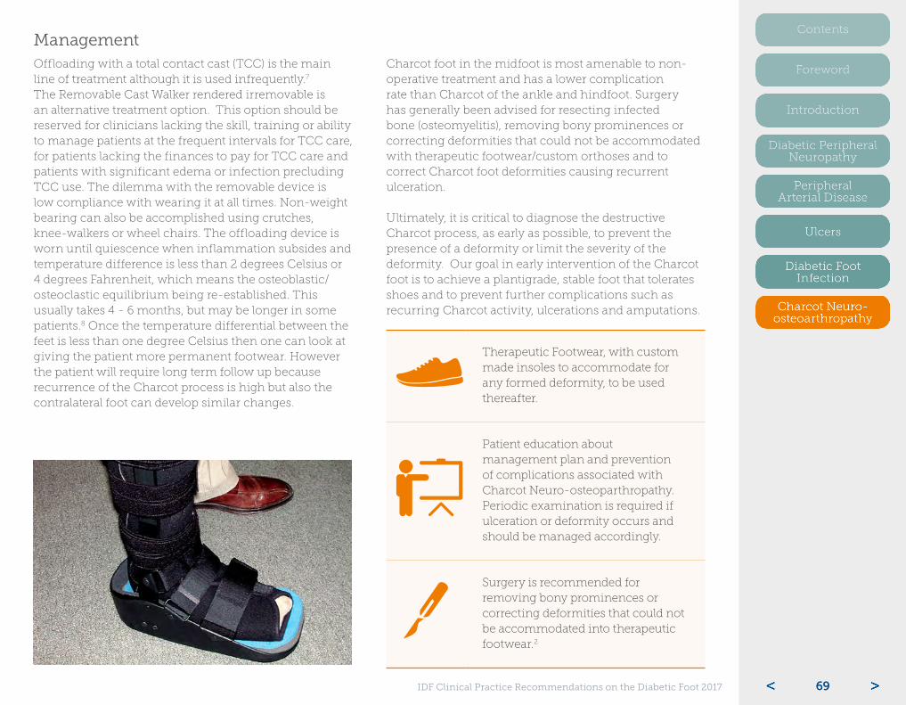
ManagementOffloading with a total contact cast (TCC) is the main line of treatment although it is used infrequently.7 The Removable Cast Walker rendered irremovable is an alternative treatment option. This option should be reserved for clinicians lacking the skill, training or ability to manage patients at the frequent intervals for TCC care, for patients lacking the finances to pay for TCC care and patients with significant edema or infection precluding TCC use. The dilemma with the removable device is low compliance with wearing it at all times. Non-weight bearing can also be accomplished using crutches, knee-walkers or wheel chairs. The offloading device is worn until quiescence when inflammation subsides and temperature difference is less than 2 degrees Celsius or 4 degrees Fahrenheit, which means the osteoblastic/osteoclastic equilibrium being re-established. This usually takes 4 - 6 months, but may be longer in some patients.8 Once the temperature differential between the feet is less than one degree Celsius then one can look at giving the patient more permanent footwear. However the patient will require long term follow up because recurrence of the Charcot process is high but also the contralateral foot can develop similar changes.
Charcot foot in the midfoot is most amenable to non-operative treatment and has a lower complication rate than Charcot of the ankle and hindfoot. Surgery has generally been advised for resecting infected bone (osteomyelitis), removing bony prominences or correcting deformities that could not be accommodated with therapeutic footwear/custom orthoses and to correct Charcot foot deformities causing recurrent ulceration.
Ultimately, it is critical to diagnose the destructive Charcot process, as early as possible, to prevent the presence of a deformity or limit the severity of the deformity. Our goal in early intervention of the Charcot foot is to achieve a plantigrade, stable foot that tolerates shoes and to prevent further complications such as recurring Charcot activity, ulcerations and amputations.
Therapeutic Footwear, with custom made insoles to accommodate for any formed deformity, to be used thereafter.
Patient education about management plan and prevention of complications associated with Charcot Neuro-osteoparthropathy. Periodic examination is required if ulceration or deformity occurs and should be managed accordingly.
Surgery is recommended for removing bony prominences or correcting deformities that could not be accommodated into therapeutic footwear.2
6969IDF Clinical Practice Recommendations on the Diabetic Foot 2017

References:1. Güven MF, Karabiber A, Kaynak G, Öğüt T. Conservative and
surgical treatment of the chronic Charcot foot and ankle. Diabetic foot & ankle. 2013 Aug 2;4.
2. Rogers LC, Frykberg RG, Armstrong DG, Boulton AJ, Edmonds M, Van GH, Hartemann A, Game F, Jeffcoate W, Jirkovska A, Jude E. The Charcot foot in diabetes. Diabetes Care. 2011 Sep 1;34(9):2123-9.
3. Mascarenhas JV, Jude EB. Pathogenesis and medical management of diabetic Charcot neuroarthropathy. Medical Clinics of North America. 2013 Sep 30;97(5):857-72.
4. Rajbhandari S, Jenkins RD, Davies C, Tesfaye S. Charcot neuroarthropathy in diabetes mellitus. Diabetologia. 2002 Aug 1;45(8):1085-96.
5. Ndip A, Williams A, Jude EB, Serracino-Inglott F, Richardson S, Smyth JV, Boulton AJ, Alexander MY. The RANKL/RANK/OPG signaling pathway mediates medial arterial calcification in diabetic Charcot neuroarthropathy. Diabetes. 2011 Aug 1;60(8):2187-96.
6. La Fontaine J, Harkless LB, Sylvia VL, Carnes D, Heim-Hall J, Jude E. Levels of Endothelial Nitric Oxide Synthase and Calcitonin Gene–related Peptide in the Charcot Foot: A Pilot Study. The Journal of Foot and Ankle Surgery. 2008 Oct 31;47(5):424-9.
7. Game FL, Catlow R, jones GR. Edmonds ME, Jude EB, Rayman G, Jeffcoate WJ. Audit of acute Charcot’s disease in the UK: the CDUK study. Diabetologia 2012;55:32-35
8. Armstrong DG, Todd WF, Lavery LA, Harkless LB, Bushman TR. The natural history of acute Charcot's arthropathy in a diabetic foot specialty clinic. Journal of the American Podiatric Medical Association. 1997 Jun;87(6):272-8.
7070IDF Clinical Practice Recommendations on the Diabetic Foot 2017