identification of upar-positive chemoresistant cells in
TRANSCRIPT

Identification of uPAR-positive Chemoresistant Cells inSmall Cell Lung CancerMargarita Gutova1*, Joseph Najbauer1, Anna Gevorgyan1, Marianne Z. Metz1, Yehua Weng1, Chu-Chih Shih1, Karen S. Aboody1,2*
1 Division of Hematology/Hematopoietic Cell Transplantation, City of Hope National Medical Center and Beckman Research Institute, Duarte,California, United States of America, 2 Division of Neurosciences, City of Hope National Medical Center and Beckman Research Institute, Duarte,California, United States of America
Background. The urokinase plasminogen activator (uPA) and its receptor (uPAR/CD87) are major regulators of extracellularmatrix degradation and are involved in cell migration and invasion under physiological and pathological conditions. The uPA/uPAR system has been of great interest in cancer research because it is involved in the development of most invasive cancerphenotypes and is a strong predictor of poor patient survival. However, little is known about the role of uPA/uPAR in small celllung cancer (SCLC), the most aggressive type of lung cancer. We therefore determined whether uPA and uPAR are involved ingeneration of drug resistant SCLC cell phenotype. Methods and Findings. We screened six human SCLC cell lines for surfacemarkers for putative stem and cancer cells. We used fluorescence-activated cell sorting (FACS), fluorescence microscopy andclonogenic assays to demonstrate uPAR expression in a subpopulation of cells derived from primary and metastatic SCLC celllines. Cytotoxic assays were used to determine the sensitivity of uPAR-positive and uPAR-negative cells to chemotherapeuticagents. The uPAR-positive cells in all SCLC lines demonstrated multi-drug resistance, high clonogenic activity and co-expression of CD44 and MDR1, putative cancer stem cell markers. Conclusions. These data suggest that uPAR-positive cellsmay define a functionally important population of cancer cells in SCLC, which are resistant to traditional chemotherapies, andcould serve as critical targets for more effective therapeutic interventions in SCLC.
Citation: Gutova M, Najbauer J, Gevorgyan A, Metz MZ, Weng Y, et al (2007) Identification of uPAR-positive Chemoresistant Cells in Small Cell LungCancer. PLoS ONE 2(2): e243. doi:10.1371/journal.pone.0000243
INTRODUCTIONSmall cell lung cancer (SCLC) is the most aggressive type of lung
cancer and has a uniformly poor prognosis. Metastases develop
quickly, primarily to bone marrow and brain, and are usually
present at the time of diagnosis. In untreated patients, median
survival is two months from the onset of symptoms [1].
In several types of tumors increased levels of urokinase
plasminogen activator (uPA) and its receptor uPAR (CD87)
strongly correlate with poor prognosis and unfavorable clinical
outcome [2,3,4,5,6]. uPA and uPAR are instrumental in
controlling membrane-associated extracellular proteolysis and
transmembrane signaling, thus affecting cell migration and
invasion under physiological and pathological conditions
[2,7,8,9,10]. uPAR over-expression in malignant cells results from
activation of several oncogenic pathways, including MAPK, RTK,
ERK2 and FAK [2,7,9]. Multiple oncogenic mutations, including
p53 in cancer cells lead to uncontrolled expression of uPA/uPAR
[11]. Inhibition of uPAR in a mouse model of non-small cell lung
cancer and other tumors inhibited tumor growth, invasion,
angiogenesis and metastasis [12,13,14]. Increased levels of uPAR
are correlated with higher mortality in patients with squamous cell
and non-small cell lung cancer [15,16], however little is known
about the role of uPA/uPAR expression in SCLC.
A recent study by Alfano et al underlines the importance of
uPAR signaling in prevention of apoptosis by resistance of cancer
cells to anoikis (apoptosis induced by loss of anchorage). uPAR
expression promotes cell survival by activating anti-apoptosis
factor Bcl-xL transcription through the MEK/ERK- and PI3K/
Akt-dependent pathways [17]. Therefore, we hypothesize that
uPAR expression may be involved in development of drug-
resistant cancer phenotype in SCLC.
We report here the presence of a rare population of uPAR-
positive cells in human SCLC cell lines that demonstrate
significant drug resistance to traditional chemotherapeutic
agents such as 5-fluorouracil (5-FU), cisplatin and etoposide.
The uPAR-positive cells expressed stem- and cancer cell markers,
including CD44 and MDR1. Identification and targeting of
uPAR-positive cells in SCLC may provide valuable insight into
biology of human lung cancer and may establish novel critical
targets for more effective anticancer therapies.
METHODS
Immunostaining and Flow Cytometry AnalysisPrimary (lung) small cell lung carcinoma (SCLC) cell lines (H1688,
H1417, H69AR), bone marrow (BM) metastatic SCLC (H211,
H1882) and brain metastatic SCLC (H250) cell lines were
obtained from human primary lung and metastatic tissues (
ATCC), grown in RPMI 1640 modified medium (ATCC, N: 30–
2001) supplemented with 10% Fetal Bovine Serum (FBS). The BM
metastatic cell line (H1882) was cultured in complete HITES
medium (D-MEM/F-12, N: 30–2006 supplemented with insulin
5 mg/mL, transferrin 10 mg/mL, sodium selenite 30 nM, hydro-
cortisone 10 nM, b-estradiol 10 nM, L-glutamine 2 mM, HEPES
10 mM and 5% FBS). Cells were grown for two weeks and were
Academic Editor: Christopher Arendt, Sanofi-Aventis, United States of America
Received January 15, 2007; Accepted January 31, 2007; Published February 28,2007
Copyright: � 2007 Gutova et al. This is an open-access article distributed underthe terms of the Creative Commons Attribution License, which permitsunrestricted use, distribution, and reproduction in any medium, provided theoriginal author and source are credited.
Funding: This work was supported by Stop Cancer Foundation, The Rosalindeand Arthur Gilbert Foundation, and Marcus Foundation.
Competing Interests: The authors have declared that no competing interestsexist.
* To whom correspondence should be addressed. E-mail: [email protected](MG); [email protected] (KA)
PLoS ONE | www.plosone.org 1 February 2007 | Issue 2 | e243

analyzed by flow cytometry using the following antibodies: CD59
(CBL467P), CD109 (CBL585P), CD62E (CBL180F) from Che-
micon, CD87 (3936CJ) from American Diagnostica, CXCR4
(FAB170F) from R&D Systems, CD24 (555427), CD90 (555596),
CD38 (347680), CD44 (555478), CD45 (555482), CD13 (555394),
CD49b (555498), CD29 (555443), CD3 (30104X) from BD
Pharmingen, ABCG2/BCRP1 (10400) from Stem Cell Technol-
ogies, CD133/2 (clone 293C3) and CD133/1 (clone AC133) from
Miltenyi Biotec, CD34 (347660) from Becton Dickinson, CD105
(326–050) from Alexis, MNF116 (F0859), Cyt18 (F7212) from
DACO, and CD166 (3FT) from RDI. For FACS analysis each cell
line was detached by trypsinization and re-suspended in staining
buffer (SB) (HBSS, Irvine Scientific, 9228) supplemented with 2%
FBS and 10 mM HEPES at a density of 56106 cells/ml. Fifty ml
(2.56104 cells) was added to each well of a 96-well v-shaped plate.
Antibodies (FITC- or PE-conjugated) were added in concentra-
tions recommended by the manufacturer (20 ml/106 cells).
Antibodies to CD133, CD34, CD44, CD87 and MDR1 have
been individually titrated. The 96-well plates were placed on ice
and cells were stained with antibodies for 30 min in dark. After
staining, 150 ml of wash buffer (HBSS, supplemented with 15%
FBS and 10 mM HEPES)/well was added and the plates were
centrifuged at 5006 g for 5 min at 4uC. The cell pellets were re-
suspended in SB, supplemented with propidium iodide (PI) (1 mg/
ml) to exclude nonviable cells, followed by flow cytometric
analysis.
Cell Staining and SortingSCLC cell lines (H211, H69AR, H1417) were grown in RPMI
1640 medium and 46106 cells were collected and then re-
suspended in 800 ml of SB, followed by staining with uPAR
(CD87)-FITC-conjugated antibody as described above. uPAR-
positive and uPAR-negative cells were sorted by FACS, followed
by culture in ‘‘base’’ methylcellulose media (Stem Cell Technol-
ogies, 04100) for 16 days at 37uC, 5% CO2.
Clonogenic Assay of SCLC Cell LinesComplete RPMI 1640 was added to MethoCult H4100 medium
(40 ml) to achieve a final volume of 100 ml. uPAR-positive and
uPAR-negative cells derived from SCLC cell lines (H211, H69AR,
H1417) were plated in MethoCult medium in triplicates containing
36103, 16103, or 16102 cells/ml. Cells were cultured at 37uC, 5%
CO2 in a humidified incubator for 16 days. Colonies were counted
using an inverted brightfield microscope at 46magnification.
Cytotoxicity AssayCells derived from three SCLC lines were immunostained and
sorted by flow-cytometry analysis (as described above). Cell lines
(H211, H69AR, H1417) were counted and placed into 96-well
plates (46103 cells/well, in triplicates) with final concentrations of
drugs 0, 3, 10, 100, 200 mg/ml. After incubation for 72 hr, both
viable and dead cells were counted by using Guava ViaCount
assay, and only the viable cells were included in data analysis. The
Guava ViaCount assay distinguishes between viable and non-
viable cell based on the differential permeability of DNA-binding
dyes in the ViaCount reagent, and thus fluorescence of the dyes
allows quantitative assessment of both viable and non-viable cells
in suspension. Alternatively, non-sorted cells were applied to 48-
well plates (16104 cells/well) and treated with cisplatin, etoposide
and their combination in concentrations of 0, 3, 10, 100 mg/ml.
The seeding densities per unit area (mm2) were the same for both
the 48- and 96-well plates (density = 125 cells/mm2). After 72 hrs
of incubation, viable cells were stained with uPAR-FITC
antibodies and evaluated by flow cytometry.
Double-labeling for Flow CytometryCell lines were stained with CD44-PE and MDR1-PE, were washed
and then stained with uPAR-FITC (16105 cells/1 mg of antibody).
Iso-type matched PE- or FITC-conjugated antibodies were used as
controls. Stained cells were analyzed by flow cytometry.
Figure 1. Flow cytometric analysis of uPAR expression in SCLC-derived cell lines. (A, B, C) Lung-derived SCLC cell lines, (D, E) metastatic bone marrowand (F) metastatic brain cell lines. All cells were cultured in RPMI 1640 medium, stained with uPAR-FITC antibody and analyzed by flow cytometry(lower panels). Control staining was performed using FITC-conjugated, isotype-matched mouse IgG (upper panels). A small population of uPAR-positive cells was detected in all cell lines examined, and is indicated as percent of R1-gated viable cells. Results shown are representative of threeindependent experiments.doi:10.1371/journal.pone.0000243.g001
uPAR-positive Cells in SCLC
PLoS ONE | www.plosone.org 2 February 2007 | Issue 2 | e243

RESULTS
Flow Cytometry Analysis of Human SCLC Cell LinesTo identify the most invasive SCLC phenotypes, we screened six
human SCLC cell lines (H1688, H1417, H69AR derived from
primary tumor site in lung, H250 from metastases to brain, and
H211, H1882 from metastases to bone marrow) with a panel of
antibodies for surface determinants of tumor cells, including
uPAR, CD13, CD29, CD44, CXCR4, CD105, CD109, CD166,
and for stem cell markers CD34, CD90, CD133, ABCG2/
BCRP1. SCLC cell lines displayed heterogeneous phenotypes with
regard to tumor surface determinants. All six SCLC cell lines were
positive for CD29 (20–99%), CD44 (8–98%), CD105 (3–34%),
CD166 (85–98%), and negative for CD90, and CXCR4. uPAR
(CD87) was the only cell surface antigen expressed on a small sub-
population of cells (1–4%) in each of the six SCLC cell lines, when
analyzed by FACS using anti-uPAR-FITC antibody (Figure 1,
lower panels).
Control samples stained with isotype-matched IgG-FITC were
negative for uPAR expression (Figure 1, upper panels). The
consistent presence of a small uPAR-positive subpopulation of
cells in all primary (lung, Figure 1, lower panels A, B, C) and
metastatic (bone marrow; Figure 1, lower panels D, E, and brain; F)
SCLC cell lines, in contrast to other markers, which varied in
abundance, suggests that uPAR-positive cells may comprise
a functionally unique subpopulation of cells.
Chemoresistance of uPAR-positive CellsWe hypothesized that the uPAR-expressing cell population may be
resistant to chemotherapeutic agents. We performed cytotoxicity
assays on three selected cell lines using non-sorted (bulk) as well as
sorted uPAR-positive and uPAR-negative cell populations. In-
creasing concentrations of 5-fluorouracil (5-FU) (10, 100, 200 mg/
ml) were added to these cell cultures and incubated for 72 hrs,
followed by counting of both viable and killed cells. Data was
normalized to 100%, which signified the number of uPAR-positive
and uPAR-negative cells without drug added. A cell-killing effect
was detected in all three non-sorted cell lines (H211, H69AR,
H1417), where 40–80% of cells were killed by 5-FU (Figure 2A).
Importantly, uPAR-positive sorted cells from these three cell lines
displayed significantly increased resistance to 5-FU, with only 40–
50% of cells being killed in the case of H211 and H1417
(Figure 2B). Although the H69AR cell line also showed differential
killing of uPAR-positive and uPAR-negative cells (e.g., 10 mg/ml
5-FU killed 30% of uPAR-positve cells versus 85% of uPAR-
Figure 2. Cytotoxic effect of 5-FU on non-sorted and sorted (uPAR-positive and uPAR-negative populations) derived from SCLC cell lines. (A) 16104
cells (H211, H69AR, H1417) were placed in wells of a 48-well plate in triplicates and incubated for 72 hr in the presence of varying concentrations of 5-FU. (B) SCLC cell lines were FACS sorted after staining with anti-uPAR antibodies and were plated at the same seeding density (46103/well of 96-wellplate) and treated with 5-FU at 0, 10, 100, 200 mg/ml for 72 hr. Cell survival was evaluated after adding Guava ViaCount reagent and counting viableand dead cells. Only viable cells were included in data analysis, and 100% viability was defined as number of viable cells cultured in absence of 5-FU.Statistical analysis (2-way ANOVA) of uPAR(+) and uPAR(2) data sets revealed significant differences among viability of uPAR(+) and uPAR(2) cells(P = 0.0002, 0.0027, 0.0008 for H211, H69AR, H1417 cells, respectively). The data points represent averages6SD of three independent experiments.doi:10.1371/journal.pone.0000243.g002
uPAR-positive Cells in SCLC
PLoS ONE | www.plosone.org 3 February 2007 | Issue 2 | e243
negative cells), at high concentration of 5-FU (200 mg/ml), the
killing effect for both populations (H69AR) was the same
(,100%). Statistical analysis (2-way ANOVA) of uPAR(+) and
uPAR(2) data sets revealed significant differences in survival of
uPAR(+) and uPAR(2) cells after treatment with 5-FU
(P = 0.0002, 0.0027, 0.0008 for H211, H69AR, H1417 cells,
respectively) (Figure 2B).
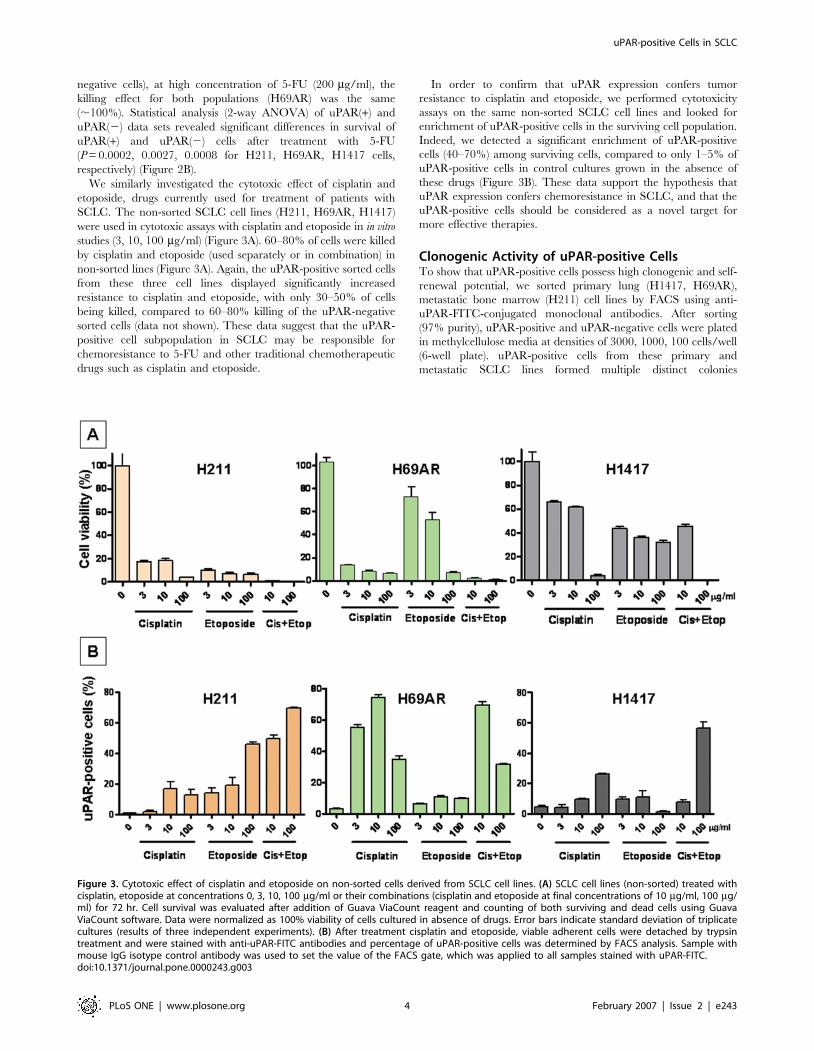
We similarly investigated the cytotoxic effect of cisplatin and
etoposide, drugs currently used for treatment of patients with
SCLC. The non-sorted SCLC cell lines (H211, H69AR, H1417)
were used in cytotoxic assays with cisplatin and etoposide in in vitro
studies (3, 10, 100 mg/ml) (Figure 3A). 60–80% of cells were killed
by cisplatin and etoposide (used separately or in combination) in
non-sorted lines (Figure 3A). Again, the uPAR-positive sorted cells
from these three cell lines displayed significantly increased
resistance to cisplatin and etoposide, with only 30–50% of cells
being killed, compared to 60–80% killing of the uPAR-negative
sorted cells (data not shown). These data suggest that the uPAR-
positive cell subpopulation in SCLC may be responsible for
chemoresistance to 5-FU and other traditional chemotherapeutic
drugs such as cisplatin and etoposide.
In order to confirm that uPAR expression confers tumor
resistance to cisplatin and etoposide, we performed cytotoxicity
assays on the same non-sorted SCLC cell lines and looked for
enrichment of uPAR-positive cells in the surviving cell population.
Indeed, we detected a significant enrichment of uPAR-positive
cells (40–70%) among surviving cells, compared to only 1–5% of
uPAR-positive cells in control cultures grown in the absence of
these drugs (Figure 3B). These data support the hypothesis that
uPAR expression confers chemoresistance in SCLC, and that the
uPAR-positive cells should be considered as a novel target for
more effective therapies.
Clonogenic Activity of uPAR-positive CellsTo show that uPAR-positive cells possess high clonogenic and self-
renewal potential, we sorted primary lung (H1417, H69AR),
metastatic bone marrow (H211) cell lines by FACS using anti-
uPAR-FITC-conjugated monoclonal antibodies. After sorting
(97% purity), uPAR-positive and uPAR-negative cells were plated
in methylcellulose media at densities of 3000, 1000, 100 cells/well
(6-well plate). uPAR-positive cells from these primary and
metastatic SCLC lines formed multiple distinct colonies
Figure 3. Cytotoxic effect of cisplatin and etoposide on non-sorted cells derived from SCLC cell lines. (A) SCLC cell lines (non-sorted) treated withcisplatin, etoposide at concentrations 0, 3, 10, 100 mg/ml or their combinations (cisplatin and etoposide at final concentrations of 10 mg/ml, 100 mg/ml) for 72 hr. Cell survival was evaluated after addition of Guava ViaCount reagent and counting of both surviving and dead cells using GuavaViaCount software. Data were normalized as 100% viability of cells cultured in absence of drugs. Error bars indicate standard deviation of triplicatecultures (results of three independent experiments). (B) After treatment cisplatin and etoposide, viable adherent cells were detached by trypsintreatment and were stained with anti-uPAR-FITC antibodies and percentage of uPAR-positive cells was determined by FACS analysis. Sample withmouse IgG isotype control antibody was used to set the value of the FACS gate, which was applied to all samples stained with uPAR-FITC.doi:10.1371/journal.pone.0000243.g003
uPAR-positive Cells in SCLC
PLoS ONE | www.plosone.org 4 February 2007 | Issue 2 | e243
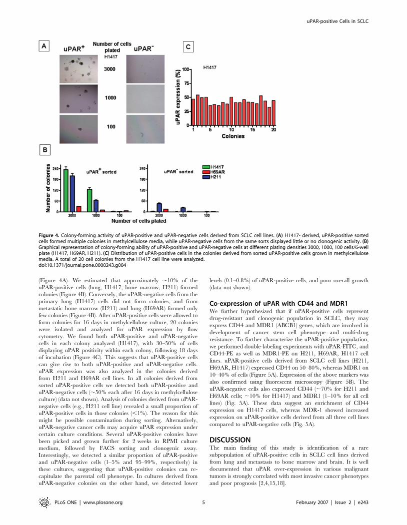
(Figure 4A). We estimated that approximately ,10% of the
uPAR-positive cells (lung, H1417; bone marrow, H211) formed
colonies (Figure 4B). Conversely, the uPAR-negative cells from the
primary lung (H1417) cells did not form colonies, and from
metastatic bone marrow (H211) and lung (H69AR) formed only
few colonies (Figure 4B). After uPAR-positive cells were allowed to
form colonies for 16 days in methylcellulose culture, 20 colonies
were isolated and analyzed for uPAR expression by flow
cytometry. We found both uPAR-positive and uPAR-negative
cells in each colony analyzed (H1417), with 30–50% of cells
displaying uPAR positivity within each colony, following 18 days
of incubation (Figure 4C). This suggests that uPAR-positive cells
can give rise to both uPAR-positive and uPAR-negative cells.
uPAR expression was also analyzed in the colonies derived
from H211 and H69AR cell lines. In all colonies derived from
sorted uPAR-positive cells we detected both uPAR-positive and
uPAR-negative cells (,50% each after 16 days in methylcellulose
culture) (data not shown). Analysis of colonies derived from uPAR-
negative cells (e.g., H211 cell line) revealed a small proportion of
uPAR-positive cells in those colonies (,1%). The reason for this
might be possible contamination during sorting. Alternatively,
uPAR-negative cancer cells may acquire uPAR expression under
certain culture conditions. Several uPAR-positive colonies have
been picked and grown further for 2 weeks in RPMI culture
medium, followed by FACS sorting and clonogenic assay.
Interestingly, we detected a similar proportion of uPAR-positive
and uPAR-negative cells (1–5% and 95–99%, respectively) in
these cultures, suggesting that uPAR-positive colonies can re-
capitulate the parental cell phenotype. In cultures derived from
uPAR-negative colonies on the other hand, we detected lower
levels (0.1–0.8%) of uPAR-positive cells, and poor overall growth
(data not shown).
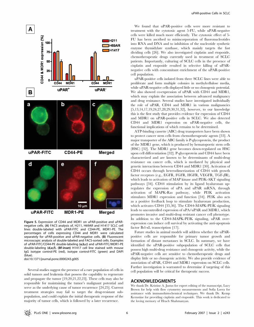
Co-expression of uPAR with CD44 and MDR1We further hypothesized that if uPAR-positive cells represent
drug-resistant and clonogenic population in SCLC, they may
express CD44 and MDR1 (ABCB1) genes, which are involved in
development of cancer stem cell phenotype and multi-drug
resistance. To further characterize the uPAR-positive population,
we performed double-labeling experiments with uPAR-FITC, and
CD44-PE as well as MDR1-PE on H211, H69AR, H1417 cell
lines. uPAR-positive cells derived from SCLC cell lines (H211,
H69AR, H1417) expressed CD44 on 50–80%, whereas MDR1 on
10–40% of cells (Figure 5A). Expression of the above markers was
also confirmed using fluorescent microscopy (Figure 5B). The
uPAR-negative cells also expressed CD44 (,70% for H211 and
H69AR cells; ,10% for H1417) and MDR1 (1–10% for all cell
lines) (Fig. 5A). These data suggest an enrichment of CD44
expression on H1417 cells, whereas MDR-1 showed increased
expression on uPAR-positive cells derived from all three cell lines
compared to uPAR-negative cells (Fig. 5A).
DISCUSSIONThe main finding of this study is identification of a rare
subpopulation of uPAR-positive cells in SCLC cell lines derived
from lung and metastasis to bone marrow and brain. It is well
documented that uPAR over-expression in various malignant
tumors is strongly correlated with most invasive cancer phenotypes
and poor prognosis [2,4,15,18].
Figure 4. Colony-forming activity of uPAR-positive and uPAR-negative cells derived from SCLC cell lines. (A) H1417- derived, uPAR-positive sortedcells formed multiple colonies in methylcellulose media, while uPAR-negative cells from the same sorts displayed little or no clonogenic activity. (B)Graphical representation of colony-forming ability of uPAR-positive and uPAR-negative cells at different plating densities 3000, 1000, 100 cells/6-wellplate (H1417, H69AR, H211). (C) Distribution of uPAR-positive cells in the colonies derived from sorted uPAR-positive cells grown in methylcellulosemedia. A total of 20 cell colonies from the H1417 cell line were analyzed.doi:10.1371/journal.pone.0000243.g004
uPAR-positive Cells in SCLC
PLoS ONE | www.plosone.org 5 February 2007 | Issue 2 | e243
Several studies suggest the presence of a rare population of cells in
solid tumors and leukemia that possess the capability to regenerate
and propagate the tumor [19,20,21,22,23]. These cells may also be
responsible for maintaining the tumor’s malignant potential and
serve as the underlying cause of tumor recurrence [24,25]. Current
treatment strategies may fail to target the drug-resistant sub-
population, and could explain the initial therapeutic response of the
majority of tumor cells, which is followed by a later recurrence.
We found that uPAR-positive cells were more resistant to
treatment with the cytotoxic agent 5-FU, while uPAR-negative
cells were killed much more efficiently. The cytotoxic effect of 5-
FU has been ascribed to misincorporation of fluoronucleotides
into RNA and DNA and to inhibition of the nucleotide synthetic
enzyme thymidylate synthase, which mainly targets the fast
dividing cells [26]. We also investigated cisplatin and etoposide,
chemotherapeutic drugs currently used in treatment of SCLC
patients. Importantly, culturing of SCLC cells in the presence of
cisplatin and etoposide resulted in selective killing of uPAR-
negative cells with concomitant enrichment of the uPAR-positive
cell population.
uPAR-positive cells isolated from three SCLC lines were able to
proliferate and form multiple colonies in methylcellulose media,
while uPAR-negative cells displayed little or no clonogenic potential.
We also showed co-expression of uPAR with CD44 and MDR1,
which may explain the association between advanced malignancy
and drug resistance. Several studies have investigated individually
the role of uPAR, CD44 and MDR1 in various malignancies
[5,13,14,17,19,24,27,28,29,30,31,32], however, to our knowledge
this is the first study that provides evidence for expression of CD44
and MDR1 on uPAR-positive cells in SCLC. We also detected
CD44 and MDR1 expression on uPAR-negative cells, the
functional implications of which remains to be determined.
ATP-binding cassette (ABC) drug transporters have been shown
to protect cancer stem cells from chemotherapeutic agents [33]. A
major transporter of the ABC family is P-glycoprotein, the product
of the MDR1 gene, which is produced by hematopoetic stem cells
(HSC) [32]. The MDR1 gene becomes down-regulated on HSC
upon cell differentiation [32]. P-glycoprotein and CD44 have been
characterized and are known to be determinants of multi-drug
resistance on cancer cells, which is mediated by physical and
genetic interactions between CD44 and MDR1 [30]. Activation of
CD44 occurs through heterodimerization of CD44 with growth
factor receptors (e.g., EGFR, FGFR, HGFR, VEGFR, TGF-bR),
which leads to activation of MAP kinase and PI3K-AKT signaling
pathways [34]. CD44 stimulation by its ligand hyaluronan up-
regulates the expression of uPA and uPAR mRNA, through
activation of MAPK-Ras pathway, while PI3K activation
stimulates MDR1 expression and function [34]. PI3K also acts
as a positive feedback loop to stimulate hyaluronan production,
which activates CD44 [35,36]. The CD44-MAPK-PI3K signaling
leads to uncontrolled expression of uPA/uPAR and MDR1, which
promotes invasive and multi-drug resistant cancer cell phenotype.
In addition to the CD44-MAPK-PI3K signaling, uPAR over-
expression can induce cell survival by activating the anti-apoptosis
factor Bcl-xL transcription [17].
Future studies in animal models will address whether the uPAR-
positive cells are responsible for primary tumor growth and
formation of distant metastases in SCLC. In summary, we have
identified the uPAR-positive subpopulation of SCLC cells that
possess high multi-drug resistance and clonogenic activity, while the
uPAR-negative cells are sensitive to chemotherapeutic drugs and
display little or no clonogenic activity. We also provide evidence of
association of uPAR, CD44 and MDR1 expression on SCLC cells.
Further investigation is warranted to determine if targeting of this
cell population will be critical for therapeutic success.
ACKNOWLEDGMENTSWe thank Dr. Kristine A. Justus for expert editing of the manuscript, Lucy
Brown for help with flow cytometry measurements and Sofia Loera for
assistance with immunohistochemical techniques. We thank Dr. Kemp
Kernstine for providing cisplatin and etoposide. This work is dedicated to
the loving memory of Hrach Shahmanyan.
Figure 5. Expression of CD44 and MDR1 on uPAR-positive and uPAR-negative cells. (A) FACS analysis of, H211, H69AR and H1417 SCLC celllines double-labeled with uPAR-FITC and CD44-PE, MDR1-PE. Thepercentages of cells expressing CD44 and MDR1 were calculatedseparately for uPAR-positive and uPAR-negative cells. (B) Fluorescentmicroscopic analysis of double-labeled and FACS-sorted cells. Examplesof uPAR-FITC/CD44-PE double-labeling (a,b,c) and uPAR-FITC/MDR1-PEdouble-labeling (d,e,f). (Bf-inset) H1417 cell line stained with mouseIgG isotype control-PE (red), isotype control-FITC (green) and DAPI(blue).doi:10.1371/journal.pone.0000243.g005
uPAR-positive Cells in SCLC
PLoS ONE | www.plosone.org 6 February 2007 | Issue 2 | e243
Author Contributions
Conceived and designed the experiments: KA JN MG AG. Performed the
experiments: MM MG YW. Analyzed the data: KA JN MG AG.
Contributed reagents/materials/analysis tools: KA CS. Wrote the paper:
JN MG AG.
REFERENCES1. Pisick E, Jagadeesh S, Salgia R (2003) Small cell lung cancer: from molecular
biology to novel therapeutics. J Exp Ther Oncol 3: 305–318.2. Aguirre Ghiso JA, Alonso DF, Farias EF, Gomez DE, de Kier Joffe EB (1999)
Deregulation of the signaling pathways controlling urokinase production. Itsrelationship with the invasive phenotype. Eur J Biochem 263: 295–304.
3. Aref S, El-Sherbiny M, Mabed M, Menessy A, El-Refaei M (2003) Urokinaseplasminogen activator receptor and soluble matrix metalloproteinase-9 in acute
myeloid leukemia patients: a possible relation to disease invasion. Hematology 8:
385–391.4. Foekens JA, Peters HA, Look MP, Portengen H, Schmitt M, et al. (2000) The
urokinase system of plasminogen activation and prognosis in 2780 breast cancerpatients. Cancer Res 60: 636–643.
5. Meijer-van Gelder ME, Look MP, Peters HA, Schmitt M, Brunner N, et al.
(2004) Urokinase-type plasminogen activator system in breast cancer: associationwith tamoxifen therapy in recurrent disease. Cancer Res 64: 4563–4568.
6. Rigolin GM, Tieghi A, Ciccone M, Bragotti LZ, Cavazzini F, et al. (2003)Soluble urokinase-type plasminogen activator receptor (suPAR) as an in-
dependent factor predicting worse prognosis and extra-bone marrow in-volvement in multiple myeloma patients. Br J Haematol 120: 953–959.
7. Alfano D, Franco P, Vocca I, Gambi N, Pisa V, et al. (2005) The urokinase
plasminogen activator and its receptor: role in cell growth and apoptosis.Thromb Haemost 93: 205–211.
8. Allgayer H (2006) Molecular regulation of an invasion-related molecule–optionsfor tumour staging and clinical strategies. Eur J Cancer 42: 811–819.
9. Blasi F, Carmeliet P (2002) uPAR: a versatile signalling orchestrator. Nat Rev
Mol Cell Biol 3: 932–943.10. Montuori N, Visconte V, Rossi G, Ragno P (2005) Soluble and cleaved forms of
the urokinase-receptor: degradation products or active molecules? ThrombHaemost 93: 192–198.
11. Czekay RP, Kuemmel TA, Orlando RA, Farquhar MG (2001) Direct binding ofoccupied urokinase receptor (uPAR) to LDL receptor-related protein is required
for endocytosis of uPAR and regulation of cell surface urokinase activity. Mol
Biol Cell 12: 1467–1479.12. Lakka SS, Gondi CS, Dinh DH, Olivero WC, Gujrati M, et al. (2005) Specific
interference of urokinase-type plasminogen activator receptor and matrixmetalloproteinase-9 gene expression induced by double-stranded RNA results
in decreased invasion, tumor growth, and angiogenesis in gliomas. J Biol Chem
280: 21882–21892.13. Muralikrishna PS, Gondi CS, Lakka SS, Julta A, Estes N, et al. (2005) RNA
interference-directed knockdown of urokinase plasminogen activator andurokinase plasminogen activator receptor inhibits prostate cancer cell invasion,
survival and tumorigenicity in vivo. J Biol Chem.14. Rao JS, Gondi C, Chetty C, Chittivelu S, Joseph PA, et al. (2005) Inhibition of
invasion, angiogenesis, tumor growth, and metastasis by adenovirus-mediated
transfer of antisense uPAR and MMP-9 in non-small cell lung cancer cells. MolCancer Ther 4: 1399–1408.
15. Almasi CE, Hoyer-Hansen G, Christensen IJ, Dano K, Pappot H (2005)Prognostic impact of liberated domain I of the urokinase plasminogen activator
receptor in squamous cell lung cancer tissue. Lung Cancer 48: 349–355.
16. Werle B, Kotzsch M, Lah TT, Kos J, Gabrijelcic-Geiger D, et al. (2004)Cathepsin B, plasminogenactivator-inhibitor (PAI-1) and plasminogenactivator-
receptor (uPAR) are prognostic factors for patients with non-small cell lungcancer. Anticancer Res 24: 4147–4161.
17. Alfano D, Iaccarino I, Stoppelli MP (2006) Urokinase signaling through its
receptor protects against anoikis by increasing BCL-xL expression levels. J BiolChem 281: 17758–17767.
18. Lakka SS, Bhattacharya A, Mohanam S, Boyd D, Rao JS (2001) Regulation of
the uPA gene in various grades of human glioma cells. Int J Oncol 18: 71–79.
19. Al-Hajj M, Wicha MS, Benito-Hernandez A, Morrison SJ, Clarke MF (2003)
Prospective identification of tumorigenic breast cancer cells. Proc Natl Acad
Sci U S A 100: 3983–3988.
20. Galli R, Binda E, Orfanelli U, Cipelletti B, Gritti A, et al. (2004) Isolation and
characterization of tumorigenic, stem-like neural precursors from human
glioblastoma. Cancer Res 64: 7011–7021.
21. Kondo T, Setoguchi T, Taga T (2004) Persistence of a small subpopulation of
cancer stem-like cells in the C6 glioma cell line. Proc Natl Acad Sci U S A 101:781–786.
22. Singh SK, Hawkins C, Clarke ID, Squire JA, Bayani J, et al. (2004)
Identification of human brain tumour initiating cells. Nature 432: 396–401.
23. Dick JE (2003) Breast cancer stem cells revealed. Proc Natl Acad Sci U S A 100:3547–3549.
24. Patrawala L, Calhoun T, Schneider-Broussard R, Li H, Bhatia B, et al. (2006)
Highly purified CD44+ prostate cancer cells from xenograft human tumors are
enriched in tumorigenic and metastatic progenitor cells. Oncogene 25:
1696–1708.
25. Kim CF, Jackson EL, Woolfenden AE, Lawrence S, Babar I, et al. (2005)
Identification of bronchioalveolar stem cells in normal lung and lung cancer.
Cell 121: 823–835.
26. Longley DB, Johnston PG (2005) Molecular mechanisms of drug resistance.J Pathol 205: 275–292.
27. Margheri F, D’Alessio S, Serrati S, Pucci M, Annunziato F, et al. (2005) Effects
of blocking urokinase receptor signaling by antisense oligonucleotides in a mouse
model of experimental prostate cancer bone metastases. Gene Ther 12:
702–714.
28. Draffin JE, McFarlane S, Hill A, Johnston PG, Waugh DJ (2004) CD44
potentiates the adherence of metastatic prostate and breast cancer cells to bone
marrow endothelial cells. Cancer Res 64: 5702–5711.
29. Jin L, Hope KJ, Zhai Q, Smadja-Joffe F, Dick JE (2006) Targeting of CD44
eradicates human acute myeloid leukemic stem cells. Nat Med 12: 1167–1174.
30. Miletti-Gonzalez KE, Chen S, Muthukumaran N, Saglimbeni GN, Wu X, et al.
(2005) The CD44 receptor interacts with P-glycoprotein to promote cell
migration and invasion in cancer. Cancer Res 65: 6660–6667.
31. Toole BP (2004) Hyaluronan: from extracellular glue to pericellular cue. NatRev Cancer 4: 528–539.
32. Zhou S, Schuetz JD, Bunting KD, Colapietro AM, Sampath J, et al. (2001) The
ABC transporter Bcrp1/ABCG2 is expressed in a wide variety of stem cells and
is a molecular determinant of the side-population phenotype. Nat Med 7:
1028–1034.
33. Dean M, Fojo T, Bates S (2005) Tumour stem cells and drug resistance. Nat Rev
Cancer 5: 275–284.
34. Kobayashi H, Suzuki M, Kanayama N, Nishida T, Takigawa M, et al. (2002)
CD44 stimulation by fragmented hyaluronic acid induces upregulation of
urokinase-type plasminogen activator and its receptor and subsequentlyfacilitates invasion of human chondrosarcoma cells. Int J Cancer 102: 379–389.
35. Zoltan-Jones A, Huang L, Ghatak S, Toole BP (2003) Elevated hyaluronan
production induces mesenchymal and transformed properties in epithelial cells.
J Biol Chem 278: 45801–45810.
36. Kamikura DM, Khoury H, Maroun C, Naujokas MA, Park M (2000) Enhanced
transformation by a plasma membrane-associated met oncoprotein: activation of
a phosphoinositide 39-kinase-dependent autocrine loop involving hyaluronic acid
and CD44. Mol Cell Biol 20: 3482–3496.
uPAR-positive Cells in SCLC
PLoS ONE | www.plosone.org 7 February 2007 | Issue 2 | e243