identification of the major allergen of malassezia globosa relevant for atopic dermatitis
TRANSCRIPT

Journal of Dermatological Science 55 (2009) 185–192
Identification of the major allergen of Malassezia globosa relevant foratopic dermatitis
Yoshio Ishibashi a, Hiroshi Kato a, Yoko Asahi a, Takashi Sugita b, Akemi Nishikawa a,*a Department of Immunobiology, Meiji Pharmaceutical University, 2-522-1 Noshio, Kiyose 204-8588, Tokyo, Japanb Department Microbiology, Meiji Pharmaceutical University, 2-522-1 Noshio, Kiyose 204-8588, Tokyo, Japan
A R T I C L E I N F O
Article history:
Received 4 December 2008
Received in revised form 11 April 2009
Accepted 20 May 2009
Keywords:
Allergen
Malassezia globosa
Atopic dermatitis
IgE
Proteomics
A B S T R A C T
Background: Malassezia globosa constitutes a part of the normal flora of human skin, but may induce IgE
production in atopic dermatitis (AD). However, information on M. globosa allergens is scant.
Objective: To identify the major M. globosa allergens by using proteomic analysis.
Methods: Immunoglobulin E (IgE) immunoblotting and cross-inhibition tests for M. globosa allergens
were performed using sera from AD patients and control subjects. These allergens were identified and
characterized using the proteomics approach involving a combination of two-dimensional (2D)
electrophoresis, mass spectrometry, and bioinformatics tools. We cloned the cDNA of this allergen using
sequences obtained by 50- and 30-rapid amplification of cDNA ends polymerase chain reaction.
Results: The sera of the AD patients had IgE-reactive 40–45-kDa protein components. By 2D
immunoblotting, we detected a 42-kDa protein spot with an isoelectric point (pI) of 4.8; the protein
was highly reactive to IgE and was designated MGp42. Full-length MGp42 cDNA contained a 1908-bp
open reading frame encoding 635 amino acid residues (calculated molecular mass, 69.7 kDa; pI, 6.02).
The N-terminal MGp42 sequence started from the 250th residue (Asp-250) of the deduced amino acid
sequence and consisted of 386 amino acid residues; these results are consistent with those of 2D
immunoblotting. MGp42 showed sequence similarity to members of the heat shock protein 70 (hsp70)
family. Immunoblot inhibition tests revealed no IgE cross-reactivity between MGp42 and human HSP70.
Conclusions: MGp42 may be a cleavage product of intact HSP70. This novel M. globosa allergen could be
useful for the diagnosis of AD.� 2009 Japanese Society for Investigative Dermatology. Published by Elsevier Ireland Ltd. All rights reserved.
Contents lists available at ScienceDirect
Journal of Dermatological Science
journal homepage: www. int l .e lsev ierhea l th .com/ journa ls / jods
1. Introduction
The lipophilic yeasts of the genus Malassezia are naturalinhabitants of the human skin, preferentially inhabiting seborrheicskin areas such as the head and neck; however, these organismsare associated with several skin diseases such as pityriasisversicolor, pityrosporum folliculitis, and seborrheic dermatitis[1–3]. They are also considered to be one of the factors thatexacerbate atopic dermatitis (AD) located primarily in the headand neck area. This consideration is based on the finding that over90% of AD patients exhibited specific serum immunoglobulin E(IgE) antibodies against Malassezia yeasts [4–8]. In particular, ADpatients with head and neck dermatitis have high levels ofMalassezia-specific IgE [7,9]. In addition, treatment with antifungalagents such as ketoconazole results in clinical improvement ofhead and neck AD in association with decreased Malassezia
colonization [5,10,11]. The genus Malassezia consists of 13 species,
* Corresponding author. Tel.: +81 424 95 8740; fax: +81 424 95 8740.
E-mail address: [email protected] (A. Nishikawa).
0923-1811/$36.00 � 2009 Japanese Society for Investigative Dermatology. Published b
doi:10.1016/j.jdermsci.2009.05.005
including Malassezia furfur, Malassezia sympodialis, Malassezia
globosa, Malassezia restricta, Malassezia slooffiae, Malassezia obtusa,and Malassezia pachydermatis [12–17]. Of these, M. globosa and M.
restricta may play an important role in the pathogenesis of AD [12].Although a number of Malassezia allergens have been identifiedand studied [18–21], data on allergens of M. globosa is unavailable.The identification and characterization of clinically relevantMalassezia allergens remain incomplete; this limits our under-standing of the roles of these allergens in the immunopathogenicmechanisms involved in AD.
The identification and characterization of allergens requireextensive effort and a large amount of raw material. Newlydeveloped proteomics approaches involving the combined applica-tion of separation techniques, mass spectrometry (MS), andbioinformatics tools have been proposed for the identificationand characterization of proteins in complex biological mixturesunder various experimental conditions [22,23]. In this study, wepropose the use of a proteomics approach combining two-dimensional (2D) western blotting and matrix-assisted laserdesorption ionization time-of-flight MS (MALDI–TOF–MS) for theidentification of M. globosa allergens. In addition, we cloned and
y Elsevier Ireland Ltd. All rights reserved.

Y. Ishibashi et al. / Journal of Dermatological Science 55 (2009) 185–192186
sequenced the gene encoding the major allergen of M. globosa byusing 50/30-RNA ligase-mediated rapid amplification of cDNA endspolymerase chain reaction (50/30-RLM-RACE PCR). Comparison ofthis sequence with known protein sequences revealed that the M.
globosa allergen is highly similar to members of the heat shockprotein 70 (hsp70) family. This study suggests that the majorallergen of M. globosa is a cleavage product of a protein belonging tothe hsp70 family.
2. Materials and methods
2.1. Yeast strains, culture conditions, and extract preparation
M. globosa CBS7966 was cultured on modified Leeming andNotman agar (LNA; 20 g glucose, 50 g malt extract, 1 g poly-peptone, and 20 g bile salts) (Oxoid, Hampshire, United Kingdom)containing 1% Tween-40, 0.2% glycerol, and 50 mg/mL chloram-phenicol (Sankyo, Tokyo, Japan) at 32 8C [24]. After cultivation for 5days, the yeasts were harvested, washed 5 times with Dulbecco’sphosphate-buffered saline (PBS), suspended in an equal volume ofPBS, and then disrupted using a French pressure cell (Amicon) at14,500 psi. The crude lysate was centrifuged at 100,000 � g for30 min, and the supernatant was collected. The protein concen-tration of the crude extract was estimated using a Protein AssayRapid Kit (Wako Pure Chemical Industries Ltd., Osaka, Japan)according to the manufacturer’s instructions.
2.2. Patient sera
Sera from 28 AD patients, including 12 males and 16 females,whose ages ranged from 20 to 64 years (mean age, 35.4 years) wereused in this study. Allergic response was confirmed on the basis ofthe patient’s clinical history and diagnosis, and characterized usingthe AlaSTAT system (Diagnostic Product Corporation) for measur-ing IgE reactivity. The diagnoses were made according to thecriteria of Hanifin and Rajka [25]. The initial inclusion criterion forthis study was an M. furfur-specific IgE titer of >3 IU/mL, asdetermined using the AlaSTAT system. The severity of AD wasminor in 6 cases, moderate in 18 cases, and severe in 4 cases. Serafrom 12 healthy adult subjects (mean age, 26.8 years) were used ascontrols. This study was approved by the ethics committee of MeijiPharmaceutical University, and informed consent was obtainedfrom the patients or their relatives.
2.3. Immunoblotting
Crude extracts (100 mg) were subjected to sodium dodecylsulphate (SDS)–polyacrylamide (with a linear 10–20% gradient)slab–gel electrophoresis and subsequently transferred to a poly-vinylidine fluoride (PVDF) membrane [26]. The blotted membraneswere incubated with blocking buffer (PBS containing 5% skim milkpowder) for 30 min at room temperature, cut into 2-mm wide strips,and used for immunoblotting. IgE-binding components weredetected using an AlaBlot kit, according to the manufacturer’sinstructions. In brief, the membrane strips were incubated overnightat 4 8C with each patient’s serum samples diluted at a ratio of 1:10 inPBS. After incubation, the strips were washed 3 times with washingbuffer and subsequently incubated with anti-human IgE alkalinephosphatase conjugate for 1 h. After incubation, the IgE-bindingcomponents were visualized using a substrate solution containing5-bromo-4-chloro-3-indolyl phosphate and nitroblue tetrazolium.
2.4. Immunoblot inhibition analysis
Immunological cross-reactivity was examined by performingimmunoblot inhibition experiments [21]. To inhibit specific IgE
antibodies, the pooled sera from the AD patients were diluted to aratio of 1:10 in PBS and incubated overnight at 4 8C with 0.1, 1, and10 mg/mL of recombinant human HSP70 (StressMarq BiosciencesInc.) as an inhibitor. Unabsorbed sera and sera absorbed with100 mg/mL of crude extract obtained from M. globosa were run inparallel as controls. The membrane strips were incubated over-night at 4 8C with the mixtures and subsequently developed asdescribed above.
2.5. Sodium metaperiodate treatment
Carbohydrates of antigenic components present on the blottedmembrane strips were disrupted by incubation with 50 mMsodium metaperiodate for 20 h at 4 8C in the dark [27]. After themembrane strips were washed, they were processed for antibodyreactivity, as described above.
2.6. 2D electrophoresis
Crude extracts were subjected to duplicate 2D electrophoresis:one sample was used for immunodetection, and the other, forproteomic analysis. Isoelectric focusing was performed using anEttan IPGphor II system (Amersham Pharmacia Biotech, Piscataway,NJ, USA) with 7-cm strips of immobilized pH gradient (IPG; pH 3–10).Weusedaprogrammed voltagegradientof300Vfor30 min,whereinthe voltage was linearly increased to 1000 V within 30 min andmaintained at 5000 V for 1.2 h to generate a total of approximately2 kVh. After isoelectric focusing, the IPG strips were washed for15 min first with equilibration buffer (0.05 M Tris, 6 M urea, 30%glycerol, and 1% SDS) containing 32 mM dithiothreitol and then withequilibrationbuffercontaining216 mMiodoacetamide.The proteinswere then separated according to their molecular weight on a 10–20% gradient slab gel under standard SDS-PAGE conditions. The gelswere either immunoblotted as described below or stained withCoomassie brilliant blue (Daiichi Pure Chemicals, Japan). For specificIgE immunodetection, the proteins on the 2D gel were blotted onto aPVDF membrane and stained with Cypro Ruby to enable visualiza-tion. The membrane was then blocked with skimmed milk andincubated overnight with patient serum diluted at a ratio of 1:5 inTris-buffered saline (TBS). After the membrane was washed, it wasincubated with anti-human IgE peroxidase conjugate for 1 h. IgE-binding proteins were detected using the ECL Plus western blottingsystem (Amersham), according to the manufacturer’s instructions.
2.7. Sequencing of internal amino acid residues
Protein spots corresponding to the IgE-reactive componentwere excised from the 2D gel for de novo sequence analysis [28].Excised spots were trypsinized in gel, desalted, and analyzed byorthogonal matrix-assisted laser desorption ionization quadrupoletime-of-flight (oMALDI Qq-ToF) MS, which was performed on aQSTAR Pulsar i instrument (Applied Biosystems, Foster City, CA,USA). Data were screened against the SWISS-PROT database byusing the MASCOT search program. Ions of interest were obtainedafter isolation of the appropriate derivatized precursor ions usingtimed-ion selection and were used for post-source decay (PSD)analysis. The fragment ions were refocused onto the final detectorby incrementally increasing the voltage applied to the reflectron.The individual segments were subsequently stitched together witha software developed by PerSeptive Biosystems, and the PSD massspectra were searched using the MS–Tag program.
2.8. N-terminal amino acid sequencing
After 2D electrophoresis, the blotted proteins were visualizedby Coomassie blue staining, and the protein spots containing the
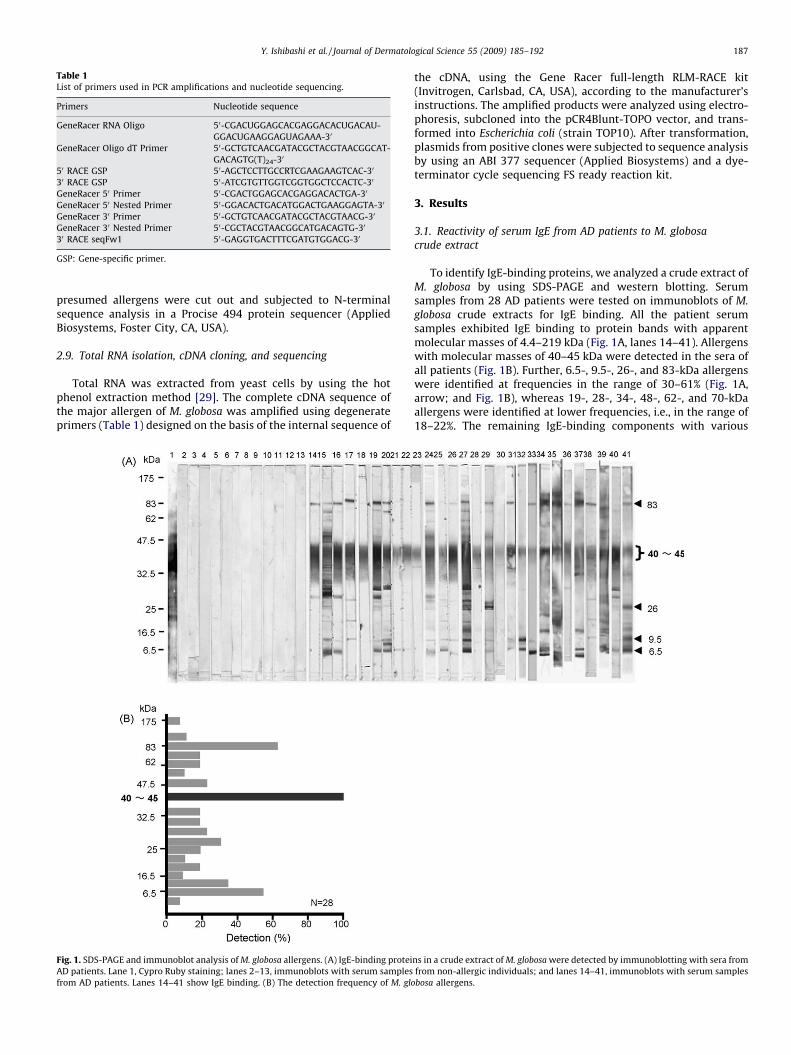
Table 1List of primers used in PCR amplifications and nucleotide sequencing.
Primers Nucleotide sequence
GeneRacer RNA Oligo 50-CGACUGGAGCACGAGGACACUGACAU-
GGACUGAAGGAGUAGAAA-30
GeneRacer Oligo dT Primer 50-GCTGTCAACGATACGCTACGTAACGGCAT-
GACAGTG(T)24-30
50 RACE GSP 50-AGCTCCTTGCCRTCGAAGAAGTCAC-30
30 RACE GSP 50-ATCGTGTTGGTCGGTGGCTCCACTC-30
GeneRacer 50 Primer 50-CGACTGGAGCACGAGGACACTGA-30
GeneRacer 50 Nested Primer 50-GGACACTGACATGGACTGAAGGAGTA-30
GeneRacer 30 Primer 50-GCTGTCAACGATACGCTACGTAACG-30
GeneRacer 30 Nested Primer 50-CGCTACGTAACGGCATGACAGTG-30
30 RACE seqFw1 50-GAGGTGACTTTCGATGTGGACG-30
GSP: Gene-specific primer.
Y. Ishibashi et al. / Journal of Dermatological Science 55 (2009) 185–192 187
presumed allergens were cut out and subjected to N-terminalsequence analysis in a Procise 494 protein sequencer (AppliedBiosystems, Foster City, CA, USA).
2.9. Total RNA isolation, cDNA cloning, and sequencing
Total RNA was extracted from yeast cells by using the hotphenol extraction method [29]. The complete cDNA sequence ofthe major allergen of M. globosa was amplified using degenerateprimers (Table 1) designed on the basis of the internal sequence of
Fig. 1. SDS-PAGE and immunoblot analysis of M. globosa allergens. (A) IgE-binding protei
AD patients. Lane 1, Cypro Ruby staining; lanes 2–13, immunoblots with serum samples
from AD patients. Lanes 14–41 show IgE binding. (B) The detection frequency of M. glo
the cDNA, using the Gene Racer full-length RLM-RACE kit(Invitrogen, Carlsbad, CA, USA), according to the manufacturer’sinstructions. The amplified products were analyzed using electro-phoresis, subcloned into the pCR4Blunt-TOPO vector, and trans-formed into Escherichia coli (strain TOP10). After transformation,plasmids from positive clones were subjected to sequence analysisby using an ABI 377 sequencer (Applied Biosystems) and a dye-terminator cycle sequencing FS ready reaction kit.
3. Results
3.1. Reactivity of serum IgE from AD patients to M. globosa
crude extract
To identify IgE-binding proteins, we analyzed a crude extract ofM. globosa by using SDS-PAGE and western blotting. Serumsamples from 28 AD patients were tested on immunoblots of M.
globosa crude extracts for IgE binding. All the patient serumsamples exhibited IgE binding to protein bands with apparentmolecular masses of 4.4–219 kDa (Fig. 1A, lanes 14–41). Allergenswith molecular masses of 40–45 kDa were detected in the sera ofall patients (Fig. 1B). Further, 6.5-, 9.5-, 26-, and 83-kDa allergenswere identified at frequencies in the range of 30–61% (Fig. 1A,arrow; and Fig. 1B), whereas 19-, 28-, 34-, 48-, 62-, and 70-kDaallergens were identified at lower frequencies, i.e., in the range of18–22%. The remaining IgE-binding components with various
ns in a crude extract of M. globosa were detected by immunoblotting with sera from
from non-allergic individuals; and lanes 14–41, immunoblots with serum samples
bosa allergens.

Fig. 2. Immunoblot analysis and 2D electrophoresis of M. globosa allergens. (A) A 2D membrane stained with Cypro Ruby. (B) Representative immunoblots probed individually
with 8 serum samples from AD patients. The immunoreactive protein spot corresponding to the major allergen from M. globosa, designated MGp42 has been circled. The
detection frequency of each spot when using the sera of 8 AD patients was as follows: MGp42 spot (8/8), spot 1 (5/8), spot 2 (2/8), spot 3 (1/8), spot 4 (2/8), spot 5 (1/8), spot 6
(2/8), spot 7 (2/8), spot 8 (3/8), spot 9 (2/8), spot 10 (2/8), spot 11 (3/8), spot 12 (2/8), spot 13 (3/8), spot 14 (2/8), spot 15 (2/8), spot 16 (2/8), spot 17 (1/8), spot 18 (1/8), spot 19
(2/8), spot 20 (3/8), spot 21 (3/8), spot 22 (2/8), spot 23 (3/8), and spot 24 (5/8).
Y. Ishibashi et al. / Journal of Dermatological Science 55 (2009) 185–192188
molecular masses, such as the 17-, 55-, and 126-kDa components,were detected at frequencies lower than 11%. No IgE binding wasobserved when the sera of healthy donors were used (Fig. 1A, lanes2–13). For our analysis, we selected the 40–45-kDa proteinbecause it is a novel IgE-binding protein identified in the sera of ADpatients at a considerably high frequency.
3.2. 2D immunoblotting
To better characterize the M. globosa allergens detected in thesera from AD patients, crude protein extracts were subjected to 2D-gel electrophoresis and subsequently, to immunoblotting. Fig. 2Ashows the 2D-gel profile of the crude extract of M. globosa, in which>80 distinct protein spots were detected on Cypro Ruby staining.To identify the spots corresponding to allergens, the IgE-bindingspots on the 2D gel were visualized by immunoblotting with 8individual serum samples from AD patients; at least 20 differentreactive spots with molecular masses of 6–90 kDa and isoelectricpoint (pI) values ranging from 3 to 10 were observed (Fig. 2B). Ahighly reactive protein spot with a molecular mass of 42 kDa and apI of 4.8, designated MGp42, was detected in each of the 8 serumsamples from the AD patients, whereas several spots, such as spots
Fig. 3. MALDI–TOF–MS profiles of a tryptic digest of the spot corresponding to the major a
analyzed by MALDI–TOF–MS, and prominent mass peaks were selected for database se
1 and 24, were detected in 63% (5 of 8) of the serum samples fromthe AD patients. Other spots were detected at lower frequencies (in1–3 of 8 serum samples). No positive spots were detected in serafrom non-allergic individuals (data not shown).
3.3. Determination of internal and N-terminal amino acid sequences
of the M. globosa allergen
To identify the IgE-reactive spot, it was excised and digested in-gel with trypsin; the resultant peptide mixtures were analyzed byMALDI–TOF–MS. The MS profile of the peptides (molecular mass,42-kDa; pI, 4.8; MGp42) showed multiple peaks in the range of1000–2700 Da (Fig. 3). Of these, 3 peptides showing prominentpeaks were selected for comparison with established databases,and the protein with the highest correlation to the spot was foundto be a member of the hsp family. To further characterize theinternal sequence of the spot, the signals at 1183.63, 1330.63, and1445.76 Da were selected for PSD analysis. The amino acidsequences of the fragments are shown in Table 2A.
After western blotting, analysis of the N-terminal amino acidsequence of MGp42 was performed, and it yielded the sequenceDLTTN (Table 1B).
llergen from M. globosa. The spot was digested in situ with trypsin. The peptides were
arches.

Table 2Determination of amino acid sequences of the major allergen of M. globosa Cf.
(A) Internal amino acid sequences of the major allergen of M. globosa (MGp42)
determined by MALDI–TOF–MS and PSD analysis.a
Peak Amino acid sequence Similarity
m/z 1183 (276) (IorL)SG(IorL)PPAPR Heat shock protein
70 of Guancha lacunosa
m/z 1330 D(299) SYTYN(IorL)R None
m/z 1445 (314) D(IorL)V(IorL)VGGSTR ACR038Wp of Ashbya
gossypii ATCC10895
(B) The N-terminal amino acid sequence of the major allergen of M. globosa
(MGp42) as determined by Edman degradation and mass spectrometry.b
N-terminal amino acid sequence DLTTN
a The sequences of the identified peptides were compared with the peptide
sequences of annotated coding regions by using the MASCOT search. The underlined
parts indicate sequences similar to those of known peptides. Full-length MGp42
cDNA contained 1908 bp of open reading frame encoding 635 amino acid residues
(Fig. 4) with a calculated molecular mass of 69.7 kDa and a pI of 4.92.b Cf. The MGp42 protein consisted of 386 amino acid residues (Fig. 4) with a
calculated molecular mass of 42.1 kDa and a pI of 4.75; these results are consistent
with those of 2D immunoblotting.
Y. Ishibashi et al. / Journal of Dermatological Science 55 (2009) 185–192 189
3.4. Sequence analysis of full-length cDNA
To obtain full-length MGp42 cDNA, we cloned and sequenced50- and 30-RACE clones. Fig. 4 shows the nucleotide sequence of thefull-length cDNA and the deduced amino acid sequence. The full-
Fig. 4. The nucleotide sequence and deduced amino acid sequence of the gene encoding t
amino acid sequence established by Edman degradation. The internal amino acid seque
length cDNA contained 1908 bp of open reading frame (ORF)encoding 635 amino acid residues with a calculated molecularmass of 69.7 kDa and a pI of 4.92. The N-terminal sequence of themature protein, MGp42, started from the 250th residue (Asp-250)of the deduced amino acid sequence. This protein consisted of 386amino acid residues and had a calculated molecular mass of42.1 kDa and a pI of 4.75, which is consistent with the results of 2Dimmunoblotting.
3.5. Sequence similarities and comparisons between HSPs
We used the basic local alignment search tool (BLAST) andfound that MGp42 showed high similarity to proteins from the hsp70 family (Fig. 5). For instance, MGp42 and HSP70 from Penicillium
citrinum (formally designated Pen c 19, accession no. U64207)share 75% identical residues. MGp42 and HSP70 from Alternaria
alternata (formally designated Alt a 3, accession no. U87807) share59% identity, whereas MGp42 and the HSP from M. sympodialis
(formally designated Mala s 10, accession no. AJ428052) share only22% sequence identity. Further, MGp42 shares 65% identity withthe HSP70 of Homo sapiens (accession no. DQ451402).
3.6. Evaluation of cross-reactivity of MGp42 with human HSP70
The IgE immunoblot inhibition studies using pooled sera fromAD patients revealed that human HSP70 did not inhibit the bindingof IgE to the 42-kDa band, corresponding to MGp42, even at high
he major allergen from M. globosa. The grey boxed region represents the N-terminal
nces determined using MALDI–TOF–MS and PSD analysis are indicated in the box.

Fig. 5. Comparison of the amino acid sequence of the M. globosa allergen with the sequences of allergens from other species. The M. globosa allergen, designated MGp42, and
HSP70 from Penicillium citrinum (formally designated Pen c 19, accession no. U64207) share 75% identical residues. MGp42 and HSP70 from Alternaria alternata (formally
designated Alt a 3, accession no. U87807) share 59% identical residues, whereas MGp42 and an hsp from M. sympodialis (formally designated Mala s 10, accession no.
AJ428052) share 22% identity. MGp42 and HSP70 from Homo sapiens (accession no. DQ451402) share 65% similarity. Amino acid residues identical to those of MGp42 are
highlighted black and in reverse type, while homologous residues are highlighted grey and in black type.
Y. Ishibashi et al. / Journal of Dermatological Science 55 (2009) 185–192190
concentrations of up to 10 mg/mL (Fig. 6). This result indicates thatno cross-reactivity exists between MGp42 and human HSP70.Preadsorption of the pooled sera with the crude extract from M.
globosa, which was used as the control, completely blocked IgEreactivity to MGp42.
Fig. 6. Immunoblotting of pooled human sera competitively inhibits binding of
MGp42 to human recombinant HSP70 protein. Lane 1, unabsorbed serum; lane 2,
serum absorbed with 0.1 mg/mL inhibitor; lane 3, serum absorbed with 1 mg/mL
inhibitor; lane 4, serum absorbed with 10 mg/mL inhibitor; and lane 5, serum
absorbed with 100 mg/mL of the M. globosa extract. Immunoblots were probed with
alkaline phosphatase-labeled goat anti-human IgE.
4. Discussion
Many studies have attempted to find a differential pattern ofMalassezia colonization on skin affected by AD [30]. However, theresults of culture-based methods have differed among studies [30–32]. This may be due to the difficulties in culturing Malassezia
species. Recently, a molecular-based (nested PCR) analysis hasrevealed that M. globosa and M. restricta are detected inapproximately 90% AD patients, while other Malassezia speciesare detected in less than 40% AD patients [12]. By using aquantitative real-time PCR method, Sugita et al. [33] have shownthat M. globosa and M. restricta account for 80% of all Malassezia
colonization on skin affected by AD. Further, it has been reportedthat AD patients have specific serum IgE antibodies againstMalassezia [7,8]. Treatment of AD patients with antifungal agentsdecreases Malassezia colonization and the severity of eczematouslesions, suggesting a major role for Malassezia species in AD [5,34].Malassezia extracts have been shown to contain 13 different

Y. Ishibashi et al. / Journal of Dermatological Science 55 (2009) 185–192 191
IgE-binding components (Mala f 2–4, Mala s 1, and Mala s 5–13)[18–21]. However, none of the IgE-binding components from M.
globosa have been identified and studied. In the present study, weused the proteomics approach to identify and characterize anallergen from M. globosa.
Immunoblot analysis showed that IgE-reactive protein compo-nents with molecular masses of 40–45 kDa were present in all theserum samples obtained from the AD patients. On 2D immuno-blots, we observed an IgE-binding protein with a molecular mass of42 kDa and a pI of 4.8; this protein was designated MGp42. The IgEreactivity of MGp42 was retained even when its carbohydratemoieties were disrupted by sodium metaperiodate treatment (datanot shown), suggesting that the carbohydrates in this compounddo not function as antigenic determinants. Proteomics analysisinvolving the MALDI–TOF–MS PSD method revealed that MGp42showed high sequence similarity to HSP70. On the basis of theinternal amino acid sequences of MGp42, the full-length cDNA ofMGp42 was cloned and sequenced by 50/30-RLM-RACE PCRanalysis. The full-length cDNA contained a 1908-bp ORF encoding635 amino acid residues with a calculated molecular mass of69.7 kDa and a pI of 6.02. Recently, the genomes of M. globosa andM. restricta were sequenced using the whole-genome shotgunmethod [35]. The ORF sequence of full-length MGp42 cDNAmatched the gene sequence of M. globosa (MGL_0058). The N-terminal sequence of the mature MGp42 protein started from the250th residue, i.e., Asp-250, of the deduced amino acid sequence. Itconsisted of 386 amino acid residues, and its molecular mass and pI
were consistent with the results of 2D immunoblotting. Theseresults suggest that MGp42 may be a cleavage product of intactHSP70.
We found that the amino acid sequences of MGp42 from M.
globosa and Mala s 10 (HSP from M. sympodialis) show lowhomology to each other (22%) even though they belong to the samegenus. The diversity of HSP70 isoforms is probably attributable tothe presence of multiple HSP70 gene copies [36,37]. For instance,the hsp70 family of Saccharomyces cervisiae contains at least 10members, including cytosolic HSP70 [38]. Cytosolic HSP70 isfurther divided into 2 groups: Ssa and Ssb. The essential Ssaproteins are most closely related to the major cytosolic hsp70s ofmammalian cells, while Ssb proteins differ from the members ofthe Ssa group by 37% [39]. Alternatively, the less-conserved COOH-terminal domain of HSP70 might account for isoform diversity[40].
Proteins of the hsp70 family appear to be phylogeneticallyhighly conserved across human, fungi, and bacterial species, thusfacilitating immunological cross-reactions between pathogens andhuman HSP70 [41]. Autoreactivity to human proteins has beenpostulated as a decisive pathogenetic factor for patients with AD[42,43]. AD patients reacting to human manganese superoxidedismutase (MnSOD) are sensitized against the MnSOD of M.
sympodialis (Mala s 11) [44]. Since MnSODs of fungal and humanorigin are highly similar in terms of their structure, sensitization ismost likely induced by exposure to fungal MnSOD of M. sympodialis
present in the environment. This results in molecular mimicry dueto secondary autoreactivity of fungal MnSOD with its humancounterpart and may contribute to the perpetuation of inflam-matory skin reactions [45]. Similarly, thioredoxins (Trxs) of M.
sympodialis (Mala s 13) and Aspergillus fumigates (Asp f 28–29) havebeen identified as novel IgE-binding proteins [46]. Mala s 13 shares45% sequence identity with human Trx. Trxs of 3 different origins(M. sympodialis, A. fumigates, and human) represent cross-reactivestructures recognized by serum IgE from individuals sensitized toTrx of M. sympodialis [46,47]. In the present study, however,immunoblot inhibition analysis revealed no IgE cross-reactivitybetween MGp42 and recombinant human HSP70. This finding is inagreement with the result that an IgE-reactive component with a
molecular mass of approximately 70 kDa was detected at lowfrequencies (18%) (Fig. 1). Thus, it seems possible that the epitopesof MGp42 recognized by serum IgE of AD patients are masked bysteric hindrance in intact HSP70 and exposed because ofconformation changes during HSP70 cleavage. We cannot ruleout the possibility that a cleavage product of human HSP70 cross-reacts with MGp42. Further studies involving epitope mapping andstructural analysis of MGp42 are required to clarify the molecularbasis of interaction between MGp42 and IgE antibodies. Inaddition, the IgE cross-reactivities between MGp42 and homo-logues of other pathogens must be explored in future studies.
Several research groups have attempted to produce recombi-nant Malassezia allergens (rMala s1, rMala s5–11) for diagnosticpurposes [19,45,46,48]. Using M. sympodialis extract and 3recombinant allergens (rMala s 1, rMala s 5, and rMala s 6),Johansson et al. [49] obtained positive reactions with specific IgEtests, skin prick tests (SPTs), and/or atopy patch tests (APTs) in 67%of the AD patients examined in their study.
No studies have investigated the clinical application of M.
globosa MGp42 for the diagnosis of AD. Recombinant MGp42allergens are required for preparing a reliable diagnostic tool forAD. Further studies are required to assess the clinical relevance ofpositive results of SPTs and APTs and of specific IgE levels to therecombinant MGp42 protein, and to evaluate the practicalapplication of these findings in the clinical management ofpatients with AD.
Proteomics is a powerful tool for the identification of allergenicproteins from Malassezia species and will greatly help furtherstudies addressing the mechanism underlying allergy sensitiza-tion.
Acknowledgment
This study was supported in part by research grants from theMinistry of Education, Culture, Sports, Science and Technology ofJapan for an Open Research Center Project.
References
[1] Faergemann J, Aly R, Maibach HI. Quantitative variations in distribution ofPityrosporum orbiculare on clinically normal skin. Acta Derm Venereol1983;63:346–8.
[2] Faergemann J, Tjernlund U, Scheynius A, Bernander S. Antigenic similaritiesand differences in genus Pityrosporum. J Invest Dermatol 1982;78:28–31.
[3] Kieffer M, Bergbrant IM, Faergemann J, Jemec GB, Ottevanger V, Stahl Skov P,et al. Immune reactions to Pityrosporum ovale in adult patients with atopic andseborrheic dermatitis. J Am Acad Dermatol 1990;22:739–42.
[4] Waersted A, Hjorth N. Pityrosporum orbiculare—a pathogenic factor in atopicdermatitis of the face, scalp and neck? Acta Derm Venereol Suppl (Stockh)1985;114:146–8.
[5] Clemmensen OJ, Hjorth N. Treatment of dermatitis of the head and neck withketoconazole in patients with type 1 sensitivity to Pityrosporum orbiculare.Semin Dermatol 1983;2:26–9.
[6] Roberts SO. Pityrosporum orbiculare: incidence and distribution on clinicallynormal skin. Br J Dermatol 1969;81:264–9.
[7] Kawano S, Nakagawa H. The correlation between the levels of anti-Malasseziafurfur IgE antibodies and severities of face and neck dermatitis of patients withatopic dermatitis. Arerugi 1995;44:128–33.
[8] Kawano S, Nakagawa H. IgE antibodies to Malassezia furfur in atopic derma-titis: comparison between CAP RAST and AlaSTAT. Jpn J Dermatol1996;106:1289–93.
[9] Bayrou O, Pecquet C, Flahault A, Artigou C, Abuaf N, Leynadier F, et al. Head andneck atopic dermatitis and malassezia-furfur-specific IgE antibodies. Derma-tology 2005;211:107–13.
[10] Nikkels AF, Pierard GE. Framing the future of antifungals in atopic dermatitis.Dermatology 2003;206:398–400.
[11] Back Bartosik J. Systemic ketoconazole for yeast allergic patients with atopicdermatitis. J Eur Acad Dermatol Venereol 2001;15:34–8.
[12] Sugita T, Suto H, Unno T, Tsuboi R, Ogawa H, Shinoda T, et al. Molecularanalysis of Malassezia microflora on the skin of atopic dermatitis patients andhealthy subject. J Clin Microbiol 2001;39:3486–90.
[13] Gueho E, Midgley G, Guillot J. The genus Malassezia with description of fournew species. Antonie van Leeuwenhoek 1996;69:337–55.

Y. Ishibashi et al. / Journal of Dermatological Science 55 (2009) 185–192192
[14] Hirai A, Kano R, Makimura K, Duarte ER, Hamdan JS, Lachance MA, et al.Malassezia nana sp. nov., a novel lipid-dependent yeast species isolated fromanimals. Int J Syst Evol Microbiol 2004;54:623–7.
[15] Sugita T, Takashima M, Shinoda T, Suto H, Unno T, Tsuboi R, et al. New yeastspecies, Malassezia dermatis, isolated from patients with atopic dermatitis.J Clin Microbiol 2002;40:1363–7.
[16] Sugita T, Tajima M, Takashima M, Amaya M, Saito M, Tsuboi R, et al. A newyeast, Malassezia yamatoensis, isolated from a patient with seborrheic derma-titis, and its distribution in patients and healthy subjects. Microbiol Immunol2004;48:579–83.
[17] Sugita T, Takashima M, Kodama M, Tsuboi R, Nishikawa A. Description of a newyeast species, Malassezia japonica, and its detection in patients with atopicdermatitis and healthy subjects. J Clin Microbiol 2003;41:4695–9.
[18] Lindborg M, Magnusson CG, Zargari A, Schmidt M, Scheynius A, Crameri R,et al. Selective cloning of allergens from the skin colonizing yeast Malasseziafurfur by phage surface display technology. J Invest Dermatol 1999;113:156–61.
[19] Schmidt M, Zargari A, Holt P, Lindbom L, Hellman U, Whitley P, et al. Thecomplete cDNA sequence and expression of the first major allergenic proteinof Malassezia furfur. Mal f 1Eur J Biochem 1997;246:181–5.
[20] Rasool O, Zargari A, Almqvist J, Eshaghi H, Whitley P, Scheynius A. Cloning,characterization and expression of complete coding sequences of three IgEbinding Malassezia furfur allergens, Mal f 7, Mal f 8 and Mal f 9.. Eur J Biochem2000;267:4355–61.
[21] Andersson A, Rasool O, Schmidt M, Kodzius R, Fluckiger S, Zargari A, et al.Cloning, expression and characterization of two new IgE-binding proteinsfrom the yeast Malassezia sympodialis with sequence similarities to heat shockproteins and manganese superoxide dismutase. Eur J Biochem 2004;271:1885–94.
[22] Conrads TP, Anderson GA, Veenstra TD, Pasa-Tolic L, Smith RD. Utility ofaccurate mass tags for proteome-wide protein identification. Anal Chem2000;72:3349–54.
[23] Peng J, Gygi SP. Proteomics: the move to mixtures. J Mass Spectrom 2001;36:1083–91.
[24] Kato H, Sugita T, Ishibashi Y, Nishikawa A. Detection and quantification ofspecific IgE antibodies against eight Malassezia species in sera of patients withatopic dermatitis by using an enzyme-linked immunosorbent assay. MicrobiolImmunol 2006;50:851–6.
[25] Hanifin JM, Rajka G. Diagnostic features of atopic dermatitis. Acta DermVenereol (Stockh) 1980;92:44–7.
[26] Kato H, Sugita T, Ishibashi Y, Nishikawa A. Evaluation of the levels of specificIgE against Cryptococcus diffluens and Cryptococcus liquefaciens in patients withatopic dermatitis. Microbiol Immunol 2007;51:945–50.
[27] Chardin H, Mayer C, Senechal H, Poncet P, Clement G, Wal JM, et al. Polyga-lacturonase (pectinase), a new oilseed rape allergen. Allergy 2003;58:407–11.
[28] Moritz RL, Eddes JS, Reid GE, Simpson RJ. S-pyridylethylation of intact poly-acrylamide gels and in situ digestion of electrophoretically separated proteins:a rapid mass spectrometric method for identifying cysteine-containing pep-tides. Electrophoresis 1996;17:907–17.
[29] Hoffman C. Preparation of yeast DNA, RNA and proteins. In: Ausubel F, Brent R,Kingston R, Moore D, Seidman J, Smith J, editors. The Current Protocols inMolecular Biology. New York: J Wiley & Sons; 1997. p. 13.11.12–..
[30] Faergemann J. Atopic dermatitis and fungi. Clin Microbiol Rev 2002;15:545–63.
[31] Gupta AK, Kohli Y, Summerbell RC, Faergemann J. Quantitative culture ofMalassezia species from different body sites of individuals with or withoutdermatoses. Med Mycol 2001;39:243–51.
[32] Nakabayashi A, Sei Y, Guillot J. Identification of Malassezia species isolatedfrom patients with seborrhoeic dermatitis, atopic dermatitis, pityriasis versi-color and normal subjects. Med Mycol 2000;38:337–41.
[33] Sugita T, Tajima M, Tsubuku H, Tsuboi R, Nishikawa A. Quantitative analysis ofcutaneous malassezia in atopic dermatitis patients using real-time PCR.Microbiol Immunol 2006;50:549–52.
[34] Mukai H, Kaneko S, Saito N, Nagase A, Arai S, Hiramatsu M, et al. Clinicalsignificance of Malassezia furfur specific IgE antibody in atopic dermatitis.Arerugi 1996;46:26–33.
[35] Xu J, Saunders CW, Hu P, Grant RA, Boekhout T, Kuramae EE, et al. Dandruff-associated Malassezia genomes reveal convergent and divergent virulencetraits shared with plant and human fungal pathogens. Proc Natl Acad Sci U S A2007;104:18730–5.
[36] Boorstein WR, Ziegelhoffer T, Craig EA. Molecular evolution of the HSP70multigene family. J Mol Evol 1994;38:1–17.
[37] Craig EA, Baxter BK, Becker J, Hatladay J, Ziegelhoffer T. In: Morimoto RI,Tissieres A, Georgopoulos C, editors. the Biology of Heat Shock Proteins andMolecular Chaperones. Plain view, NY: Cold Spring Harbor Lab Press; 1994. p.31–52.
[38] Matthews RC, Maresca B, Burnie JP, Cardona A, Carratu L, Conti S, et al. Stressproteins in fungal diseases. Med Mycol 1998;36:45–51.
[39] Lopez-Buesa P, Pfund C, Craig EA. The biochemical properties of the ATPaseactivity of a 70-kDa heat shock protein (Hsp70) are governed by the C-terminaldomains. Proc Natl Acad Sci U S A 1998;95:15253–8.
[40] Bukau B, Horwich AL. The Hsp70 and Hsp60 chaperone machines. Cell1998;92:351–66.
[41] Daugaard M, Rohde M, Jaattela M. The heat shock protein 70 family highlyhomologous proteins with overlapping and distinct functions. FEBS Lett2007;581:3702–10.
[42] Mittermann I, Aichberger KJ, Bunder R, Mothes N, Renz H, Valenta R, et al.Autoimmunity and atopic dermatitis. Curr Opin Allergy Clin Immunol2004;4:367–71.
[43] Zeller S, Glaser AG, Vilhelmsson M, Rhyner C, Crameri R, et al. Immunoglo-bulin-E-mediated reactivity to self antigens: a controversial issue. Int ArchAllergy Immunol 2008;145:87–93.
[44] Andersson A, Rasool O, Schmidt M, Kodzius R, Fluckiger S, Zargari A, et al.Cloning, expression and characterization of two new IgE-binding proteinsfrom the yeast Malassezia sympodialis with sequence similarities to heat shockproteins and manganese superoxide dismutase. Eur J Biochem 2004;271:1885–94.
[45] Schmid-Grendelmeier P, Fluckiger S, Disch R, Trautmann A, Wuthrich B, BlaserK, et al. IgE-mediated and T cell-mediated autoimmunity against manganesesuperoxide dismutase in atopic dermatitis. J Allergy Clin Immunol 2005;115:1068–75.
[46] Limacher A, Glaser AG, Meier C, Schmid-Grendelmeier P, Zeller S, Scapozza L,et al. Cross-reactivity and 1.4-A crystal structure of Malassezia sympodialisthioredoxin (Mala s 13), a member of a new pan-allergen family. J Immunol2007;178:389–96.
[47] Glaser AG, Menz G, Kirsch AI, Zeller S, Crameri R, Rhyner C, et al. Auto- andcross-reactivity to thioredoxin allergens in allergic bronchopulmonary asper-gillosis. Allergy 2008;63:1617–23.
[48] Schmid-Grendelmeier P, Scheynius A, Crameri R. The role of sensitization toMalassezia sympodialis in atopic eczema. Chem Immunol Allergy 2006;91:98–109.
[49] Johansson C, Sandstrom MH, Bartosik J, Sarnhult T, Christiansen J, Zargari A,et al. Atopy patch test reactions to Malassezia allergens differentiate subgroupsof atopic dermatitis patients. Br J Dermatol 2003;148:479–88.