identification of brain-derived neurotrophic factor ... · bdnf m cat contains a 4.4-kb hindhi/xbai...
TRANSCRIPT

Identification of Brain-derived Neurotrophic Factor Promoter Regions Mediating Tissue-specific, Axotomy-, and Neuronal Activity-induced Expression in Transgenic Mice T6nis Timmusk,*~ Urban Lendahl ,§ Hiroshi Funakoshi ,* Ernes t Arenas,* H/ikan Persson,*t and Madis Metsis*
* Laboratory of Molecular Neurobiology, Department of Medical Biochemistry and Biophysics, Karolinska Institute, S-171 77 Stockholm, Sweden; ~ Laboratory of Molecular Genetics, Institute of Chemical Physics and Biophysics, EE0026 TaJlinn, Estonia; and § Laboratory of Developmental Biology, Department of Cell and Molecular Biology, Karolinska Institute, S-171 77 Stockholm, Sweden
Abstract. The structure of rat brain-derived neuro- trophic factor (BDNF) gene is complex; four 5' exons are linked to separate promoters and one 3' exon is encoding the BDNF protein. To analyze the relative importance of the regulatory regions in vivo, we have generated transgenic mice with six different promoter constructs of the BDNF gene fused to the chloram- phenicol acetyl transferase reporter gene. High level and neuronal expression of the reporter gene, that in many respects recapitulated BDNF gene expression, was achieved by using 9 kb of genomic sequences covering the promoter regions that lie adjacent to each other in the genome (promoters I and II and promoters
HI and IV, respectively) and by including sequences of BDNF intron-exon splice junctions and 3' untranslated region in the constructs. The genomic regions respon- sible for the in vivo upregulation of BDNF expression in the axotomized sciatic nerve and in the brain after kainic acid-induced seizures and KCl-induced spread- tug depression were mapped. These data show that regulation of the different aspects of BDNF expression is controlled by different regions in vivo, and they sug- gest that these promoter constructs may be useful for targeted expression of heterologous genes to specific regions of the central and peripheral nervous systems in an inducible manner.
B RAin-derived neurotrophic factor (BDNF) ~ (Barde et al., 1982; Leibrock et al., 1989) is a member of the neurotrophin family that includes three other struc-
turally related proteins: NGF, neurotrophin-3, and neu- rotrophin-4 (reviewed in Korsching, 1993; Persson, 1993). In cell culture, BDNF promotes the survival of retinal gan- glion cells (Johnson et al., 1986), basal forebrain cholinergic neurons (Alderson et al., 1990), embryonic mesencephalic dopaminergic neurons (Hyman et al., 1991; Kniisel et al., 1991), and embryonic cerebellar granule neurons (Segal et al., 1992). BDNF also promotes neuronal differentiation in neural crest cell cultures (Sieber-Blum, 199D and enhances early maturational change in dorsal root ganglion cell cul-
t HAkan Persson died on 16 May 1993. Address correspondence to T6nis Timmusk, Laboratory of Molecular
Neurobiology, Department of Medical Biochemistry and Biophysics, Karolinska Institute, S-171 77 Stockholm, Sweden. Tel.: (46) 8-728-7657. Fax: (46) 8-341-960.
L Abbreviations used in this paper: BDNF, brain-derived neurotrophic fac- tor; CAT, chloramphenicol acetyl transferase; KA, kainic acid; NRSE, neural-restrictive silencer element; SD, spreading depression; UTR, un- translated region.
rares (Wright et al., 1992). Recently, it was shown that en- dogenous BDNF is atrophic factor in activity-mediated neu- ronal survival of cultured embryonic cortical neurons (Ghosh et al., 1994). In vivo BDNF increases the survival of em- bryonic neurons in the dorsal root ganglia, nodose ganglia (Hofer and Barde, 1988), basal forebrain cholinergic neu- rons (Kntisel et al., 1992), as well as developing and ax- otomized motoneurons (Oppenheimet al., 1992; Sendtner et al., 1992; Yan et al., 1992; Koliatsos et al., 1993). BDNF null-mutated mice develop several sensory deficits, but no obvious defects were seen for motoneurons or central do- paminergic neurons (Ernfors et al., 1994; Jones et al., 1994).
Except for the striatum and septum, BDNF mRNA is ex- pressed in neurons throughout the adult rat brain with the highest level in the hippocampal formation (Ernfors et al., 1990a, b; Hofer et al., 1990; Phillips et al., 1990; Wetmore et al., 1990), where also BDNF-like immunoreactivity has been demonstrated 0Vetmore et al., 1991). Glutamatergic, GABA-ergic, and cholinergic neurotransmitter systems have been shown to be involved in the regulation of BDNF mRNA expression (Zafra et al., 1990, 1991; Ernfors et al., 1991; Isackson et al., 1991; Dugich-Djordejevic et al., 1992a, b; Lindefors et al., 1992; Lindvall et al., 1992). Activation of
© The Rockefeller University Press, 0021-9525/95/01/185/15 $2.00 The Journal of Cell Biology, Volume 128, Numbers 1 &2, January 1995 185-199 185
Dow
nloaded from https://rupress.org/jcb/article-pdf/128/1/185/383948/185.pdf by guest on 16 D
ecember 2019

glutamate receptors of both NMDA and non-NMDA sub- types has been implicated in the increase of BDNF mRNA after administration of kainlc acid (KA) or after epileptic and ischemic insults (Zafra et al., 1990, 1991; Ernfors et al., 1991; Lindvall et al., 1992). However, the molecular mecha- nisms governing the increase of BDNF mRNA levels, as well as the mechanisms responsible for the tissue-specific and de- velopmentally regulated expression of BDNF mRNA, are unknown.
It has recently been shown that the organization of BDNF gene is complex (Tunmusk et al., 1993). Four short untrans- lated 5' exons and one 3' exon encoding the mature BDNF protein are present within the rat BDNF gene with a separate promoter upstream of each 5' exon. Alternative promoter us- age, differential splicing and the use of two different polyade- nylation sites within each of the four transcription units generate altogether eight different BDNF mRNAs (Tunmusk et al., 1993). Promoters I-HI are used predominantly in the brain, while promoter IV is more active in peripheral tis- sues. Moreover, the levels of BDNF mRNAs transcribed from promoters I-HI are markedly elevated in a promoter- specific subset of hippocampal and neocortical neurons in different models of neuronal activation, while only a modest increase was seen for promoter IV specific mRNA (Falken- berg et al., 1993; Metsis et al., 1993; Timmusk et al., 1993; Kokaia et al., 1994).
In this report, we have analyzed the potential of the pro- moter regions of the rat BDNF gene to drive expression of a reporter gene in transgenic mice. Tissue-specific and high level expression of the bacterial reporter gene chioramphenl- col acetyl transferase (CAT) in correct neuronal populations of transgenic mice brain was achieved by using 9 kb of genomic sequences covering the promoter regions that lic close in the genome (promoters I and II and promoters HI and IV, respectively) and by including sequences of BDNF introns with splice donor and acceptor sites and 3' untrans- lated region (UTR) in the constructs. We also identified the promoter regions that are involved in the regulation of BDNF induction in different experimental models, i.e., (a) in the cerebral cortex after KCl-induced cortical spreading depres- sion; (b) in the hippocampus and cerebral cortex after KA- induced seizures; and (c) in the axotomized sciatic nerve.
Materials and Methods
General Methods All molecular biology procedures were performed according to standard practices (Sambrook et ai., 1989). Plasmid DNA was purified by alkaline lysis and CsCI centrifugation. Labeling of cRNA and cDNA probes, RNA isolation, CAT assays, RNase protection assay, RNA quantification, and in situ hybridization were performed as shown earlier (Thnmusk et ul., 1993, 1994a). BDNF exon-specific cRNA probes were synthesized from the same fragments of rat BDNF gene as described (Timmttsk ct al., 1993, 1994a). As a probe for CAT, the 0.8-kb coding sequence of it was digested out from pBLCAT2 (Luckow and Schutz, 1987) using XhoH restriction enzyme, sub- cloned into the Eco RV site of pBSKS vector (Stratagene, La Jolla, CA), and used further to synthesize cRNA or cDNA probes. As a probe for mouse BDNF, a 0.34-kb NarI fragment of mouse BDNF cDNA (Hofer et al., 1990) was used as a template.
Tmnsgenic Mice Transgenic mice were generated and analyzed for transgene integration as previously described (Nilsson and Lendahl, 1993). Vector sequences were
removed from the BDNF promoter constructs and the transgene was in- jected into the male pronucleus of one-cell mouse embryos obtained from superovulated Fl(C57BL6 X CBA). Injected eggs were transferred at the one- or two-cell stage to the oviducts of pseudoprngnant foster mothers. Tail biopsy-derived DNA isolated from the resulting offspring (F0) was screened by Southern or dot blot analysis with CAT-specific probe for the presence of the transgene. Different restriction digests were used to identify those animals that contained intact copies of the construct (data not shown). The frequencies for transgenicity were between 15 and 50%. As the integra- tion of transgene in F0 founders may be chimeric, most of the transgenic founders were maintained as lines by breeding them with wild-type mice. The analysis of transgene expression and regulation was performed with 1-2-mo-old FI animals unless specifically noted.
Animals, Pharmacological Treatments, and Surgery Adult male and female transgenic mice (body wt ffi 20-30 g) were used for all experiments. Treatment of animals with kainic acid was performed as described earlier (Zafra et al., 1990) and animals were killed 3-6 h after the injection. Spreading depression produced by repeated topical applica- tion of KCI to the cortical surface of mice during 2 h was performed as de- scribed in (Kokaia et al., 1993) and mice were killed after another 2 h. Tran- section of sciatic nerve was performed as previously described (Funakoshi et al., 1993). After all treatments mice were anesthetized with ether and decapitated. The brains, indicated brain regions, and peripheral organs were dissected and immediately frozen on dry ice.
BDNF-CAT Fusion Genes Promoters of BDNT I, H, m , and IV CAT constructs were all inserted into modified pBLCAT2 vector, replacing the thymidine kinase promoter by cloning into the XbaI and XhoI sites. All these constructs contain SV-40 small t intron and SV-40 polyadenylation signals downstream from CAT gene. The numbering and restriction enzyme sites is corresponding to the published rat BDNF gene sequence (T'munusk ct ul., 1993).
BDNF I CAT contains a 2.6-kb EcoRUCIaI fragment of exon I and its 5' flanking region. The 5' EcoRI site lies 1.5 kb upstream from the BamHI site located at the first basepair of rat BDNF gene sequence. The 3' ClaI site is located at 937 bp.
BDNF 11 CAT contains a 3.7-kb EcoRI/AccI fragment of exon 11 and its flanking region. This construct has the same 5' end as BDNF I CAT, but it extends 1.2 kb to the 3' direction. The 3' AceI site is located at 2,151 bp inside of exon II.
BDNF m CAT contains a 4.4-kb HindHi/XbaI fragment of exon HI and its 5' flanking region. The 5' HindIII site lies 3.5 kb upstream from the HinclI site located at the first basepair of rat BDNF gene sequence. The 3' XbaI site is located at 901 bp.
BDNF IV CAT contains a 5.5-kb HindiIFBam~ fragment of exon IV and its 5' flanking region. This construct has the same 5' end as BDNF HI CAT, but it extends 1.1 kb to the 3' direction. The 3' BamIH site is located at 2,051 bp inside of exon IV.
BDNF IV prox CAT contains a 0.9-kb EcoRJ/BamHI fragment of exon IV and its 5' flanking region. This construct is a 5' deletion of BDNF IV CAT.
BDNF I + II CAT and BDNF HI + IV CAT were cloned in pBSKS and they contain the following: (a) Promoter region and the 5' part of the mini- intron including splice donor sites: genomic frasments covering the 5' and 3' flanking regions of exons I and II or exons IlI and IV, respectively. (b) 3' part of the mini-intron including splice acceptor site: a 0.7-kb genomic fragment covering the Y flenking region of exon V (coding exon) including 10 bp of exon V (this is the common splice aceeptor region for all the BDNF transcripts). This region is common for both minigenes. (c) CAT 8ene (0.7-kb XhoII fragment) without SV-40 small t intron and polyadenylation signals. This region is common for both minigenes. (d) 3' UTR including polyadenylation signals: 3.2-kb genomic fragment covering BDNF 3' UTR, extending 0.1 kb downstream from the end of the 4.2-kb mRNA. This re- gion is common for both minigenes.
BDNF I + II CAT promoter region contains a 9.5-kb BamHI/SalI genomic fragment including exons I and II and the flanking regions. The 5' BamHI site lies ,o6.5 kb upstream from the BamHI site located at the first bp of rat BDNF gene sequence. The 3' SalI site lies ,~,0.7 kb down- stream from the end of exon H.
BDNF HI + IV CAT promoter region contains a 9-kb BamHI/HindHi genomic fragment including exons HI and IV and the flank/rig regions. The 5' BamHI site lies ,o6 kb upstream from the HincH site located at the first
The Journal of Cell Biology, Volume 128, 1995 186
Dow
nloaded from https://rupress.org/jcb/article-pdf/128/1/185/383948/185.pdf by guest on 16 D
ecember 2019

bp of rat BDNF gene sequence. The 3' HindM site lies ~,0.7 kb downstream from the end of exon IV.
Results
Expression of BDNF Promoter L II, III, and IV CAT Constructs in Transgenic Mice
Functional analysis of the proximal promoter regions of the BDNF gene by ~ransient transfections showed that sequences upstream of all four 5' exons promote expression of the CAT reporter gene in neuronal and nonneuronal cell lines (Tim- musk et al., 1993). To analyze the tissue specificity of the rat BDNF gene expression in vivo, transgenic mice were generated that harbored a chimeric gene composed of one of the upstream regions of the 5' exons of rat BDNF gene fused to sequences encoding the bacterial enzyme CAT. The con- structs (designated BDNF I CAT, BDNF II CAT, BDNF M CAT, and BDNF IV CAT) are represented in Fig. 1. All these fusion genes contain SV-40 small t intron and SV-40 polyad- enylation signals downstream of the CAT coding region. BDNF I CAT contains a 2.6-kb fragment of exon I and its 5' flanking region. BDNF II CAT contains a 3.7 kb fragment of exon II and its 5' flanking region. This construct has the same 5' end as BDNF I CAT, but it extends 1.2 kb to the 3' direction and has the 3' end in exon 1I. BDNF M CAT con- tains a 4.4-kb fragment of exon M and its 5' flanking region. BDNF IV CAT contains a 5.5-kb fragment of exon IV and its 5' flanking region. This construct has the same 5' end as BDNF M CAT, but it extends 1.1 kb to the 3' direction ter- minating inside exon IV.
CAT activity was measured in brain and in 10 nonneural tissues: salivary gland, thymus, heart, lung, spleen, liver, kidney, pancreas, muscle, and testis. As the mouse BDNF gene structure has not been reported, the expression patterns of the BDNF-CAT fusion genes were compared to the levels of rat BDNF mRNAs containing different 5' exons by RNase protection assay.
2.6 kb of BDNF Promoter I Targets the Expression of Reporter Gene to the Thymus of Transgenic Mice
Eight different transgenic lines expressing CAT under the control of a 2.6-kb 5' flanking sequence of exon I (BDNF I CAT) were analyzed. CAT activity was detected in the thy- mus of all of the eight lines tested. In the other organs, in- cluding the brain, the levels of CAT activity were close to or below the detection limit (Fig. 2 and Table I). We have recently shown that in the rat this promoter is active in brain and not in lung and heart (Tmunusk et al., 1993). RNase
I
10kb
I+11 L
BJ 11 B
,, Jill' 111 !1[ \
Il l I l l \ ~ I I / \
~ II I \ I I I I
I
Figure 1. Schematic representation of rat BDNF-CAT fusion genes analyzed in transgenic mice in relation to the rat BDNF gene struc- ture. The gene is shown in top with exons with their corresponding numbering as boxes and introns as lines. UTRs of the exons are in- dicated by open boxes, and the region corresponding to the prepro- BDNF protein is indicated by the filled box. Restriction sites for BamHI are indicated by B's. The BDNF CAT fusion genes are shown below the schematic map of BDNF gene. Open boxes with I, II, HI, IV, IV prox, I + II, and M + IV indicate the upstream regions of BDNF 5' exons included in BDNF I CAT, BDNF II CAT, BDNF HI CAT, BDNF IV CAT, BDNF IV prox CAT, BDNF I + il CAT, and BDNF HI + IV CAT, respectively. Broken lines show the regions of rat BDNF gene included in the BDNF-CAT fusion genes. ~,, SV-40 small t intron and polyadenylation signals. BDNF 3', 3' UTR of rat BDNF gene.
Figure 2. Expression of BDNF I CAT (CAT/), BDNF II CAT (CAT //), BDNF m CAT (CATIlI), and BDNF IV CAT (CAT/V) in the brain and nonneural tissues of transgenic mice of representative founder lines. 1 mg of protein extract was analyzed in each line by CAT assay. Samples were incubated for 2 h at 37°C. Expression of endogenous rat BDNF exon mRNAs in the same rat tissues ana- lyzed by RNase protection assay is shown in top of the CAT assays of each BDNF-CAT fusion gene.
Timmusk et al. BDNF Promoters in Transgenic Mice 187
Dow
nloaded from https://rupress.org/jcb/article-pdf/128/1/185/383948/185.pdf by guest on 16 D
ecember 2019

Table I. 7~ssue Distribution of CAT Activity in Transgenic Mice with Different BDNF-CAT Fusion Genes
CAT activity TISSUE
BRA SAL THY HEA LUN SPL LIV KID PAN MUS T/O
I 6 0 8 6 4 6 0 0 0 0 0 n = 8 (+) - + (+) (+) (+) . . . . . H 4 0 4 0 0 0 0 0 0 0 0 n ffi 18 (+) - (+) . . . . . . . . Ill 6 2 4 4 4 4 4 3 3 6 4 n = 11 + (+ ) + + + ( + ) ( + ) ( + ) ( + ) + (+ ) IV 11 8 8 8 8 8 8 8 11 8 8 n = 13 + ( + ) (+ ) (+ ) ( + ) ( + ) ( + ) ( + ) + ( + ) (+ ) I + H 9 m 9 1 7 1 0 1 1 1 1 n = 9 + + + ++ (+) + (+) - (+) (+) (+) (+) IH+IV 6 nt 6 2 5 1 1 3 2 2 1 n = 6 + + + + + ( + ) + (+) (+) (+) (+) (+) (+)
Average conversion of [14C]chloramphenicol per 1 nag protein during 2 h reaction at 37°C in the expressing founder lines: (+), <1% conversion; +, 1-5%; + +, 5-25%; + + +, >25%. I, II, HI, IV, I + If, and Ill + IV, BDNF-CAT fusion genes BDNF I CAT, BDNF II CAT, BDNF HI CAT, BDNF IV CAT, BDNF I + H CAT, and BDNF HI + IV CAT, r~e~vely , n, number of analyzed founder lines; nt, not tested; BRA, brain; SAL, salivary gland; THY, thymus; HEA, heart; LUN, lung; SPL, spleen; L/V, liver; K/D, kidney; PAN, pancreas; MUS, skeletal muscle; T/O, testis or ovary.
protection analysis of RNA from 11 different rat t issues showed that, in addition to brain, lower levels of BDNF exon I mRNA are expressed in the thymus and spleen (Fig. 2). These data demonstrate that the 2.6-kb of 5' flanking BDNF exon I sequence is sufficient to direct appropriate tissue- specific expression of reporter gene in the thymus, but not in the spleen and brain.
~7 kb of BDNF Promoter H Is Predominantly Silent in Transgenic Mice
18 lines containing CAT under the control of 3.7 kb of the 5' flanking sequences of BDNF exon II were generated and analyzed. In 14 lines, we were not able to detect any CAT activity. In the remaining four lines, CAT activity was de- tected only in the brain and thymus, and it was significantly lower than i~ the thymus of transgenic mice carrying the BDNF I CAT transgene. (Fig. 2 and Table I). RNase pro- tection analysis revealed that the endogenous expression of BDNF exon II mRNA in rat is restricted to the brain (Fig. 2). The 3.7-kb of BDNF exon II 5' flanking region is apparently lacking positive regulatory sequences that are regulating the endogenous BDNF mRNA expression from this promoter.
4.4 kb of BDNF Promoter I11 Directs CAT Expression Predominantly in Brain and Muscle
11 different founder lines carrying 4.4 kb of the upstream se- quences of BDNF exon IT[ linked to CAT gene were analyzed and in six of them CAT activity was detected (Table I). In two lines, CAT activity was highest in the muscle and brain, and lower levels were present in the lung, thymus, heart, and liver (representative founder line is shown in Fig. 2). In two other lines, CAT activity was detected only in the muscle and brain. In two fines, CAT activity was detected in most of the organs with little differences in the expression levels. In five lines, we did not detect CAT activity in any of the analyzed organs. In the rat, this promoter is active mainly in brain and less also in the heart, as revealed by Northern blotting (Tim- musk et al., 1993). RNase protection analysis of 11 rat tis-
sues confirmed that the highest levels of BDNF exon 11I mRNA are present in the brain and significantly lower levels were seen in heart. Exon llI mRNA was also detected in muscle and lung at low (close to detection limit) levels (Fig. 2). Taken together, the results demonstrate that the 4.4- kb of BDNF exon Ill 5' flanking region directed the transgenic expression of CAT in tissues that overlap with the endoge- nous expression pattern of exon III mRNA in the rat.
5.5 kb of BDNF Promoter IV Directs the Expression of Reporter Gene Preferentially in Brain and Pancreas
Promoter IV of the rat BDNF gene is a classical G/C rich promoter that directs the expression of BDNF mRNA mainly outside of the nervous system with the highest levels in lung and heart (Timmusk et al., 1993). Sensitive RNase protection analysis showed that BDNF exon IV mRNA is ex- pressed at low levels in all the rat tissues analyzed, except liver (Fig. 2). 13 different founder lines possessing the reporter gene fused to the 5' flanking sequences of BDNF exon IV were analyzed (Fig. 2 and Table I). CAT activity was detected in 11 founder lines, showing that the percentage of transgene-expressing mice was higher than for BDNF HI CAT. In seven founder lines, the levels were highest in the brain and pancreas, in two lines in the liver and in one line CAT levels were nearly equal in all the tissues analyzed. The expression pattern of BDNF IV CAT in transgenlc mice (as shown for one representative founder line in Fig. 2) suggests that the 5.5-kb of rat BDNT promoter IV region includes ele- ments essential for exon IV mRNA regulation in brain but is lacking sequences that are controlling the high level ex- pression of endogenous BDNF in the heart and lung.
Expression o fBDNF I + H CAT and I I I+ IV CAT in Transgenic Mice
Partial recapitulation of the tissue specificity of endogenous BDNF mRNA expression and comparatively low transgene expression levels in the transgenlc mice possessing the four BDNF promoter constructs described above suggested that these promoter regions of the rat BDNF gene are not
The Journal of Cell Biology, Volume 128, 1995 188
Dow
nloaded from https://rupress.org/jcb/article-pdf/128/1/185/383948/185.pdf by guest on 16 D
ecember 2019

sufficient to account for the full activity of the gene. Promoters I and II and promoters HI and IV are adjacent in the genome and display similar regulation patterns during brain development, suggesting the existence of shared regulatory sequences (Tmunusk et al., 1994a). We therefore decided to generate two new BDNF constructs, designated BDNF I + 1I CAT and BDNF HI + IV CAT (Fig. 1). The promoter region of BDNF I + II CAT consists of a 9.5-kb genomic fragment including exons I and II with their splice donor sites and the flanking regions. The promoter region of BDNF HI + IV CAT contains a 9-kb genomic fragment in- eluding exons HI and IV with the flanking regions. Several studies have shown that transgene constructs that include SV small t intron downstream of coding sequences are not op- timal for expression, while inclusion of generic introns in the constructs increases transgene expression in mice (Brinster et al., 1988; Choi et al., 1991; Palmiter et al., 1991). In addi- tion, the requirement of intragenic regulatory elements for tissue-specific and regulated expression has been described for several genes, including neural-specific ones (Vidal et al., 1990; Beaudet et al., 1992; Belecky et al., 1993; Van- selow et al., 1994; Zimmerman et al., 1994). We, therefore, decided to include BDNF mini-introns and 3' UTR in the constructs (Fig. 1). Except for the promoter regions and splice donor sites, both these constructs also contain 0.7 kb of BDNF coding exon 5' sequence with splice acceptor site and CAT coding sequence. The SV-40 polyadenylation sig- nals downstream of the CAT gene were replaced by rat BDNF genomic region covering the 3' UTR of the 4.2-kb mRNA and 0.1 kb of additional downstream sequences. The transgene expression was analyzed in seven different brain regions: cerebral cortex, hippocampus, cerebellum, stria- turn, thalamus/hypothalarnus, midbrain, and brainstem. The analyzed nonneural tissues were the same as for the four transgenic constructs described above, except that expres- sion was not analyzed in salivary gland.
Figure 3. Expression of BDNF I + II CAT (CATI + II) and BDNF HI + IV CAT (CAT III + IV ) in the different brain regions and non- neural tissues of transgenic mice of representative founder lines. 0.1 nag of protein extract was analyzed in each line by CAT assay. Sam- pies were incubated for 2 h at 37"C.
BDNF I + H CAT Directs High Level Expression of the Reporter Gene in the Brain and Thymus of Transgenic Mice
Nine founders possessing the BDNT I + II CAT were ob- tained and bred to establish F1 transgenic lines to study the expression and regulation of this promoter construct in brain and peripheral tissues (Tables I and II and Fig. 3). All the nine lines expressed high level of CAT activity in the brain. In the thymus, CAT activity was always 5-10-fold lower (Ta- ble I and Fig. 3). In seven founder lines, CAT was also ex- pressed in the lung with levels significantly lower than in the thymus. In the brain, the measured CAT activities were
Table II. CAT Activity in Different Brain Regions and the Levels of CAT mRNA in the Hippocampus of Transgenic Mice Possessing BDNF I + II CAT and BDNF III + IV CAT Fusion Genes
CAT activity BRAIN REGION
CAT mRNA x 100%
Founder line CTX HIP CBL STR THA MID STE BDNF mRNA
I+II
P4 + + + + + PI1 + + + Q2 + + + + Q4 + + + + + + Q6 + + + + Q9-1 + + + Q9-2 + + + + + Q l l + +
m + I V X7 + + Y3-1 + + + + + + Y3-2 + + + + + + Y7 +++ +++ Z1 (+) + + + Z8 ++ ++
+ + + + + + + + ++ 200-300% + nt nt nt + nt
+ + + + + + + + ++ 15-40% + + + + + + + + + 50-100% + nt nt nt + nt
+ + + + + + + + + + <5% + + + + + + + + 25-40%
(+) + (+) + (+) <5%
- - (+) + - <5%
++ +++ +++ +++ +++ 15-30% (+) +++ +++ +++ ++ 10-20%
- + + + + + + + + + + + + 300--400%
- (+) + + + + + 20-30% ++ + + + ++ ++ + + + 20-30%
Average conversion of [14C]chloramphenicol per 0.1 rag protein during 2 h reaction at 37°C in the expressing founder lines: (+), <1% conversion; + , 1-5 %; + + , 5-25%; + + + , >25%. 1 + l land I11 + IV, BDNF-CAT fusion genes BDNF I + 1I CAT and BDNF HI + IV CAT, repectively; nt, not tested; CTX, cerebral cortex; HIP, hippocampus; CBL, cerebellum; STR, striatum; THA, thalamus and hypothalumus; MID, midbrain; STE, brainstem.
Timmusk et al. BDNF Promoters in Transgenic Mice 189
Dow
nloaded from https://rupress.org/jcb/article-pdf/128/1/185/383948/185.pdf by guest on 16 D
ecember 2019

~lO0-fold higher than in the transgenic animals with BDNF I CAT and BDNF II CAT.
CAT activity was analyzed in seven different brain regions (Table II and Fig. 3). The expression patterns varied slightly from one founder line to another, but in most of them, the highest levels were seen in the hippocampus, midbraln, thai- amus/hypothalamus, and cerebral cortex. Lower CAT activ- ity was detected in the brainstem and the lowest in the stria- turn and cerebellum. In the rat brain, the highest levels of endogenous BDNF exon I mRNA, transcribed from pro- moter I, were seen in the hippocampus and cerebral cortex, and lower levels were present in the other brain regions (Timmusk et al., 1994a, b). In the cerebellum and striatum, exon I mRNA was not detected. BDNF exon H transcripts, synthesised from promoter H, are expressed throughout the rat brain, with the highest levels in cerebellum, hip- pocampus, and midbrain RNA (Timmusk et al., 1994a, b). The transgene expression pattern of BDNF I + H CAT recapitulated that of endogenous rat BDNF mRNA tran- scribed from promoters I and H of rat BDNF gene with only two minor differences: a lower expression of transgene in the cerebellum and a higher expression in the striatum (Table IT). Endogenous mouse BDNF mRNA and transgenlc CAT mRNA were quantified in the hippocampus of transgenlc animals by RNase protection using known amounts of sense BDNF and CAT mRNA, respectively. The results revealed that in several founder lines the transgene is expressed with levels comparable (founder fines Q4, Q2, and Q9-1) or up to threefold higher (founder line P4) than the endogenous mouse BDNF mRNA (Table ID. These results clearly show that the rat BDNF genomic region of BDNF I + II CAT con- struct directs high level tissue-specific expression of the reporter gene in transgenic mice.
BDNF III + IV CAT Is Expressed at High Levels in the Brain of Tmnsgenic Mice
Six different transgenic mice lines with BDNF HI + IV CAT were obtained and the offspring analyzed as shown in Tables I and II and illustrated in Fig. 3. In all these founder lines, transgene expression was highest in brain followed by thy- mus. Lower levels of CAT activity were seen in the lung of five transgenic lines, in the kidney of three lines, in the heart, pancreas and muscle of two lines, and in the spleen, liver, and testis of one line. In the rat heart and lung, BDNF mRNA is predominantly expressed from promoter IV of the BDNF gene and the total levels are comparable with BDNF mRNA in the brain. The results revealed that BDNF HI + IV CAT recapitulated the endogenous BDNF expression in the lung but not in the heart.
Analysis of CAT activity expression patterns in different brain regions of transgenic mice revealed that it was ex- pressed throughout the brain with only small differences in the expression levels. In the cerebellum, CAT activity was absent or significantly lower. Quantification of RNA showed that in the hippocampus, CAT mRNA levels were compara- ble (founder lines Z1, Z8, Y3d, and Y3-2) or higher (founder line YT) as compared to the endogenous mouse BDNF mRNA levels determined in the same animals. BDNF exon HI mRNA, that is transcribed from promoter m of the rat BDNF gene, has the most restricted expression pattern in the rat brain having highest levels in the cerebral cortex and hip- pocampus and 5-10-fold lower levels in the other brain
regions. BDNF exon IV mRNA is expressed in all rat brain regions, except striatum, without significant differences in the levels (Tunmusk et al., 1994a, b). Comparison of rat BDNF expression patterns with that of BDNF I l I + IV CAT in transgenic mice showed that the transgene is expressed in a similar manner to the rat BDNT mRNA transcribed from promoters Ill and IV. The minor differences from the endog- enous pattern were the expression of transgene in the stria- turn and lower levels of expression in the cerebellum.
5.3 kb of the Y Sequence of BDNF Exon IV Is SuffJcient to Direct Lesion-induced Expression of CAT in the Sciatic Nerve BDNF mRNA has been shown to be induced in the distal segment of az.otomized sciatic nerve (Meyer et al., 1992) by selective upregulation of exon IV mRNA levels (Funakoshi et al., 1993). To explore the possibility that the induction is caused by differential activation of BDNF promoter IV we studied if axotomy of sciatic nerve induces CAT expression
Figure 4. Expression of BDNF IV CAT in axotomized sciatic nerve of A2 founder line. (A) Time course of CAT activity changes in the distal segment of transect~ sciatic nerve. (B) CAT activities, CAT mRNA, and mouse BDNF mRNA (codin~ exon) in different seg- ments of the sciatic nerve 14 d after lesion. 0.1 mg of protein extract was analyzed in each line by CAT assay. Samples were incubated for 2 h at 37°C. (B) 10 rag of total RNA was An~yzed by RNase protection assay using cRNA probe specific for CAT or mouse BDNE CTR, control sciatic nerve; PRX, proximal segment of the transected sciatic nerve, DIS, distal segment of the transect~ sciatic nerve.
The Journal of Cell Biology. Volume 128, 1995 190
Dow
nloaded from https://rupress.org/jcb/article-pdf/128/1/185/383948/185.pdf by guest on 16 D
ecember 2019
in transgenic mice possessing BDNF IV CAT. The sciatic nerves of mice were transected and CAT activity was deter- mined in the distal part of the lesioned nerve at different time points after operation (Fig. 4 A). In the intact sciatic nerve, CAT activity was low with levels only marginally above the detection limit. An increase in CAT activity was seen 1 wk after lesion and the levels were fivefold higher 2 and 3 wk after lesion (Fig. 4 A). This time course of trans- gene expression mimics exactly the delayed induction of rat BDNF mRNA after sciatic nerve lesion.
CAT activity and CAT mRNA levels were analyzed in the distal and proximal part of the transected sciatic nerve 2 wk after lesion (Fig. 4 B). In the distal part, both CAT activity and CAT mRNA were upregulated. In the proximal segment, only CAT activity was enhanced, indicating that it is not a transcriptional regulation and could be caused by transport and accumulation of CAT protein. Similar results were ob- tained with mice from two different lines of the same con- struct. As the regulation of mouse BDNF mRNA in this ax- otomy model has not been reported, we also determined the mouse BDNF mRNA levels in the same samples (Fig. 4 B). Endogenous mouse BDNF mRNA was increased predomi- nantly in the distal segment of axotomized sciatic nerve. These data demonstrate that the 5.5-kb of BDNF exon IV the 5' sequence is sufficient to direct correctly spatiotemporally regulated expression of reporter gene in the lesioned sciatic nerve.
To study if the proximal region of promoter IV is sufficient to confer the axotomy-induced expression of reporter gene in sciatic nerve, transgenic mice possessing BDNF IV prox CAT (Fig. 1) were generated and the offspring analyzed. This BDNF-CAT fusion gene is a 4.6-kb 5' deletion of BDNF IV CAT. CAT activity was not induced in the axotomized sciatic nerve of any of the six different founder lines analyzed (data not shown). These results indicate that the regulatory se- quences involved in the induction of BDNF expression in the lesioned sciatic nerve are located in the distal part of BDNF promoter IV in the region between 0.9 and 5.5 kb upstream from the 3' end of exon IV.
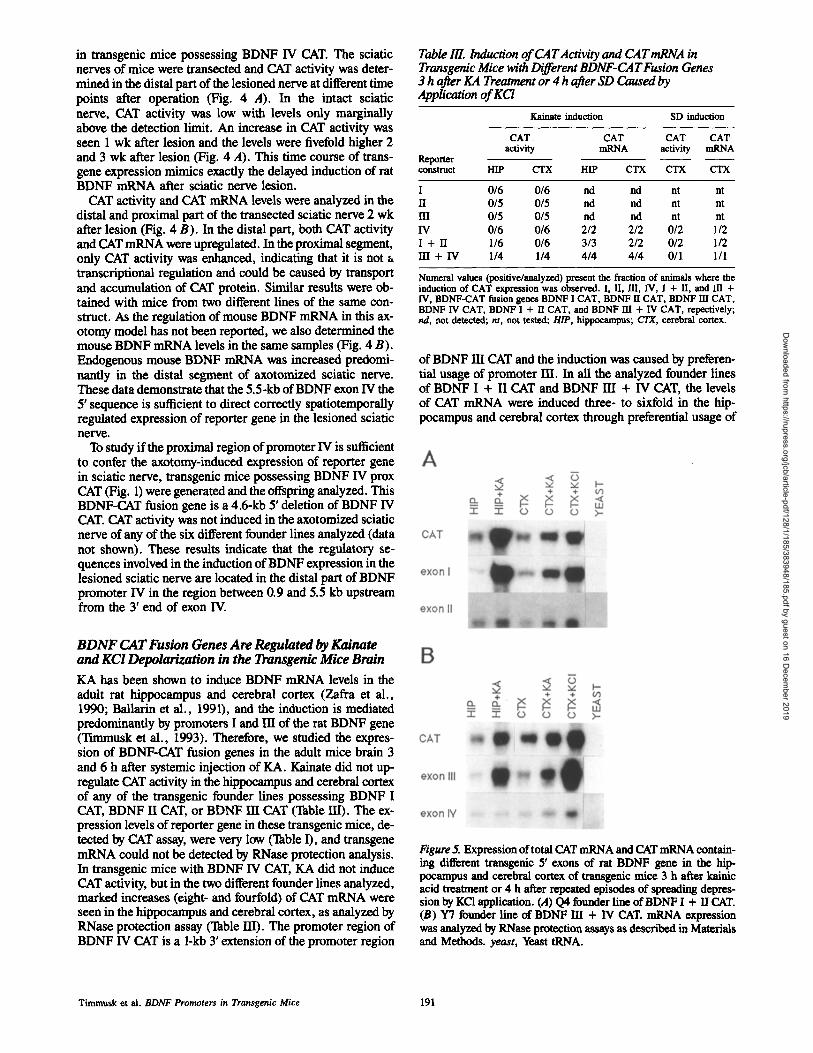
Table III. Induction of CAT Activity and CAT mRNA in Transgenic Mice with Different BDNF-CAT Fusion Genes 3 h after KA Treatment or 4 h after SD Caused by Application of KCl
Kainate induction SD induction
CAT CAT CAT CAT activity rnRNA activity rnRNA
Reporter construct HIP CTX HIP CTX CTX CTX
I 0/6 0/6 nd nd nt nt II 0/5 0/5 nd nd nt nt Ill 0/5 0/5 lad rid nt nt IV 0/6 0/6 2/2 2/2 0/2 1/2 I + lI 1/6 0/6 3/3 2/2 0/2 1/2 HI + IV 1/4 1/4 4/4 4/4 0/1 1/1
Numeral values (positive/analyzed) present the fraction of animals where the induction of CAT expression was observed. I, 11, IN, IV, I + n, and III+ IV, BDNF-CAT fusion genes BDNF I CAT, BDNF n CAT, BDNF HI CAT, BDNF IV CAT, BDNF I + II CAT, and BDNF HI + IV CAT, repectively; rid, not detected; nt, not tested; HIP, hippocampus; C/X, cerebral cortex.
of BDNF HI CAT and the induction was caused by preferen- tial usage of promoter HI. In all the analyzed founder lines of BDNF I + II CAT and BDNF HI + IV CAT, the levels of CAT mRNA were induced three- to sixfold in the hip- pocampus and cerebral cortex through preferential usage of
B D N F CAT Fusion Genes Are Regulated by Kainate and KC! Depolarization in the Transgenic Mice Brain
KA has been shown to induce BDNF mRNA levels in the adult rat hippocampus and cerebral cortex (Zafra et al., 1990; Ballarin et al., 1991), and the induction is mediated predominantly by promoters I and m of the rat BDNF gene (Tunmusk et al., 1993). Therefore, we studied the expres- sion of BDNF-CAT fusion genes in the adult mice brain 3 and 6 h after systemic injection of KA. Kainate did not up- regulate CAT activity in the hippocampus and cerebral cortex of any of the transgenlc founder lines possessing BDNF I CAT, BDNF 1I CAT, or BDNF HI CAT (Table 111). The ex- pression levels of reporter gene in these transgenlc mice, de- tected by CAT assay, were very low (Table I), and transgene mRNA could not be detected by RNase protection analysis. In transgenic mice with BDNF IV CAT, KA did not induce CAT activity, but in the two different founder lines analyzed, marked increases (eight- and fourfold) of CAT mRNA were seen in the hippocampus and cerebral cortex, as analyzed by RNase protection assay (Table HI). The promoter region of BDNF IV CAT is a 1-kb 3' extension of the promoter region
Figure 5. Expression of total CAT mRNA and CAT mRNA contain- hag different transgenic 5' exons of rat BDNF gene in the hip- pocampus and cerebral cortex of transgenic mice 3 h after kainic acid treatment or 4 h after repeated episodes of spreading depres- sion by KCI application. (,4) Q4 founder line of BDNF I + II CAT. (B) Y7 founder line of BDNF Ill + IV CAT. mRNA expression was analyzed by RNase protection assays as described in Materials and Methods. yeast, Yeast tRNA.
Timmusk et al. BDNF Promoters in Transgenic Mice 191
Dow
nloaded from https://rupress.org/jcb/article-pdf/128/1/185/383948/185.pdf by guest on 16 D
ecember 2019

Figure 6. Light-field photomicrographs of au- toradiograms of in situ hybridization showing the expression of CAT mRNA and mouse BDNF mRNA in the brain of control (A, C, and E) and kainic acid treated transgenic mice (B, D, and F). Coronal sections were hybridized to cRNA probes specific for CAT (A-D) or mouse BDNF coding exon (E and F). (A, B, E, and F) P4 founder line of I + II CAT transgenic mouse. (C and D) Y7 founder line of HI + IV CAT transgenic mouse. dg, Dentate gyrus; CA/and CA3, pyramidal layers CA1 and CA3 of the hippocampus; hi, hilar region of the hippocampus; pit, piriform cortex.
transgenic BDNF exons I and HI, respectively (Table HI and Fig. 5), that resembles the induction of rat BDNF mRNA. In contrast to this, CAT activity was induced (1.5-fold) only in the hippocampus of one of the analyzed lines with BDNF I + II CAT. In transgenic mice possessing BDNF HI + IV CAT, twofold increases of CAT activity were seen only in the hippocampus of Y7 and in the cerebral cortex of ZI founder line (Table IID.
Spreading depression (SD), caused by KCI depolarization of cortical neurons, has been shown to induce BDNF mRNA in the rat cerebral cortex (Kokaia et ai., 1993). Next, we in- vestigated if SD increases CAT expression in the cerebral cortex of transgenic lines where transgene induction by KA was seen. In one out of two different analyzed lines of BDNF I + II CAT and BDNF IV CAT, KC1 depolarization increased CAT mRNA in the cerebral cortex through preferential usage of promoters I and HI, respectively (Table HI and Fig. 5). Induction of CAT mRNA was also analyzed and observed in one founder line of BDNF HI + IV CAT and it was predomi- nantly mediated by promoter HI (Table HI and Fig. 5). The
transgene mRNA levels after KC1 depolarization were up to eightfold higher than in control animals, but no changes were seen in the CAT activities. These results demonstrate that CAT mRNA transcribed from BDNF promoter constructs was regulated in the transgenic mouse brain by two different types of neuronal activation in a manner similar to the regu- lation of rat BDNF mRNA in the same experimental models.
Transgenic Expression of BDNF I + H CAT and BDNF III + IV CAT in the Mouse Brain Is Neuron-specific and Overlaps with the Basal and Kainate-induced Expression Pattern of Endogenous Rat BDNF mRNA
The founder lines of BDNF I + II CAT and HI + IV CAT that expressed the reporter gene at the highest levels (P4 and Y7, respectively) were used to analyze the transgene expres- sion in the brain of control and and KA-treated animals by in situ hybridization using 3sS-UTP labeled cRNA probe of CAT coding sequence. To check if the KA treatment had in-
Figure Z CAT mRNA expression in the hipIxx:mnpus of P4 founder line of I + II CAT. Shown are emulsion autoradiograms obtained after hybridization of coronal sections from adult control transgenic brain or from transgenic animals 3 h after kaim'c acid trea~nent to a cRNA probe specific for CAT. Dark-field photomicrographs of the hippocampus are shown above. Note the sharp decrease of labeling at the end of CA3 region in the kainate-treated animal. Below are presented bright-field photomicrographs of higher magnification showing labeled neurons in CA1, CA3, and hilar region and in the granular layer of dentate gyrus, respectively. Note that some of the neurons have greater intensity of labeling than neighboring ones in the CA3 and briar regions of both control and kainate-treated animals. Note also that in contrast to the induction of CAT mRNA by kainate in the CA3, hilus, and dentate gyrus there is no increase in the grain density in the neurons of CA1 region.
The Journal of Cell Biology, Volume 128, 1995 192
Dow
nloaded from https://rupress.org/jcb/article-pdf/128/1/185/383948/185.pdf by guest on 16 D
ecember 2019

Timmusk et al. BDNF Promoters in Transgenic Mice 193
Dow
nloaded from https://rupress.org/jcb/article-pdf/128/1/185/383948/185.pdf by guest on 16 D
ecember 2019
duced endogenous BDNF expression, sections from the same brain were also hybridized with a mouse BDNF- specific probe (coding exon). BDNF mRNA was induced in both of the analyzed animals and the distribution of BDNF in the brain of the analyzed animal of P4 founder line is shown in Fig. 6, E and E
The transgenic line P4, expressing the BDNF I + II CAT, showed the highest levels of CAT mRNA in the pyramidal layer of CA3 and in the hilar region of the hippocmnpus (Fig. 6 A). Labeling was also seen in the granular layer of dentate gyrus, regions CA1 and CA2, external layers of the cerebral cortex, and in the piriform cortex. Lower levels of CAT mRNA were detected in other brain regions. Different neurons of the same region expressed the transgene at differ- ent levels. In the hippocampus, the granule neurons of den- rate gyms and the pyramidal neurons of CA1 and CA2 regions were labeled in a diffuse and uniform manner that contrasted with the different densities of grains found in the neurons of CA3 and especially of the hilus (Fig. 7). 3 h after systemic injection of KA, more than fivefold increases were seen in the CA3 and hiiar region, and in the granular cell layer of dentate gyms (Figs. 6 B and 7). Marked increases (about threefold) were also seen in the external layers of the cerebral cortex, in the piriform cortex, and in several nuclei of the amygdaloid complex, such as the medial and pos- teromedial amygdaloid nuclei. Examination of emulsion au- toradiograms revealed that labeling was restricted to neu- rons in control and in kainate-treated animals. These results are in agreement with the induction of rat BDNF exon I and exon II mRNAs by KA, but the relative levels of the endoge- nous mRNA, particularly in the dentate gyrus, are higher (Metsis et al., 1993; Tunmusk et al., 1993). On the cellular level, a differential response to KA was seen in all these regions. In addition to the uniform increase in the labeling pattern, individual cells were found to be more densely la- beled than neighboring ones (Fig. 7). In the internal layers of cerebral cortex and in the CA1 region of the hippocampus, no increase of CAT mRNA levels were seen.
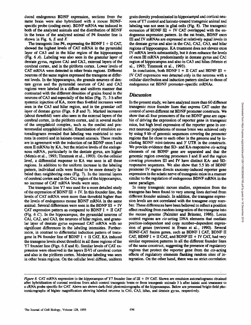
The transgenic line Y7 was used for a more detailed study of the expression of BDNF HI + IV. In this founder line, the levels of CAT mRNA were more than threefold higher than the levels of endogenous mouse BDNF mRNA in the same animal. Several differences were seen in the BDNF ffI + IV CAT expression pattern as compared to BDNF I + II CAT (Fig. 6 C). In the hippocampus, the pyramidal neurons of CA1, CA2, and CA3, the neurons of hilar region, and granu- lar layer of dentate gyrus expressed CAT mRNA with no significant differences in the labeling intensities. Further- more, in contrast to differential induction pattern of trans- gene in P4 founder line of BDNF I + II CAT, KA induced the transgene levels about threefold in all these regions of the Y7 founder line (Figs. 6 E and 8). Similar levels of CAT ex- pression were observed in the layers II-VI of cerebral cortex and also in the piriform cortex. Moderate labeling was seen in other brain regions. On the cellular level diffuse, uniform
grain density predominated in hippocampal and cortical neu- rons of Y7 control and kainate-treated transgenic animal and labeling was not seen in glial cells (Fig. 8). The transgenic exression of BDNF HI + IV CAT overlapped with the en- dogenous expression pattern. In the rat brain, BDNF exon HI and IV mRNAs are expressed at relatively equal levels in the dentate gyms and also in the CA1, CA2, CA3, and hilar regions of hippocampus. KA treatment does not elevate exon IV mRNA levels substantially, but it does enhance the levels of exon HI mRNA predominantly in dentate gyrus and CA1 region of hippocampus and also in CA3 and hilus (Metsis et al., 1993; Timmusk et al., 1993).
In conclusion, both BDNF I + II CAT and BDNF HI + IV CAT expression was detected only in the neurons with a cellular distribution and induction pattern similar to those of endogenous rat BDNF promoter-specific mRNAs.
Discussion
In the present study, we have analyzed more than 60 different transgenic mice founder lines that express CAT under the control of seven different rat BDNF promoter constructs. We show that all four promoters of the rat BDNF gene are capa- ble of driving the expression of reporter gene in transgenic mice, but high level expression of the reporter gene in cor- rect neuronal populations of mouse brain was achieved only by using 9 kb of genomic sequences covering the promoter regions that lie close to each other in the geuome and by in- cluding BDNF minl-introns and 3' UTR in the constructs. We provide evidence that SD- and KA-responsive cis-acting elements of rat BDNT gene are separated and that the genomic region covering promoters I and II and the region covering promoters HI and IV have distinct KA- and SD- responsive sequences. We also show that 5.5 kb of BDNF promoter IV region directs axotomy-induced reporter gene expression in the sciatic nerve of transgenic mice in a manner similar to the regulation of endogenous BDNF mRNA in the same paradigm.
In many transgenic mouse studies, expression from the transgene has been found to vary among lines derived from different founder animals. In addition, the transgene expres- sion levels are not correlated with the transgene copy num- ber. These differences have been believed to reflect a position effect resulting from random integration of the transgene into the mouse genome (Pnlmiter and Brinster, 1986). Locus control regions are cis-acting DNA elements that mediate position-indepondent and copy number-dependent expres- sion of genes (reviewed in Evans et al., 1990). Several BDNF-CAT fusion genes, such as BDNF I CAT, BDNF II CAT, BDNF I + 1I CAT, and BDNF HI + IV CAT, had very similar expression patterns in all the different founder lines of the same construct, suggesting the presence of regulatory regions that protect the reporter gene from the cis-acting effects of regulatory elements flanking random sites of in- tegration. On the other hand, there was no strict correlation
Figure 8. CAT mRNA expression in the hippocampus of Y7 founder line of HI + IV CAT. Shown are emulsion autoradiograms obtained after hybridization of coronal sections from adult control transgenic brain or from transgenic animals 3 h after kainic acid treatment to a cRNA probe specific for CAT. Above are shown dark-field photomicrographs of the hippocampus. Below are presented bright-field pho- tomicrographs of higher magnification showing labeled neurons in CAI, CA3, hilus, and dentate gyrus, respectively.
The Journal of Cell Biology, Volume 128, 1995 194
Dow
nloaded from https://rupress.org/jcb/article-pdf/128/1/185/383948/185.pdf by guest on 16 D
ecember 2019

Timmusk ct al. BDNF Promoters in Transgenic Mice 195
Dow
nloaded from https://rupress.org/jcb/article-pdf/128/1/185/383948/185.pdf by guest on 16 D
ecember 2019

between the integrated copy number of transgene and its ex- pression levels (data not shown). It is possible that a locus control region exists also in the BDNF gene and only part of it was included in these BDNF promoter constructs, ex- pressed in a position-independent manner, but with expres- sion levels that were not correlated with the transgene copy number.
1Issue-specific Expression of BDNF-CAT Fusion Genes in Transgenic Mice When fused separately to bacterial CAT gene the upstream regions of exons I, II, and HI of rat BDNF gene were able to direct the transgene expression in tissues that partially overlapped with the endogenous sites of particular BDNF promoter activities. 2.6 kb of exon I 5' flanking sequence recapitulated BDNF expression in the thymus, but not in the brain and spleen. Similarly, 4.5 kb of exon HI 5' flanking re- gion was insufficient to restrict transgene expression to cor- rect tissues, but predominant transgenic expression in the brain and muscle suggests that some regulatory elements are present in the analyzed promoter HI sequence. And finally, 5.5 kb of promoter IV recapitulated endogenous expression in the brain, but not in the heart and lung. These results sug- gest that cis-acting elements that are regulating the expres- sion of rat BDNF gene in different tissues (cell populations) are separated in the genome. Examples of genes for which similar regulation strategy is used are Thy-1, where elements responsible for the brain, thymus, kidney, and spleen expres- sion are all separated (Vidal et al., 1990), and nestin, where independent regulatory elements control the expression in the neural stem cells and muscle precursors (Zimmerman et ai., 1994). An exceptional case was the 3.7-kb of the exon II 5' sequence, which, when linked to the reporter gene, was silent in the transgenic mice. This may be explained by the presence of a negative control element as a neural-restrictive silencer element (NRSE) has been described in the SCG10 gene (Mori et al., 1992) and the rat type lI sodium channel gene (Kraner et al., 1992) and a highly homologous se- quence is present in the 5' region of rat BDNF exon H (Tim- musk et al., 1993). Therefore, it is possible that because of the absence of positive neural-specific regulatory sequences in the BDNF promoter II construct, this NRSE is involved in the suppression of transgene expression.
Neuronal-specific and high level expression of the reporter gene was achieved by introducing two new fusion genes, BDNF I + II CAT and BDNF HI + IV CAT, into transgenic mice. These constructs contain 9 kb of genomic sequences covering the BDNF promoter regions that lie close to each other in the genome, and sequences of BDNF intron-exon splice junctions and 3' UTR. In the brain, no significant differences from the pattern of endogenous BDNF expres- sion were seen for either of these constructs with the excep- tion of higher levels of CAT in striatum and lower in cerebel- lum. Although the expression levels of transgene in the same brain region varied from one founder line to another of the same construct, the highest expression was always in the hip- pocampus and the absolute levels were comparable, or even higher, than the endogenous levels of mouse BDNF mRNA. The cellular expression patterns were similar to those of rat BDNF promoter specific mRNAs. In the brain, BDNF I + II CAT and BDNF HI + IV CAT were expressed only in the neurons with the highest levels in the hippocampal pyrami-
dal, granular, and hilar neurons, as well as in the cortical neurons. Transgene expression was not detected in glial cells. In nonneural tissues, BDNF I + II CAT correctly mimicked the expression pattern of BDNF exon I and exon H mRNAs, but BDNF 1II + IV CAT transgenic expression was comparatively low in heart and lung, where high levels of BDNF mRNA are transcribed from promoters HI and IV in the rat.
We cannot precisely determine which regions of BDNF I + H CAT and BDNF HI + IV CAT were responsible for the high level and accurate pattern of expression in brain neu- rons, because 5' flanking regions, introns, and 3' UTR, not present in the first four analyzed BDNF constructs, were in- cluded in these BDNF-CAT fusion genes. Regulatory ele- ments that drive neuron-specific expression in transgenic mice have been shown to be located in the 5' flanking regions and in the intragenic regions (reviewed in Belecky, 1993; Beaudet, 1992). Correct expression pattern in transgenic mice has often been achieved when the entire region covering the gene, as well as its 5' and 3' flanking sequences, has been included in the transgene (Patil et al., 1990; Kaneda et al., 1991). For several neural genes, comparatively short ge- nomic regions of 5' flanking sequences have been shown to drive the expression to all neurons, or neurons and some ectopic tissues, recapitulating partially the endogeneous ex- pression pattern. Furthermore, inclusion of longer 5' se- quences (Vandaele et al., 1991) or intragenic regions (Ang et al., 1993; Belecky et al., 1993; Vanselow et al., 1994) has restricted the expression to correct neurons. Similarly, the shorter BDNF promoter-CAT fusion genes partially recapit- ulated the expression of endogenous BDNF gene, but higher level and more precise expression patterns were achieved with the longest constructs. Although both BDNF I + II and BDNF HI + IV CAT contained ~15 kb of genomic se- quences, they did not completely mimick the complex tissue- specific pattern of BDNF gene. Therefore, it is reasonable to assume that additional regulatory regions are involved in the regulation of the rat BDNF gene.
5.5 kb of BDNF Promoter IV Region Directs Axotomy-induced Expression of Reporter Gene in the Sciatic Nerve Transection of the rat sciatic nerve leads to a marked in- crease of BDNF mRNA in the distal segment of nerve, prob- ably in Schwann cells, which is thought to play a role in providing trophic support to injured peripheral nerves. The increase is seen only 1 wk after lesion, with progressively higher levels until 3 wk after lesion (Meyer et al., 1992; Funakoshi et al., 1993). Only BDNF exon IV mRNA is in- creased in the lesioned sciatic nerve, suggesting that the in- duction is caused by selective activation of BDNF promoter IV (Funakoshi et al., 1993). Here, we show that 5.5 kb of the 5' flanking sequence of BDNF exon IV directs axotomy- induced expression of CAT mRNA and CAT acti~,ity in the sciatic nerve of two different founder lines that were ana- lyzed. The time course and spatial pattern of transgene ex- pression were similar to BDNF expression in rat and in mouse. Both CAT and endogenous BDNF were induced pre- dominantly in the distal segment of sciatic nerve and the up- regulation was observed first at 7 d after lesion, peaking at 2 wk, and remained elevated also 3 wk after lesion. As previ- ously shown, there is also a marked increase of NGF mRNA
The Journal of Cell Biology, Volume 128, 1995 196
Dow
nloaded from https://rupress.org/jcb/article-pdf/128/1/185/383948/185.pdf by guest on 16 D
ecember 2019

in the nonneuronal cells of lesioned sciatic nerve. In contrast to BDNF mRNA, the increase in NGF mRNA is biphasic with a rapid transient increase followed by a second increase that lasts for several weeks (Heumann et al., 1987). It has been suggested that the induction of NGF expression in the lesioned sciatic nerve is mediated by the immediate early gene c-fos through a functional AP-l-binding site in the NGF gene (Hengerer et al., 1990). The delayed induction of BDNF and promoter IV-CAT fusion gene suggests that c-fos and other transcription factors encoded by immediate early genes are not involved in the regulation of BDNF expression after sciatic nerve transection. We have shown that adrenal- ectomy attenuated the increase of BDNF mRNA in the le- sioned nerve and that a putative binding site for the glucocor- ticoid receptor is present 420 bp upstream from the cap site of promoter IV, suggesting that glucocorticoids could be in- volved in the upregulation of BDNF mRNA (Funakoshi et al., 1993). In this study, we show that 0.9 kb of the exon IV 5' sequence is not sufficient to mediate the induction of the reporter gene in lesioned sciatic nerve, suggesting that this putative binding site for the glucocorticoid receptor is not in- volved in the regulation of BDNF induction in this experi- mental model. Work is in progress to determine which par- ticular sequence elements are involved in the regulation of BDNF expression in lesioned sciatic nerve.
Regulation of the Expression of BDNF-CAT Fusion Genes in the Hippocampus and Cerebral Cortex after KA Seizures and KCl-mediated Depolarization BDNF mRNA levels in the hippocampus and cerebral cortex of adult rat brain have been shown to be markedly but tran- siently induced by several stimuli such as seizures induced by administration ofKA (Zafra et al., 1990; Ballarin et al., 1991; Dugich-Djordejevic et al., 1992a, b), pentylentetrazol (Humpel et al., 1993), pilocarpine (Berzaghi et al., 1993; Metsis et al., 1993) focal electrolytic lesions (Isackson et al., 1991), kindling after electric stimulation (Ernfors et al., 1991), ischemic insults and insulin-induced hypoglycemic coma (Ernfors et al., 1991; Lindvall et al., 1992; Comelli et al., 1993), and cortical SD induced by application of KCl (Kokaia et al., 1993). These data have led to the hypothesis that BDNF protein could play a neuroprotective role after brain insults.
We investigated if the expression of BDNF-CAT fusion genes is induced by systemic kainic acid treatment, which is a potent pharmacological regulator of BDNF mRNA expres- sion. CAT mRNA was under the detection limit in the founder lines possessing BDNF I CAT, BDNF II CAT, and BDNF Ill CAT. All the other BDNF promoter constructs analyzed, BDNF IV CAT, BDNF I + II CAT, and BDNF Ill + IV CAT, directed kainate-inducible expression of reporter gene mRNA in the transgenic mice by preferential usage of exons I and Ill in these constructs. The relative in- creases of transgene mRNA varied in different founder lines of the same construct but were always smaller than for partic- ular BDNF exon mRNAs. Striking differences were seen in the regional distribution of increases of CAT mRNA after treatment with kainic acid. In the hippocampus, transgene mRNA transcribed from BDNF I + II CAT of P4 founder line was increased in the hilar region, in the granular cells of dentate gyrus and in the CA3 region. No significant induc- tion was observed in the CA1 region. Rat BDNF exon I and
exon 11 mRNAs are induced in similar hippocampal neuronal populations, but with a more marked relative increase in the granular cell layer of dentate gyrus. CAT mRNA transcribed from BDNF HI + IV CAT was increased in CA1, CA2, and CA3 regions and in dentate gyrus with no significant differ- ences in the amplitude of induction. Rat BDNF exon HI mRNA is increased in the same hippocampal neuronal popu- lations, but the increase is more marked in CA1 region.
SD, caused by KCI depolarization of cortical neurons, was tested in those founder lines that showed a marked induction of transgene mRNA after KA treatment. SD induced CAT mRNA expression in the cerebral cortex of one analyzed founder line of BDNF Ill + IV CAT and in one out of two analyzed founder lines of BDNF IV CAT. Furthermore, SD increased CAT mRNA expression in one out of two analyzed founder lines ofBDNF I + II CAT, while KA induced trans- gene expression in both of them. The results indicate that both KA- and SD-responsive cis-acting elements are present in the BDNF 5.5-kb sequence of BDNF IV CAT (containing both exon 11I and exon IV, and their 5' flanking sequences), and also in the BDNF genomic regions of BDNF I + II CAT. As these BDNF-CAT fusion genes do not have overlapping BDNF sequences, it is highly likely that different signaling pathways, linked to the responsive cis-acting elements in the cluster of promoters I and 1/, and to the sequences in the cluster of promoters HI and IV, mediate the kainate induction of rat BDNF gene. The variation of induction of BDNF I + II CAT and BDNF IV CAT by KA and SD could be the result of a different transgene integration site where the SD- but not KA-responsive cis-acting elements lost the ability to mediate transgene induction in one founder line, suggesting that KA and SD elements are different and located in different sites of the sequence of these BDNF-CAT fusion genes. Taken to- gether, our data suggest that (a) SD- and KA-responsive c/s- acting elements of rat BDNF gene are separated; and (b) genomic region covering promoters I and II and genomic re- gion covering promoters Ill and IV have distinct KA- and SD-responsive sequences.
Concerning the signaling pathways leading to the induc- tion of BDNF mRNA, it has recently been shown that in cul- tured embryonic cortical neurons, KC1 and glutamate induce BDNF expression by activation of distinct calcium channels, L-type voltage-sensitive calcium channels, and N-methyl-I)- aspartate receptors respectively (Ghosh et al., 1994). Fur- thermore, in cultured hippocampal neurons L-type calcium channels and NMDA receptors are coupled to two different signaling pathways that activate transcription through differ- ent cis-acfing regulatory elements in the c-fos promoter (Bading et al., 1993). Since KC1 and KA in this study were applied systemically, the induction of CAT expression was probably caused not only by direct activation of one particu- lar glutamate receptor, but also by indirect activation of different glutamate receptor subtypes by endogenous gluta- mate release that is thought to mediate the increase of BDNF expression in these experimental models (Zafra et al., 1990; Kokaia et al., 1993). It remains to be determined to what ex- tent the increase of BDNF gene expression is causally related to the activation of immediate-early genes, such as c-los, c-tim, zif/268, and others, in KA-mediated seizures and SD (reviewed in Morgan and Curran, 1991).
KA and SD upregulated CAT mRNA levels in transgenic mice of different BDNF-CAT fusion genes up to 10-fold, but mostly it was not paralleled by an increase of CAT protein,
Timmusk ¢t al. BDNF Promoters in Transgenic Mice 197
Dow
nloaded from https://rupress.org/jcb/article-pdf/128/1/185/383948/185.pdf by guest on 16 D
ecember 2019

as determined by CAT assay 3 or 6 h after the treatment. Re- cent]y, it was reported that the kainate-induced increase of BDNF mRNA was not paralleled by alterations of equal magnitude and distribution of BDNF immunoreactivity (Wetmore et al., 1994). The increase ofBDNF immunoreac- tivity was observed in restricted neuronal subpopulations of hippocampus, with most significant changes in the C.A3 re- gion, and to a lesser degree in other cortical areas. Similarly, it has been reported that during the acute phases of pentylen- tetrazol seizures, the increase of BDNF mRNA levels in the hippocampus is not paralleled by an increase of BDNF-like immunoreactivity (Humpel et al., 1993). This discrepancy between the induction level of CAT (and also BDNF) mRNA and protein could be explained by the results obtained using hippocampal slice culture showing that high concentrations of glutamate, KA, NMDA, and quisqualate inhibit neuronal protein synthesis by ~90% (Vornov and Coyle, 1991). We cannot rule out that CAT protein, like BDNF protein, is in- duced only in some neuronal populations, and that the CAT assay is not a suitable method to detect these subtle changes. Finally, the finding that axotomy induced CAT activity in transgenlc lines with BDNF IV CAT suggests that only moderate BDNF mRNA induction by focal lesions, like ax- otomy, or by more physiological stimuli like light (Castn~n et al., 1992), long-term potentiation (Patterson et al., 1992; Castn~n et al., 1993), improved spatial memory, and en- riched environment (Falkenberg et al., 1992), may result in elevated BDNF protein levels.
Taken together, this study is one of the first steps towards the understanding of the regulatory mechanisms governing BDNF gene expression. Our results show that the mul- tipromoter structure of the BDNF gene supports neuronal function through a complex regulation of gene expression in different tissues and cell populations. The transgenic mice with the various BDNF-CAT fusion genes provide the oppor- tunity to study the relative contributions of BDNF promoters and their particular sequence elements in the regulation of BDNT gene expression during development, as well as in different lesion models or after neuronal activation. Finally, targeted expression of neurotrophins and other heterologous genes under the control of the described BDNF promoter constructs could be used to study their function and regula- tion in the nervous system and nonneural tissues.
Prof. H/dmn Persson died during the preparation of this study and his premature death is a great loss for ~ of us. We thank E. Nilsson for excel- lent technical assistance and Dr. N. Belluardo for help and advice in animal experiments.
This study was supported by grants from Cancerfonden, The Bank of Sweden Tercentenary Foundation and funds from Karolinska Institute. T. Ttmmusk was supported by a stipend from Karolinska Institute's coopera- tion program with Baltic countries, U. Lendald was supported by Axel and Margaret Axson Johnson Foundation and Kjell and Mirta Beijer Founda- tion, H. Funakoshi was supported by Human Frontier Science Program, E. Arenas was supported by Swedish Medical Research Council and M. Metsis was supported by The Bank of Sweden Tercentenary Foundation.
Rt~rence$
Alderson, R. F., A. L. Alterman, Y.-A. Barde, and R. M. Lindsay. 1990. Brain-derived neurotrophic factor increases survival and differentiated fonc- fions of rat septal cholinergic neurons in culture. Neuron. 5:297-306.
Ang, H. L., D. A., Carter, and D. Murphy. 1993. Neuron-specific expression and physiological regulation of bovine vasopressin transgenes in mice. EMBO (Eur. Mol. Biol. Organ.) J. 12:2397-2409.
Bnding, H., D. D. Ginty, and M. E. Greenberg. 1993. Regulation of gune ex- pression in hippocampal neurons by distinct calcium signaling pathways. Science (Wash. DC). 260:181-186.
Ballarin, M., P. Ernfors, N. Lindefors, and H. Persson. 1991. Hippocampal damage and kainic acid injection induce a rapid increase in mRNA for BDNF and NGF in the rat brain. Exp. Neurol. 114:35-43.
Barde, Y.-A., D. Edgar, and H. Thnenen. 1982. Purification of a new neuro- trophic factor from mammalian brain. EMBO (Eur. MoL Biol. Organ.) J. i :549-553.
Beaudet, L., G. Charron, D. Houle, I. Tretjakoff, A. Peterson, and J. P. Julien. 1992. Intragenic regulatory elements contribute to transcripdoanl control of the neurofilament light gune. Gene (Amst.). 116:205-214.
Belecky, A. T., D. C. Wight, J. J. Kopchick, and L. M. Parysek. 1993. Intra- genic sequences are required for cell type-specific and injury-induced expres- sion of the rat paripherin gene. J. NeuroscL 13:5056-5065.
Berzaghi, M. P., J. Cooper, E. Castr~n, F. Zafra, M. Sofroniew, H. Thnenen, and D. Lindholm. 1993. Cholinergic regulation of brain-derived neuro- trophic factor (BDNF) and nerve growth factor (NGF) but not neurotro- phin-3 (NT-3) mRNA levels in the developing rat hippoc.mnpas. J. Neurosci. 13:3818-3826.
Brinster, R. L., J. M. Allen, R. R. Behringer, R. E. Gelines, and R. D. Psimiter. 1988. Introas increase transcripUonal efficiency in transgenic mice. Proc. Natl. Acad. Sci. USA. 85:836-840.
Castr6n, E., M. Pitidlnen, J. Sirvio, A. Parsadanlan, D. Lindholm, H. Thoe- nen, and P. J. Riekkinen. 1993. The induction of LTP increases BDNF and NGF mRNA but decreases NT-3 mRNA in the dentate gyrus. Neuroreport. 4:895-898.
Castr6n, E , F. Zafra, H. Thoenen, and D. Lindholm. 1992. Light regulates expression of brain-derived neurotrophic factor mRNA in rat visual cortex. Proc. Natl. Acad. Sci. USA. 89:9444-9448.
Choi, T., M. Hneng, C. Gorman, and R. Jaeulsch. 1991. A generic intron in- creases gene expression in transgeulc mice. Mol. Ceil. Biol. 11:3070-3074.
Comelli, M. C., D. Guidolin, M. S. Seren, R. Zanoni, R. Canella, R. Rubiul, and H. Manev. 1993. Time course, localization and pharmacological modu- lation of immediate early inducible genes, brain-derived neurotrophic factor and trkB messenger RNAs in the rat brain following photochemical stroke. Neuroscience. 55:473-490.
Dugich-Djordejevic, M. M., G. Tocco, D. A. Willoughby, I. Njam, G. Pesinetd, P. A. [atpchak, and F. Hefti. 1992a. BDNF mRNA expression in the developing rat brain following kainic acid-induced seizure activity. Neu- ron. 8:1127-1138.
Dugich-Djordjevich, M. M., G. Tocco, P. A. Lapchak, G. M. Pesinetti, I. Najm, M. Baudry, and F. Hefli. 1992/7. Regionally specific and rapid in- creases in brain-derived neurotrophic factor messenger RNA in the adult rat brain following seizures induced by systemic admim'stradon of kalm'c acid. Neuroscience. 47:303-315.
Ernfors, P., C. F. Ibanez, T. Ebendal, L. Olson, and H. Persson. 1990a. Mo- lecular cloning and neurotrophic activities of a protein with structural similarities to nerve growth factor: developmental and topographical expres- sion in the brain. Proc. Natl. Acad. ScL USA. 87:5454--5458.
Ernfors, P., C. Wetmore, L. Olson, and H. Persson. 1990b. Identification of cells in rat brain and peripheral tissues expressing mRNA for members of the nerve growth factor family. Neuron. 5:511-526.
Ernfors, P., J. Bengzon, Z. Kokala, H. Persson, and O. Lindvall. 1991. In- creased levels of messenger RNAs for neumuophic factors in the brain dur- ing kindling epileptogunesis. Neuron. 7:165-176.
Ernfors, P., K.-F. Lee, and R. Jeaulsch. 1994. Mice lacldng brain-derived neurotrophic factor develop with sensory deficits. Nature (Lond.). 368: 147-150.
Evans, T., G. Felsenfeld, and M. Reilman. 1990. Control of globin gune tran- scription. Annu. Rev. Cell Biol. 6:95-124.
Falkenberg, T., M. Metsis, T. Timmnsk, and N. Lindefors. 1993. Entorhinal cortex regulation of multiple braln-derived neurotrophic factor promoters in the rat hiplx3campus. Neuroscience. 57:891-896.
Falkenberg, T., A. K. Mohamed, B. Henriksson, H. Persson, B. Winblad, and N. Lindefors. 1992. Increased expression of brain-derived neurotrophic fac- tor mRNA in rat hippocmnpus is associated with improved spatial memory and enriched enviroment. NeuroacL Lett. 138:153-156.
Fonakoshi, H., J. Frisen, G. Barbany, T. Timmask, O. Zachrisson, V. M. K. Verge, and H. Persson. 1993. Differential expression of mRNAs for neu- rotrophins and their receptors after axotomy of the sciatic nerve. J. Cell Biol. 123:455--465.
Ghosh, A., J. Carnahan, andM. E. Greenberg. 1994. Requirement for BDNF in activity-dependent survival of cortical neurons. Science (Wash. DC). 263:1618-1623.
Hengerer, B., D. Lindholm, R. Heumann, U. Rather, E. F. Wagner, and H. Thnenen. 1990. Lesion-induced increase in nerve growth factor mRNA is mediated by c-fos. Proc. Natl. Acad. Sci. USA. 87:3899-3903.
Heumann; R., S. Korsching, C. Bandtlow, and H. Thoenen. 1987. Changes of nerve growth factor synthesis in nooneuronal cells in response to sciatic nerve transection. J. Cell Biol. 104:1623-1631.
Hofer, M., S. R. Pagliusi, A. Holm, J. Leibrock, and Y. A. Barde. 1990. Re- gional distribution of brain-derived neurotrephic factor mRNA in the adult mouse brain. EMBO (Eur. Mol. Biol. Organ.) Y. 9:2459-2464.
Hofer, M. M., and Y.-A. Barde. 1988. Brain-derived nemotrophic factor pre-
The Journal of Cell Biology, Volume 128, 1995 198
Dow
nloaded from https://rupress.org/jcb/article-pdf/128/1/185/383948/185.pdf by guest on 16 D
ecember 2019

vents neuronal death in vivo. Nature (Lond.). 331:261-262. Humpel, C., C. Wetmore, and L. Oison. 1993. Regulation of brain-derived
neurotrophic factor messenger RNA and protein at the cellular level in pentylenetetrazol-induced epileptic seizures. Neuroscience. 53:909-918.
Hyman, C., M. Hofer, Y. A. Barde, M. Juhnsz, G. D. Yancopoulos, S. P. Squinto, and R. M. Lindsay. 1991. BDNF is a neurotrophic factor for dopaminergic neurons of the substantia nigra. Nature (Lond.). 350:230-232.
Isackson, P. J., M. M. Huntsman, K. D. Murray, and C. M. Gall. 1991. BDNF mRNA expression is increased in adult rat forebrain after limbic seizures: temporal patterns of induction distinct from NGF. Neuron. 6:937-948.
Johnson, J. E., Y.-A. Barde, M. Schwab, and H. Thneneu. 1986. Brain-derived neurotrophic factor supports the survival of cultured rat retinal ganglion cells. J. Neurosci. 6:3031-3038.
Jones, K. R., I. Farinas, C. Backus, and L. F. Reicharth. 1994. Targeted dis- ruption of the BDNF gene perturbs brain and sensory neuron development but not motor neuron development. Cell. 76:989-999.
Kaneda, N., T. Sasaoka, K. Kobayashi, K. Kiuchi, I. Nagutsu, Y. Kurosawa, K. Fujita, M. Yokoyama, T. Nomura, M. Katsuki, M. Katsuki, and T. Nagatsu. 1991. Tissue-specific and high-level expression of the human tyro- sine hydroxylase gene in transgenic mice. Neuron. 6:583-594.
Kn~isel, B., K. D. Beck, J. W. Winslow, A. Rosenthal, L. E. Burton, H. R. Widmer, K. Nikofics, and F. He~. 1992. Brain-derived neurotrophic factor administration protects basal forebrain cholinergic but not nigral dopaminer- gic neurons from degenerative changes after axotomy in the adult rat brain. J. Neurosci. 12:4391--4402.
Kntisel, B., J. W. Winslow, A. Rosenthal, L. E. Burton, D. P. Seid, K. Nikolics, and F. Hefl.i. 1991. Promotion of central cholinergic and dopaminergic neuron differentiation by brain-derived neurotrophic factor but not neurotrophin 3. Proc. Natl. Acad. Sci. USA. 88:961-965.
Kokaia, Z., G. Gid6, T. Ringstedi, J. Bengzon, M. Kokaia, B. K. Siesj6, H. Persson, and O. Lindvall. 1993. Rapid increase of BDNF mRNA levels in cortical neurons following spreading depression: regulation by glutamatergic mechanisms independent of seizure activity. Mol. Brain Res. 19:277-286.
Kokaia, Z., M. Metsis, M. Kokaia, J. Bengzon, E. Elmer, M.-L. Smith, T. Timmusk, B. K. Siesj6, H. Persson, and O. Lindvail. 1994. Brain insults induce increased expression of the BDNF gene through differential usage of multiple promoters. Fur. J. Neurosci. 6:587-596.
Koliatsos, V. E., R. E. Clatterbuck, J. W. Winslow, M. H. Cayouette and D. L. Price. 1993. Evidence that brain-derived neurotrophic factor is a tro- phic factor for motor neurons in vivo. Neuron. 10:359-367.
Korsching, S. 1993. The neurotrophic factor concept: a reexamination. J. Neu- rosci. 13:2739-2748.
Kraner, S. D., J. A. Chong, H.-J. Tsay, and G. Mandel. 1992. Silencing the type II sodium channel gene: a model of neural-specific gene regulation. Neu- ron. 9:37-44.
Leibrock, J., A. H. Lottspeich, M. Hofer, B. Heugerer, P. Masiakowski, H. Thoeneu, and Y.-A. Barde. 1989. Molecular cloning and expression of brain-derived neurotrophic factor. Nature (Lond.). 341:149-152.
Lindefors, N., P. Ernfors, T. Falkeuberg, and H. Persson. 1992. Septsl cholinergic afferents regulate expression of brain-derived neurotrophic fac- tor and b-nerve growth factor mRNA in rat hippocampus. F_zp. Brain Res. 88:771-779.
Lindvall, O., P. Ernfors, J. Bengzon, Z. Kokaia, M. L. Smith, B. K. Siesj6, and H. Persson. 1992. Differential regulation of mRNAs for nerve growth factor, brain-derived neurotmphic factor, and neurotrophin 3 in the adult rat brain following cerebral ischemia and hypogiycemic coma. Proc. Natl. Acad. Sci. USA. 89:648-652.
Luckow, B., and G. Schntz. 1987. CAT constructions with multiple unique re- striction sites for the functional analysis of eucariotic promoters and regula- tory elements. Nucleic Acid~ Res. 15:5490.
Metsis, M., T. Timmusk, E. Arenas, and H. Persson. 1993. Differential usage of multiple brain-derived neurotrophic factor promoters in the rat brain fol- lowing neuronal activation. Proc. Natl. Acad. Sei. USA. 90:8802-8806.
Meyer, M., C. Matsuoka, L. Wetmore, L. Olson, and H. Thoenen. 1992. En- hanced synthesis of brain-derived neurotrophic factor in lesioned peripheral nerve: differential mechanisms are responsible for the regulation of BDNF and NGF mRNA. J. Cell Biol. 119:151-164.
Morgan, J. I., and T. Curran. 1991. Stimulus-transcription coupling in the ner- vous system: involvment of the inducible proto-oncogene fos and jun. Annu. Rev. Neurosci. 14:421-451.
Mori, S., C. Schoenherr, D. J. Vandenberg, and D. J. Anderson. 1992. A com- mon silencer element in the SCGI0 and type II Na channel genes binds a factor present in nonneuronai cells but not in neuronal cells. Neuron. 9:45-54.
Nilsson, E., and U. Leudahl. 1993. Transient expression of a human beta-aetin promoter/lacZ gene introduced into mouse embryos correlates with a low de- gree of methylation. Mol. Reprod. Dev. 34:149-157.
Oppenheim, R. W., Y. Qin-Wei, D. Prevette, and Q. Yan. 1992. Brain-derived neurotrophic factor rescues developing avian motoneurons from cell death.
Nature (Lond.). 360:755-757. Palmiter, R. D., and R. L. Brinster. 1986. Germ-line transformation of mice.
Annu. Rev. Genet. 20:465-469. Paimiter, R. D., E. P. Sandgreu, M. R. Avarbock, D. D. Allen, and R. L. Brin-
ster. 1991. Heterologous introns can enhance expression of transgenes in trace. Proc. Natl. Acad. Sci. USA. 88:478-485.
Patti, N., E. Lacy, and M. V. Chao. 1990. Specific neuronal expression of hu- man NGF receptors in the basal forebrain and cerebellum of transgenic mice. Neuron. 4:437-447.
Patterson, S. L., L. M. (]rover, P. A. Schwarzkroin, and M. Bothwell. 1992. Neurotrophin expression in rat hippocampal slices: A stimulus paradigm in- ducing LTP in CA1 evokes increases in BDNF and NT-3 mRNAs. Neuron. 9:1081-1088.
Persson, H. 1993. Neurotrophin production in the brain. Semin. Neurasci. 5:227-237.
Phillips, H. S., J. M. Hains, G. R. Laramee, A. Rosenthai, and J. W. Winslow. 1990. Widespread expression of BDNF but not NT3 by target areas of basal forebrain chollnergic neurons. Science (Wash. DC). 250:290-294.
Sambrook, J., E. J. Fritsch, and T. Maniatis. 1989. Molecular Cloning: A Lab- oratory Manual. Cold Spring Harbor Laboratory, Cold Spring Harbor, NY.
Segal, R. A., H. Takahashi, and R. D. McKay. 1992. Changes in neurotrophin responsiveness during the development of cerebellar granule neurons. Neu- ron. 9:1041-1052.
Sendtner, M., B. HolUnann, R. Kolbeck, H. Thnenen, and Y.-A. Barde. 1992. Brain-derived neurotrophic factor prevents the death of motoneurons in new- born rats after nerve section. Nature (Lond.). 360:757-759.
Sieber-Blum, M. 1991. Role of the neurotrophic factors BDNF and NGF in the commitment of pluripotent neural crest cells. Neuron. 6:949-955.
Timmusk, T., K. Palm, M. Metsis, T. Reintam, V. Paalme, M. Sanrma, and H. Persson. 1993. Multiple promoters direct tissue-specific expression of the rat BDNF gene. Neuron. 10:475-489.
Timmusk, T., N. Belluardo, H. Persson, and M. Metsis. 1994a. Developmen- tal regulation of brain-derived neurotrophic factor messenger RNAs tran- scribed from different promoters in the rat brain. Neuroscience. 60:287-291.
Timmusk, T., H. Persson, and M. Metsis. 1994b. Analysis of transcriptional initiation and translatability of brain-derived nenrotrophic factor mRNAs in the rat brain. Neurosci. Left. 177:27-31.
Vandaele, S., D. T. Nordquist, R. M. Feddersen, I. Tretjakoff, A. C. Peterson, and H. T. Orr. 1991. Purkinje cell protein-2 regulatory regions and trans- gene expression in cerebellar compartments. Genes & Dev. 5:1136-1148.
Vanseiow, J., E. Grabczyk, J. Ping, M. Beetscber, S. Tang, and M. C. Fish- man. 1994. GAP-43 transgeaic mice: dispersed genomic sequences confer a GAP-43-llke expression pattern during development and regeneration. J. Neurosci. 14:499-510.
Vidal, M., R. Morris, F. Grosveld, and E. Spanopoulou. 1990. Tissue-specific control elements of the Thy-1 gene. EMBO (Eur. Mol. Biol. Organ.) J. 9:833-840.
Vornov, J. J., and J. T. Coyle. 1991. Glutamate neurotoxicity and the inhibition of protein synthesis in the hippocampal slice. J. Neurochem. 56:996-1006.
Wetmore, C., P. Ernfors, H. Persson, and L. Olson. 1990. Localization of brain-derived neurotrophic factor mRNA to neurons in the brain by in situ hybridization. Exp. Neurol. 109:141-152.
Wetmore, C., Y. H. Can, R. F. Pettersson, and L. Olson. 1991. Brain-derived neurotrophic factor: subcellular compartmentalization and interneuronal transfer as visualized with anti-peptide antibodies. Proc. Natl. Acad. Sci. USA. 88:9843-9847.
Weunore, C., L. OIson, and A. J. Bean. 1994. Regulation of brain-derived neu- rotrophic factor (BDNF) expression and release from hippocampal neurons is mediated by non-NMDA type glutamate receptors. J. Neurosci. 14: 1688-1700.
Wright, E., K. S. Vogel, and A. M. Davies. 1992. Neurotrophic factors pro- mote the maturation of developing sensory neurons before they become de- pendent on these factors for survival. Neuron. 9:139-150.
Yan, Q., J. Elliott, and W. D. Snider. 1992. Brain-derived neurotrophic factor rescues spinal motor neurons from axotomy-induced cell death. Nature (Lond.). 360:753-755.
Zafra, F., E. Castren, H. Thoenen, and D. Lindbohn. 1991. Interplay between glUtRmRte and gamma-aminobutyric acid transmitter systems in the physio- logical regulation of brain-derived neurotrophic factor and nerve growth fac- tor synthesis in hippocampal neurons. Proc. Natl. Acad. Sci. USA. 88: 10037-10041.
Zafra, F., B. Hengerer, J. Leibrock, H. Tboenen, and D. Lindholm. 1990. Ac- tivity dependent regulation of BDNF and NGF mRNAs in the rat hip- pocampus is mediated by non-NMDA glutamate receptors. EMBO (Eur. Mol. Biol. Organ.)J. 9:3545-3550.
Zimmerman, L., U. Leudabl, M. Cunningham, R. McKay, B. Parr, B. Gavin, J. Mann, G. Vassileva, and A. McMahon. 1994: Independent regulatory ele- ments in the nestin gene direct transgene expression to neural stem cells or muscle precursors. Neuron. 12:11-24.
Timmusk et al. BDNF Promoters in Transgenic Mice 199
Dow
nloaded from https://rupress.org/jcb/article-pdf/128/1/185/383948/185.pdf by guest on 16 D
ecember 2019