identification and validation of targets for cancer immunotherapy
TRANSCRIPT

Chapter 12
Identification and Validation of Targets for CancerImmunotherapy: From the Bench-to-Bedside
Ghazala Khan, Suzanne E. Brooks, Frances Denniss,Dagmar Sigurdardottir and Barbara-ann Guinn
Additional information is available at the end of the chapter
http://dx.doi.org/10.5772/54698
1. Introduction
The link between immune responses and cancer is evident from findings such as, a com‐promised immune system resulting in an increased tumour incidence [1] and cancer pa‐tient sera evidencing recognition of autologous cancer antigens [2]. The identification oftumour associated antigens (TAAs) plays a central role in our understanding of how can‐cer cells can inhibit the immune system and how we can overcome this tumour immunesuppression to break tolerance and achieve cancer destruction [3]. Antibodies reactingwith TAAs on the surface of cancer cells provoke an extremely effective immune re‐sponse [4] which can be exquisitely specific to the tumour cells present in the body.However not many surface proteins are present on tumour cells and limited otherwise inexpression to healthy non-essential tissues.
TAAs are most often proteins which have acquired mutations or have elevated expressionlevels which are expressed at the sub-cellular level. The ideal immunotherapy targets shouldalso play a role in tumour progression [5]. For example p53 [6] is one of the most desirabletargets for immunotherapy – targeting p53 can kill both the evolving tumour cell populationand any cancer “stem” cell which harbours this as an early tumourigenesis aberration andsupports further tumour growth. In addition, a number of tumour antigens have been shownto be useful biomarkers for cancer diagnosis [7] and survival [8].
In this chapter, we will examine how tumour antigens are identified and characterised todemonstrate their potential as immunotherapy targets and examine their role as biomarkersfor treatment response and patient survival, and targets for personalised therapies.
© 2013 Khan et al.; licensee InTech. This is an open access article distributed under the terms of the CreativeCommons Attribution License (http://creativecommons.org/licenses/by/3.0), which permits unrestricted use,distribution, and reproduction in any medium, provided the original work is properly cited.

2. Which tumour antigens?
Many of the tumour antigens identified by serological identification of antigens by recombi‐nant expression cloning (SEREX) can be classified into one or more categories which are:cancer-testis, mutational, differentiation, amplified/overexpressed, splice variant and viralantigens [9]. Cancer-testis (CT) antigens [10] have been found to be highly expressed in tu‐mours but not in normal tissues with the exception of immunologically protected sites(those tissues which lack major histocompatibility complex (MHC) class I and therefore donot present self-antigens). CT antigens make attractive targets as due to their limited expres‐sion, they should therefore induce specific anti-tumour immune responses and less toxicityto healthy tissue [11]. However some debate remains as to the definition of a CT antigen [12]with some suggestion of differential levels of expression and others suggesting no expres‐sion in normal tissues expect those in immunologically protected sites (such as ovary, pla‐centa and testis). TAAs that are frequently found in tumours and provide excellent targetsfor immunotherapy include Wilms tumour 1 [13] and PRAME [14].
3. Identification of tumour antigens
Strategies are required to help identify potential targets which can be used for cancer immuno‐therapy. Some of the most commonly used and successful techniques are described as follows:-
3.1. Reverse-Transcription-Polymerase Chain Reaction (RT-PCR) and real-time PCR (RQ-PCR)
Reverse-transcription-polymerase chain reaction (RT-PCR) and real-time PCR (RQ-PCR) hasbeen used to examine known TAA expression in a range of solid and haematological malig‐nancies [15-19]. Although this has provided important expression information and a goodstarting point to identify potential antigenic targets in a range of cancers, these studies areentirely limited to tumour antigens which had already been discovered.
3.2. Representational difference analysis
Representational difference analysis was developed by Thierry Boon’s group and used todiscover a number of CT antigens [20, 21] including the MAGE family of antigens, typicallyfrom melanoma with one exception, RAGE, from renal cancer. Briefly, total RNA was ex‐tracted from normal tissue (driver) and a tumour sample (tester) and used to construct dou‐ble-stranded cDNA. Both cDNA samples were digested with the restriction enzymes DpnIIand ligated to adapters which contained primer binding sites. The fragments were amplifiedby PCR, the adapters removed and new adapters for unrelated primers ligated to the tester.The tester and driver were then mixed and hybridized leading to three combinations ofproduct: driver-driver (no amplification), tester-driver (linear amplification) and tester-test‐er (exponential amplification). A further two hybridization and amplification steps generategreater variation in the products which are subsequently cloned and sequenced.
Novel Gene Therapy Approaches298

3.3. Serological identification of antigens by recombinant expression cloning (SEREX)
Serological identification of antigens by recombinant expression cloning (SEREX) provided amuch needed boost to the area of antigen identification at a time when few cancer antigenidentification options existed [2]. SEREX was not limited to immunogenic cancers such asmelanoma and has now been used to identify more than 2,000 antigens [22-23] in a largerange of different solid [24-26] and haematological malignancies [27-30]. cDNA libraries arecreated from tumour samples, cell lines or healthy normal donor cells (such as testes). RNAfrom these cells were reverse transcribed and inserted as cDNA into phage vectors and ex‐pressed as recombinant proteins on the capsid surface of phage which survived on permis‐sive E.coli. Expressed proteins were transferred to nitrocellulose membranes and followingthe removal of excess E.coli waste, phage plaques were immunoscreened using pre-clearedpatient sera. Any positive plaques were isolated, eluted and used for secondary confirmato‐ry screening, prior to cDNA sequencing of phagemid inserts [31].
3.4. Serological proteome analysis (SERPA)
Serological proteome analysis (SERPA) was first described by Klade et al in 2001 [32]. Proteinswere extracted from primary tumours or cell lines, separated concurrently on two 2D gels andtransferred to nitrocellulose membranes. A third gel is stained with Coomassie Blue as a prepa‐rative gel. The membranes are incubated with cancer patient’s sera and normal control. The twogels are directly compared and any bright spots on the cancer sera membrane were cut from thepreparative gel and indentified using mass spectrometry [33, 34].
3.5. CDNA microarrays
The differential expression of tumour antigens and/or protein biomarkers between cell and dis‐ease subtypes have been directly compared on cDNA microarrays and has allowed our im‐proved understanding of lymphomas [35] and aided our development of personalisedtherapies [36]. Microarray technology is able to distinguish between different subtypes of a par‐ticular cancer as well as identify the expression of novel antigens [37]. Minimal residual diseaseis a very important tool in the detection of impending relapse in patients who have had someform of treatment. Markers for minimal residual disease in acute lymphocytic leukaemia wereidentified by gene profiling [38]. cDNA microarray has been used to identify the frequency ofelevated tumour antigen expression in acute myeloid leukaemia [28] and also associations be‐tween specific cytogenetic abnormalities and relative levels of tumour antigen expression [39].Micorarray has also been used to elucidate the possible function of tumour antigens such asSynovial Sarcoma X breakpoint 2 Interacting Protein (SSX2IP) in the subversion of cells har‐bouring cytogenetic abnormalities (t(8;21) associated with mitotic spindle failure and the asso‐ciation between the elevated expression of some tumour antigens (SSX2IP, RHAMM andSURVIVIN) at disease presentation and patient survival [8] in acute myeloid leukaemia.
3.6. Mass Spectroscopy (MS)
Mass Spectroscopy (MS) involves the analysis of peptides eluted from the MHC of antigenpresenting cells [40-42] or proteins in serum [43]. This area is reviewed more completely in the
Identification of Targets for Immunotherapyhttp://dx.doi.org/10.5772/54698
299

following reviews [44,45]. By using mass spectrometry, it has been demonstrated that as manyas 10,000 different peptide species are presented by individual class I MHC alleles [46]. Thetechnique, its strengths and limitations are extensively reviewed [47].
3.7. Protein microarrays
Protein microarrays involve the immunoscreening of protein arrays (approximately 9,000full length proteins and functional domains) which may be purchased from companies suchas Invitrogen, Functional Genomics or Cambridge Protein Arrays. Antibodies in sera frompatients [33,48,49] can be detected using generic secondary antibodies (fluorescently conju‐gated anti-human IgG) and visualised on microarray scanners.
4. Validation of the expression of tumour antigens in tumour cells
Once TAAs have been identified their expression in tumour cells needs to be confirmed.There are a number of assays which can be used to validate the expression of antigens intumour cells. Many of the most frequently used rely on an available antibody which hasbeen validated [50,51]. Techniques frequently used include:-
4.1. Reverse Transcription (RT-PCR)/Real-time PCR
Total RNA is extracted from cells and used to make cDNA using reverse transcriptase. ThecDNA product is amplified by PCR and run on an agarose gel to identify the presence of thetranscribed gene in the cell [52]. This technique is sensitive and real-time PCR can providerelative quantitation, however both techniques only indicate the presence/level of gene ex‐pression and not protein translation, which can vary greatly between antigens.
4.2. Enzyme-Linked Immunosorbent Assay (ELISA)
Enzyme-Linked Immunosorbent Assay (ELISA) is a straightforward procedure which can beused to detect an antigen using an antibody [53]. The antigen is attached to the bottom of a 96-well plate, or bound by a capture antibody on the bottom of a plate (in the case of a sandwichELISA). The protein of interest is then incubated with a chemically labelled detection anti‐body. In most experiments the chemical label is an enzyme and a substrate is added which willproduce a colour change detectable by a microplate reader. The technique is sensitive and quan‐titative when used in conjunction with appropriate protein concentration controls but is betterfitted to the analysis of protein in urine and blood, rather than in tissues.
4.3. Immunoblotting
Other systems which use antigen-antibody interactions are techniques such as Western blot.Extracted proteins from tumours or cells are separated by 2-dimensional electrophoretic gelsand then blotted onto nitrocellulose membranes. The membranes are incubated with primaryand then secondary antibody. The secondary antibody is covalently labelled with an enzymewhich reacts with a substrate solution generating colour, which then can be measured [54].
Novel Gene Therapy Approaches300
4.4. Immunoprecipitation
The protein of interest can be purified by incubating lysed cell extracts with its specificantibody in solution. Once the antibody has bonded with the protein, the resulting complexcan be precipitated using agarose or G Sepharose beads which remove the required protein.The complex can be separated using sodium dodecyl sulphate-polyacrylamide gel electro‐phoresis [55]. The sample can then be used to determine how much protein is present relativeto other cells or other treatment conditions but denaturation is often required and details aboutsub-cellular localisation are not possible.
4.5. Immunocytochemistry/histochemistry
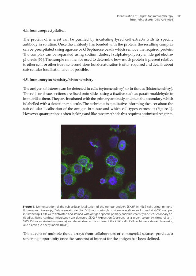
The antigen of interest can be detected in cells (cytochemistry) or in tissues (histochemistry).The cells or tissue sections are fixed onto slides using a fixative such as paraformaldehyde toimmobilise them. They are incubated with the primary antibody and then the secondary whichis labelled with a detection molecule. The technique is qualitative informing the user about thesub-cellular localisation of the antigen in tissue and which cell types express it (Figure 1).However quantitation is often lacking and like most methods this requires optimised reagents.
Figure 1. Demonstration of the sub-cellular localisation of the tumour antigen SSX2IP in K562 cells using immuno‐fluoresence microscopy. Cells were air dried for 4-18hours onto glass microscope slides and stored at -20oC wrappedin saranwrap. Cells were defrosted and stained with antigen specific primary and fluorescently labelled secondary an‐tibodies. Using confocal microscopy we detected SSX2IP expression (observed as a green colour by virtue of anti-SSX2IP-fluorescein isothiocyanate) was detectable on the surface of the K562 cells. Cell nuclei were stained blue using4,6'-diamino-2-phenylindole (DAPI).
The advent of multiple tissue arrays from collaborators or commercial sources provides ascreening opportunity once the cancer(s) of interest for the antigen has been defined.
Identification of Targets for Immunotherapyhttp://dx.doi.org/10.5772/54698
301

4.6. Flow cytometry
This technique allows the analyses of cells with a variety of parameters such as extracellularor intracellular markers, granularity, size and shape. Cancer cells are labelled with fluorescentantibodies for the required antigen. The cells are passed in a stream and intersected with alaser beam. The intensity of the fluorescence is measured and plotted in the form of dot plotsand histograms. This technique is sensitive and informative to allow specific cell types to be“gated” by virtue of size, granularity and detectable protein expression. Machines can measureup to 19 parameters in the most sophisticated machines allowing multiple proteins and celltypes to be analysed simultaneously [56]. However the technique requires validated antibodiesthat have been shown to be appropriate for fluorescence activated cell sorting analysis andenough tumour cells in suspension for analysis.
5. Identification of HLA-binding epitopes — in vitro assays
Immune responses in the body can ensure that any foreign matter is eliminated effectively.Class I and II major histocompatability complexes (MHC) are present on the surface ofnucleated cells and present processed peptides from proteins inside the cell to T cells. T cellscan destroy infected cells if peptides in the context of “danger” are detected [57]. MHC inhumans is known as the human leukocyte antigen (HLA) system. MHC class I HLA moleculesare highly polymorphic and generally the best defence against infections.
5.1. MHC Peptide binding assay
Peptide antigens are stripped from the HLA class I molecules by mild acid treatment, cells arethen incubated with a fluorescent reference peptide together with different concentrations ofthe peptide of interest. The efficiency with which the required peptide competes for bindingto the HLA class I molecules is examined by measuring the amount of HLA-bound referencepeptide with fluorescence activated cell sorting analysis [58].
5.2. T2 in vitro HLA-A2 binding assay
T2 in vitro HLA-A2 binding assay is more frequently used to determine the strength of peptidebinding to the most common HLA molecule in Caucasian populations. The HLA-A2 express‐ing, TAP-1 deficient human T-cell line T2 is used as an assay of HLA-A2 peptide bindingefficiency. T2 cells are washed and resuspended in serum-free RPMI media and plated in 96-well microtitre plates. Human β2-microglobulin and often nonamers (nine amino acids longpeptides) are added and the cells are incubated overnight at 37°C/5% CO2. The cells are washedand probed with a HLA-A2-specific monoclonal antibody and appropriate secondary anti‐body prior to flow cytometry. Only HLA-A2 molecules bound to peptide are stabilised anddetectable on the cell surface. Results are reported as a relative mean fluorescence index (MFI),calculated as the MFI of peptide-pulsed T2 cells compared with the MFI of unpulsed T2 cells[59]. Time course assays may be used to indicate how long the peptide remains on the HLA-
Novel Gene Therapy Approaches302

A2, indicating how long T cells will have to interact with peptide bound HLA-A2 before thecomplex falls apart.
6. In silico identification of epitopes
There are a number of databases which can be mined to find epitopes which have already beenshown to bind to HLA molecules. These have been used to identify established epitopes thatmay be used in immunotherapy strategies.
6.1. The SYFPEITHI
The SYFPEITHI database allows the prediction of MHC class I and II binding ligands fordifferent mammalian species. When a search is carried out using a protein sequence, aprediction is made based on the amino acids in the anchor and auxiliary anchor posi‐tions and other frequent amino acids which can bind to MHC molecules. A score is thencalculated which follows certain rules which are: a numerical value of 10 is given to ami‐no acids that regularly arise in anchor positions, the value 8 is set for amino acids occur‐ring in a significant number of ligands, six is for unusual anchors such as auxiliaryanchors and less frequent residues of the same set have a value of four. Preferred aminoacids have coefficients between 1–4 depending on the signal strength in pool sequencingor the occurrence of individual sequences. Amino acids that are considered as having anadverse effect on binding have a coefficient of –1 to –3 [60]. SYFPEITHI database getsupdated regularly and has been used to identify various ligands; p28 peptide as an epit‐ope for the CT antigen PLAC1 in breast cancer [61], p101-111 is the first CTA-derivedpeptide which induces CD4(+), CD8(+), and B-cell responses in vitro [62], p43-57 epitopestimulates T cells in HCA587-derived tumours [63] and PASD1(1) – PASD1(5) [51].
6.2. Bioinformatics and Molecular Analysis Section (BIMAS)
Bioinformatics and Molecular Analysis Section (BIMAS) develops computational processes toanalyse data generated from molecular biology and genetics research; and provides bioinfor‐matics guidance, support and resources for the collection, management, and display ofbiological sequence and genomic information for scientists involved in genomics and geneticanalysis [64]. Other online software which can be used to identify epitopes includes EpiJen,Rankpep, nHLApred, NetCTL and Multipred [65].
7. Cell based assays — In vitro demonstration of T cell reactivity
There are a number of assays which can be used to determine if T cells are activated in responseto antigen.
Identification of Targets for Immunotherapyhttp://dx.doi.org/10.5772/54698
303

7.1. Carboxyfluorescein diacetate Succinimidyl Ester (CFSE)
Carboxyfluorescein diacetate succinimidyl ester (CFSE) is a cytoplasmic dye which is absorbedby all cell types. Once the CFSE labelled cells divide, the dye is shared amongst the daughtercells equally therefore the fluorescence is halved after each round of the cell cycle. Thisdifference in fluorescence can be measured. The more cells proliferate, the greater the decreasein the fluorescent signal. The fluorescence peaks can be measured by flow cytometry [66]. CFSElabelling is increasingly used to measure target tumour cell killing [67], superseding radiationbased assays, as well as T cell proliferation in response to tumour cells in vivo [68]. CFSElabelling can also be performed in vivo where the dye is injected into the host animal’s spleenor lymph nodes, however the labelling is not uniform and it is sometimes difficult to obtainindividual peaks once lymphocyte cell division has occurred [66].
7.2. Lymphocyte proliferation assays
Lymphocyte proliferation assays can be used to determine activation of T cells. Peripheralblood mononuclear cells are isolated and cultured in microtitre plates. The specific antigen isincubated with the cells, which causes the T cells to divide and grow. The MTT colorimetricassay is based upon (3-(4,5-Dimethylthiazol-2-yl)-2,5-diphenyltetrazolium bromide, a yellowtetrazolium salt which gets cleaved by enzymes in the mitochondria to produce blue formazan.Viable, dividing cells will create more formazan which can be quantified using a plate reader.MTT assay is very convenient, however some considerations need to be assessed to avoid falsepositives such as cell densities, correct culture medium, filtration of media to remove precip‐itate, optimisation of MTT concentrations and incubation times [69].
7.3. [3H]-Thymidine incorporation assay
[3H]-Thymidine incorporation assay is based on the use of [3H]-thymidine a radioactivemolecule which can be incorporated into DNA during the S-phase of cell division. Asnew DNA is synthesised, occasional thymidine bases are replaced by [3H]-thymidine andsubsequently the incorporated radioactivity is measured, following washes to remove un‐incorporated radioactivity, using a Scintillation Counter [70]. As [3H]-thymidine is radio‐active an analogue called bromodeoxyuridine (BrdU) was developed to replace it inassays. BrdU integrates into DNA strands and can be measured using immunohisto‐chemistry and flow cytometry protocols using fluorescent conjugates and can be ob‐served over a longer duration. BrdU is also used to look at the number of cells in eachpart of the cell cycle by flow cytometry [71]. However BrdU has been found to be moretoxic than [3H]-thymidine, possibly because it is structurally very different to the originalDNA nucleotides. It also adversely affects cell division, the pattern of cell migration, fi‐nal position of migrating cells and the fate of labelled cells [72].
7.4. Peptide-MHC (pMHCs)
Peptide-MHC (pMHCs) based assays circumvent issues caused by measuring T cell prolifer‐ation. T cell proliferation assays can provide information on whether an immune response has
Novel Gene Therapy Approaches304

been generated but won’t determine which T cells, if they are indeed T cells, have beenactivated. pMHCs, often referred to as tetramers, can be used to identify antigen specific Tcells. They are produced through the refolding of β2-microglobulin and heavy chains in MHCmolecules with the appropriate epitope of interest. The pMHC is then labelled with biotinusing BirA enzyme. A streptavidin molecule conjugated to a fluorescent detector binds to four(tetramers) pMHCs or can used to create multimers (for example dimers, pentamers, dex‐tramers) of these constructed MHC molecules courtesy of the biotin-avidin interaction [73]. Tcell populations are added to this mixture and T cells with the specific receptor for the epitopeof interest will bind and be measurable by flow cytometry [74]. Shen et al [74] have found thatcross-reactive T cells i.e. T cells which recognise two different antigens can be identifiedproviding an extra tool in vaccine development. In some cases antigen specific T cells may notbind tetramers due to being undifferentiated and unable to accumulate T cell receptor (TCR)molecules close to the antigen. Another reason could be low affinity between TCR and MHC[75]. Other techniques based on the use of pMHCs include pMHC arrays [76] (Section 7.5),NACS [77] and the combinatorial approach [78,79]. These techniques all provide high through‐put analysis of multiple T cell populations with a variety of pros and cons to each techniqueincluding issues with background, specificity/binding capacity of individual pMHC com‐plexes, activated induced cell death of pMHC bound T cells, internalisation of pMHCsfollowing T cell binding [80], cost and labour intensity. Sequencing of TCRs (2-3 million every2-3 days) by companies such as TRON gGmbH (Johannes Gutenberg University Mainz,Germany) and Adaptive Biotechnologies (Seattle, USA) will provide a new way of analysingT cell populations which will be informative with regards to which TCRs are present but notnecessarily whether they are present on mature, anergic, activated or functional T cells norwhich sub-group of T cells are harbouring them (helper T cells, cytotoxic T cells, Th17 cells orindeed regulatory T cells (Tregs)). This technology allows the first opportunity to examine anextremely large number of TCRs in a very short time and will revolutionise how we examineT cell responses in patients in the future.
7.5. pMHC arrays
pMHC or tetramer arrays [76,81] (Figure 2) provides a strategy to determine which spe‐cific CD8+ T cell populations are present in the peripheral blood of patients. Antigensidentified by the techniques described already can be used to help expand the pMHC ar‐ray for future studies. In addition, the pMHC array provides a means to investigate epit‐ope spreading and changes in T cell specificities with disease progression. The techniquebenefits from the low number of purified CD8+ T cells required for each array (0.5-2 x10⁶), which can be purified from 20ml of patient peripheral blood using StemCell CD8+
negative isolation beads providing “untouched” T cells (Bonney, Guinn et al, in prepara‐tion). The purified CD8+ T cells are then lipophyllically dyed with DiD (MolecularProbes), washed and incubated with the pMHC array. The pMHC array has a detectionlimit of 0.02% matching the sensitivity we can reproducibly achieve with flow cytometrywhen analysing patient samples. Where sample availability permits, pMHC array datashould be validated by flow cytometry [82] using the same pMHC tetramers as in thepMHC array. The pMHC array has the added advantage that it can be used for the ini‐
Identification of Targets for Immunotherapyhttp://dx.doi.org/10.5772/54698
305

tial screening of a relatively small number of CD8+ T cells against a large number ofpMHCs on the array, and a short-list of T cell populations which are shown to exist onthe pMHC array can then be quantitated by flow cytometry (limiting the amount of sam‐ple required in subsequent studies). The pMHC array can be used to analyse patientsamples at a number of disease time-points (presentation, post-treatment (surgery and/orradiotherapy) and with disease progression) to examine how T cell responses to tumourantigens change with treatment, to examine epitope spreading and to correlate changingimmune responses with clinical responses.
39
Figure 2. pMHC arrays for the simultaneous detection of T cell populations in patient
peripheral blood. (A) Using a QArray2 printer and HPLF 0.3mm solid tip pins (Genetix) we
printed multiplexed AF532-conjugated pMHCs at a concentration of 0.5mg/ml so that 1ng
was placed in each spot (shown as coloured ovals). Each spot is 400uM wide with a 700uM
inter-spot distance. (B) At least six spots per pMHC are applied in each of two independent
sites. Each hydrogel slide can hold up to 1,000 spots of tetramer in total. (C) We applied 0.8-
1.5 x 10⁶ lipophillically-dyed CD8
+ T cells/slide (seen in red). pMHCs included a range of
HLA restrictions and had already been shown to work in flow cytometry studies,(i) pMHCs
are spotted across the gel, (seen as green spots) (ii) even some HLA-A2 positive patients
show no reactivity with any pMHCs (iii) while others show the presence of multiple T cell
populations which recognise tumour antigens. Based on figure in [144].
Figure 2. pMHC arrays for the simultaneous detection of T cell populations in patient peripheral blood. (A) Using aQArray2 printer and HPLF 0.3mm solid tip pins (Genetix) we printed multiplexed AF532-conjugated pMHCs at a con‐centration of 0.5mg/ml so that 1ng was placed in each spot (shown as coloured ovals). Each spot is 400uM wide witha 700uM inter-spot distance. (B) At least six spots per pMHC are applied in each of two independent sites. Each hydro‐gel slide can hold up to 1,000 spots of tetramer in total. (C) We applied 0.8-1.5 x 10⁶ lipophillically-dyed CD8+ T cells/slide (seen in red). pMHCs included a range of HLA restrictions and had already been shown to work in flow cytometrystudies,(i) pMHCs are spotted across the gel, (seen as green spots) (ii) even some HLA-A2 positive patients show noreactivity with any pMHCs (iii) while others show the presence of multiple T cell populations which recognise tumourantigens. Based on figure in [144].
Novel Gene Therapy Approaches306

7.6. Intracellular cytokine staining assay
Intracellular cytokine staining assay detects particular cytokines released by immunecells which can provide a useful insight into the responding T cell populations. Such cy‐tokines could include interferon-gamma (IFNγ), interleukin-2 (IL-2), IL-4 and tumour ne‐crosis factor-α. Cells are plated and incubated with the antigen to stimulate cytokineproduction. To prevent the cytokine from exiting the cell a transport inhibitor is addede.g. brefeldin A. The cells are then fixed in paraformaldehyde and permeabilized to al‐low the anti-cytokine antibody to bind. The results are analysed by flow cytometry. Theuse of intracellular cytokine staining assay to detect the cytokine IFNγ shows high repro‐ducibility and linearity with little background [83]. Duration of culture prior to antigenstimulation, as well as the cytokine accumulation period, are critical parameters of thesemethods. In both murine and cattle models, following 2-6 hours in culture, T cells pro‐duced a mixture of cytokines IFNγ, IL-2 and tumour necrosis factor-α, however follow‐ing 6-16 hours of culture only IFN-γ cytokine was found [84].
Using multiple peptides from distinct TAAs to stimulate immune cells may prove the mosteffective for peptide vaccines. A cocktail of four multiple myeloma antigen peptides were usedto stimulate T lymphocytes from HLA-A2 positive people to induce IFNγ production, cellproliferation and cytotoxicity against HLA-A2 positive multiple myeloma patients' cells [85].Indeed long peptides may offer the advantage of allowing the immune system to choose theepitope(s) it can best process and present from a peptide sequence and induce an effectivecytotoxic T cell response in the presence of longer CD4+ helper motifs [86]. Converselysometimes longer proteins can inhibit CD8+ T cells responses [87] but this may vary dependingon the constituents of individual protein sequences.
8. Cell based assays — In vivo assays
There are a number of approaches that can be taken when using mouse models to detectT cell immune responses. Animals can be used in transplantable tumour (xenografts)models or genetically engineered tumour models. In xenograft models human tumourcells are taken and injected into immunodeficient mice so that complex immune respons‐es involving multiple cell types can be investigated. In contrast, in genetically engineeredmodels, genes known to cause cancer are activated or tumour suppressor genes are“switched off” to allow their effects on tumour growth to be examined. In additiontransgenic mice can be used to examine T cell responses to epitopes presented on MHCmolecules as described in this section.
8.1. Immunodeficient mice
Immunodeficient mice such as athymic nude mice, severely compromised immunodefi‐cient (SCID) mice and non-obese diabetic severe combined immunodeficiency mice willaccept xenografts of human cells [88]. Depending upon the number of cells injected, or
Identification of Targets for Immunotherapyhttp://dx.doi.org/10.5772/54698
307
the size of the tumour transplanted, the tumour can develop over weeks to months andthe response to appropriate therapeutic agents studied in vivo [89]. Indeed such modelshave been used to examine the effectiveness of various immunotherapeutic strategies in‐cluding whole cell vaccines [90], dendritic cell (DC) vaccines [91], peptide vaccines [92]and DNA vaccines [67,87,93].
8.2. Genetically modified mice
Genetically modified mice may have genes which are overexpressed or deleted and the effectsof these genes on tumour development can be studied. Examples include p53 null andheterogenous mice [94,95] demonstrated that these genes can act as oncogenes and lead totumour development. Possible therapies for these oncogenes/tumour suppressor genes aretested for their response in an in vivo, full organism context [89] and examples include Ad-p53,AAV-HGFK1 [96].
HLA-A2 transgenic mice have genes inserted into the DNA so they will express the HLAmolecules known in mice as H-2. In order to prevent the presentation of murine H-2-restrictedcytotoxic T lymphocyte (CTL) epitopes in HLA-A2 (AAD) transgenic mice, HLA-A2.1transgenic/H-2 class I knockout mice (HHD mice) were created [97]. In HHD mice, the H-2class I gene is knocked out, and a chimeric HLA-A2.1 monochain (HHD) is produced by linkingthe C terminal of the human β2-microglobulin (unit of the class 1 MHC) covalently to the N-terminus of the chimeric HLA-A2 heavy chain (which contain the α1&2 domains of HLA-A2.1and the α3 domain of H-2Db) by means of a peptide bond. This guarantees the sole expressionof the HHD molecule on the cell surface, making sure that any identified CTL epitopes areHLA-A2 restricted [98]. HHD mice allow epitopes which are presented on human HLA-A2 tobe examined for their ability to induce T cell responses in a variety of studies; for exampleSTEAP, a prostate tumour antigen has been shown to be targeted by anti-tumour T cells [99]and DNA vaccines encoding Wilms tumour antigen 1 induce cytotoxic responses in mice [100]using this model system.
9. Modes of immunotherapy
One of the biggest debates in cancer immunotherapy remains which mode will be themost effective. The National Cancer Institute have suggested that immunotherapy stud‐ies should focus on a limited number of antigenic targets to maximise the chances ofsuccess [101]. However for some cancers effective immunotherapy targets have yet to bediscovered (i.e. ovarian cancer, adult acute lymphocytic leukaemia) and better targetsmay yet be determined.
When T cells were found to be able to identify cancer cells [102] it was thought that T celltherapies would be the most effective with the aim being to stimulate CD8+/CTL cells to killtumour cells. This can be achieved by a number of ways such as through the use of DCs [103],peptide vaccines [85], DNA vaccines [104] and natural killer cells [105]. In recent years
Novel Gene Therapy Approaches308

monoclonal antibodies (mAb) have become standard treatments for cancer. Ultimately if thereis an antibody and it recognises a surface antigen solely on cancer and non-essential cells thenthis will likely be the most effective way to cause tumour destruction. mAbs are derived byvaccinating an animal with the target antigen and testing to see if the B cells are producingantibodies against it. Then the B cells are extracted from its spleen and infused with myelomacells to produce hybridomas. Hybridomas divide perpetually and produce the mAb to theantigen in large amounts [106].
Rosenberg et al [107] showed that only 2.6% of immunotherapy clinical trials hadworked and therefore an overhaul was needed in the practice of immunotherapy. Subse‐quently the same group showed that adoptive T cell therapy could be very promisingwith cell numbers being returned to the patient [108] and their status – activated but notmature [109] and cell numbers being the main issues. It is also likely that the best strat‐egy may include a combination of conventional and immunotherapy techniques [110] oreven a combination of immunotherapy techniques as demonstrated in increasing num‐bers of mouse models [111] and clinical trials [112].
DCs are antigen presenting cells therefore they have received some attention for possible usein cancer immunotherapy. DCs pulsed by peptide and injected into the skin showed a responserate of 28%. This percentage increased to 35.7% when immature DCs are injected straight intothe tumour and even higher to 40% for advanced pancreatic cancer [113].
Tumour-infiltrating T cells (TIL) therapy has been used in stage IV melanoma patients. TILsare obtained from the blood, lymph nodes or from a tumour tissue biopsy. TILs are isolated,activated and expanded using IL-2 in vitro. The patient undergoes lympho-depleting chemo‐therapy prior to the T cells being injected back in to the blood [114].
When a tumour antigen is secreted into the circulation in high levels immune tolerancecan be induced in the thymus. CD8α−Sirpα+, a subset of DCs, are able to capture tumourantigens in the blood, which can induce tolerance through Tregs or negative selection.Tregs are cells which are part of the tolerance system which prevents autoimmunity [115,116]. Simultaneous Treg depletions (using anti-CD25 antibodies for instance) may aid theeffectiveness of immunotherapy in some cancer types where Treg infiltration into the tu‐mour is rife [117,118].
There are a number of reviews in this area of research which aim to look into effectiveimmunotherapy strategies for the future. These include cellular immunotherapy [119],whole call vaccines [120], multidrug resistance [121] and DCs [122]. Targeted therapeuticstrategies along with ever improving designs in clinical trials pave the way for furthersuccess [123].
10. Clinical trials
Clinical trials are undertaken after a large amount of data has been obtained on the antigen ofinterest in the lab. This data is required to ensure treatment safety and efficacy as far as is
Identification of Targets for Immunotherapyhttp://dx.doi.org/10.5772/54698
309

possible. It remains imperative in most countries that treatments have been tested on liveanimals prior to first-in-man clinical trials and that favourable results are apparent in orderfor treatments to be taken into clinical trials. People, often patients, are recruited as compen‐sated or full volunteers. The drug is given to participants initially to show that it is safe andthen that it is effective. Dose escalation is also important so that an effective and safe dosagein humans is used.
Clinical trials have four phases, very basically as follows: І – evaluation of safety, ІІ –safety and efficacy (with Phase I/II often including dose escalation), ІІІ – efficacy in alarge cohort of patients and ІV – post-approval studies. Phase І trials look at the safetyand the best dose of the drug to administer. Such trials often involve 13 patients or less,and these patients are often have late stage cancer and are refractory to all other treat‐ments with little chance of recovery. Phase ІІ trials start to look at the efficacy of themedicine and often involve 20 patients with late stage disease. Phase І and ІІ trials haveto be completed successfully in order for testing to proceed to Phase ІІІ. Current “bestpractise” treatment is compared to the new drug being tested in phase ІІІ trials. Only ifthe new drug offers an improvement over best practise does the new medicine have achance of becoming the standard treatment. At this time the drug will need to be li‐censed and approved by the authoritative body e.g. the US Food and Drug Administra‐tion, and once licensed, phase ІV trials investigate the long term benefits and unexpectedside effects. In some cases the drug may go through one of the phases more than oncebefore moving forward and even then may get rejected [124].
It is very important that trials follow certain rules for the results to be considered legitimate.Phase ІІІ trials need to be randomised i.e. a computer randomly puts people into one of twogroups. These can also be double-blind trials so that neither the patient nor the investigatorknows which treatment is being given, thus avoiding any bias. In some cases there may notbe any treatment to compare a new therapy against, in which case a placebo (inactive treat‐ment) is given to one group [125]. In the UK the Medicines and Healthcare products RegulatoryAgency is responsible for the regulation of medicines and medical devices and equipment usedin healthcare, and the investigation of harmful incidents as well as overseeing the use of bloodand blood products [126].
Cohen et al [127] have created an online website called BreastCancerTrials.org which matchespatients to current trials taking place depending on the information they provide. Thisprovides a valuable source for cancer patients who may want initial guidance on which clinicaltrials may be beneficial to them.
11. Assays to demonstrate efficacy of the response
Assays tend to reflect the immunotherapy strategy employed with the efficacy of antibodytherapies being measured by tumour destruction, ertumaxomab destroys tumour cellsexpressing HER2/neu [128], bispecific antibodies represent a new class of anticancer thera‐peutics [129] and antibody-targeted delivery of a vaccine can improve tumour cell killing [130].
Novel Gene Therapy Approaches310
11.1. Enzyme-Linked Immunosorbent Spot (ELISPOT) assay
Enzyme-Linked Immunosorbent Spot (ELISPOT) assay was developed by Cecil Czer‐kinskdy in 1983 [131] and shows most similarity to the ELISA technique. It is based onthe use of a 96 well plate with a polyvinyl-difluoride membrane to which antigen specif‐ic monoclonal “capture” antibodies are attached. The cells are grown in media on thecapture antibody coated membrane usually for several hours to overnight and secretedprotein (often cytokines such as IFNγ) bind to the capture antibody. A second “detec‐tion” antibody specific to the protein is used. This is often conjugated to an enzyme al‐lowing a chemical reaction to occur. Black spots form on the membrane whereverprotein is present and these can be counted by an ELISPOT reader [131]. ELISPOT as‐says are one of the most sensitive ex vivo detection methods available with low detectionthresholds in peripheral blood. ELISPOT is also able to identify patients with allergiesthrough the detection of drug-specific T cells in their blood [132].
11.2. Cultured ELISPOT
Cultured ELISPOT measures memory T cells. Cells are stimulated and plated on a 24-wellplate. After an incubation period half of the cell culture supernatant is removed and replacedwith Lymphocult (an IL-2 containing growth factor supplement). Fresh Lymphocult is addedagain on day 7. On day 9, the cells are incubated overnight. On day 10, around 2.5x10⁴ of theoriginally plated cells are plated for a standard ELISPOT assay. Cultured ELISPOT assaysrevealed the existence of longer-lasting T cell memory responses [133].
12. Conclusions
This chapter has focussed predominantly on the identification of epitopes within tumourantigens and their validation as they enter clinical trials. Focus on clinical trials using antibodytherapies, DCs, natural killer cells, and adoptive therapy among many other options are thefocus of other excellent reviews in the field [134-136].
Cancer immunotherapy is a vital area of research that continues to progress at a pace.Our understanding of the immune response and its potential to recognise and kill tu‐mour cells with mans guidance offers hope to the patients for whom few other treat‐ment options exist. Many tumour antigens have been identified, but some cancers stilllack antigen targets that are expressed in the majority of the cancer cells by the majorityof cancer patients. New techniques to extend antigen discovery will allow the improve‐ment of immunotherapy strategies while the identification of new biomarkers will assistin the development of personalised therapies. Personalised therapies will decrease thecost (quantitative and qualitative) of treatment on patients who are unlikely to respondto it, allowing patients to avoid unpleasant and harmful side effects while maximisingpatient quality of life.
Identification of Targets for Immunotherapyhttp://dx.doi.org/10.5772/54698
311

Target antigen Mode of
immunotherapy
Patient group Phase of
clinical
trial
Outcome Reference
CD22 Monoclonal antibody
conjugated to
calecheamicin
Refractory and
relapsed acute
lymphocytic
leukaemia
Phase 2 18% patients had complete
response, 39% had marrow
complete response, 39% had
resistant disease, and 4% died
within 4 weeks of starting
treatment
[137]
TG4010 targeting MUC1 &
IL-2
Poxvirus (modified
vaccinia virus Ankara)
in combination with
first-line
chemotherapy
Advanced non-
small-cell lung
cancer
Phase 2B 6-month progression-free
survival was 43·2% in the
TG4010 plus chemotherapy
group, and 35·1% in the
chemotherapy alone group
[138]
LY6K and TTK peptide vaccines in
combination with
CpG-7909
Metastatic
oesophageal
squamous cell
carcinoma
Phase 1 Vaccination with peptides in
combination with CpG-7909
increased and activated pDC
populations and NK cell
populations
[139]
CTLA-4 Monoclonal antibody
with glycoprotein
100 (gp100) peptide
vaccine
Previously treated
metastatic
melanoma
Phase 3 The median overall survival
was 10.0 months among
patients receiving ipilimumab
plus gp100, as compared with
6.4 months among patients
receiving gp100 alone
[140]
Prostate-specific antigen Poxviral vaccines Prostate cancer Phase 2 The primary end point was
progression-free survival
which was similar in the two
groups (treated, controls).
However, at 3 years post
study, treated patients had a
overall survival rate of 30%
compared to 17% of controls
[141]
CD20 Monoclonal antibody Relapsed or
refractory follicular
lymphoma
Phase 1/2 Treatment caused immediate
and profound B-cell depletion,
and 65% of patients reverted
to negative BCL2 status.
[142]
CA-125 Abagovomab, an
anti-idiotype
antibody
Ovarian Cancer Phase 1 Improved CA125-specific
cellular cytotoxicity might
indicate that longer
vaccination (nine injections)
would be preferred to short
(six injections)
[143]
Table 1. Examples of completed clinical trials showing the cancer antigen targets. Representation of the variousmodes of immunotherapy employed to date, cancer patients treated and the outcome of the trials.
Novel Gene Therapy Approaches312

Abbreviations
BIMAS: Bioinformatics and Molecular Analysis Section; BrdU: bromodeoxyuridine; CFSE: Car‐boxyfluorescein diacetate succinimidyl ester; CTL: Cytotoxic T lymphocyte; CT: cancer-testis;DC: dendritic cell; ELISA: Enzyme-linked immunosorbent assay; ELISPOT: Enzyme-linked im‐munosorbence assay; HLA: human leukocyte antigen; IFNγ: interferon-gamma; IL: interleukin;mAb: monoclonal antibodies; MFI: mean fluorescence index; MHC: major histocompatibilitycomplex; PCR: polymerise chain reaction; pMHC: peptide and major histocompatibility com‐plex; SEREX: Serological identification of antigens by recombinant expression cloning; SSX2IP:Synovial Sarcoma X breakpoint 2 Interacting Protein; TAA: tumour associated antigens; TCR: Tcell receptor; TIL: Tumour-infiltrating T cells; Tregs: regulatory T cells.
Acknowledgements
SEB was funded by Cancer Research U.K. Research by our group has been supported byLeukaemia and Lymphoma Research, Wessex Cancer Trust and a Wessex Medical ResearchInnovations grant.
Author details
Ghazala Khan1, Suzanne E. Brooks2, Frances Denniss3, Dagmar Sigurdardottir4 andBarbara-ann Guinn1,2,3*
*Address all correspondence to: [email protected]
1 Division of Science, University of Bedfordshire, Park Square, Luton, UK
2 Cancer Sciences Division (MP824), Southampton University Hospitals, University ofSouthampton, Southampton, UK
3 Department of Haematological Medicine, King’s College London School of Medicine, TheRayne Institute, London, UK
4 Department of Immunology, Institute for Cell Biology, University of Tübingen, Tübingen,Germany
References
[1] Penn, C.G. Halgrimson, T.E. Starzl, De novo malignant tumors in organ transplantrecipients, Transplant. Proc. 1 (1971) 773-778.
Identification of Targets for Immunotherapyhttp://dx.doi.org/10.5772/54698
313

[2] U. Sahin, O. Tureci, H. Schmitt, B. Cochlovius, T. Johannes, R. Schmits, F. Stenner, G.Luo, I. Schobert, M. Pfreundschuh, Human neoplasms elicit multiple specific immuneresponses in the autologous host, Proc. Natl. Acad. Sci. U. S. A. 25 (1995) 11810-11813.
[3] L.T. Nguyen, A.R. Elford, K. Murakami, K.M. Garza, S.P. Schoenberger, B. Odermatt,D.E. Speiser, P.S. Ohashi, Tumor growth enhances cross-presentation leading to limitedT cell activation without tolerance, J. Exp. Med. 4 (2002) 423-435.
[4] E.M. Tan, Autoantibodies as reporters identifying aberrant cellular mechanisms intumorigenesis, J. Clin. Invest. 10 (2001) 1411-1415.
[5] J.Y. Zhang, K.S. Looi, E.M. Tan, Identification of tumor-associated antigens as diag‐nostic and predictive biomarkers in cancer, Methods Mol. Biol. (2009) 1-10.
[6] T. Soussi, p53 Antibodies in the sera of patients with various types of cancer: a review,Cancer Res. 7 (2000) 1777-1788.
[7] E. Haralambieva, K.A. Pulford, L. Lamant, S. Pileri, G. Roncador, K.C. Gatter, G. Delsol,D.Y. Mason, Anaplastic large-cell lymphomas of B-cell phenotype are anaplasticlymphoma kinase (ALK) negative and belong to the spectrum of diffuse large B-celllymphomas, Br. J. Haematol. 3 (2000) 584-591.
[8] B. Guinn, J. Greiner, M. Schmitt, K.I. Mills, Elevated expression of the leukemia-associated antigen SSX2IP predicts survival in acute myeloid leukemia patients wholack detectable cytogenetic rearrangements, Blood. 113 (2009) 1203-1204.
[9] Y.T. Chen, Identification of human tumor antigens by serological expression cloning:an online review on SEREX, Cancer Immun. (2004) http://www.cancerimmunity.org/serex
[10] M. Kalejs, J. Erenpreisa, Cancer/testis antigens and gametogenesis: a review and "brain-storming" session, Cancer. Cell. Int. 1 (2005) 4.
[11] S.H. Lim, Y. Zhang, J. Zhang, Cancer-testis antigens: the current status on antigenregulation and potential clinical use, Am. J. Blood Res. 1 (2012) 29-35.
[12] M.J. Scanlan, C.M. Gordon, B. Williamson, S.Y. Lee, Y.T. Chen, E. Stockert, A. Jung‐bluth, G. Ritter, D. Jager, E. Jager, A. Knuth, L.J. Old, Identification of cancer/testis genesby database mining and mRNA expression analysis, Int. J. Cancer. 4 (2002) 485-492
[13] Van Driessche, Z.N. Berneman, V.F. Van Tendeloo, Active specific immunotherapytargeting the Wilms' tumor protein 1 (WT1) for patients with hematological malignan‐cies and solid tumors: lessons from early clinical trials, Oncologist. 2 (2012) 250-259.
[14] F. Wadelin, J. Fulton, P.A. McEwan, K.A. Spriggs, J. Emsley, D.M. Heery, Leucine-richrepeat protein PRAME: expression, potential functions and clinical implications forleukaemia, Mol. Cancer. (2010) 226.
[15] A.M. Krackhardt, M. Witzens, S. Harig, F.S. Hodi, A.J. Zauls, M. Chessia, P. Barrett,J.G. Gribben, Identification of tumor-associated antigens in chronic lymphocyticleukemia by SEREX, Blood. 6 (2002) 2123-2131.
Novel Gene Therapy Approaches314
[16] M. Forgber, U. Trefzer, W. Sterry, P. Walden, Proteome serological determination oftumor-associated antigens in melanoma, PLoS One. 4 (2009) e5199.
[17] K. Wang, X. Xu, Y. Nie, L. Dai, P. Wang, J. Zhang, Identification of tumor-associatedantigens by using SEREX in hepatocellular carcinoma, Cancer Lett. 2 (2009) 144-150.
[18] J. Qian, J. Xie, S. Hong, J. Yang, L. Zhang, X. Han, M. Wang, F. Zhan, J.D. ShaughnessyJr, J. Epstein, L.W. Kwak, Q. Yi, Dickkopf-1 (DKK1) is a widely expressed and potenttumor-associated antigen in multiple myeloma, Blood. 5 (2007) 1587-1594.
[19] S.P. Adams, S.S. Sahota, A. Mijovic, B. Czepulkowski, R.A. Padua, G.J. Mufti, B.A.Guinn, Frequent expression of HAGE in presentation chronic myeloid leukaemias,Leukemia. 16 (2002) 2238-2242.
[20] V. Martelange, C. De Smet, E. De Plaen, C. Lurquin, T. Boon, Identification on a humansarcoma of two new genes with tumor-specific expression, Cancer Res. 14 (2000)3848-3855.
[21] K.L. Tyson, P.L. Weissberg, C.M. Shanahan, Heterogeneity of gene expression inhuman atheroma unmasked using cDNA representational difference analysis, Physiol.Genomics. 2 (2002) 121-130.
[22] http://www.licr.org/D_programs/d4a1i_SEREX.php
[23] S. Zhou, T Yi, B Zhang, F Huang, H Huang, J Tang, X Zhao. Mapping the high through‐put SEREX technology screening for novel tumor antigens, Comb Chem High Through‐put Screen. 15 (2012) 202-15.
[24] D. Jäger, Potential target antigens for immunotherapy identified by serologicalexpression cloning (SEREX), Methods Mol Biol. 360 (2007) 319-26.
[25] M.J. Edelman, L. Hodgson, P.Y. Rosenblatt, R.H. Christenson, E.E. Vokes, X. Wang, R.Kratzke, CYFRA 21-1 as a prognostic and predictive marker in advanced non-small-cell lung cancer in a prospective trial: CALGB 150304, J. Thorac. Oncol. 4 (2012) 649-654.
[26] K. Imai, S. Hirata, A. Irie, S. Senju, Y. Ikuta, K. Yokomine, M. Harao, M. Inoue, Y. Tomita,T. Tsunoda, H. Nakagawa, Y. Nakamura, H. Baba, Y. Nishimura, Identification of HLA-A2-restricted CTL epitopes of a novel tumour-associated antigen, KIF20A, overex‐pressed in pancreatic cancer, Br. J. Cancer. 2 (2011) 300-307.
[27] A.P. Liggins, B.A. Guinn, C.S. Hatton, K. Pulford, A.H. Banham, Serologic detection ofdiffuse large B-cell lymphoma-associated antigens, Int. J. Cancer. 110 (2004) 563-569.
[28] B.A. Guinn, E.A. Bland, U. Lodi, A.P. Liggins, K. Tobal, S. Petters, J.W. Wells, A.H.Banham, G.J. Mufti, Humoral detection of leukaemia-associated antigens in presenta‐tion acute myeloid leukaemia, Biochem. Biophys. Res. Commun. 335 (2005) 1293-1304.
[29] S. Patel, H. Chen, L. Monti, E. Gould, E. Haralambieva, J. Schmid, D. Toomey, W.Woessmann, G. Roncador, C.S. Hatton, A.P. Liggins, R. Taramelli, A.H. Banham, F.Acquati, D. Murphy, K. Pulford, RNASET2 - An autoantigen in anaplastic large celllymphoma identified by protein array analysis, J Proteomics. 75 (2012) 5279-5292.
Identification of Targets for Immunotherapyhttp://dx.doi.org/10.5772/54698
315

[30] S. Tschiedel, C. Gentilini, T. Lange, C. Wolfel, T. Wolfel, V. Lennerz, S. Stevanovic, H.G.Rammensee, C. Huber, M. Cross, D. Niederwieser, Identification of NM23-H2 as atumour-associated antigen in chronic myeloid leukaemia, Leukemia. 8 (2008)1542-1550.
[31] Y. Wang, Q. Gu, B. Liu, Z. Zhu, Perspectives of SEREX-defined antigens in diagnosisand immunotherapy for gastric cancer, Cancer. Biol. Ther. 9 (2004) 806-811.
[32] C.S. Klade, T. Voss, E. Krystek, H. Ahorn, K. Zatloukal, K. Pummer, G.R. Adolf,Identification of tumor antigens in renal cell carcinoma by serological proteomeanalysis, Proteomics. 7 (2001) 890-898.
[33] C.G. Gunawardana, E.P. Diamandis, High throughput proteomic strategies foridentifying tumour-associated antigens, Cancer Lett. 249 (2007) 110-119.
[34] C.A. Casiano, M. Mediavilla-Varela, E.M. Tan, Tumor-associated antigen arrays for theserological diagnosis of cancer, Mol. Cell. Proteomics. 5 (2006) 1745-1759.
[35] M. Nishiu, R. Yanagawa, S. Nakatsuka, M. Yao, T. Tsunoda, Y. Nakamura, K. Aozasa,Microarray analysis of gene-expression profiles in diffuse large B-cell lymphoma:identification of genes related to disease progression, Jpn. J. Cancer Res. 8 (2002)894-901.
[36] D.J. Brennan, C. Kelly, E. Rexhepaj, P.A. Dervan, M.J. Duffy, W.M. Gallagher, Contri‐bution of DNA and tissue microarray technology to the identification and validationof biomarkers and personalised medicine in breast cancer, Cancer Genomics Proteo‐mics. 3 (2007) 121-134.
[37] C. De Pitta, L. Tombolan, M. Campo Dell'Orto, B. Accordi, G. te Kronnie, C. Romualdi,N. Vitulo, G. Basso, G. Lanfranchi, A leukemia-enriched cDNA microarray platformidentifies new transcripts with relevance to the biology of pediatric acute lymphoblasticleukemia, Haematologica. 7 (2005) 890-898.
[38] J.S. Chen, E. Coustan-Smith, T. Suzuki, G.A. Neale, K. Mihara, C.H. Pui, D. Campana,Identification of novel markers for monitoring minimal residual disease in acutelymphoblastic leukemia, Blood. 7 (2001) 2115-2120.
[39] B.A. Guinn, L. Bullinger, N.S. Thomas, K.I. Mills, J. Greiner, SSX2IP expression in acutemyeloid leukaemia: an association with mitotic spindle failure in t(8;21), and cell cyclein t(15;17) patients, Br. J. Haematol. 140 (2008) 250-251.
[40] V. Dutoit, C. Herold-Mende, N. Hilf, O. Schoor, P. Beckhove, J. Bucher, K. Dorsch, S.Flohr, J. Fritsche, P. Lewandrowski, J. Lohr, H.G. Rammensee, S. Stevanovic, C.Trautwein, V. Vass, S. Walter, P.R. Walker, T. Weinschenk, H. Singh-Jasuja, P.Y.Dietrich, Exploiting the glioblastoma peptidome to discover novel tumour-associatedantigens for immunotherapy, Brain. 135 (2012) 1042-1054.
[41] J.S. Stickel, A.O. Weinzierl, N. Hillen, O. Drews, M.M. Schuler, J. Hennenlotter, D.Wernet, C.A. Muller, A. Stenzl, H.G. Rammensee, S. Stevanovic, HLA ligand profiles
Novel Gene Therapy Approaches316

of primary renal cell carcinoma maintained in metastases, Cancer Immunol. Immun‐other. 9 (2009) 1407-1417.
[42] A.J. Knights, A.O. Weinzierl, T. Flad, B.A. Guinn, L. Mueller, G.J. Mufti, S. Stevanovic,G. Pawelec, A novel MHC-associated proteinase 3 peptide isolated from primarychronic myeloid leukaemia cells further supports the significance of this antigen forthe immunotherapy of myeloid leukaemias, Leukemia. 20 (2006) 1067-1072.
[43] Mohamedali, B.A. Guinn, S. Sahu, N.S. Thomas, G.J. Mufti, Serum profiling revealsdistinctive proteomic markers in chronic myeloid leukaemia patients, Br. J. Haematol.144 (2009) 263-265.
[44] N. Hillen, S. Stevanovic S. Contribution of mass spectrometry-based proteomics toimmunology. Expert Rev Proteomics. 3 (2006) 653-64.
[45] L.J. Stern, Characterizing MHC-associated peptides by mass spectrometry, J. Immunol.5 (2007) 2667-2668.
[46] A.L. Zarling, S.B. Ficarro, F.M. White, J. Shabanowitz, D.F. Hunt, V.H. Engelhard,Phosphorylated peptides are naturally processed and presented by major histocom‐patibility complex class I molecules in vivo, J. Exp. Med. 12 (2000) 1755-1762.
[47] J.R. Yates, C.I. Ruse, A. Nakorchevsky, Proteomics by mass spectrometry: approaches,advances, and applications, Annu. Rev. Biomed. Eng. (2009) 49-79.
[48] R. Chen, M. Snyder, Yeast proteomics and protein microarrays, J. Proteomics. 11 (2010)2147-2157.
[49] Anonymous ProtoArray® Human Protein Microarrays | Life Technologies http://www.invitrogen.com/site/us/en/home/Products-and-Services/Applications/Protein-Expression-and-Analysis/Biomarker-Discovery/ProtoArray.html?CID=fl-protoarray.
[50] C.D. Cooper, A.P. Liggins, K. Ait-Tahar, G. Roncador, A.H. Banham, K. Pulford,PASD1, a DLBCL-associated cancer testis antigen and candidate for lymphomaimmunotherapy, Leukemia. 12 (2006) 2172-2174.
[51] K. Ait-Tahar, A.P. Liggins, G.P. Collins, A. Campbell, M. Barnardo, C. Lawrie, D. Moir,C. Hatton, A.H. Banham, K. Pulford, Cytolytic T-cell response to the PASD1 cancertestis antigen in patients with diffuse large B-cell lymphoma, Br. J. Haematol. 4 (2009)396-407.
[52] W.L. Chen, K.T. Kuo, T.Y. Chou, C.L. Chen, C.H. Wang, Y.H. Wei, L.S. Wang, The roleof cytochrome c oxidase subunit Va in non-small cell lung carcinoma cells: associationwith migration, invasion and prediction of distant metastasis, BMC Cancer. 12 (2012)273.
[53] http://www.piercenet.com/browse.cfm?fldID=F88ADEC9-1B43-4585-922E-836FE09D8403#detectionmethods
[54] B. Magi, S. Liberatori, Immunoblotting techniques, Methods Mol. Biol. (2005) 227-254.
Identification of Targets for Immunotherapyhttp://dx.doi.org/10.5772/54698
317
[55] L.A. Goff, J. Davila, M.R. Swerdel, J.C. Moore, R.I. Cohen, H. Wu, Y.E. Sun, R.P. Hart,Ago2 immunoprecipitation identifies predicted microRNAs in human embryonic stemcells and neural precursors, PLoS One. 9 (2009) e7192.
[56] S.P. Perfetto, P.K. Chattopadhyay, M. Roederer. Seventeen-colour flow cytometry:unravelling the immune system. Nature Reviews Immunol. 4 (2004) 648-655.
[57] P. Matzinger, The danger model: a renewed sense of self, Science. 296 (2002) 301-305.
[58] J.H. Kessler, W.E. Benckhuijsen, T. Mutis, C.J. Melief, S.H. van der Burg, J.W. Drijfhout,Competition-based cellular peptide binding assay for HLA class I, Curr. Protoc.Immunol. (2004) Unit 18.12.
[59] B.M. Olson, T.P. Frye, L.E. Johnson, L. Fong, K.L. Knutson, M.L. Disis, D.G. McNeel,HLA-A2-restricted T-cell epitopes specific for prostatic acid phosphatase, CancerImmunol. Immunother. 6 (2010) 943-953.
[60] H. Rammensee, J. Bachmann, N.P. Emmerich, O.A. Bachor, S. Stevanovic, SYFPEITHI:database for MHC ligands and peptide motifs, Immunogenetics. 3-4 (1999) 213-219.
[61] W. Liu, M. Zhai, Z. Wu, Y. Qi, Y. Wu, C. Dai, M. Sun, L. Li, Y. Gao, Identification of anovel HLA-A2-restricted cytotoxic T lymphocyte epitope from cancer-testis antigenPLAC1 in breast cancer, Amino Acids. 6 (2012) 2257-2265.
[62] F. Neumann, B. Kubuschok, K. Ertan, C. Schormann, S. Stevanovic, K.D. Preuss, W.Schmidt, M. Pfreundschuh, A peptide epitope derived from the cancer testis antigenHOM-MEL-40/SSX2 capable of inducing CD4(+) and CD8(+) T-cell as well as B-cellresponses, Cancer Immunol. Immunother. 9 (2011) 1333-1346.
[63] W. Wen, L. Zhang, J. Peng, J. Chen, J. Hao, X. Li, X. Qian, P. Zeng, Y. Zhang, Y. Yin,Identification of promiscuous HLA-DR-restricted CD4(+) T-cell epitopes on the cancer-testis antigen HCA587, Cancer. Sci. 8 (2011) 1455-1461.
[64] K.C. Parker, M.A. Bednarek, J.E Coligan, Scheme for ranking potential HLA-A2 bindingpeptides based on independent binding of individual peptide side-chains, J Immunol152 (1994) 163–175
[65] N. Seyed, F. Zahedifard, S. Safaiyan, E. Gholami, F. Doustdari, K. Azadmanesh, M.Mirzaei, N. Saeedi Eslami, A. Khadem Sadegh, A. Eslami Far, I. Sharifi, S. Rafati, Insilico analysis of six known Leishmania major antigens and in vitro evaluation of specificepitopes eliciting HLA-A2 restricted CD8 T cell response, PLoS Negl Trop. Dis. 9 (2011)e1295.
[66] B. Asquith, C. Debacq, A. Florins, N. Gillet, T. Sanchez-Alcaraz, A. Mosley, L. Willems,Quantifying lymphocyte kinetics in vivo using carboxyfluorescein diacetate succini‐midyl ester (CFSE), Proc. Biol. Sci. 1590 (2006) 1165-1171.
[67] J.N. Radcliffe, J.S. Roddick, F.K. Stevenson, S.M. Thirdborough, Prolonged antigenexpression following DNA vaccination impairs effector CD8+ T cell function andmemory development, J. Immunol. 12 (2007) 8313-8321.
Novel Gene Therapy Approaches318

[68] D. Bagnara, M.S. Kaufman, C. Calissano, S. Marsilio, P.E. Patten, R. Simone, P. Chum,X.J. Yan, S.L. Allen, J.E. Kolitz, S. Baskar, C. Rader, H. Mellstedt, H. Rabbani, A. Lee,P.K. Gregersen, K.R. Rai, N. Chiorazzi, A novel adoptive transfer model of chroniclymphocytic leukemia suggests a key role for T lymphocytes in the disease, Blood. 20(2011) 5463-5472.
[69] P.W. Sylvester, Optimization of the tetrazolium dye (MTT) colorimetric assay forcellular growth and viability, Methods Mol. Biol. (2011) 157-168.
[70] R.K. Paul, A. Ramamoorthy, J. Scheers, R.P. Wersto, L. Toll, L. Jimenez, M. Bernier, I.W.Wainer, Cannabinoid receptor activation correlates with the pro-apoptotic action of thebeta2-adrenergic agonist, (R,R')-4-methoxy-1-naphthylfenoterol, in HepG2 hepatocar‐cinoma cells, J. Pharmacol. Exp. Ther. 343 (2012) 157-166.
[71] B. Piovesan, N. Pennell, N.L. Berinstein, Human lymphoblastoid cell lines expressingmutant p53 exhibit decreased sensitivity to cisplatin-induced cytotoxicity, Oncogene.18 (1998) 2339-2350.
[72] Duque, P. Rakic, Different effects of bromodeoxyuridine and [3H]thymidine incorpo‐ration into DNA on cell proliferation, position, and fate, J. Neurosci. 42 (2011)15205-15217.
[73] S. Sims, C. Willberg, P. Klenerman, MHC-peptide tetramers for the analysis of antigen-specific T cells, Expert Rev. Vaccines. 7 (2010) 765-774.
[74] Z.T. Shen, M.A. Brehm, K.A. Daniels, A.B. Sigalov, L.K. Selin, R.M. Welsh, L.J. Stern,Bi-specific MHC heterodimers for characterization of cross-reactive T cells, J. Biol.Chem. 43 (2010) 33144-33153.
[75] N. Khan, M. Cobbold, J. Cummerson, P.A. Moss, Persistent viral infection in humanscan drive high frequency low-affinity T-cell expansions, Immunology. 4 (2010) 537-548.
[76] D.S. Chen, Y. Soen, T.B. Stuge, P.P. Lee, J.S. Weber, P.O. Brown, M.M. Davis, Markeddifferences in human melanoma antigen-specific T cell responsiveness after vaccina‐tion using a functional microarray, PLoS Med. 10 (2005) e265.
[77] G.A. Kwong, C.G. Radu, K. Hwang, C.J. Shu, C. Ma, R.C. Koya, B. Comin-Anduix, S.R.Hadrup, R.C. Bailey, O.N. Witte, T.N. Schumacher, A. Ribas, J.R. Heath, Modularnucleic acid assembled p/MHC microarrays for multiplexed sorting of antigen-specificT cells, J. Am. Chem. Soc. 28 (2009) 9695-9703.
[78] S.R. Hadrup, T.N. Schumacher, MHC-based detection of antigen-specific CD8+ T cellresponses, Cancer Immunol. Immunother. 9 (2010) 1425-1433.
[79] E.W. Newell, L.O. Klein, W. Yu, M.M. Davis, Simultaneous detection of many T-cellspecificities using combinatorial tetramer staining, Nat. Methods. 7 (2009) 497-499.
[80] J.A. Whelan, P.R. Dunbar, D.A. Price, M.A. Purbhoo, F. Lechner, G.S. Ogg, G. Griffiths,R.E. Phillips, V. Cerundolo, A.K. Sewell, Specificity of CTL interactions with peptide-
Identification of Targets for Immunotherapyhttp://dx.doi.org/10.5772/54698
319

MHC class I tetrameric complexes is temperature dependent, J. Immunol. 8 (1999)4342-4348.
[81] Y. Soen, D.S. Chen, D.L. Kraft, M.M. Davis, P.O. Brown, Detection and characterizationof cellular immune responses using peptide-MHC microarrays, PLoS Biol. 3 (2003) E65.
[82] J. Dittmann, K. Keller-Matschke, T. Weinschenk, T. Kratt, T. Heck, H.D. Becker, S.Stevanovic, H.G. Rammensee, C. Gouttefangeas, CD8+ T-cell response against MUC1-derived peptides in gastrointestinal cancer survivors, Cancer Immunol. Immunother.8 (2005) 750-758.
[83] I.E. Flesch, N.A. Hollett, Y.C. Wong, D.C. Tscharke, Linear fidelity in quantification ofanti-viral CD8(+) T cells, PLoS One. 6 (2012) e39533.
[84] D.A. Kaveh, A.O. Whelan, P.J. Hogarth, The duration of antigen-stimulation signifi‐cantly alters the diversity of multifunctional CD4 T cells measured by intracellularcytokine staining, PLoS One. 6 (2012) e38926.
[85] J. Bae, R. Smith, J.F. Daley, N. Mimura, Y.T. Tai, K.C. Anderson, N.C. Munshi, Myelo‐ma-specific multiple peptides able to generate cytotoxic T lymphocytes: A potentialtherapeutic application in multiple myeloma and other plasma cell disorders, Clin.Cancer Res. 18 (2012) 4850-4860.
[86] S. Zwaveling, S.C. Ferreira Mota, J. Nouta, M. Johnson, G.B. Lipford, R. Offringa, S.H.van der Burg, C.J. Melief, Established human papillomavirus type 16-expressingtumors are effectively eradicated following vaccination with long peptides, J. Immunol.1 (2002) 350-358.
[87] J. Rice, S. Buchan, F.K. Stevenson, Critical components of a DNA fusion vaccine able toinduce protective cytotoxic T cells against a single epitope of a tumor antigen. JImmunol. 169 (2002) 3908-13.
[88] C.L. Morton, P.J. Houghton, Establishment of human tumor xenografts in immunode‐ficient mice, Nat. Protoc. 2 (2007) 247-250.
[89] Richmond, Y. Su, Mouse xenograft models vs GEM models for human cancer thera‐peutics, Dis. Model. Mech. 2-3 (2008) 78-82.
[90] B.A. Guinn, M.A. DeBenedette, T.H. Watts, N.L. Berinstein, 4-1BBL cooperates withB7-1 and B7-2 in converting a B cell lymphoma cell line into a long-lasting antitumorvaccine, J. Immunol. 162 (1999) 5003-5010.
[91] S. Spranger, B. Frankenberger, D.J. Schendel, NOD/scid IL-2Rg(null) mice: a preclinicalmodel system to evaluate human dendritic cell-based vaccine strategies in vivo, J.Transl. Med. (2012) 30.
[92] Y. Motomura, Y. Ikuta, T. Kuronuma, H. Komori, M. Ito, M. Tsuchihara, Y. Tsunoda,H. Shirakawa, H. Baba, Y. Nishimura, T. Kinoshita, T. Nakatsura, HLA-A2 and -A24-restricted glypican-3-derived peptide vaccine induces specific CTLs: preclinical studyusing mice, Int. J. Oncol. 5 (2008) 985-990.
Novel Gene Therapy Approaches320

[93] B.B. Williams, M. Wall, R.Y. Miao, B. Williams, I. Bertoncello, M.H. Kershaw, T.Mantamadiotis, M. Haber, M.D. Norris, A. Gautam, P.K. Darcy, R.G. Ramsay, Induc‐tion of T cell-mediated immunity using a c-Myb DNA vaccine in a mouse model ofcolon cancer, Cancer Immunol. Immunother. 11 (2008) 1635-1645.
[94] L.D. Attardi, S.W. Lowe, J. Brugarolas, T. Jacks, Transcriptional activation by p53, butnot induction of the p21 gene, is essential for oncogene-mediated apoptosis, EMBO J.14 (1996) 3693-3701.
[95] E.L. Volk, L. Fan, K. Schuster, J.E. Rehg, L.C. Harris, The MDM2-a splice variant ofMDM2 alters transformation in vitro and the tumor spectrum in both Arf- and p53-nullmodels of tumorigenesis, Mol. Cancer. Res. 6 (2009) 863-869.
[96] B. Nie, Z. Shen, J.B. Wen, O.G. Wong, W.D. Hsueh, L.F. Huo, H.F. Kung, B. Jiang, M.C.Lin, AAV-HGFK1 and Ad-p53 cocktail therapy prolongs survival of mice with coloncancer, Mol. Cancer. Ther. 9 (2008) 2855-2865.
[97] S. Pascolo, N. Bervas, J.M. Ure, A.G. Smith, F.A. Lemonnier, B. Perarnau, HLA-A2.1-restricted education and cytolytic activity of CD8(+) T lymphocytes from beta2microglobulin (beta2m) HLA-A2.1 monochain transgenic H-2Db beta2m doubleknockout mice, J. Exp. Med. 12 (1997) 2043-2051.
[98] S. Peng, C. Trimble, L. He, Y.C. Tsai, C.T. Lin, D.A. Boyd, D. Pardoll, C.F. Hung, T.C.Wu, Characterization of HLA-A2-restricted HPV-16 E7-specific CD8(+) T-cell immuneresponses induced by DNA vaccines in HLA-A2 transgenic mice, Gene Ther. 1 (2006)67-77.
[99] P.M. Alves, O. Faure, S. Graff-Dubois, S. Cornet, I. Bolonakis, D.A. Gross, I. Miconnet,S. Chouaib, K. Fizazi, J.C. Soria, F.A. Lemonnier, K. Kosmatopoulos, STEAP, a prostatetumor antigen, is a target of human CD8+ T cells, Cancer Immunol. Immunother. 12(2006) 1515-1523.
[100] C. Chaise, S.L. Buchan, J. Rice, J. Marquet, H. Rouard, M. Kuentz, G.E. Vittes, V.Molinier-Frenkel, J.P. Farcet, H.J. Stauss, M.H. Delfau-Larue, F.K. Stevenson, DNAvaccination induces WT1-specific T-cell responses with potential clinical relevance,Blood. 7 (2008) 2956-2964.
[101] M.A. Cheever, J.P. Allison, A.S. Ferris, O.J. Finn, B.M. Hastings, T.T. Hecht, I. Mellman,S.A. Prindiville, J.L. Viner, L.M. Weiner, L.M. Matrisian, The prioritization of cancerantigens: a national cancer institute pilot project for the acceleration of translationalresearch, Clin. Cancer Res. 17 (2009) 5323-5337.
[102] T. Wolfel, E. Klehmann, C. Muller, K.H. Schutt, K.H. Meyer zum Buschenfelde, A.Knuth, Lysis of human melanoma cells by autologous cytolytic T cell clones. Identifi‐cation of human histocompatibility leukocyte antigen A2 as a restriction element forthree different antigens, J. Exp. Med. 3 (1989) 797-810.
[103] I.G. Zizzari, F. Veglia, F. Taurino, H. Rahimi, E. Quaglino, F. Belleudi, F. Riccardo, M.Antonilli, C. Napoletano, F. Bellati, P. Benedetti-Panici, M.R. Torrisi, L. Frati, M. Nuti,
Identification of Targets for Immunotherapyhttp://dx.doi.org/10.5772/54698
321

A. Rughetti, HER2-based recombinant immunogen to target DCs through FcgammaRsfor cancer immunotherapy, J. Mol. Med. (Berl). 12 (2011) 1231-1240.
[104] T. Nguyen-Hoai, G. Baldenhofer, M.S. Ahmed, M. Pham-Duc, M. Gries, M. Lipp, B.Dorken, A. Pezzutto, J. Westermann, CCL19 (ELC) improves TH1-polarized immuneresponses and protective immunity in a murine Her2/neu DNA vaccination model, J.Gene Med. 2 (2012) 128-137.
[105] M.W. Anderson, S. Zhao, A.G. Freud, D.K. Czerwinski, H. Kohrt, A.A. Alizadeh, R.Houot, D. Azambuja, I. Biasoli, J.C. Morais, N. Spector, H.F. Molina-Kirsch, R.A.Warnke, R. Levy, Y. Natkunam, CD137 Is Expressed in Follicular Dendritic Cell Tumorsand in Classical Hodgkin and T-Cell Lymphomas: Diagnostic and TherapeuticImplications, Am. J. Pathol. 3 (2012) 795-803.
[106] Tazi, H. Nafil, L. Mahmal, Monoclonal antibodies in hematological malignancies: past,present and future, J. Cancer. Res. Ther. 4 (2011) 399-407.
[107] S.A. Rosenberg, J.C. Yang, N.P. Restifo, Cancer immunotherapy: moving beyondcurrent vaccines, Nat. Med. 9 (2004) 909-915.
[108] L. Gattinoni, S.E. Finkelstein, C.A. Klebanoff, P.A. Antony, D.C. Palmer, P.J. Spiess,L.N. Hwang, Z. Yu, C. Wrzesinski, D.M. Heimann, C.D. Surh, S.A. Rosenberg, N.P.Restifo, Removal of homeostatic cytokine sinks by lymphodepletion enhances theefficacy of adoptively transferred tumor-specific CD8+ T cells, J. Exp. Med. 7 (2005)907-912.
[109] C.A. Klebanoff, L. Gattinoni, D.C. Palmer, P. Muranski, Y. Ji, C.S. Hinrichs, Z.A.Borman, S.P. Kerkar, C.D. Scott, S.E. Finkelstein, S.A. Rosenberg, N.P. Restifo, Deter‐minants of successful CD8+ T-cell adoptive immunotherapy for large establishedtumors in mice, Clin. Cancer Res. 16 (2011) 5343-5352.
[110] Z. Peng, Current status of gendicine in China: recombinant human Ad-p53 agent fortreatment of cancers, Hum. Gene Ther. 9 (2005) 1016-1027.
[111] Bose, D.B. Lowe, A. Rao, W.J. Storkus, Combined vaccine+axitinib therapy yieldssuperior antitumor efficacy in a murine melanoma model, Melanoma Res. 3 (2012)236-243.
[112] D. Karan, P. Van Veldhuizen, Combination immunotherapy with prostate GVAX andipilimumab: safety and toxicity, Immunotherapy. 6 (2012) 577-580.
[113] Nakamura, M. Kanazawa, Y. Sato, A. Irisawa, T. Takagi, T. Ogata, S. Kashimura, A.Kenjo, H. Suzuki, M. Shibata, T. Shimura, H. Ohira, M. Goto, S. Takenoshita, H. Ohto,Clinical evaluation of dendritic cells vaccination for advanced cancer patients atFukushima Medical University, Fukushima J. Med. Sci. 1 (2012) 40-48.
[114] P. Kvistborg, C.J. Shu, B. Heemskerk, M. Fankhauser, C.A. Thrue, M. Toebes, N. vanRooij, C. Linnemann, M.M. van Buuren, J.H. Urbanus, J.B. Beltman, P. thor Straten, Y.F.Li, P.F. Robbins, M.J. Besser, J. Schachter, G.G. Kenter, M.E. Dudley, S.A. Rosenberg,
Novel Gene Therapy Approaches322

J.B. Haanen, S.R. Hadrup, T.N. Schumacher, TIL therapy broadens the tumor-reactiveCD8+ T cell compartment in melanoma patients, Oncoimmunology. 4 (2012) 409-418.
[115] R. Pacholczyk, J. Kern, The T-cell receptor repertoire of regulatory T cells, Immunology.4 (2008) 450-458.
[116] T. Baba, S. Badr Mel, U. Tomaru, A. Ishizu, N. Mukaida, Novel Process of IntrathymicTumor-Immune Tolerance through CCR2-Mediated Recruitment of Sirpalpha(+)Dendritic Cells: A Murine Model, PLoS One. 7 (2012) e41154.
[117] W. Jing, X. Yan, W.H. Hallett, J.A. Gershan, B.D. Johnson, Depletion of CD25(+) T cellsfrom hematopoietic stem cell grafts increases posttransplantation vaccine-inducedimmunity to neuroblastoma, Blood. 25 (2011) 6952-6962.
[118] Baba, S. Watanabe, Y. Saida, T. Tanaka, T. Miyabayashi, J. Koshio, K. Ichikawa, K.Nozaki, T. Koya, K. Deguchi, C. Tan, S. Miura, H. Tanaka, J. Tanaka, H. Kagamu, H.Yoshizawa, K. Nakata, I. Narita, Depletion of radio-resistant regulatory T cells enhan‐ces antitumor immunity during recovery from lymphopenia, Blood. 120 (2012) 2417-27.
[119] E.L. Smits, C. Lee, N. Hardwick, S. Brooks, V.F. Van Tendeloo, K. Orchard, B.A. Guinn,Clinical evaluation of cellular immunotherapy in acute myeloid leukaemia, CancerImmunol. Immunother. 60 (2011) 757-769.
[120] B.P. Keenan, E.M. Jaffee, Whole cell vaccines--past progress and future strategies,Semin. Oncol. 3 (2012) 276-286.
[121] T.J. Curiel, Immunotherapy: a useful strategy to help combat multidrug resistance,Drug Resist Updat. 1-2 (2012) 106-113.
[122] Palucka, J. Banchereau, Cancer immunotherapy via dendritic cells, Nat. Rev. Cancer.4 (2012) 265-277.
[123] Mellman, G. Coukos, G. Dranoff, Cancer immunotherapy comes of age, Nature. 7378(2011) 480-489.
[124] http://cancerhelp.cancerresearchuk.org/trials/types-of-trials/phase-1-2-3-and-4-trials
[125] http://www.macmillan.org.uk/Cancerinformation/Cancertreatment/Clinicaltrials/Understandingtrials/Trialdesign.aspx
[126] http://www.mhra.gov.uk/#page=DynamicListMedicines
[127] E. Cohen, J. Belkora, J. Tyler, J. Schreiner, M.J. Deering, L. Grama, B. Duggan, J. Illi, J.Pederson, A. Anand, A. Teng, E. McCreary, D. Moore, D. Tripathy, M. Hogarth, M.Lieberman, J. Park, L. Esserman, Adoption, acceptability, and accuracy of an onlineclinical trial matching website for breast cancer, J. Med. Internet Res. 4 (2012) e97.
[128] Jager, A. Schoberth, P. Ruf, J. Hess, H. Lindhofer, The trifunctional antibody ertumax‐omab destroys tumor cells that express low levels of human epidermal growth factorreceptor 2, Cancer Res. 10 (2009) 4270-4276.
Identification of Targets for Immunotherapyhttp://dx.doi.org/10.5772/54698
323
[129] E.A. Rossi, D.M. Goldenberg, T.M. Cardillo, R. Stein, C.H. Chang, Hexavalent bispecificantibodies represent a new class of anticancer therapeutics: 1. Properties of anti-CD20/CD22 antibodies in lymphoma, Blood. 24 (2009) 6161-6171.
[130] A.J. Marks, M.S. Cooper, R.J. Anderson, K.H. Orchard, G. Hale, J.M. North, K. Gane‐shaguru, A.J. Steele, A.B. Mehta, M.W. Lowdell, R.G. Wickremasinghe, Selectiveapoptotic killing of malignant hemopoietic cells by antibody-targeted delivery of anamphipathic peptide, Cancer Res. 6 (2005) 2373-2377.
[131] C.C. Czerkinsky, L.A. Nilsson, H. Nygren, O. Ouchterlony, A. Tarkowski, A solid-phase enzyme-linked immunospot (ELISPOT) assay for enumeration of specificantibody-secreting cells, J. Immunol. Methods. 1-2 (1983) 109-121.
[132] D.G. Ebo, J. Leysen, C. Mayorga, A. Rozieres, E.F. Knol, I. Terreehorst, The in vitrodiagnosis of drug allergy: status and perspectives, Allergy. 10 (2011) 1275-1286.
[133] J.M. Vuola, S. Keating, D.P. Webster, T. Berthoud, S. Dunachie, S.C. Gilbert, A.V. Hill,Differential immunogenicity of various heterologous prime-boost vaccine regimensusing DNA and viral vectors in healthy volunteers, J. Immunol. 1 (2005) 449-455.
[134] A.M. Scott, J.D. Wolchok, L.J. Old, Antibody therapy of cancer, Nat. Rev. Cancer. 4(2012) 278-287.
[135] J.E. Boudreau, A. Bonehill, K. Thielemans, Y. Wan, Engineering dendritic cells toenhance cancer immunotherapy, Mol. Ther. 5 (2011) 841-853.
[136] J. Copier, M. Bodman-Smith, A. Dalgleish, Current status and future applications ofcellular therapies for cancer, Immunotherapy. 4 (2011) 507-516.
[137] H. Kantarjian, D. Thomas, J. Jorgensen, E. Jabbour, P. Kebriaei, M. Rytting, S. York, F.Ravandi, M. Kwari, S. Faderl, M.B. Rios, J. Cortes, L. Fayad, R. Tarnai, S.A. Wang, R.Champlin, A. Advani, S. O'Brien, Inotuzumab ozogamicin, an anti-CD22-calecheami‐cin conjugate, for refractory and relapsed acute lymphocytic leukaemia: a phase 2study, Lancet Oncol. 4 (2012) 403-411.
[138] E. Quoix, R. Ramlau, V. Westeel, Z. Papai, A. Madroszyk, A. Riviere, P. Koralewski,J.L. Breton, E. Stoelben, D. Braun, D. Debieuvre, H. Lena, M. Buyse, M.P. Chenard, B.Acres, G. Lacoste, B. Bastien, A. Tavernaro, N. Bizouarne, J.Y. Bonnefoy, J.M. Limacher,Therapeutic vaccination with TG4010 and first-line chemotherapy in advanced non-small-cell lung cancer: a controlled phase 2B trial, Lancet Oncol. 12 (2011) 1125-1133.
[139] Iwahashi, M. Katsuda, M. Nakamori, M. Nakamura, T. Naka, T. Ojima, T. Iida, H.Yamaue, Vaccination with peptides derived from cancer-testis antigens in combinationwith CpG-7909 elicits strong specific CD8+ T cell response in patients with metastaticesophageal squamous cell carcinoma, Cancer. Sci. 12 (2010) 2510-2517.
[140] F.S. Hodi, S.J. O'Day, D.F. McDermott, R.W. Weber, J.A. Sosman, J.B. Haanen, R.Gonzalez, C. Robert, D. Schadendorf, J.C. Hassel, W. Akerley, A.J. van den Eertwegh,J. Lutzky, P. Lorigan, J.M. Vaubel, G.P. Linette, D. Hogg, C.H. Ottensmeier, C. Lebbe,
Novel Gene Therapy Approaches324
C. Peschel, I. Quirt, J.I. Clark, J.D. Wolchok, J.S. Weber, J. Tian, M.J. Yellin, G.M. Nichol,A. Hoos, W.J. Urba, Improved survival with ipilimumab in patients with metastaticmelanoma, N. Engl. J. Med. 8 (2010) 711-723.
[141] P.W. Kantoff, T.J. Schuetz, B.A. Blumenstein, L.M. Glode, D.L. Bilhartz, M. Wyand, K.Manson, D.L. Panicali, R. Laus, J. Schlom, W.L. Dahut, P.M. Arlen, J.L. Gulley, W.R.Godfrey, Overall survival analysis of a phase II randomized controlled trial of aPoxviral-based PSA-targeted immunotherapy in metastatic castration-resistantprostate cancer, J. Clin. Oncol. 7 (2010) 1099-1105.
[142] Hagenbeek, O. Gadeberg, P. Johnson, L.M. Pedersen, J. Walewski, A. Hellmann, B.K.Link, T. Robak, M. Wojtukiewicz, M. Pfreundschuh, M. Kneba, A. Engert, P. Sonneveld,M. Flensburg, J. Petersen, N. Losic, J. Radford, First clinical use of ofatumumab, a novelfully human anti-CD20 monoclonal antibody in relapsed or refractory follicularlymphoma: results of a phase 1/2 trial, Blood. 12 (2008) 5486-5495.
[143] J. Pfisterer, A. du Bois, J. Sehouli, S. Loibl, S. Reinartz, A. Reuss, U. Canzler, A. Belau,C. Jackisch, R. Kimmig, K. Wollschlaeger, V. Heilmann, F. Hilpert, The anti-idiotypicantibody abagovomab in patients with recurrent ovarian cancer. A phase I trial of theAGO-OVAR, Ann. Oncol. 10 (2006) 1568-1577.
[144] B.A. Guinn, A. Mohamedali, N.S. Thomas, K.I. Mills, Immunotherapy of myeloidleukaemia, Cancer Immunol. Immunother. 7 (2007) 943-957.
Identification of Targets for Immunotherapyhttp://dx.doi.org/10.5772/54698
325
