iabp to prevent pulmonary edema under va-ecmo - critical care
TRANSCRIPT

IABP to prevent
pulmonary edema
under VA-ECMO
Alain Combes
Service de Réanimation
iCAN, Institute of Cardiometabolism and Nutrition
Hôpital Pitié-Salpêtrière, AP-HP, Paris
Université Pierre et Marie Curie, Paris 6 www.reamedpitie.com

Conflict of interest
Principal Investigator: HEROICS trial HVHF after complicated heart surgery
NCT01077349
Sponsored by GAMBRO
Principal Investigator: EOLIA trial VV ECMO in ARDS
NCT01470703
Sponsored MAQUET, Getinge Group
Received honoraria from MAQUET, Getinge Group
Gambro

Pulmonary edema
under VA ECMO


Pulmonary edema
Due to an increase in LV afterload created by the backward ECMO flow
More frequent With peripheral ECMO
If no residual LV ejection
Increase in LV afterload Aortic/Mitral regurgitation, LV dilation
↗ LV end-diastolic pressure, ↗ PCWP
Pulmonary edema
Laminar flow Alteration of microcirculation?

Pulmonary edema under VA-ECMO
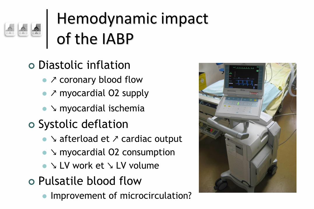
Diastolic inflation
↗ coronary blood flow
↗ myocardial O2 supply
↘ myocardial ischemia
Systolic deflation
↘ afterload et ↗ cardiac output
↘ myocardial O2 consumption
↘ LV work et ↘ LV volume
Pulsatile blood flow
Improvement of microcirculation?
Hemodynamic impact of the IABP

Retrospective study

December 2007 to December 2012
457 peripheral VA-ECMO
90 patients with laminar flow
LVEF <15%
ITV < 8 cm
Δ SBP-DBP <15mmHg

2007-2012 457 PVA ECMO
Post-cardiotomy 117 Refractory MOF<48h 96 Chronic pulmonary disease 26 Massive mitral regurgitation 21 Refractory septic shock 14 Femoro-axillary cannulation 10 ARDS 8 Isolate RV dysfunction 8 Prior impella implantation 5
Pulse pressure≥ 15 mmHg 48 or TVI ≥8 cm
IABP for APO 5 APO after IABP explantation 5 ECMO centralisation for other reasons than APO 4
90 patients
No-IABP 56
IABP 34

Patients’s characteristics
Parameter Median (25th-75th)
No-IABP n=56
IABP n=34
p
Age, y 46,5 (32-54) 51,5 (43-59,5) * 0,02 Male, % 57 % 73 % 0,12 BMI 24,6 (20,6-27,8) 26,6 (24,8-30,7) * 0,02 Charlson score 2 (1-3) 2 (1-3) 0,14 Year 2009 (2008-2010) 2011 (2010-2012) ***p<0,0001 Saps-II score 65 (56-72,5) 75 (54-81) 0,11 SOFA score 11 (6,5-13) 11 (8-14,5) 0,39 Etiology, %
Cardiac arrest AMI Myocarditis DCM
28,5% 35,7% 32,1% 32,1%
41,1% 64,7% 11,7% 23,5%
0,22
** 0,007 *0,03 0,38
Inotrope score, g/kg/min 76,5 (41-127,5) 49 (30-77) *0,03 LVEF, % 10 (7-10) 10 (6-11) 0,73 TVI, cm 4 (2-5) 4 (2-5) 0,38 Mechanical ventilation, % 87,5 % 94,1 % 0,42 PaO2/FiO2 ratio 270 (155-326) 296 (170-376) 0,69 PEEP, cmH2O 4 (2-5) 4 (4-6) 0,14 pH 7,25 (7,18-7,35) 7,25 (7,12-7,32) 0,40 Lactatemia, mmoles/L 8,0 (5,7-10,9) 7,3 (5,0-11,5) 0,98 Q ECMO, L/min 3,97 (3,33-4,45) 4,22 (3,73-4,61) 0,09

Radiologic component of the LIS
No IABP
IABP
Radiologic score
Time from ECMO implantation
D0 D1 D2 D3 D7 D150
1
2
3
4
*** *** ***
***
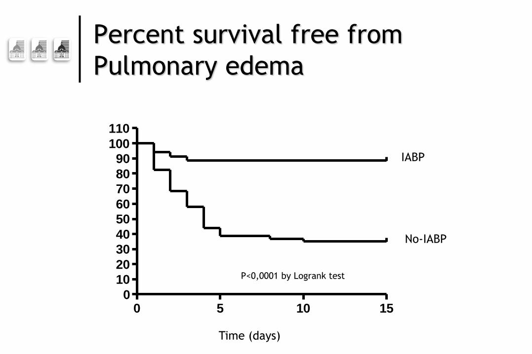
Percent survival free from
Pulmonary edema
0 5 10 150
10
20
30
40
50
60
70
80
90
100
110
IABP
No-IABP
Time (days)
P<0,0001 by Logrank test

Impact of IABP use
2006 2007 2008 2009 2010 2011 2012 20130
25
50
75
IABP
switch for central ECMO
Major pulmonary edema

Need for central ECMO
- +0
25
50
75
100
IABP
44.6% vs 5.9%
***, p=0.0001

Independent predictors of pulmonary
edema occurrence
Variables OR IC 95% P
No IABP 18.87 4.3-90.97 <0.0001
pH <7.25 at ECMO
initiation 4.47 1.19-16.80 0.027
Inotropic Score >66
at ECMO initiation 5.0 1.43-18.87 0.013

Outcomes
Parameter Median (25th-75th)
No-IABP n=56
IABP n=34
p
IABP duration, d - 5,5 (3,5-7,5)
ECMO duration, d 5 (2,5-7,5) 6 (4-10) 0,06
Follow-up, % Death under peripheral ECMO Death under central ECMO Death under temporary assistance Myocardial recovery Cardiac transplant Bridge to long term assistance
12,5 14,2 32,1 35,7 17,9 14,3
26,5
0 26,5 35,3 5,9 32,3
0,09
- 0,57 0,97 0,10 0,04*
Weaning from MV under ECMO, % 30,4 38,2 0,44
Time on MV under ECMO, % 100 (66,3-100) 100 (40,4-100) 0,52
ICU mortality, % 35,7 29,4 0,54
ICU duration, d 14 (6-30) 10 (6-21) 0,24
Temporary circulatory assistance duration, d
9 (5,5-13) 6 (4-10) 0,13
Mechanical ventilation duration, d 6 (3-15) 5 (3-12,5) 0,29
RRT duration, d 5,5 (1-42) 2 (0-6) 0,69

Prospective study

Study objectives
Evaluate the impact of the combination of
Peripheral veno-arterial ECMO
Counterpulsation with IABP
On general hemodynamics and
microcirculation
In patients with refractory cardiogenic
shock

Methods
Prospective monocenter crossover study
12 months-study period
12 patients
Admitted for refractory cardiogenic shock
requiring emergent peripheral veno-arterial ECMO
Low or non-ejecting heart : laminar blood flow
Evaluation
Evaluation under IABP support, after 30 min
interruption and 30 min after restarting the IABP
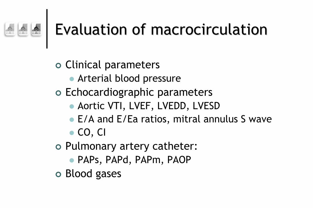
Evaluation of macrocirculation
Clinical parameters
Arterial blood pressure
Echocardiographic parameters
Aortic VTI, LVEF, LVEDD, LVESD
E/A and E/Ea ratios, mitral annulus S wave
CO, CI
Pulmonary artery catheter:
PAPs, PAPd, PAPm, PAOP
Blood gases

Sidestream Dark Field
imaging SDF
Sublingual videomicroscopy
sequences
Semi-quantitative and
dynamic evaluation of
microcirculation
Evaluation of microcirculation

InSpectra® StO2 - NIRS
Early indicator of tissular hypoperfusion?
• Models : arteriopathy, septic shock
Parameters :
• Baseline StO2
• Vascular occlusion test VOT : T1, T2
• Hyperemia (StO2 overshoot)
Thenar eminence microcirculation

Equanox ® Nonin
Early indicator of brain hypoperfusion
• Models : cardiac surgery
Parameters :
• Left and right rSO2
Brain microcirculation

Variable Value Range
Age, yr 57 ± 14 28–75
Men, n (%) 9 (75%)
SAPS2 79 ± 16 65–106
Before inclusion
Days of ECMO 6.3 ± 5.9 1–21
Days of IABP 4.7 ± 4.4 1–17
Diagnosis, n (%)
Acute myocardial infarction 8 (67%)
Acute valvular dysfunction 2 (17%)
Dilated cardiomyopathy 1 (8%)
Fulminant myocarditis 1 (8%)
During study protocol
ECMO flow, L/min 4.3 ± 0.9 3–5.5
Catecholamines
Dobutamine (n = 4), µg/kg/min 7.5 ± 3.0 5–10
Norepinephrine (n = 1), mg/h 0,6
Epinephrine (n = 5), mg/h 3.0 ± 4.0 0.35–10
Patients on mechanical ventilation, n (%) 12 (100%)
Patients’ characteristics

Parameter IABP on IABP off IABP
restart P
Heart rate 99 ± 21 101 ± 17 103 ± 20 0.07
SBP (mmHg) 103 ± 20 102 ± 20 100 ± 22 0.75
DBP (mmHg) 74 ± 17 88 ± 16 72 ± 16 0.02
MBP (mmHg) 87 ± 14 92 ± 16 84 ± 16 0.06
Pulse pressure (mmHg) 29 ± 22 15 ± 13 29 ± 24 0.02
DBP increase (mmHg) 134 ± 40 – 125 ± 26 –
Hemodynamic data

Pulmonary artery catheter
Parameter IABP on IABP off IABP
restart P
SBP, mmHg 24 ± 9 29 ± 11 23 ± 10 0.01
DBP, mmHg 16 ± 7 19 ± 10 16 ± 9 0.04
MBP, mmHg 19 ± 8 24 ± 10 19 ± 9 0.02
PAOP, mmHg 15 ± 8 19 ± 10 15 ± 8 0.01
Central venous oxygen saturation, % 73 ± 11 73 ± 15 75 ± 12 0.43

IABP to prevent pulmonary
edema under peripheral ECMO P
ulm
onary
A
rtery
Occlu
sio
n P
ressure

Parameter IABP on IABP off IABP
restart P
LVEDD (mm) 52 ± 14 55 ± 13 47 ± 13* 0.003$
LVESD (mm) 50 ± 14 51 ± 13 42 ± 13* 0.05$
Velocity–time integral (mm) 25 ± 13 25 ± 14 26 ± 15 0.85
Cardiac output (l/min) 0.79 ± 0.46 0.77 ± 0.78 0.81 ± 0.74 0.85
Diastolic velocity
Transmitral early peak (E) (cm/s) 49 ± 19 61 ± 25 51 ± 26 0.07
Transmitral late (A) (cm/s) 32 ± 13 31 ± 9 31 ± 12 0.10
E/A 1.26 ± 0.38 1.95 ± 0.66 1.33 ± 0.36 0.003
Lateral mitral early annular (Ea) (cm/s) 6.4 ± 2.8 6.4 ± 2.5 5.8 ± 1.9 0.44
E/Ea 8.6 ± 4.0 9.8 ± 2.6 9.2 ± 4.3 0.31
S (cm/s) 4.9 ± 2.5 5.0 ± 2.9 4.9 ± 2.3 0.90
Echocardiographic data

Near-infrared spectroscopy IABP on IABP off IABP
restart P
Thenar
Baseline StO2, % 82 ± 6 79 ± 8 82 ± 6 0.41
Tissue desaturation during VOT, (%/s) –0.13 ± 0.06 –0.13 ± 0.06 –0.14 ± 0.08 0.56
Range –0.04;–0.23 –0.02;–0.24 –0.03;–0.28
Tissue resaturation after VOT (%/s) 1.26 ± 0.76 1.28 ± 0.70 1.28 ± 0.58 0.21
Range 0.56;–3.20 0.67;2.55 0.57;2.95
Cerebral hemisphere rSO2
Right, % 69.1 ± 5.3 69.4 ± 5.1 69.9 ± 5.3 0.76
Left, % 67.4 ± 5.5 68.6 ± 4.0 68.9 ± 5.3 0.24
NIRS data

200
300
400
100
50
0
FCD
(cm
/cm
²)M
FIP
PV
(%
)
IABP on
He
tero
gen
eit
y in
dex
IABP off IABP on again
200
300
400
100
50
0
FCD
(cm
/cm
²)M
FIP
PV
(%
)
IABP on
Het
ero
gen
eity
ind
ex
IABP off IABP on again
200
300
400
100
50
0
FCD
(cm
/cm
²)M
FIP
PV
(%
)
IABP on
He
tero
gen
eit
y in
de
x
IABP off IABP on again
200
300
400
100
50
0
FCD
(cm
/cm
²)M
FIP
PV
(%
)
IABP on
He
tero
gen
eit
y in
dex
IABP off IABP on again
SDF sublingual imaging

If Pulmonary edema
occurs…

Impella 5.0

Conclusion
For cardiogenic shock patients with little/no residual LV ejection while on peripheral VA-ECMO
Restoring pulsatility and decreasing LV afterload with IABP Associated with smaller LV dimensions and
lower pulmonary artery pressures
But no impact on microcirculation parameters
IABP might prevent severe hydrostatic pulmonary edema in this context

May 21-24 2014