i-trendz - siemens · welcome to the new issue of i-trendz, ... s1000tm helx) 2. hockey stick...
TRANSCRIPT

s
i-trendzsharing imaging ideas and innovations
Issue 01 / October 2015
www.siemens.co.in/healthcare
1 Foreword and Musculoskeletal Ultrasound introduction
2 Transducer Innovations
3 5 Case 1: Intra-muscular Lipoma
Case 2: Ganglion
Case 3: Hematoma
Case 4: Post Vaccination Myositis in an infant
Dear Clinicians,
Welcome to the new issue of i-trendz, the clinical platform that helps you keep track of the latest trends and practices in medical imaging. The current issue highlights the utility of High Resolution Ultrasonography in musculoskeletal imaging (MSUS).
Musculoskeletal disorders pose a persistent and pervasive challenge to the healthcare system, impacting the quality of life, across the globe. These are the most common causes of long term morbidity and pain; affecting all ages and groups. With the rise in geriatric population and lifestyle disorders, the burden of disease is going to increase. However, the complications can mostly be avoided with timely diagnosis and effective management.
Siemens has designed specialized ultrasound solutions to address the challenges of musculoskeletal imaging, thus enabling a quick and accurate diagnosis in every patient. We would like to thank Dr. Mukund Joshi and Dr. Ashwin Lawande of Dr. Joshi’s Clinic, Mumbai and Dr. Ganesh Rao B of RAGAVS Diagnostics & Research Center, Bangalore for sharing their valuable clinical inputs and case studies with us.
From basic 2D and Doppler imaging to advanced clinical
applications like CadenceTM ultrasound contrast and ARFI elastography, Siemens assures the best in class with robust and reliable technologies. SieScapeTM (Extended FOV) technology allows display of 2D panoramic images upto 240 cm in length and curvature of 180 degrees. It enables demonstrating very large contiguous sections of anatomy without distorting structural relationships and preserving spatial resolution. ClarifyTM - Vascular Enhancement feature automatically reduces noise and artifacts; providing clear definition of micro and macro vasculature. Advanced SieClear™ spatial compounding is a real time compounding technique for enhanced border definition and improved tissue contrast. Dynamic TCETM speckle reduction provides fine texture details in nerves, ligaments and tendons.
18L6 HD transducer from Siemens, is the industry’s benchmark with 576 crystal elements, providing ultra-high details of complex masses and subtle lesions which is crucial for musculoskeletal imaging. Siemens offers the specialized hockey stick probes - 14L5 SP and VF13-5 SP, developed for imaging difficult small areas like adjacent to malleoli in ankle, foot and wrist. Matrix transducer 9L4 provides uniform near-far field imaging with ARFI elastography and contrast capabilities. Our most recent innovation, the wireless transducer technology on ACUSON FreeStyleTM system is one of a kind in interventional setting like guided injections and biopsies.
We hope you have an insightful reading. Your feedback and suggestions are welcome and are extremely valuable to us. If you wish to share any interesting cases, please send in your inputs to [email protected]
Richard Guest,CEO-Siemens Healthcare, India
Transducer Innovations continued
4 Advanced Ultrasound Applications 7
8
Case 5: Rotator Cuff Tear
Case 6: Biceps Subluxation-Instability
Quiz
6
Advanced SieClear™ spatial compounding Dynamic tissue contrast enhancement: Distal Quadriceps Insertion – Suprapatellar Synovitis
Multi-modality review Right Shoulder SieScape™ Panoramic imaging: Quadriceps

Page 2 i-trendz Issue 01 / October 2015
Siemens Answers Special Clinical Needs of MSUS
Musculoskeletal sonography has been a highly specialized stream of radiology, demanding special set of requirements in the ultrasound system as well as the operator skill. Development of dedicated solutions that are apt to the anatomy and function of musculoskeletal system, as well as easy to use and maneuver is thus crucial to advance the prospects of ultrasound in this field.
Excellent image quality
and Doppler sensitivity to demonstrate microvasculature and inflammatory arthropathies
• 18L6 high definition probe
• Hockey stick probes: 14L5 SP and VF13-5 SP
• Multi-D Matrix probes
• Advanced SieClear compounding and DTCE speckle reduction: provides superior contrast and detail resolution needed to distinguish the subtle changes and fine tissue texture in tendons, nerves and ligaments
• Virtual Format/Trapezoid format, SieScapeTM panoramic imaging
Advanced applications for specialized studies and research
• CadenceTM contrast applications for contrast enhanced ultrasound: CadenceTM CPS, Contrast harmonic imaging and Contrast Dynamics for quantification
• Complete suite of elastography applications: eSie TouchTM, ARFI - Virtual TouchTM imaging and quantification, Virtual Touch™ IQ
• syngo® Velocity Vector Imaging
• Multi-modality Review with CT/MR
Enhanced needle visualization for procedures, portability, intervention workflow needs
• Wireless ultrasound technology in ACUSON FreestyleTM system
Transducer innovations:
1. High Definition Transducer Technology - 18L6HD: more clinical information in every single image
High density of elements, matched with system channels, maximizes information density acquired by HD transducers.
Higher degree of compounding,
resulting in increased detail
resolution
Maximizes information density,
yielding sharper images
Conventional
transducer array
HD transducer
array
High density (HD) element array
Finer pitch (size) elements
More elements
��
Widest bandwidth of 6 - 18 MHz
Widest linear field of view of 58 mm; Industry’s best
• It covers more anatomy in a single image, reducing the need for additional image acquisition
Highest number of array elements: 576 elements
Fine Pitch Allows Beam Steering at Greater Angles →
• Increased detail resolution
• Improved speckle presentation
• Increased contrast resolution
• Clearer border definition and continuity
Improved color sensitivity
Enhanced ergonomics –
• Palmer grip for improved stress distribution
• ElastoGrip coating for non slip usage
• Extra long cable ~2.1m for ease of use
Available on ACUSON S family ultrasound systems (S3000TM HELX, S2000TM HELX, S1000TM HELX)
2. Hockey Stick Transducers - 14L5 SP and VF13-5 SP:
These are specialized high frequency linear array transducers developed for MSUS, with a small footprint (typically 25–30 mm), small field-of-view, and easy maneuverability.
{14L5 SP → available for ACUSON S family of ultrasound systems
VF13-5 SP → ACUSON X 300TM PE ultrasound system

Page 3 i-trendz Issue 01 / October 2015
They are especially useful at sites where it is difficult to maneuver probe and obtain adequate skin contact with routine larger transducers (i.e. soft tissues adjacent to bony prominences like clavicle, malleoli). That makes them ideal for imaging the ankle, foot, hand and elbow.
3. Multi-D Matrix Transducer - 14L5 probe and 9L4 probe:
Uniform image from near field to far field
Virtual format supported on 14L5
9L4 supports ARFI elastography
4. MultiHertz™ multiple frequency imaging:
Improve the penetration on larger shoulders and legs with just a touch of a button on the premium ACUSON S family of systems, as well as Acuson X600TM, Acuson X700TM and Acuson X300TM PE systems.
5. Hanafy Lens Technology and Micro-Pinless Connectors
Acoustic EfficiencyMicro-Pinless connectors
Hanafy Lens
Maximizes efficiency of transmission of ultrasound into the body
• Multiple matching layers
• Multilayer ceramic (crystals)
Unique, patented design
512 pinless connections
Unmatched signal to noise
Patented lens crystal design
Adapts focus throughout field, delivering the highest resolution
6. Wireless Transducer, L13-5 and L8-3 on ACUSON FreestyleTM platform:
The ACUSON Freestyle™ ultrasound system is the world’s first ultrasound system with wireless transducers.
Using proprietary ultra-wideband radio technology, the transducers are able to send images at a high sustainable data rate back to the system, all while maintaining excellent image quality and high frame rates.
Operation of all imaging parameters on the system from up to 3 meters/10 feet away from the system
Removable battery which offers up to 90 minutes of continuous scan time between charges
For proper and complete disinfection, the probe and battery should be immersed separately.
NeedleV: Enhanced needle visualization during procedures both In-plane and Out of Plane
Figure: ACUSON FreestyleTM system with three wireless transducers
Figure: Median nerve using L13-5 on ACUSON FreestyleTM
Figure: Botulinum injection in leg for exertional compartment syndrome; NeedleV feature to enhance needle visualization

Page 4 i-trendz Issue 01 / October 2015
Advanced Applications
1. Contrast enhanced ultrasound applications :
CadenceTM CPS, Contrast Harmonic Imaging, Contrast Dynamics
CadenceTM CPS application for CEUS offers an extremely sensitive tool for detecting microvascularity and can aid in assessing inflammatory diseases, rheumatoid arthritis, focal lesions and malignancies.
Contrast Dynamics application provides quantification capability which can potentially help in therapeutic monitoring of the disease activity, e.g. antiTNF treatment.
Contrast Harmonic Imaging is Siemens unique technology with even higher spatial and temporal resolution, particularly suited in low flow lesions and small vascularities, chronic infammation.
2. Ultrasound Elastography applications
ARFI - Virtual TouchTM imaging (VTI) Virtual TouchTM quantification (VTQ) Virtual TouchTM IQ (VTIQ)
eSie TouchTM elastography
Elastography is an emerging new tool for evaluating various MSK conditions like muscle fibrosis, muscular dystrophy, spasticity in upper motor neuron disease, tumors; as the pathology often brings a change in tissue mechanical properties. Preliminary studies show potential use of elastography to differentiate inflammatory from infectious synovitis; to differentiate malignant from benign synovial masses, monitoring the process of healing.
Figure: Tendon fibrosis depicted as dark on VTI image → stiff
Figure: Tendon fibrotic site showing high Shear wave velocity on VTQ image → stiff
3. Multi-modality Review:
The multi-modality review provides a comparison with MR/CT images side-by-side with the ultrasound image. This capability can provide better precision of anatomy from MR/CT available with real time sonography. It is available on the ACUSON S Family systems.
Figure: Multi-Modality Review of shoulder- MRI with real time US
4. syngo® Velocity Vector ImagingTM:
It is an investigational tool in MSUS that can help track muscle motion in neuromuscular disorders, identify muscle deformations.

Page 5 i-trendz Issue 01 / October 2015
The intra-operative image confirms the findings.
Figure: Intra-op image showing the tumor
Comments: The large footprint high frequency linear array probe 18L6 was used for superior depth resolution. Scanning at a frequency of 15MHz helped in tracing the nerve which normally measures 0.8mm. The superior B-mode resolution of Siemens made a difference in identifying nerve separate from the lesion.
Case studies
Case studies 1 through 4 courtesy:
L-R: Dr. Mukund Joshi: [email protected] Dr. Ashwin Lawande: [email protected] Dr. Joshi’s clinic, Mumbai
Case 1: Intra-muscular Lipoma
A 54yrs old lady going to gymnasium developed left upper lateral forearm swelling with pain.
Adv: HFUS
Ultrasound Findings: A well defined homogenous echogenic solid mass in the deep head of supinator was seen with no calcification or necrosis. No significant vascularity was depicted on Doppler → consistent with an intramuscular lipoma.
The posterior interosseous nerve traversing along the periphery of this lesion was seen. It was normal in calibre and did not show any signs of compression.
Figure: An intramuscular homogeneous hyper-echoic mass and relation to posterior interosseous nerve (PIN)
Figure: The mass with relation to surrounding structures [SS- superficial supinator, DS- deep supinator]

Page 6 i-trendz Issue 01 / October 2015
Patient presented with complaint of painful Rt. wrist hard swelling, along radial and volar aspect for 15 days.
Adv: HFUS
US findings: A well defined anechoic cystic lesion in the subcutaneous tissue plane superficial to FCR tendon and the
radial artery. A branch of radial is seen along the lateral aspect of the lesion. No communication with the joint observed. Color Doppler does not show any intralesional vascularity. Findings → consistent with diagnosis of ganglion
Comments: Hockey stick probe 14L5SP provides exquisite soft tissue detail and ease of maneuverability around wrist.
Case 2: Ganglion
Case 3: Hematoma
Figure: Anechoic cystic mass on wrist → Ganglion
Figure: No vascularity seen in mass lesion on Doppler
A middle aged lady presented with history of fall on the knee with swelling along the medial aspect.
Adv: HFUS
US findings with 18L6HD: A well defined hypoechoic fluid collection in the subcutaneous tissue plane along the medial knee. Thin internal septae were seen with low level echoes. No
vascularity detected on Doppler. Findings → suggested a few days old hematoma.
Comments: The superior B mode resolution helped us in showing the classical reticular pattern of the organizing hematoma. The extended field of view shows the entire hematoma in one image.
Figure: Ultrasound depicting a hematoma with classical reticular pattern
Case 4: Post Vaccination Myositis in an infant
A 4 month old boy developed swelling in the upper thigh post vaccination and associated fever.
Adv. Sonography to rule out an abscess.
US Findings: A bulky and hypoechoic Vastus Lateralis muscle seen with 18L6 HD. There was no abscess formation visualized. Increased vascularity was seen in the edematous muscle on Doppler. The femoral vessels were patent.
Findings → suggested post vaccination myositis.
Comments: Superior B mode resolution, user friendly workflows and easy to handle probes on Siemens ACUSON S2000™ system offer great convenience and efficiency in pediatric and neonatal patients.

Page 7 i-trendz Issue 01 / October 2015
Figure: A partial thickness tear of supraspinatus
Figure: Subacromial impingement of rotator-cuff at 40 degree abduction; mild subdeltoid fluid collection
Figure: Edematous bulky vastus medialis Figure: Increased vascularity in the edematous muscle on Doppler
Case studies 5 and 6 courtesy:
Dr. Ganesh Rao B: [email protected]
RAGAVS Diagnostics & Research Center, Bangalore
Case 5: Rotator Cuff Tear
A 37 year old female presented with an insidious pain in shoulder with difficulty to raise the arm. There was a history of fall. The patient was referred for high frequency ultrasound scan of shoulder with clinical diagnosis of impingement.
US Findings (18L6 HD probe): A partial thickness, partial width interstitial tear of Supraspinatus tendon was seen at insertion, measuring 7x2.5mm, not reaching the capsular or bursal aspect of tendon. Tear is 9mm posterior to biceps tendon. Subacromial impingement of rotator-cuff was noted at 40 degree abduction. Mild sub-detloid fluid collection and bicepital tenosynovitis was also detected.
Comments: Musculoskeletal high frequency ultrasound is an extremely useful tool to diagnose shoulder disease, with very high accuracy; and sensitivity reaching close to 100% in detecting full thickness tears, as demonstrated across a number of studies
It can detect and classify tear and also monitor the progression
Offers dynamic information e.g. stretch, compression response
Targeted scanning on the symptomatic region
The normal side can be readily compared
Page 8 i-trendz Issue 01 / October 2015
Quiz #1
1. What is highest available frequency on an ultrasound transducer for musculoskeletal imaging:
a. 10MHz
b. 18MHz
c. 17MHz
d. 16MHz
e. 20MHz
2. Which of the following is true about Siemens ACUSON FreestyleTM system:
a. World’s first wireless ultrasound system
b. Probe can be easily sterilized for intervention environment
c. NeedleV features allow both in-plane and out-of plane needle visualization
d. Probes can perform up to 90 minutes continuous scanning
e. All of the above
3. Siemens offers the following technologies for MSK ultrasound imaging:
a. High frequency High definition transducer 18L6HD and Hockey stick probes 14L5 SP and VF13-5 SP
b. Ultrasound CadenceTM contrast and ARFI elastography applications
c. SieScapeTM panoramic imaging for extended field of view
d. syngo® Velocity Vector imagingTM
e. All of the above
Please send your answers along with your contact details and the lucky winners will receive a special prize. E-mail to [email protected]
Read further on Siemens Ultrasound: http://www.healthcare.siemens.com/ultrasound#
If you have interesting cases to be shared, please e-mail us at [email protected]
Our next edition will be equally exciting with other modalities/applications.
Compiled by Clinical Marketing Team, Healthcare, Siemens Ltd., 130, P. B. Marg, Worli, Mumbai - 400 018.Tel.: 91 22 3967 7000. Fax: 91 22 3967 7500. www.siemens.co.in/healthcare
Customer Care No.: 1-800-419-7477 Download i-trendz issues from siemens.co.in/healthcare
Technological innovations spur new clinical applications. This gives the medical community an edge in diagnosis and helps detect / treat diseases at an early stage. This in turn will help the society at large. These are the primary objectives with which we developed i-trendz. We would like to know how we can make this initiative more valuable for your practice and the wellbeing of patients.
© 2015 by Siemens Ltd., India
All trademarks are properties of Siemens AG or associate companies. Third-party trademarks are properties of their respective companies. The content of this publication does not necessarily reflect the opinion of the publisher. Reproduction of articles in whole or in part requires the permission of the editorial office. This also applies to storage in electronic databases and on the internet.
Intended for use by Registered Medical Practitioners or Hospital Staff only. The medical information in this newsletter is for individuals of the healthcare community and not for general public. The information and reference materials contained here are intended solely for the general information of the reader and is neither intended to dictate what constitutes reasonable, appropriate or best care for any given health issue, nor is it intended to be used as a substitute for the independent judgment of a physician or a surgeon for any given health issue.
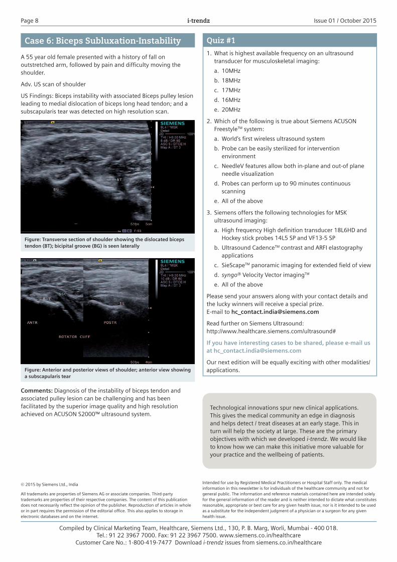
Case 6: Biceps Subluxation-Instability
A 55 year old female presented with a history of fall on outstretched arm, followed by pain and difficulty moving the shoulder.
Adv. US scan of shoulder
US Findings: Biceps instability with associated Biceps pulley lesion leading to medial dislocation of biceps long head tendon; and a subscapularis tear was detected on high resolution scan.
Figure: Transverse section of shoulder showing the dislocated biceps tendon (BT); bicipital groove (BG) is seen laterally
Figure: Anterior and posterior views of shoulder; anterior view showing a subscapularis tear
Comments: Diagnosis of the instability of biceps tendon and associated pulley lesion can be challenging and has been facilitated by the superior image quality and high resolution achieved on ACUSON S2000™ ultrasound system.