i synthesis and characterization of surface...
TRANSCRIPT

i
SYNTHESIS AND CHARACTERIZATION OF SURFACE-ACTIVATED
MULTIWALLED CARBON NANOTUBES-POLYMER COMPOSITE
ELECTROSPUN NANOFIBER
FATIRAH BINTI FADIL
UNIVERSITI TEKNOLOGI MALAYSIA

ii
SYNTHESIS AND CHARACTERIZATION OF SURFACE-ACTIVATED
MULTIWALLED CARBON NANOTUBES-POLYMER COMPOSITE
ELECTROSPUN NANOFIBER
FATIRAH BINTI FADIL
A thesis submitted in fulfilment of the
requirements for the award of the degree of
Doctor of Philosophy (Chemistry)
Faculty of Science
Universiti Teknologi Malaysia
APRIL 2016

iv
لرلٱ رلٱ مي ح لمرلٱ هلٱ مي Bismillah Hir-Rahman Nir-Rahim.
In the name of Allah, the Beneficent, the Merciful.
Dedication To my parents: The reason of what I become today. Thanks for your great support and continuous care. To my husband: Thanks for everlasting support and unconditional love my dear soulmate. To my siblings: I am really greatful of having both of you in my life. There is no better friend than a sister. Love you guys.

v
ACKNOWLEDGEMENTS
In the name of Allah, the Most Gracious, the Most Merciful,
Alhamdulillah. With His blessing on me, I have completed my PhD
research journey toward the end. My thanks are due first to my amazing supervisor,
Assoc. Prof. Dr. Nor Aziah Buang for her guidance and encourangement, for
always making time to offer me advice, from the beginning and until to the very
end. Special thanks go also to Prof. Seeram Ramakrishna and Dr. Molamma P.
Prabhakaran from National University of Singapore (NUS) for their invaluable
help, knowledge, given me oppurtunity to work in an international environment at
their place. Many thanks to all the people I have work and collaborated with,
especially Mr. KH Ang from Nanolab Instrument (M) Sdn. Bhd for providing
industrial instrument training. I owe many thanks to all the past and present lab
members of Inorganic Laboratory for the enjoyable experience of working and
having good time with them. I would like to thank all the people who I have met
over this four years, who have enriched me in so many different ways. Thanks to
Dr. Fatiha Ismail for her lasting friendship amd support, constant guidance despite
the distance and also my other collangues; Azizul, Amin, Syikin, Johari, Akmaliah,
Syafiq, Khairullah, Rijal and Amirul. Lastly, my really heartfelt thanks to my dear
husband, Mohd Firdaus A.Aziz, Ibu, Ayah, Along and Adik for their love and
support through my years of studying. Many thanks to all of you. Alhamdulillah.

vi
ABSTRACT
The major problem in the development of polymer nanofiber composites with
the infusion of multiwalled carbon nanotubes (MWCNTs) is to ensure good dispersion
of the MWCNTs within the polymer matrix. This study reports an effective approach to
activate the surface of MWCNTs by a non-covalent binding strategy, and incorporation
of MWCNTs in poly (L-lactide-co-ε-caprolactone) (PLCL) using electrospinning
process. The debundling of the MWCNTs aggregates through the non-covalent
surfactant attachment on their outer layers was studied using surfactants with different
ionic characters, which were sodium dodecyl sulphate (anionic, SDS), cetyltrimethyl
ammonium bromide (cationic, CTAB), and polysorbate 80 (non-ionic, Tween-80)
surfactants. Results obtained from the Atomic Force Microscopy (AFM) analysis of
surface roughness of the surfactant-MWCNTs aggregates show different contours
which were assigned to the size of the aggregates, distribution and orientation of the
deposited surfactants on the surfaces of MWCNTs. The dispersion behavior of the
respective surfactant molecules studied showed that the non-ionic surfactant molecules
of Tween-80 have better adsorption coverage on MWCNTs surface due to the
hydrophobic interactions between the liquid-solid interfaces, rather than the ionic
surfactants of SDS and CTAB. The orientation of the adsorbed surfactants on the
surfaces of MWCNTs was found to be strongly associated with the surfactant affinity,
which was contributed by the surfactants head groups ionization. The surface
morphology of each adsorbed surfactant molecule onto MWCNTs surface was
determined by the Field Emission Scanning Electron Microscopy (FESEM) analysis.
Furthermore, the infusion of the Tween-80-MWCNTs usability as the nanofiller
component to produce electrospun polymer nanofiber composites was conducted using
a customized electrospinning reactor system. The inclusion of Tween-80-MWCNTs
resulted in superior electrospun MWCNTs-PLCL nanofiber composite with tensile
stress value of 5.82-15.95 MPa, with the incorporation of MWCNTs ranging from
0.1wt% to 1.0wt%. Characterization by Transmission Electron Microscopy (TEM)
depicted the homogenous distribution of MWCNTs within the polymer matrix. The
manipulation of the electrospinning operational parameters in producing different
structural features of the polymer nanofibers from PLCL was successful in producing
both solid and porous structured nanofibers through the variation of solvent
composition used. The solid PLCL nanofibers were formulated from the optimized
polymer solution of 11wt% (w/v) of PLCL in dichloromethane/ dimethyl formamide
(DCM/DMF) (70:30) at an applied voltage of 14kV with spinning solution flow rate of
1.0 mL/hr. While the porous PLCL nanofibers were formulated from the optimized
polymer solution of 11wt% (w/v) of PLCL in DCM/acetone (70:30) at an applied
voltage of 14kV with spinning solution flow rate of 1.0 mL/hr. The substitution of
DMF to acetone in binary solvent system has resulted in highly-porous PLCL
nanofibers. The AFM characterization revealed the differences in the surface roughness
and pore depths of both dense and porous PLCL electrospun nanofibers fabricated.

vii
ABSTRAK
Masalah utama dalam pembangunan gentian nano polimer komposit dengan
penyatuan tiub nano karbon dinding berlapis (MWCNTs) adalah untuk memastikan
penyebaran yang baik MWCNTs di dalam matriks polimer. Kajian ini melaporkan
pendekatan efektif untuk mengaktifkan permukaan MWCNTs dengan menerokai
strategi pengikatan bukan kovalen, dan penggabungan MWCNTs ke dalam poli (L-
lactid-co-ε-kaprolakton) (PLCL) menggunakan proses pemintalan elektro.
Penyahgumpalan agregat MWCNTs dengan melekatkan surfaktan secara bukan
kovalen pada lapisan luarnya telah dikaji dengan menggunakan surfaktan yang berbeza
sifat ionik iaitu natrium dodesil sulfat (anionik, SDS), setiltrimetil ammonium bromida
(kationik, CTAB), dan polisorbat 80 (bukan ionik, Tween-80). Keputusan yang
diperoleh daripada analisis mikroskop daya atom (AFM) terhadap kekasaran
permukaan agregat surfaktan-MWCNTs menggambarkan perbezaan bentuk kontur
yang merujuk kepada saiz agregat, taburan dan orientasi surfaktan yang terenap pada
permukaan MWCNTs. Perilaku penyerakan bagi setiap molekul surfaktan yang dikaji
menunjukkan bahawa molekul surfaktan bukan ionik Tween-80 mempunyai liputan
penjerapan yang lebih baik pada permukaan MWCNTs disebabkan oleh interaksi
hidrofobik antara muka cecair-pepejal, berbanding surfaktan ionik SDS dan CTAB.
Orientasi surfaktan terjerap pada permukaan MWCNTs didapati berkaitan rapat dengan
afiniti surfaktan, yang disumbangkan oleh pengionan kumpulan kepala surfaktan.
Morfologi permukaan bagi setiap molekul surfaktan terjerap di permukaan MWCNTs
telah ditentukan oleh analisis mikroskopi pengimbasan elektron pancaran medan
(FESEM). Tambahan pula, kebolehgunaan gabungan Tween-80-MWCNTs sebagai
komponen bahan pengisi nano bagi menghasilkan pintalan elektro komposit gentian
nano polimer telah dijalankan menggunakan sistem reaktor pintalan elektro yang
ditempah khas. Perangkuman Tween-80-MWCNTs telah menghasilkan pintal elektro
gentian nano MWCNTs-PLCL komposit terbaik dengan nilai tegasan tegangan 5.82-
15.95 MPa, dengan julat penggabungan MWCNTs antara 0.1wt% hingga 1.0wt%.
Pencirian menggunakan mikroskopi penghantaran elektron (TEM) menunjukkan
taburan MWCNTs yang sekata dalam matriks polimer. Manipulasi terhadap parameter
operasi pemintalan elektro untuk menghasilkan gentian nano polimer daripada PLCL
dengan ciri struktur yang berbeza telah berjaya menghasilkan gentian nano yang
berstruktur padat dan berliang melalui penggunaan pelbagai komposisi pelarut. Gentian
nano PLCL padat telah diformulasikan daripada larutan polimer 11wt% (w/v) PLCL
yang dioptimumkan dalam diklorometana/ dimetilformamida (DCM/DMF) (70:30)
pada voltan gunaan 14kV dengan kadar aliran 1.0 mL/jam. Manakala gentian nano
PLCL berliang telah diformulasi daripada larutan polimer 11wt% (w/v) PLCL yang
dioptimumkan dalam DCM/aseton (70:30) pada voltan gunaan 14kV dengan kadar
aliran larutan pemintalan 1.0 mL/jam. Penggantian DMF dengan aseton dalam sistem
pelarut dedua telah menghasilkan gentian nano PLCL yang amat berliang. Pencirian
AFM telah mendedahkan perbezaan kekasaran permukaan dan kedalaman liang bagi
kedua-dua pintalan elektro gentian nano PLCL padat dan berliang yang telah
difabrikasi.

viii
TABLE OF CONTENTS
CHAPTER TITLE PAGE
DECLARATION
DEDICATION
ACKNOWLEDGEMENTS
ABSTRACT
ABSTRAK
TABLE OF CONTENTS
LIST OF TABLES
LIST OF FIGURES
LIST OF ABBREVIATIONS
LIST OF APPENDICES
ii
iii
iv
v
vi
vii
xiii
xiv
xix
xxi
1
INTRODUCTION
1.1 Nanofibers technology
1.2 Biodegradable polyester in nanofibers technology
1.3 Composite fabrication process
1.4 Synthetic composite
1.5 Carbon nanotubes as multifunctional filler
1.6 Surfactant assisted dispersion of carbon nanotubes
1.7 Electrospinning in nanofibers technology
1.8 Problem statement
1.9 Objectives of the research
1.10 Scope of the research
1.11 Significance of the research
1
1
2
5
5
6
7
8
9
11
11
13

ix
2
3
LITERATURE REVIEW
2.1 Carbon nanotubes in polymer composite application
2.2 Carbon nanotubes structure and its properties
2.3 Current approaches for the dispersion of carbon
nanotubes
2.3.1 Role of surfactant in assisting the
dispersion of carbon nanotubes
2.3.2 Types of surfactant and their characteristics
2.3.3 Fluid surfaces and interface
2.4 Electrospun polymer nanofibers composite
2.5 Structure formation in polymer nanofibers
2.6 Electrospinning processing parameters
2.6.1 Needle diameter
2.6.2 Flow rate
2.6.3 Applied voltage
2.6.4 Spinneret tip-to-collector
2.6.5 Collector
2.6.6 Spinning solution
EXPERIMENTAL
3.1 Experimental outline
3.2 Preparation of surfactant activated MWCNTs
3.3 Preparation of surfactant solutions
3.4 Characterization of surfactant activated MWCNTs
3.4.1 Surface topography of surfactant-MWCNTs
by atomic force microscope (AFM)
3.4.2 Surface morphology of surfactant-MWCNTs
by field emission scanning electron
microscope (FESEM)
3.4.3 Diffraction pattern of surfactant-MWCNTs
by X-Ray diffraction (XRD)
3.4.4 Thermodynamic properties of surfactant-
MWCNTs by differential scanning
15
15
18
21
23
24
26
29
32
35
35
35
36
37
37
38
42
42
43
44
46
46
47
47

x
4
calorimeter (DSC)
3.4.5 Chemical properties of surfactant-MWCNTs
by Fourier transform infra-red (FTIR)
3.4.6 Graphitization of surfactant-MWCNTs by
Raman spectroscopy
3.4.7 Thermal stability of surfactant-MWCNTs by
thermal gravimetric analysis (TGA)
3.4.8 Dispersion test of surfactant-MWCNTs by
dynamic light scattering (DLS) analysis
3.5 Fabrication of custom-built electrospinning unit
3.6 Optimization of spinning solution
3.7 Electrospinning of nanofibers and MWCNTs
composite nanofibers of PLCL
3.8 Characterization of electrospun nanofibers
3.8.1 Surface morphology of electrospun
nanofibers by scanning electron microscope
(SEM)
3.8.2 Structural morphology of electrospun
nanofibers by transmission electron
microscopy (TEM)
3.8.3 Chemistry properties of electrospun
nanofibers by attenuated total reflectance
Fourier transform infra-red (ATR-FTIR)
3.8.4 Tensile measurement of electrospun
nanofibers by tensile analyzer
3.8.5 Electrical conductivity of electrospun
nanofibers by electrical impedence
spectroscopy (EIS)
3.9 Research flow chart
SURFACE ACTIVATION OF MWCNTs THROUGH
NON-COVALENT ATTACHMENT OF
SURFACTANT
48
49
49
50
50
51
53
54
55
55
56
56
56
57
58
59

xi
5
4.1 Chapter overview
4.2 AFM analysis of surfactant-MWCNTs aggregates
4.3 FESEM analysis of surfactant-MWCNTs aggregates
4.4 XRD analysis of surfactant-MWCNTs aggregates
4.5 Raman spectroscopy analysis of surfactant-
MWCNTs aggregates
4.6 Non-covalent interaction between surfactant and
MWCNTs
4.7 DSC thermogram of surfactant-MWCNTs
aggregates
4.8 TGA analysis of surfactant-MWCNTs aggregates
4.9 Dispersion stability of surfactant-MWCNTs
aggregates
4.10 Surface tension of surfactant-MWCNTs solution
4.11 Chapter summary
OPTIMIZATION OF ELECTROSPINNING
OPERATIONAL PARAMETERS FOR THE
FABRICATION OF POLY (L-LACTIDE)-CO- ɛ-
CAPROLACTONE) NANOFIBERS
5.1 Chapter overview
5.2 Optimization of solvent
5.2.1 Electrospun PLCL nanofibers in single
solvent
5.2.2 Electrospun PLCL nanofibers in
DMF/acetone solution
5.2.3 Electrospun PLCL nanofibers in DCM/DMF
solution
5.2.4 Electrospun PLCL nanofibers in
DCM/acetone solution
5.3 Influence of dielectric constant and boiling point of
solvents in electrospinning
5.4 Optimization of polymer concentration
59
60
69
73
75
77
79
81
85
89
92
94
94
95
96
99
101
103
105
112
xii
6
5.5 Relation of electrospinning jets and nanofibers
structure
5.5.1 Branched jet
5.5.2 Single jet
5.5.3 Lateral jet
5.5.4 Pendant droplet jet
5.6 Optimization of electrospinning operational
parameters
5.6.1 Optimization of applied voltage
5.6.2 Optimization of distance spinneret tip-to-
collector
5.6.3 Optimization of flow rate
5.6.4 Optimization of needle diameter
5.7 Chapter summary
CHARACTERISTIC OF PLCL NANOFIBERS
COMPOSITE INFUSED WITH MWCNTs
6.1 Chapter overview
6.2 SEM analysis of MWCNTs-PLCL nanofibers
composite
6.2.1 Solid MWCNTs-PLCL nanofibers
composite
6.2.2 Porous MWCNTs-PLCL nanofibers
composite
6.3 Fragmentation of electrospun MWCNTs-PLCL
nanofibers composite
6.4 TEM analysis of solid MWCNTs-PLCL nanofibers
composite
6.5 ATR-FTIR analysis of solid MWCNTs-PLCL
nanofibers composite
6.6 Mechanical characteristic of solid MWCNTs-PLCL
nanofibers composite
6.7 Electrical conductivity of solid MWCNTs-PLCL
117
118
119
119
121
122
122
124
126
128
129
131
131
132
132
135
139
143
145
147

xiii
7
nanofibers composite
6.8 Reinforcement and ballistic behavior of MWCNTs-
polymer nanofibers
6.9 Chapter summary
CONCLUSION AND RECOMMENDATIONS
7.1 Conclusion
7.2 Recommendations
150
152
154
155
155
158
REFERENCES
Appendices A-F
160
177-193
xiv
LIST OF TABLES
TABLE NO. TITLE PAGE
2.1 Chemical oxidation process of carbon nanotubes 22
2.2 Typical surface and interfacial tensions of liquids at 20°C
(Myers, 2006)
27
2.3 Properties table of common solvents (Reichardt, 1990)
41
3.1 Preparation of surfactant solution
44
4.1 Enthalpy changes in surfactant-MWCNTs aggregates
79
5.1 Spinning solution parameter for PLCL
96
5.2
Properties table of DCM, acetone and DMF (Reichardt,
1990)
105
5..3 Calculated dielectric constant value of binary solvent
composition
110
5.4 Nanofibers form and diameter size at different PLCL
concentration
112
6.1
Tensile properties of PLCL and MWCNTs-PLCL
nanofibers composite
148
6.2 Electrical conductivity of PLCL and MWCNTs-PLCL
nanofibers composite
150

xv
LIST OF FIGURES
FIGURE NO. TITLE PAGE
1.1 Molecular structures of ε-caprolactone monomer, L-
lactide monomer and PLCL copolymer
3
1.2 The degradation mechanism of PLCL by hydrolysis
process
4
1.3 (a) Illustration model of cabon nanotubes by rolling
sheets of graphene into a cylinder of nanometer size
diameter (b) The structure of CNTs explored by high-
resolution transmission electron microscopy. This
figure is adapted from Endo (2010)
6
1.4 (a) Photograph of spider web (b) electrospun
nanofiber web
9
2.1 Illustration of (a) carbon black (b) carbon fiber (c)
carbon nanotubes dispersion in polymeric matrix.
16
2.2 The molecular dynamics model of carbon nanotubes
subjected to ballistic impact (a) initial model (b) a
deformed carbon nanotubes at its maximum energy
absorption. This figure is adapted from Mylvaganam
and Zhang (2007)
17
2.3 Illustration of carbon atom in hexagonal framework
arrangement
20
2.4 Simplified surfactant structure
23
2.5 Cohesive forces between water molecules
26
2.6 The development of the critical micelle concentration
(CMC). This figure is adapted from Emmert (2015)
28

xvi
2.7 MWCNTs composited nanofibers at different
MWCNTs loading of (a) 1 wt% (b) 3 wt% (c) 5wt%
and (d) 10 wt%. This figure is adapted from Keulder
(2013)
31
2..8
Electrospinning design in (a) vertical upward, (b)
vertical downward and (c) horizontal arrangement.
This figure is adapted from Sahay et al., (2011)
33
2.9
2.10
Jet formations in the electrospinning process. This
figure is adapted from Ramakrishna et al., (2005)
Material for collector cover (a) Aluminium foil (b)
plastic (c) metal mesh and (d) wipe tissue
34
38
3.1
(a) The schematic diagram and (b) photograph of the
fabricated custom-built electrospinning unit
51
4.1
AFM images of purified MWCNTs 61
4.2
AFM image of CTAB-MWCNTs aggregates 61
4.3
AFM images of SDS-MWCNTs aggregates 62
4.4
AFM images of Tween-80-MWCNTs aggregates 62
4.5
AFM surface contour measurement and the illustration
of adsorbed surfactant orientation on MWCNTs solid
surface (a) CTAB-MWCNTs (b) SDS-MWCNTs and
(c) Tween-80-MWCNTs
63
4.6
Molecular modeling of the material arrangement (a)
dome surface contour (b) pointy surface contour (c)
horizontal positioning (d) upward positioning. This
figure is adapted from Della Pia et al. (2012)
65
4.7
Molecular structure of SDS 66
4.8
SDS arrangement on hydrophobic surface. This figure
is adapted from De Aguiar et al. (2011)
66
4.9
Molecular structure of CTAB
67
4.10
Molecular structure of Tween-80
68
4.11
FESEM micrograph of Tween-80-MWCNTs
aggregates at magnification of 50000x
70
4.12
FESEM micrograph of SDS-MWCNTs aggregates at
magnification of 50000x
71

xvii
4.13
FESEM micrograph of CTAB-MWCNTs aggregates
at magnification of 50000x
72
4.14
XRD pattern of (a) purified MWCNTs, (b) SDS-
MWCNTs (c) Tween-80-MWCNTs and (d) CTAB-
MWCNTs
74
4.15
Raman spectra of (a) purified MWCNTs (b) SDS-
MWCNTs, (c) Tween-80-MWCNTs and (d) CTAB-
MWCNTs
76
4.16
TG and DTG thermogram of purified MWCNTs 82
4.17
TG and DTG thermogram of SDS-MWCNTs 83
4.18
TG and DTG thermogram of CTAB-MWCNTs 84
4.19
TG and DTG thermogram of Tween-80-MWCNTs
85
4.20
The illustration of the type of polydispersion of the
particle distribution in suspension
86
4.21
Distribution of (a) Tween-80-MWCNTs, (b) CTAB-
MWCNTs and (c) SDS-MWCNTs aggregates with
mono-modal, bi-modal and poly-modal histogram
88
4.22
Graph of surface tension measurement versus
surfactant concentration of surfactant-MWCNTs
mixture in (a) SDS, (b) CTAB and (c) Tween-80
solution
89
4..23
Resonance of sulphate ion 91
5.1
SEM micrographs of PLCL nanofibers in single
solvent of DMF (a1) (a2), acetone (b1) (b2) and DCM
(c1) (c2) at magnification of 1000x and 5000x,
respectively
98
5.2
SEM micrographs of electrospun PLCL nanofibers in
DMF/acetone binary solvent ratio of 7:3 (a1) (a2), 1:1
(b1) (b2) and 3:7 (c1) (c2) at magnification of 1000x
and 3000x respectively
100
5.3
SEM micrographs of electrospun PLCL nanofibers in
DCM/DMF binary solvent ratio of 7:3 (a1) (a2), 1:1
(b1) (b2) and 3:7 (c1) (c2) at magnification of 1000x
and 3000x respectively
102
5.4
SEM micrographs of electrospun PLCL nanofibers in
DCM/acetone binary solvent ratio of 7:3 (a1) (a2), 1:1

xviii
(b1) (b2) and 3:7 (c1) (c2) at magnification of 1000x
and 3000x respectively
104
5.5
The electrospinnability of PLCL in series of binary
solvent consisting of DCM, DMF and acetone
111
5.6
SEM micrograph of 10 wt% (a), 11 wt% (b), 12 wt%
(c) and 13 wt% (d) of electrospun PLCL nanofibers at
magnification of 3000x
113
5.7
PLCL nanofibers thickness
115
5.8
PLCL nanofibers diameter size distribution and
nanofibers web thickness in (a) 10 wt% (b) 11 wt% (c)
12 wt% (d) 13 wt%
116
5.9
(a) multiple branches jet (b) solidified jet branches at
needle end of branched jet
118
5.10 (a) primary jet (b) solidified jet at needle end of single
jet
119
5.11
(a) multiple jet (b) solidified jet at needle end of lateral
jet
120
5.12
(a) droplet jet (b) solidified jet at needle end of
pendant droplet jet
121
5.13
SEM micrograph of PLCL nanofibers at (a) 11 kV (b)
12 kV (c) 13 kV (d) 14 kV (e) 15 kV (f) 16 kV at
magnification of 3000x
123
5.14
SEM micrographs of PLCL nanofibers collected at
distance tip-to-collector of 10 cm (a1) (a2), 8 cm (b1)
(b2) and 6 cm (c1) (c2) with magnification of 1000x
and 3000x, respectively
125
5.15
SEM micrograph of PLCL nanofibers at (a) 0.8
mL/hr, (b) 1.0 mL/hr (c) 1.2 mL/hr (d) 1.4 mL/hr of
spinning solution flow rate at magnification of 3000x
127
5.16
SEM micrograph of PLCL nanofibers produced using
a (a) 0.45 mm (b) 0.5 mm (c) 0.7 mm (d) 0.8 mm of
needle internal diameter at magnification of 10000x
129
6.1
SEM micrograph of electrospun solid nanofiber (a)
PLCL (b) MWCNTs-PLCL with (0.1 wt%) (c) (0.2
wt%) (d) (0.3 wt%) (e) (0.4 wt%) (f) (0.5 wt%) and
(g) (1.0 wt%) of MWCNTs at magnification of
20000x
133

xix
6.2
SEM micrograph of electrospun porous nanofibers (a)
PLCL (b)/MWCNTs-PLCL with (0.1wt%) (c) (0.2
wt%) (d) (0.3wt%) (e) (0.4wt%) (f) (0.5wt%) and (g)
(1.0 wt%) of MWCNTs at magnification of 10000x
136
6.3
Simulated electrospun fibers from the spinneret (top)
to the collector plate (bottom) with reference of the
phase diagram where (i), (ii) and (iii) represent the
elongation of fluids during the spinning. Electrospun
fibers in dark regions represent solvent-rich regions
and bright regions represent polymer-rich regions.
This figure is adapted from Dayal et al. (2007)
138
6.4
SEM micrographs of solid MWCNTs-PLCL
nanofibers composite with the occurrence of fibers cut
off at magnification of (a) 20000x, (b) 40000x and (c)
80000x
139
6.5
SEM micrographs of porous MWCNTs-PLCL
nanofibers composite with the occurrence of fibers cut
off at magnification of (a) 3000x, (b) 5000x and (c)
10000x
140
6.6
Illustration of the proposed mechanism of the jet’s
axial stress and entanglement before (dashed line) and
after (solid line) the jet break. This figure is adapted
from Dayal et al. (2007)
141
6.7
AFM 3D images of the fragmented solid MWCNTs-
PLCL nanofibers composite
142
6.8
TEM micrograph of electrospun PLCL nanofibers
composite at scale bar of (a1) 100 nm (a2) 20 nm (a3)
10 nm and MWCNTs-PLCL nanofibers composite at
scale bar of (b1) 100 nm (b2) 20 nm (b3) 10 nm
144
6.9
ATR-FTIR spectra of MWCNTs, PLCL nanofibers
and MWCNTs-PLCL nanofibers composites of 0.1-
1.0 wt%
146
6.10
Stress-strain curves of MWCNTs-PLCL nanofibers
composite
148
6.11
Molecular dynamic simulation of CNTs under axial
compression, a-d is the possible buckling of CNTs
morphology changes corresponding to strain. This
figure is adapted from Yakobson et al. (1996)
152

xx
LIST OF ABBREVIATIONS
PLCL - Poly ε-caprolactone co- L-lactide
CFRP - Carbon Fiber Reinforced Polymer
CNTs - Carbon nanotubes
MWCNTs - Multiwalled carbon nanotubes
FTIR - Fourier Transform Infrared
TGA - Thermal Gravimetric Analysis
XRD - X-ray Diffraction
FESEM - Field Emission Scanning Electron Microscope
AFM - Atomic Force Microscopy
DCM - Dichloromethane
DMF - Dimethyl Formamide
TEM - Transmission Electron Microscope
SWCNTs - Singlewalled Carbon Nanotubes
PS - Poly Styrene
PMMA - Poly Methyl Methacrylate
HCl - Hydrochloric acid
STM - Scanning Tunnelling Microscope
CCVD - Catalytic Chemical Vapor Deposition
H2SO4 - Sulphuric acid
HNO3 - Nitric acid
H2O2 - Hydrogen peroxide
SOCl2 - Thionyl chloride
SLS - Sodium lauryl sulphate
SDS - Sodium dodecyl sulphate
CV - Cyclic voltammetry
DA - Dopamine
NaDDBS - Sodium dodecyl benzene sulfonate

xxi
PVA - Poly vinyl alcohol
DMAcAM - Dimethylacetamide
THF - Tetrahydrofuran
DMSO - Dimethyl sulfoxide
Tween-80 - Polyoxyethylene (20) sorbitan monooleate
CTAB - Cetyl trimethylammonium bromide
CMC - Critical micelle concentration
KBr - Potassium bromide
DTA - Differential thermal analysis
DLS - Dynamic light scattering
ATR-FTIR - Attenuated Total Reflection- Fourier Transform Infrared
D-band - Defect band
G-band - Graphite band
Z-average - Cumulant size
PU - Polyurethane

xxii
LIST OF APPENDICES
APPENDIX TITLE PAGE
A FESEM micrograph and EDX value of purified
MWCNTs
177
B DSC profile of Tween-80-MWCNTs aggregates 178
C DSC profile of CTAB-MWCNTs aggregates 179
D DSC profile of SDS-MWCNTs aggregates 180
E Publications and presentations 181
F Awards 183

CHAPTER 1
INTRODUCTION
1.1 Nanofibers technology
Nanofiber technology is one of the nanotechnology divisions where fibrous
materials are fabricated at nano-scale dimension. Electrospun polymer nanofibers
were formed from the highly charged polymer solution that which electrically
heated, extruded, evaporated and cooled upon exposed in air through a technique
called electrospinning. Electrospinning is a powerful technique in producing
polymeric based fibers at nano to sub-micron level range of fibers diameter. They
have attracted significant attention across multiple fields of chemistry, biochemistry
and engineering because of the uniqueness rheological, mechanical and biomedical
properties that are inaccessible from the casting polymers (Cameron and Shaver,
2011). The electrospun nanofibers from biopolymer based have offered new avenue
in the field of tissue regeneration by producing scaffolds with the critical similarities
to the native tissue based on the interconnectivity and its dimensions.
Electrospun polymer nanofibers were commonly produced from variety of
polymers including, natural or synthetic polymer source, with or without filler, in
melt or with solvent, on condition that the polymer solution was conductive enough
to be drawn as fibrous form. Polymer based liquid precursor from high molecular
weight polymers and high polymer concentrations are advantageous for the
electrospinning process since polymer chain entanglements and overlapping are
important for the formation of uniform nanofibers (Celebioglu and Uyar, 2012). On
the other hand, the use of solvent in preparing the spinning solution is basically to

2
induce the polymer solution transformation process from droplet into fibrous form
under high voltage atmosphere (Leach et al., 2011). The values of the applied high
voltage used during the electrospinning process which are normally within the range
of 10-30 kV, managed to draw electrospun nanofibers with the fibers diameter size
ranging from 100-1000 nm (Ramakrishna et al., 2005).
1.2 Biodegradable polyester in nanofibers technology
The synthetic biodegradable polymers offer an alternative over the non-
degradable polymer materials which mostly used in the biomedical applications. The
solution of biodegradable polymers can be processed into different solid forms
through electrospinning technique. For instance, they can either be processed into
multi- or nanofilaments for surgical sutures or exhibit porous scaffolds with desired
pore morphology, which is specifically conducive for implants and tissue growth.
Besides, they can also be spun into the micro/nanospheres form for controlled drug
delivery process application (Makadia and Siegel, 2011, Kulshrestha and Mahapatro,
2008).
Polyester is a category of synthetic biodegrable polymers which contain ester
functional groups in their main chain. Polyester is widely used as cushioning and
insulating materials in pillows, comforters and padding. Nowadays, polyester is the
main family of synthetic biodegradable polymers that is used as commodity plastics
packaging materials and even in the biomedical field application. The ring opening
polymerization of cyclic esters provides an access to biodegradable, bioassimilable
and renewable materials that made from the polyester polymer. In the previous study,
most biomedical applications and investigation were now concerned on the
polyglycolide, polylactide, poly (-ε-caprolactone) and their others copolymers (Ulery
et al., 2011, Diaz et al., 2014). These are the most common of the synthetic polyester
polymers that have been used intensively as their consumption was approved by the
health authorities in various countries (Rentsch et al., 2012, Vroman and Tighzert,
2009, Chen et al., 2012).

3
On the other hand, poly (L-lactide)-co- ε-caprolactone) (PLCL) is a polyester
copolymer which has exhibited an intermediate strength of its mechanical properties.
PLCL was synthesized by ring-opening copolymerization of L-lactide and ε-
caprolactone using coordination catalysts. PLCL is one of the compatible synthetic
polymers for medical use as referred from its biocompatibility and slow
biodegradability properties (Fernández et al., 2012, Baimark and Molloy, 2004,
Garkhal et al., 2007, Jeong et al., 2004). Figure 1.1 illustrates the molecular structure
of ε-caprolactone monomer unit, L-lactide monomer unit and PLCL copolymers.
Figure 1.1 Molecular structures of ε-caprolactone monomer, L-lactide monomer
and PLCL copolymer
The advantage of PLCL properties over both poly carprolactone (PCL) and
poly lactide (PLA) is that PLCL combines the desirable mechanical properties of
PCL, with higher degradation and biocompatibility of PLA. The degradation process
of PLCL proceeds via simple hydrolysis of random polymer chain scission nucleated
on the ester part. For L-lactide-rich fragments, hydrolysis process is continues until L-
lactic acid is formed. ε-caprolactone-rich fragments on the other hand were
hydrolyzed to produce ε-hydroxycaproic acid as shown in Figure 1.2. Both L-lactic
acid and ε-hydroxycaproic acid will then metabolized and excreted from human body
without any adverse toxicological effects (Baimark and Molly, 2004).

4
Figure 1.2 The degradation mechanism of PLCL by hydrolysis process
There are variety of PLCL available which differ in their ratio of lactide to
caprolactone (LA: CL) which are (1:99), (30:70) and (50:50) (Joeng et al., 2004,
Sanna et al., 2011, Lim et al., 2004). Since PLCL is composed of the soft matrix of
ε-caprolactone monomer units and the hard domains of the additional L-lactide
monomer units, PLCL has the tendency to exhibit merely stiff or rubber-like
elasticity in its physically cross-linked structure (Inai et al., 2005). The mechanical
strength and elasticity of PLCL is likely varied accordingly to the (LA: CL) co-
monomers ratios. Hence, the use of PLCL with specific (LA: CL) co-monomers ratio
were depend on its specific applications. For example, the use of PLCL with higher
lactide composition is preferred for slow delivery of drugs instead of PLCL with
lower lactide composition (Makadia and Siegel, 2011).
Even so, there are methods that can be done to modify the PLCL mechanical
properties. An alternative way for enhancing the mechanical properties of the
polymer is known as composite fabrication process.

5
1.3 Composite fabrication process
Composite fabrication process is described as a technique used for the
formation of blended materials which composed of two or more constituent materials
which known as composite materials (Malhotra et al., 2012, Klaus et al., 2005). A
composite material is composed from the primary phase which is the polymer matrix
materials, while the secondary phase is the reinforcing materials which is used to
fortify the matrix in terms of strength and stiffness. These combined materials work
together to give the superior properties to the properties of the individual components
(Heinrich and Vilgis, 2002).
1.4 Synthetic composite
The synthetic composite is a part of man’s technology which has been
produced thousands of years before, for examples mud bricks and concretes. One of
the examples of the first modern man-made composite is fiberglass. The primary
phase in the fiberglass is plastic, whereas the secondary phase is glass. The glass was
made into fine threads and often woven into a sort of cloth and wool as to be used in
the fabrication process of fiberglass. While the plastic matrix holds the glass fibers
together and protects them from damage by sharing the applied force on them.
Fiberglass has been widely used in many applications such as in building panel,
roofing, pipes and automobiles (Kieronski et al., 2004). With further development in
the composite technology, the advanced composites are now made by using carbon
fibers instead of fiberglass. This microscale carbon material is much lighter and
stronger than fiberglass and is practically used in expensive sports equipment such as
golf sport accessories and in the vehicle body parts in the automotive industry
(Allhoff and Moore, 2009, Szeteiová, 2010). Since then, the carbon-fiber-reinforced
polymer composites (CFRP) have remained as the major standard for polymer
composite based materials in high performance applications.
Recently, carbon nanotubes (CNTs) are one of the desired carbon materials to
be use in the composite since they have the ability to serve as multifunctional

6
nanofiller (Fiorito, 2008). The demands of having CNTs as the nanofiller have
captured huge interest among the researchers to study the insertion of this carbon
material in the composite. The prospect of carbon nanotubes in making advanced
composite was blooming as they are even lighter and tougher compared to the carbon
fibers (Ajayan et al., 2000).
1.5 Carbon nanotubes as multifunctional filler
Carbon nanotubes are one-dimensional carbon allotropes which have
outstanding characteristics based on its tensile strength, elastic modulus and
flexibility. In general, carbon nanotubes can be visualized as a rolled nanoscale
graphene layers in a form of cylinders of micrometer length (Bokobza, 2007). Figure
1.3 shows the illustration of the carbon nanotubes structure.
Figure 1.3 (a) Illustration model of cabon nanotubes by rolling sheets of
graphene into a cylinder of nanometer size diameter (b) The structure of CNTs
explored by high-resolution transmission electron microscopy. This figure is adapted
from Endo (2010)
The utilization of carbon nanotubes as multifunctional filler has opened a new
dimension for the development of nanocomposite at present. A shift to nanoscale
fillers offers the potential for lower filler content (as low as 2-5 wt %) compared to
7
the traditional microscale fillers. Based from the previous study, the percentage
composition of the microscale filler in the composite was formulated in the range of
10-70 wt% (Ma et al., 2010). Besides of offering lower content of filler consumption,
carbon nanotubes also has excellent flexibility and strength to control structural
deformation of the composite. Therefore, the nanosized dimension of carbon
nanotubes might improves the physical properties of the nanocomposite, by blocking
the micro-cracking occurence, even at the lower percentage loading of carbon
nanotubes (Barraza et al., 2002, Borowski et al., 2015).
Apart from the above mentioned of carbon nanotubes characteristic, they are
also have high aspect ratio properties, according to the proportional relationships
between the width and the length of its tubular structures. As their aspect ratio is
high, the van der Waals forces among the carbon-carbon atoms which build the
hexagonal structure framework of carbon nanotubes become stronger, caused to the
agglomeration of carbon nanotubes floss. Therefore, controlling the amount loading
of carbon nanotubes is a critical aspect in the manufacturing of carbon nanotubes-
polymer composite as they are not easily dispersed in any medium or solvent due to
the van der Waals forces. Thus, the uniform dispersion of carbon nanotubes in a
viscous polymer matrix is extremely difficult to be prepared. However, the best
possible processing technique in assisting carbon nanotubes dispersion still remains a
challenge.
1.6 Surfactant assisted dispersion of carbon nanotubes
Shi et al., (2013) and Moniruzzaman and Winey (2006) highlighted the
significant challenges on the dispersion of carbon nanotubes that must be overcome
for the effective use of carbon nanotubes. Carbon nanotubes dispersion can be
improved over prior processing techniques known as surface activation. These are
including the use of strong oxidant such as concentrated acid and strong oxidizing
agent. The surface activation using strong oxidant is known as the most frequently
used technique for dispersing carbon nanotubes (Martínez-Hernández et al., 2010).

8
However, this oxidizing treatment normally caused severe damages to the structure
of carbon nanotubes (Matarredona et al., 2003).
Since carbon nanotubes lack in active groups and has high surface energy
(Lau et al., 2003), the use of wetting agent in the surface activation is probably the
best method for dispersing carbon nanotubes. Surfactant is an excellent wetting agent
which is able to preserve important properties of carbon nanotubes. Despite of
scissoring the length of carbon nanotubes as the oxidizing treatment, the surfactant
molecules, will be attached to the surface of carbon nanotubes by non-covalent
interaction, making this technique as a better strategy for dispersion of carbon
nanotubes in smaller aggregates (Angelikopoulus and Bock, 2012). The mechanism
for the dispersion is expected to be primarily due to hydrophilic and hydrophobic
interactions, where attraction between the surface of carbon nanotubes and the
hydrophobic segment of surfactant facilitates adsorption, while the hydrophilic group
of surfactant associates with water, forming a stable suspension of carbon nanotubes
aggregates (Blanch et al., 2010). The stabilization of carbon nanotubes floss in
smaller aggregates is vital for controlling the rheological of the carbon nanotubes-
polymer composite especially in controlling the structural formation of the
electrospun polymer composite nanofibers by electrospinning.
1.7 Electrospinning in nanofibers technology
Electrospinning is a technique use for the making of nanofibers by utilizing
electrostatic interaction, which was discovered and patented by Formhals in 1934.
Through the discovery, he pointed out that as when a polymer solution is subjected to
an electric field, the consistent electrostatic forces will gradually cause the polymer
solution to be drawn into a fibrous form (Formhals, 1934, Formhals, 1939). This
technique has the ability to transform polymer solution into nanofibers structure in
the form of interconnected web, which is in similar to the spider web-like structure as
depicted in Figure 1.4.
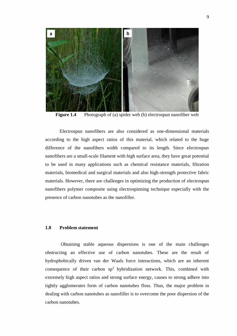
9
Figure 1.4 Photograph of (a) spider web (b) electrospun nanofiber web
Electrospun nanofibers are also considered as one-dimensional materials
according to the high aspect ratios of this material, which related to the huge
difference of the nanofibers width compared to its length. Since electrospun
nanofibers are a small-scale filament with high surface area, they have great potential
to be used in many applications such as chemical resistance materials, filtration
materials, biomedical and surgical materials and also high-strength protective fabric
materials. However, there are challenges in optimizing the production of electrospun
nanofibers polymer composite using electrospinning technique especially with the
presence of carbon nanotubes as the nanofiller.
1.8 Problem statement
Obtaining stable aqueous dispersions is one of the main challenges
obstructing an effective use of carbon nanotubes. These are the result of
hydrophobically driven van der Waals force interactions, which are an inherent
consequence of their carbon sp2 hybridization network. This, combined with
extremely high aspect ratios and strong surface energy, causes to strong adhere into
tightly agglomerates form of carbon nanotubes floss. Thus, the major problem in
dealing with carbon nanotubes as nanofiller is to overcome the poor dispersion of the
carbon nanotubes.

10
The dispersion technique via non-covalent interaction between surfactant and
carbon nanotubes surface has the advantage of preserving the conjugated π system of
carbon nanotubes, upholding their electrical and mechanical properties. Affecting
factor in the dispersion using surfactant is surfactant concentration, as there will
always be an optimum surfactant concentration for a specific loading of carbon
nanotubes. At very low surfactant concentration, the dispersion quality will be poor
because carbon nanotubes are still in a form of entangled bundles. While, at very
high surfactant concentration, dispersion quality becomes poor, as the surfactant are
starting to form micelles. Besides, the other factors such as the influence of
surfactant affinity towards the formation of surfactant-carbon nanotubes aggregates,
the size of carbon nanotubes aggregation and the preferred orientation of
accumulated surfactant molecules on the surface of carbon nanotubes also need to be
investigate.
Another importance of having well dispersed form of cabon nanotubes is to
avoid the problem of clogging during electrospinning. Clogging of the spinneret tip
through gelation of the spinning solution can be very disruptive to the spinning
process as it causes production losses. This issue is more apparent when higher
concentration spinning solution with filler addition is used, which is likely due to
higher viscosity, contributes to the clogging of the spinneret tip. The other problems
arises in electrospinning process are including the formation of beads, the
uncontrolled of nanofibers diameter size, the jet discontinuity and the instability of
nanofibers structure retention. As a results, both of mechanical and physical
properties of electrospun nanofibers composite would be poor. Besides, the effect of
polymer concentration, solvent used and amount of carbon nanotubes filler loading
towards the formation of the electrospun composite nanofibers has been few and
inconclusive. Hence, appropriate sets of electrospinning process parameters are
necessary to identifiey the significant factors in optimizing the fabrication of
electrospun composite nanofibers with the infusion of carbon nanotubes as
nanofiller.

11
1.9 Objectives of the research
The aim of this research is to determine the fundamental principle in the
electrospinning technology for the fabrication of nanofibrous materials of
electrospun nanofibers composite of poly (L-lactide)-co- ε-caprolactone) (PLCL)
infused with multiwalled carbon nanotubes (MWCNTs). The objectives that will
fulfill the aim of this research are listed as follows:
1. To synthesize the surface activated-MWCNTs using surfactant with
different affinity.
2. To investigate the surfactant behavior on the surface of the MWCNTs by
various characterization instruments.
3. To customize a lab-scale electrospinning system and optimization of the
operational parameters for the production of PLCL-MWCNTs nanofibers
composite.
4. To investigate the effect of solvent and amount of surfactant surface
activated-MWCNTs loading in the formation of PLCL-MWCNTs
nanofibers composite.
5. To study the physical and mechanical properties of the electrospun
PLCL-MWCNTs nanofibers composite using various characterization
instruments.
1.10 Scope of the research
The first scope of this research is modification of the MWCNTs hydrophobic
properties through MWCNTs surface activation process via non-covalent interaction
using surfactants. The purpose of the modification process is to have smaller size of
stabilized MWCNTs aggregates as nanofiller in the polymer composite. Three types
of surfactants with different ionic character, which are anionic, cationic and non-
ionic, were utilized for the surface modification of the MWCNTs. The conceptual
arguments on the surfactant structure and behavior of the hydrophobic tails, head

12
charged and its polarity effect towards the dispersion of carbon nanotubes were
studied in depth using various spectroscopic techniques and particle size analyzer.
The analysis studied was chemical characterization by Fourier Transform
Infrared (FTIR) spectroscopy and Raman spectroscopy. The thermal stability of
MWCNTs after surface activation was investigated using Thermal Gravimetric
Analysis (TGA). While, the morphology of MWCNTs upon surface modification
was analysed using X-ray Diffraction (XRD), and Field Emission Scanning Electron
Microscope (FESEM). Atomic Force Microscope (AFM) was used to examine the
MWCNTs aggregation size upon surface modification process, and then roughly
determine the smallest size of MWCNTs colloidal among the different surfactant
used in surface activation process.
Besides, this research is working on the customization of the lab-scale
electrospinning reactor system by assembling the basic component for a low-cost
electrospinning set-up. Since there are few arrangements in the electrospinning
reactor, the customized electrospinning system in this study is arranged in vertical
position especially to reduce the applied voltage by following the gravitational field.
The optimization of the operating parameters involves in the electrospinning
process was part of the scope in this research study. In this research, the preparation
of the spinning solution using various solvent mixtures was explored. The binary
solvent system comprised of DCM, DMF and acetones were used to investigate the
suitability of solvent for the electrospinning of PLCL. The control steps over the
electrospinning operating parameter by stabilizing the jet formation is also part of
this research study. The influence of the related operational parameters for the
electrospinning processing including the spinneret-to-collector distance, applied
current voltage and flow rate on the electrospun nanofiber structure are
alsoinvestigated in this research study. The use of surface activated MWCNTs as the
nanofiller in the electrospun nanofibers composite was investigated. The effect on
the different loading of MWCNTs on the physico-chemical and mechanical
properties of the MWCNTs-PLCL nanofibers composite was explored and for this

13
part, the smallest MWCNTs aggregates upon the surface activation is utilize in the
electospinning of the nanofibers composite.
Lastly, this research also is focused on the evaluation and characterization of
the electrospun nanofibers composite produced. Morphological study of PLCL
nanofibers and the infusion of MWCNTs within the MWCNTs-PLCL nanofibers
composite were analysed using the Transmission Electron Microscope (TEM),
Scanning Electron Microscope (SEM) and Atomic Force Microscope (AFM). The
mechanical testing based on tensile measurement of the nanofibers sample was
pursued to study the mechanical properties enhancement of the electrospun
composite nanofibers reinforced with MWCNTs.
1.11 Significance of the research
The significance of this research is the contribution in the optimization of
carbon nanotubes dispersion technique using various type of surfactants, as an
alternative to replace the conventional surface activation technique using oxidants.
The influence of surfactant affinity in altering the dispersibility of carbon nanotubes
has contributed an added value to the limited discussion in articles for established
publications. An in depth discussions on the chemistry of the surfactant-MWCNTs
interaction, cluster formation of MWCNTs aggregation and surfactant orientation on
MWCNTs surface were highlighted in this study. The knowledge from the
fundamental point of view has provided better insight to a better understanding
towards the surfactant assisted in the dispersion of carbon nanotubes.
Another significant finding of this research is the establishment in the
fabrication of nanofibrous materials of electrospun nanofibers composite of poly (L-
lactide)-co-ε-caprolactone) (PLCL) infused with multiwalled carbon nanotubes
(MWCNTs) using electrospinning technology. By the end of this research, an
optimized lab-scale electrospinning reactor system is fabricated for the production of
electrospun nanofibers purposes. A different structural form of PLCL-MWCNTs

14
electrospun nanofibers also can be spun by manipulating the optimized operational
parameters of the electrospinning technique, using the right formulation of the
specific nanofibers features needed. The difference in the solvent properties and their
dielectric constant values in the three different binary solvent mixtures have
contributed to the evolution of the PLCL nanofibers morphologies.

160
61
REFERENCES
Ajayan, P. M., Schadler, L. S., Giannaris, C. and Rubio, A. (2000). Single-walled
carbon nanotube–polymer composites: Strength and weakness. Advanced
materials. 12: 750–753.
Ajayan, P. M., Stephan, O., Colliex, C. and Trauth, D. (1994). Aligned carbon
nanotube arrays formed by cutting a polymer resin-nanotube composite.
Science. 265(5176): 1212-1214.
Ajayan, P. M., Terrones, M., De la Guardia, A., Huc, V., Grobert, N., Wei, B. Q. and
Ebbesen, T. W. (2002). Nanotubes in a flash-ignition and
reconstruction. Science. 296(5568): 705-705.
Allhoff, F., Lin, P. and Moore, D. (2009). What is nanotechnology and why does it
matter? from science to ethics. United Kingdom: John Wiley & Sons. 1st
Edition. 3-16.
Angelikopoulos, P. and Bock, H. (2012). The science of dispersing carbon nanotubes
with surfactants. Physical chemistry chemical physics. 14(27): 9546-9557.
Antonucci, V., Giordano, M., Nicolais, L., Calabro, A., Cusano, A., Cutolo, A. and
Inserra, S. (2003). Resin flow monitoring in resin film infusion process.
Journal of materials processing technology. 143: 687-692.
Baimark, Y. and Molloy, R. (2004). Synthesis and characterization of poly (L-
lactide-co-ε-caprolactone) copolymers: Effects of stannous octoate initiator
and diethylene glycol co-initiator concentrations. Science asia. 30: 327-334.
Banhart, F. (2015). Chains of carbon atoms: A vision or a new nanomaterial?
Beilstein journal of nanotechnology. 6(1): 559-569.
Barraza, H. J., Pompeo, F., O'Rea, E. A. and Resasco, D. E. (2002). SWNT-filled
thermoplastic and elastomeric composites prepared by miniemulsion
polymerization. Nano letters. 2(8): 797-802.
Baumgarten, P. K. (1971). Electrostatic spinning of acrylic microfibers. Journal of
colloid and interface science. 36(1): 71-79.

161
Bhadeshia, H. K. D. H. (2005). Bulk nanocrystalline steel. Ironmaking &
steelmaking. 32(5): 405-410.
Bhardwaj, N. and Kundu, S. C. (2010). Electrospinning: a fascinating fiber
fabrication technique. Biotechnology advances. 28(3): 325-347.
Blanch, A. J., Lenehan, C. E., and Quinton, J. S. (2010). Optimizing surfactant
concentrations for dispersion of single-walled carbon nanotubes in aqueous
solution. The journal of physical chemistry B. 114(30): 9805-9811.
Bokobza, L. (2007). Multiwall carbon nanotube elastomeric composites: A
review. Polymer. 48(17): 4907-4920.
Borowski, E., Soliman, E., Kandil, U. F. and Taha, M. R. (2015). Interlaminar
Fracture Toughness of CFRP Laminates Incorporating Multi-Walled Carbon
Nanotubes. Polymers, 7(6):1020-1045.
Buchko, C. J., Chen, L. C., Shen, Y. and Martin, D. C. (1999). Processing and
microstructural characterization of porous biocompatible protein polymer thin
films. Polymer. 40(26): 7397-7407.
Burlatsky, S. F., Atrazhev, V. V., Dmitriev, D. V., Sultanov, V. I., Timokhina, E. N.,
Ugolkova, E. A. and Vincitore, A. (2013). Surface tension model for
surfactant solutions at the critical micelle concentration. Journal of colloid
and interface science. 393: 151-160.
Cameron, D. J. A. and Shaver, M. P. (2011). Aliphatic polyester polymer stars:
synthesis, properties and applications in biomedicine and
nanotechnology. Chemical society reviews. 40(3): 1761-1776.
Celebioglu, A. and Uyar, T. (2012). Electrospinning of nanofibers from non-
polymeric systems: polymer-free nanofibers from cyclodextrin derivatives.
Nanoscale. 4(2): 621-631.
Chakoli, A. N., Cai, W., Jiehe, S. and Feng, J. T. (2009). Efficient load transfer to
functionalized carbon nanotubes as reinforcement in polymer
nanocomposites. International journal of modern physics B. 23(06n07):
1401-1406.
Chen, Q., Zhu, C. and Thouas, G. A. (2012). Progress and challenges in biomaterials
used for bone tissue engineering: bioactive glasses and elastomeric
composites. Progress in biomaterials. 1(1): 1-22.

162
Chen, Z. G., Mo, X. M. and Qing, F. L. (2007). Electrospinning of collagen-chitosan
complex. Materials letter. 61: 3490-3494.
Cheng, X., Zhong, J., Meng, J., Yang, M., Jia, F., Xu, Z. and Xu, H. (2011).
Characterization of multiwalled carbon nanotubes dispersing in water and
association with biological effects. Journal of nanomaterials. 2011: 14-25.
Choudhary, V. and Gupta, A. (2011). Polymer/carbon nanotube nanocomposites. In
Yellampalli, S. (Ed) Carbon Nanotubes-Polymer Nanocomposites. (pp 65-
90). Croatia: INTECH Open Access Publisher.
Coleman, B. D. (1986). Necking and drawing in polymeric fibers under tension. In
C. M. Dafermos, D. D. Joseph, and F. M. Leslie (Eds) The Breadth and
Depth of Continuum Mechanics. (pp 19-41). New York: Springer Berlin
Heidelberg.
Cox, H. L. (1952). The elasticity and strength of paper and other fibrous
materials. British journal of applied physics. 3(3): 72-79.
Dao, A. T. and Jirsak, O. (2010). Roller Electrospinning in Various Ambient
Parameters. Nanocon2010 conference. Czech Republic. EU2010.
Darani, K. K. and Reza Mozafari, M. (2010). Supercritical fluids technology in
bioprocess industries: a review. Journal of biochemical technology. 2(1):
144-152.
Datsyuk, V., Landois, P., Fitremann, J., Peigney, A., Galibert, A. M., Soula, B. and
Flahaut, E. (2009). Double-walled carbon nanotube dispersion via surfactant
substitution. Journal of materials chemistry. 19(18): 2729-2736.
Dayal, P., Liu, J., Kumar, S. and Kyu, T. (2007). Experimental and theoretical
investigations of porous structure formation in electrospun fibers.
Macromolecules. 40(21): 7689-7694.
De Aguiar, H. B., Strader, M. L., de Beer, A. G. and Roke, S. (2011). Surface
structure of sodium dodecyl sulfate surfactant and oil at the oil-in-water
droplet liquid/liquid interface: a manifestation of a nonequilibrium surface
state. The journal of physical chemistry B. 115(12): 2970-2978.
Della Pia, E. A., Macdonald, J. E., Elliott, M. and Jones, D. D. (2012). Direct
Binding of a Redox Protein for Single‐Molecule Electron Transfer
Measurements. Small. 8(15): 2341-2344.

163
Deng, W., Lobovsky, A., Iacono, S. T., Wu, T., Tomar, N., Budy, S. M. and Smith,
D. W. (2011). Poly (acrylonitrile–co-1-vinylimidazole): A new melt
processable carbon fiber precursor. Polymer. 52(3): 622-628.
Díaz, A., Katsarava, R. and Puiggalí, J. (2014). Synthesis, properties and applications
of biodegradable polymers derived from diols and dicarboxylic acids: From
polyesters to poly (ester amide)s. International journal of molecular
sciences. 15(5): 7064-7123.
Emmert, K. (2015). Determining the Critical Micelle Concentration of Polymer
Matrix for Drug Delivery Purposes. Honors Research Projects. University of
Akron.
Endo, M. (2010). Progress and perspectives in the carbon nanotube world,
AZoNano.com, Jun 23, 2010.
Endoh, K. and Suga, H. (1999). Phase diagram of salt–water system determined by
TG-DTA. Thermochimica acta. 327(1): 133-137.
Fendler, J. H. (1996). The colloidal domain: Where physics, chemistry, biology, and
technology meet. By D. Fennell Evans and Hakån Wennerström. New York:
VCH Publishers.
Fernández, J., Etxeberria, A. and Sarasua, J. R. (2012). Synthesis, structure and
properties of poly (L-lactide-co-ε-caprolactone) statistical copolymers.
Journal of the mechanical behavior of biomedical materials. 9: 100-112.
Fiorito, S. (2008). Carbon nanotubes: angels or demons?. 1st Edition. Singapore:
Pan Stanford Publishing. 1-13.
Fong, H. (2007). Electrospun polymer, ceramic, carbon/graphite nanofibers and their
application, in: H.S. Nalwa (Eds)., Polymeric nanostructures and their
applications. California: American Scientific Publisher. Stevenson Ranch.
451-474.
Fong, H., Chun, I. and Reneker, D. H. (1999). Beaded nanofibers formed during
electrospinning. Polymer. 40(16): 4585-4592.
Formhals, A. (1934). Process and apparatus for preparing artificial threads. U.S
Patent No. 1975704
Formhals, A. (1939). Method and apparatus for spinning. US patent 2160962.
Frazer, L. (2004). New spin on an old fiber. Environmental health perspectives.
112(13): A754- 757.

164
Friedrich, K., Fakirov, S. and Zhang, Z. (Eds). (2005) Polymer Composites: From
Nano- to Macro-Scale, New York: Springer. 1-7
Fujigaya, T. and Nakashima, N. (2015). Non-covalent polymer wrapping of carbon
nanotubes and the role of wrapped polymers as functional dispersants.
Science and technology of advanced materials. 16(2): 024802-024821.
Gao, H., Kong, Y., Cui, D. and Ozkan, C. S. (2003). Spontaneous insertion of DNA
oligonucleotides into carbon nanotubes. Nano letters. 3(4): 471-473.
Garkhal, K., Verma, S., Jonnalagadda, S. and Kumar, N. (2007). Fast degradable
poly (L‐lactide‐co‐ε‐caprolactone) microspheres for tissue engineering:
Synthesis, characterization, and degradation behavior. Journal of polymer
science part A: Polymer chemistry. 45(13): 2755-2764.
Geng, Y., Liu, M. Y., Li, J., Shi, X. M. and Kim, J. K. (2008). Effects of surfactant
treatment on mechanical and electrical properties of CNT/epoxy
nanocomposites. Composites part A: Applied science and manufacturing.
39(12): 1876-1883.
Gite, B. E. and Margaj, M. S. R. (2013). Carbon Fibre As A Recent Material Use In
Construction. Amrutvahini college of engineering, sangamner: 1-6.
Greenfeld, I. and Zussman, E. (2013). Polymer entanglement loss in extensional
flow: Evidence from electrospun short nanofibers. Journal of polymer science
Part B: Polymer physics. 51(18): 1377-1391.
Gupta, P. and Wilkes, G. L. (2003). Some investigations on the fiber formation by
utilizing a side-by-side bicomponent electrospinning approach. Polymer.
44(20): 6353-6359.
Haggenmueller, R., Gommans, H. H., Rinzler, A. G., Fischer, J. E. and Winey, K. I.
(2000). Aligned single-wall carbon nanotubes in composites by melt
processing methods. Chemical physics letters. 330(3): 219-225.
Haggenmueller, R., Zhou, W., Fischer, J. E. and Winey, K. I. (2003). Production and
characterization of polymer nanocomposites with highly aligned single-
walled carbon nanotubes. Journal of nanoscience and nanotechnology. 3(1-
2): 105-110.
Harris, P. J. F. (2001). Carbon nanotubes and related structures: new materials for the
twenty-first century. United Kingdom: Cambridge university press.
Heinrich, G., Klüppel, M. and Vilgis, T. A. (2002). Reinforcement of elastomers.
Current opinion in solid state and materials science. 6(3): 195-203.

165
Hiemenz, P. C. and R. Rajagopalan. (1997). Principles of colloid and surface
chemistry. New York: Marcel Dekker. 3rd Edition. 25-45.
Hohman, M. M., Shin, M., Rutledge, G. and Brenner, M. P. (2001). Electrospinning
and electrically forced jets. I. Stability theory. Physics of fluids. 13(8): 2201-
2220.
Holmberg, K., Joènsson, B., Kronberg, B. and Lindman, B. (2002). Surfactant and
Polymers In Aqueous Solution. West Sussex: John Wiley and Sons. 108-109.
Hou, H and Ge, J. J. (2005). Electrospun polyacrylonitrile nanofibers containing a
high concentration of well-aligned multiwalled carbon nanotubes. Chemistry
of materials. 17(5): 967-973.
Hsu, C. M. and Shivkumar, S. (2004). N, N‐Dimethylformamide Additions to the
Solution for the Electrospinning of Poly (ε‐caprolactone) Nanofibers.
Macromolecular materials and engineering. 289(4): 334-340.
Hu, C. and Hu, S. (2009). Carbon nanotube-based electrochemical sensors: principles
and applications in biomedical systems. Journal of sensors. 9: 1-40.
Hu, C. Y., Xu, Y. J., Duo, S. W., Zhang, R. F. and Li, M. S. (2009). Non‐Covalent
Functionalization of Carbon Nanotubes with Surfactants and
Polymers. Journal of the chinese chemical society. 56(2): 234-239.
Huang, W., Zou, T., Li, S., Jing, J., Xia, X. and Liu, X. (2013). Drug-loaded zein
nanofibers prepared using a modified coaxial electrospinning
process. Journal of the american association of pharmaceutical scientists.
14(2): 675-681.
Iijima, S. (1991). Helical microtubules of graphitic carbon. Nature, 354(6348): 56-
58.
Inai, R., Kotaki, M. and Ramakrishna, S. (2005). Deformation behavior of
electrospun poly (L‐lactide‐co‐ε‐caprolactone) nonwoven membranes under
uniaxial tensile loading. Journal of polymer science part B: polymer physics.
43(22): 3205-3212.
Jarusuwannapoom, T., Hongrojjanawiwat, W., Jitjaicham, S., Wannatong, L.,
Nithitanakul, M., Pattamaprom, C. and Supaphol, P. (2005). Effect of
solvents on electro-spinnability of polystyrene solutions and morphological
appearance of resulting electrospun polystyrene fibers. European polymer
journal. 41(3): 409-421.

166
Jeong, S. I., Kim, B. S., Kang, S. W., Kwon, J. H., Lee, Y. M., Kim, S. H. and Kim,
Y. H. (2004). In vivo biocompatibilty and degradation behavior of elastic
poly(l-lactide-co-ε-caprolactone) scaffolds. Biomaterials. 25(28): 5939-5946.
Jin, Y., Yang, D., Kang, D. and Jiang, X. (2009). Fabrication of necklace-like
structures via electrospinning. Langmuir. 26(2): 1186-1190.
Jung, W. R., Choi, J. H., Lee, N., Shin, K., Moon, J. H. and Seo, Y. S. (2012).
Reduced damage to carbon nanotubes during ultrasound-assisted dispersion
as a result of supercritical-fluid treatment. Carbon. 50: 633-636.
Kelly, A. and Tyson, A. W. (1965). Tensile properties of fibre-reinforced metals:
copper/tungsten and copper/molybdenum. Journal of the mechanics and
physics of solids. 13(6): 329-350.
Kerwin, B. A. (2008). Polysorbates 20 and 80 used in the formulation of protein
biotherapeutics: structure and degradation pathways. Journal of
pharmaceutical sciences. 97(8): 2924-2935.
Kessick, R., Fenn, J. and Tepper, G. (2004). The use of AC potentials in
electrospraying and electrospinning processes. Polymer. 45(9): 2981-2984.
Keulder, L. (2013). The preparation of polyolefin nanofibres by solution
electrospinning. Doctoral dissertation, Stellenbosch: Stellenbosch University.
Khare, R. and Bose, S. (2005). Carbon nanotube based composites-a review. Journal
of minerals & materials characterization & engineering. 4(1): 31-46.
Kieronski, E. A., Knock, K. K., Fallon, W. P., Walker, G. M. (2004). Mechanism of
adhesive bonding of fibreglass composites with peel ply surface preparation,
ASTM STP 1455: 78-91.
Kizildag, N., Beceren, Y., Kazanci, M. and Cukul, D. (2012). Effect of needle
diameter on diameter of electropsun silk fibroin nanofibers. In RMUTP.
Proceedings of the International Conference: Textiles & Fashion: 3-4.
Konya, Z., Vesselenyi, I., Niesz, K., Kukovecz, A., Demortier, A., Fonseca, A. and
Kiricsi, I. (2002). Large scale production of short functionalized carbon
nanotubes. Chemical physics letters. 360(5): 429-435.
Kulshrestha, A. S. and Mahapatro, A. (2008). Polymers for Biomedical Applications.
ACS symposium series. 977: 1–7.
167
Lau, K. K., Bico, J., Teo, K. B., Chhowalla, M., Amaratunga, G. A., Milne, W. I.,
and Gleason, K. K. (2003). Superhydrophobic carbon nanotube forests. Nano
letters. 3(12): 1701-1705.
Leach, M. K., Feng, Z. Q., Tuck, S. J., and Corey, J. M. (2011). Electrospinning
fundamentals: optimizing solution and apparatus parameters. J vis exp. 47
(e2494): 1-4.
Lee, K. H., Kim, H. Y., Khil, M. S., Ra, Y. M. and Lee, D. R. (2003).
Characterization of nano-structured poly (ε-caprolactone) nonwoven mats via
electrospinning. Polymer. 44(4): 1287-1294.
Li, X., Thompson, J. D., Zhang, Y., Brady, C. I., Zou, G., Mack, N. H., Williams, D.,
Duque, J. G., Jia, Q. and Doorn, S. K. (2011). Efficient synthesis of tailored
magnetic carbon nanotubes via a noncovalent chemical route. Nanoscale.
2011(3): 668–673.
Li, Y. and Ishida, H. (2002). A differential scanning calorimetry study of the
assembly of hexadecylamine molecules in the nanoscale confined space of
silicate galleries. Chemistry of materials. 14(3): 1398-1404.
Li, Z. Q., Lu, C. J., Xia, Z. P., Zhou, Y. and Luo, Z. (2007). X-ray diffraction
patterns of graphite and turbostratic carbon. Carbon. 45(8): 1686-1695.
Lim, J. Y., Liu, X., Vogler, E. A. and Donahue, H. J. (2004). Systematic variation in
osteoblast adhesion and phenotype with substratum surface characteristics.
Journal of biomedical materials research part A. 68(3): 504-512.
Liu, C. X. and Choi, J. W. (2012). Improved dispersion of carbon nanotubes in
polymers at high concentrations. Nanomaterials. 2(4): 329-347.
Liu, M., Yang, Y., Zhu, T. and Liu, Z. (2007). A general approach to chemical
modification of single-walled carbon nanotubes with peroxy organic acids
and its application in polymer grafting. The journal of physical chemistry C.
111(6): 2379-2385.
Liu, W., Huang, C. and Jin, X. (2014). Tailoring the grooved texture of electrospun
polystyrene nanofibers by controlling the solvent system and relative
humidity. Nanoscale research letters. 9(1): 1-10.
Louchev, O. A., Sato, Y. and Kanda, H. (2001). Multiwall carbon nanotubes: Self-
organization and inhibition of step-flow growth kinetics. Journal of applied
physics. 89(6): 3438-3446.

168
Luong-Van, E., Grøndahl, L., Chua, K. N., Leong, K. W., Nurcombe, V. and Cool,
S. M. (2006). Controlled release of heparin from poly (ε-caprolactone)
electrospun fibers. Biomaterials. 27(9): 2042-2050.
Ma, J. C. and Dougherty, D. A. (1997). The cation-π interaction. Chemical
reviews. 97(5): 1303-1324.
Ma, P. C., Siddiqui, N. A., Marom, G. and Kim, J. K. (2010). Dispersion and
functionalization of carbon nanotubes for polymer-based nanocomposites: a
review. Composites part A: applied science and manufacturing. 41(10):
1345-1367.
MacDiarmid, A. G. (2001). Synthetic metals: a novel role for organic polymers.
Synthetic metals. 125(1): 11-22.
Makadia, H. K. and Siegel, S. J. (2011). Poly lactic-co-glycolic acid (PLGA) as
biodegradable controlled drug delivery carrier. Polymers. 3(3): 1377-1397.
Malhotra, S. K., Goda, K. and Sreekala, M. S. (2012). Part one Introduction to
polymer composites. Polymer composite Volume 1, Macro and micro-
nanocomposites. New Jersey: John Wileys & Sons Inc.
Mamunya, Y. (2011). Carbon Nanotubes as Conductive Filler in Segregated Polymer
Composites-Electrical Properties. INTECH Open Access Publisher.
Crotia:Rijeka
Marquis, D. M., Chivas-Joly, C. and Guillaume, É. (2011). Properties of nanofillers
in polymer. In Dr. John Cuppoletti (Ed.) Nanocomposites and Polymers with
Analytical Methods (pp 261-284). Crotia: INTECH Open Access Publisher.
Martínez-Hernández, A. L., Velasco-Santos, C. and Castano, V. (2010). Carbon
nanotubes composites: processing, grafting and mechanical and thermal
properties. Current nanoscience. 6(1): 12-39.
Matarredona, O., Rhoads, H., Li, Z., Harwell, J. H., Balzano, L. and Resasco, D. E.
(2003). Dispersion of single-walled carbon nanotubes in aqueous solutions of
the anionic surfactant NaDDBS. The journal of physical chemistry
B. 107(48): 13357-13367.
Matsumoto, H. and Tanioka, A. (2011). Functionality in electrospun nanofibrous
membranes based on fiber’s size, surface area, and molecular orientation.
Membranes. 1(3): 249-264.

169
Maurin, G., Stepanek, I., Bernier, P., Colomer, J. F., Nagy, J. B. and Henn, F. (2001).
Segmented and opened multi-walled carbon nanotubes. Carbon. 39(8): 1273-
1278.
Megelski, S., Stephens, J. S., Chase, D. B. and Rabolt, J. F. (2002). Micro-and
nanostructured surface morphology on electrospun polymer fibers.
Macromolecules. 35(22): 8456-8466.
Meng, Z. X., Zheng, W., Li, L. and Zheng, Y. F. (2010). Fabrication and
characterization of three-dimensional nanofiber membrance of PCL-
MWCNTs by electrospinning. Materials science and engineering C. 30:
1014-1021.
Mirjalili, V., Ramachandramoorthy, R. and Hubert, P. (2014). Enhancement of
fracture toughness of carbon fiber laminated composites using multi wall
carbon nanotubes. Carbon. 79: 413-423.
Moniruzzaman, M. and Winey, K. I. (2006). Polymer nanocomposites containing
carbon nanotubes. Macromolecules. 39(16): 5194-5205.
Moore, V. C., Strano, M. S., Haroz, E. H., Hauge, R. H., Smalley, R. E., Schmidt, J.
and Talmon, Y. (2003). Individually suspended single-walled carbon
nanotubes in various surfactants. Nano letters. 3(10): 1379-1382.
Muherei, M. A. and Junin, R. (2007). Effect of electrolyte on synergism of anionic-
nonionic surfactant mixture. Journal of applied sciences. 7: 1362-1371.
Mylvaganam, K. and Zhang, L. C. (2007). Ballistic resistance capacity of carbon
nanotubes. Nanotechnology. 18(47): 475701-475704.
Nadler, M., Mahrholz, T., Riedel, U., Schilde, C. and Kwade, A. (2008). Preparation
of colloidal carbon nanotube dispersions and their characterisation using a
disc centrifuge. Carbon. 46(11): 1384-1392.
Natarajan, L., New, J., Dasari, A., Yu, S. and Manan, M. A. (2014). Surface
morphology of electrospun PLA fibers: mechanisms of pore formation. RSC
advances. 4(83): 44082-44088.
Njuguna, J., Vanli, O. A. and Liang, R. (2015). A Review of Spectral Methods for
Dispersion Characterization of Carbon Nanotubes in Aqueous Suspensions.
Journal of spectroscopy. 2015: 1-11
Pawlowski, K. J., Belvin, H. L., Raney, D. L., Su, J., Harrison, J. S., and Siochi, E. J.
(2003). Electrospinning of a micro-air vehicle wing skin. Polymer. 44: 1309-
1314.

170
Pichot, R., Watson, R. L. and Norton, I. T. (2013). Phospholipids at the interface:
current trends and challenges. International journal of molecular sciences.
14(6): 11767-11794.
Prabhakaran, M. P., Venugopal, J. R., Chyan, T. T., Hai, L. B., Chan, C. K., Lim, A.
Y. and Ramakrishna, S. (2008). Electrospun biocomposite nanofibrous
scaffolds for neural tissue engineering. Tissue engineering Part A. 14(11):
1787-1797.
Qi, Y. Y., Tai, Z. X., Sun, D. F., Chen, J. T., Ma, H. B., Yan, X. B., Liu, B. and Xue,
Q. J. (2013). Fabrication and characterization of poly (vinyl alcohol)/
graphene oxide nanofibrous biocomposite scaffolds. Journal of applied
polymer science. 127: 1885-1894.
Qian, D., Dickey, E. C., Andrews, R. and Rantell, T. (2000). Load transfer and
deformation mechanisms in carbon nanotube-polystyrene composites.
Applied physic letter. 76: 2868-2870.
Qin, X. H. and Wang, S. Y. (2008). Electrospun nanofibers from crosslinked poly
(vinyl alcohol) and its filtration efficiency. Journal of applied polymer
science. 109(2): 951-956.
Ragab, T. and Basaran, C. (2009). Joule heating in single-walled carbon
nanotubes. Journal of applied physics. 106(6): 063705-1 -063705-5.
Ramakrishna, S. and Fujihara, K (2005). An introduction to electrospinning and
nanofibers. Singapore: World Scientific Publishing. 135-137
Rana, M. and Chandra, A. (2007). Filled and empty states of carbon nanotubes in
water: Dependence on nanotube diameter, wall thickness and dispersion
interactions. Journal of chemical sciences. 119(5): 367-376.
Rastogi, R., Kaushal, R., Tripathi, S. K., Sharma, A. L., Kaur, I. and Bharadwaj, L.
M. (2008). Comparative study of carbon nanotube dispersion using
surfactants. Journal of colloid and interface science. 328(2): 421-428.
Reichardt, C. (1990) Solvents and Solvent Effects in Organic Chemistry. In Thomas
Welton (Ed). (pp 534).VCH: Weinheim (D) John Wiley & Sons.
Reneker, D. H., Kataphinan, W., Theron, A., Zussman, E. and Yarin, A. L. (2002).
Nanofiber garlands of polycaprolactone by electrospinning. Polymer. 43(25):
6785-6794.
Rentsch, B., Bernhardt, R., Scharnweber, D., Schneiders, W., Rammelt, S. and
Rentsch, C. (2012). Embroidered and surface coated polycaprolactone-co-

171
lactide scaffolds: a potential graft for bone tissue
engineering. Biomatter. 2(3): 158-165.
Rentsch, C., Rentsch, B., Breier, A., Spekl, K., Jung, R., Manthey, S. and Biewener,
A. (2010). Long‐bone critical‐size defects treated with tissue‐engineered
polycaprolactone‐co‐lactide scaffolds: A pilot study on rats. Journal of
biomedical materials research Part A. 95(3): 964-972.
Reznik, D., Olk, C. H., Neumann, D. A. and Copley, J. R. D. (1995). X-ray powder
diffraction from carbon nanotubes and nanoparticles. Physical review B.
52(1): 116-124.
Robertson, J. (2007). Growth of nanotubes for electronics. Materials today. 10(1):
36-43.
Ryan, K. P., Cadek, M., Nicolosi, V., Blond, D., Ruether, M., and Amstrong, G.
(2007). Carbon nanotubes for reinforcements of plastic? A case study with
PVA. Composite science technology. 67: 1640-1649.
S. Goyanes, G. R. Rubiolo, A. Salazar, A. Jimeno, M. A. Corcuera and I.
Mondragon. (2007). Carboxylation treatment of multiwalled carbon
nanotubes monitored by infrared and ultraviolet spectroscopies and scanning
probe microscopy. Diamond related materials. 16(2): 412-417.
Saba, N., Tahir, P. M. and Jawaid, M. (2014). A review on potentiality of nano
filler/natural fiber filled polymer hybrid composites. Polymers. 6(8): 2247-
2273.
Saeed, K. and Park, S Y. (2007). Preparation and properties of multiwalled carbon
nanotubes/polycaprolactone nanocomposites. Journal of applied polymer
science. 104: 1957-1963.
Sahay, R., Thavasi, V. and Ramakrishna, S. (2011). Design modifications in
electrospinning setup for advanced applications. Journal of Nanomaterials,
2011: 1-17.
Sanna, V., Roggio, A. M., Posadino, A. M., Cossu, A., Marceddu, S., Mariani, A.
and Sechi, M. (2011). Novel docetaxel-loaded nanoparticles based on poly
(lactide-co-caprolactone) and poly (lactide-co-glycolide-co-caprolactone) for
prostate cancer treatment: formulation, characterization, and cytotoxicity
studies. Nanoscale research letters. 6(1): 1-9.
Saville, D. A. (1997). Electrohydrodynamics: the Taylor-Melcher leaky dielectric
model. Annual review of fluid mechanics. 29(1): 27-64.
172
Scarlet, R., Manea, L. R., Sandu, I., Martinova, L., Cramariuc, O. and Sandu, I. G.
(2012). Study on the solubility of polyetherimide for nanostructural
electrospinning. Revista de chimie (Bucharest). 63: 688-692.
Sengupta, J., Jana, A., Singh, N. P., Mitra, C. and Jacob, C. (2010). Site-selective
synthesis of in situ Ni-filled multi-walled carbon nanotubes using Ni (salen)
as a catalyst source. Nanotechnology. 21(41): 415605-415610.
Shi, Y., Ren, L., Li, D., Gao, H. and Yang, B. (2013). Optimization conditions for
single-walled carbon nanotubes dispersion. Journal of surface engineered
materials and advanced technology. 3(1): 6-12.
Shrivastava, N. K., Kar, P., Maiti, S. and Khatua, B. B. (2012). A facile route to
develop electrical conductivity with minimum possible multi‐wall carbon
nanotube (MWCNT) loading in poly (methyl
methacrylate)/MWCNTnanocomposites. Polymer international. 61(11):
1683-1692.
Son, W. K, Youk, J. H., Lee, T. S and Park, W. H. (2004). Electrospinning of
ultrafine cellulose acetate fibers: studies of a new solvent system and
deacetylation of ultrafine cellulose acetate fibers. Journal of polymer science;
part B: Polymer physic. 42(1): 5-11.
Spitalsky, Z., Tasis, D., Papagelis, K. and Galiotis, C. (2010). Carbon nanotube-
polymer composite: chemistry, processing, mechanical and electrical
properties. Progress in polymer science. 35: 357-401.
Strano, M. S., Moore, V. C., Miller, M. K., Allen, M. J., Haroz, E. H., Kittrell, C. and
Smalley, R. E. (2003). The role of surfactant adsorption during
ultrasonication in the dispersion of single-walled carbon nanotubes. Journal
of Nanoscience and Nanotechnology. 3(1-2): 81-86.
Sultana, N. (2012). Biodegradable Polymer-Based Scaffolds for Bone Tissue
Engineering. Springer Science & Business Media. Surfactant science and
technology, 3rd ed. Myers, Drew. Wiley-Interscience
Szeteiová, K. (2010). Automotive materials: plastics in automotive markets
today. Institute of Production Technologies, Machine Technologies and
Materials, Faculty of Material Science and Technology in Trinova, Slovak
University of Technology Bratislava.

173
Szleifer, I. and Yerushalmi-Rozen, R. (2005). Polymers and carbon nanotubes-
dimensionality, interactions and nanotechnology. Polymer. 46(19): 7803-
7818.
Tanvir, S. and Qiao, L. (2012). Surface tension of nanofluid-type fuels containing
suspended nanomaterials. Nanoscale research letters. 7(1): 1-10.
Taylor, G. (1964). Disintegration of water drops in an electric field. In Proceedings
of the Royal Society of London A: Mathematical, Physical and Engineering
Sciences. The Royal Society. 280 (1382): 383-397.
Tedeschi, A. M., D'Errico, G., Busi, E., Basosi, R. and Barone, V. (2002). Micellar
aggregation of sulfonate surfactants studied by electron paramagnetic
resonance of a cationic nitroxide: an experimental and computational
approach. Physical chemistry chemical physics. 4(11): 2180-2188.
Tetteh, G., Khan, A. S., Delaine-Smith, R. M., Reilly, G. C. and Rehman, I. U.
(2014). Electrospun polyurethane/hydroxyapatite bioactive Scaffolds for bone
tissue engineering: The role of solvent and hydroxyapatite particles. Journal
of the mechanical behavior of biomedical materials. 39: 95-110.
Thostenson, E. T., Ren, Z. and Chou, T. W. (2001). Advances in the science and
technology of carbon nanotubes and their composites: a review. Composites
science and technology. 61(13): 1899-1912.
Troy, B. D. and Beringer, P. (2006). Remington: The science and practice of
pharmacy. Philadelphia: Lippincott Williams &Wilkins Publisher, 21st
Edition. 220-225
Tummala, N. R. (2009). SDS surfactants on carbon nanotubes: aggregate
morphology. Acs nano. 3(3): 595-602.
Ulery, B. D., Nair, L. S. and Laurencin, C. T. (2011). Biomedical applications of
biodegradable polymers. Journal of polymer science part B: polymer physics.
49(12): 832-864.
Uyar, T. and Besenbacher, F. (2008). Electrospinning of uniform polystyrene fibers:
The effect of solvent conductivity. Polymer. 49(24): 5336-5343.
Vaisman, L., Marom, G. and Wagner, H. D. (2006). Dispersions of Surface‐Modified
Carbon Nanotubes in Water‐Soluble and Water‐Insoluble Polymers.
Advanced functional materials. 16(3): 357-363.

174
Vaisman, L., Wagner, H. D. and Marom, G. (2006). The role of surfactants in
dispersion of carbon nanotubes. Advances in colloid and interface science.
128: 37-46.
Vassalli, J. T. (2008). Development of electrospun synthetic bioabsorbable fibers for
a novel bionanocomposite hernia repair material (Doctoral dissertation,
University of Missouri--Columbia).
Vigolo, B., Coulon, C., Maugey, M., Zakri, C. and Poulin, P. (2005). An
experimental approach to the percolation of sticky nanotubes. Science.
309(5736): 920-923.
Viguet-Carrin, S., Roux, J. P., Arlot, M. E., Merabet, Z., Leeming, D. J., Byrjalsen, I.
and Bouxsein, M. L. (2006). Contribution of the advanced glycation end
product pentosidine and of maturation of type I collagen to compressive
biomechanical properties of human lumbar vertebrae. Bone. 39(5): 1073-
1079.
Vroman, I. and Tighzert, L. (2009). Biodegradable polymers. Materials. 2(2): 307-
344.
Wachsmuth, J. P. (2008). Multiple Independent Extrusion Heads for Fused
Deposition Modeling (Doctoral dissertation, Virginia Polytechnic Institute
and State University).
Wang, S., Tang, L. A. L., Bao, Q., Lin, M., Deng, S., Goh, B. M. and Loh, K. P.
(2009). Room-temperature synthesis of soluble carbon nanotubes by the
sonication of graphene oxide nanosheets. Journal of the american chemical
society. 131(46): 16832-16837.
Wang, X., Ding, B., Sun, G., Wang, M. and Yu, J. (2013). Electro-spinning/netting: a
strategy for the fabrication of three-dimensional polymer nano-
fiber/nets. Progress in materials science. 58(8): 1173-1243.
Wang, X., Zhuang, J., Peng, Q. and Li, Y. D. (2005) A general strategy for
nanocrystal synthesis. Nature. 437: 121–124.
Wang, Y., Wu, J. and Wei, F. (2003). A treatment method to give separated multi-
walled carbon nanotubes with high purity, high crystallization and a large
aspect ratio. Carbon. 41(15): 2939-2948.
Wannatong, L., Sirivat, A. and Supaphol, P. (2004). Effects of solvents on
electrospun polymeric fibers: preliminary study on polystyrene. Polymer
international. 53(11): 1851-1859.

175
Wei, Z., Zhang, Q., Wang, L., Peng, M., Wang, X. and Yang, J. (2012). The
preparation and adsorption properties of electrospun aramid
nanofibers. Journal of polymer science part B: Polymer physics. 50(20):
1414-1420.
Weldon, C. B., Tsui, J. H., Shankarappa, S. A., Nguyen, V. T., Ma, M., Anderson, D.
G. and Kohane, D. S. (2012). Electrospun drug-eluting sutures for local
anesthesia. Journal of controlled release. 161(3): 903-909.
Xie, W., Gao, Z., Pan, W-P., Hunter, D., Singh, A. and Vaia, R. (2001). Thermal
degradation chemistry of alkyl quaternary ammonium montmorillonite.
Chemistry of material. 13: 2979–2990.
Xu, Y., Pehrsson, P. E., Chen, L., Zhang, R. and Zhao, W. (2007). Double-stranded
DNA single-walled carbon nanotube hybrids for optical hydrogen peroxide
and glucose sensing. The journal of physical chemistry C. 111(24): 8638-
8643.
Xu, Y., Wu, J., Wang, H., Li, H., Di, N., Song, L., Li, S., Li, D., Xiang, Y., Liu, W.,
Mo, X. M and Q. Zhou. (2013). Fabrication of electrospun poly (L-lactide-
co-ɛ-caprolactone)/ collagen nanoyarn netwok as a novel, three-dimensional,
macroporous, aligned scaffold for tendon tissue engineering, Tissue
engineering: Part C. 19(12): 925-936.
Yakobson, B. I. and Avouris, P. (2001). Mechanical properties of carbon nanotubes.
In Carbon nanotubes. Springer Berlin Heidelberg. 287-327.
Yakobson, B. I., Brabec, C. J. and Bernholc, J. (1996). Nanomechanics of carbon
tubes: instabilities beyond linear response. Physical review letters, 76(14):
2511.
Yarin, A. L., Kataphinan, W. and Reneker, D. H. (2005). Branching in
electrospinning of nanofibers. Journal of applied physics. 98(6): 064501-1-
064501-14.
Yu, J., Grossiord, N., Koning, C. E. and Loos, J. (2007). Controlling the dispersion
of multi-wall carbon nanotubes in aqueous surfactant solution. Carbon. 45(3):
618-623.
Yuan, X., Zhang, Y., Dong, C. and Sheng, J. (2004). Morphology of ultrafine
polysulfone fibers prepared by electrospinning. Polymer international.
53(11):1704–1710.

176
Zeleny, J. (1914). The electrical; discharge from liquid points and a hydrostatic
method of measuring the electric intensity at their surface. Physical Review.
3(2): 69-91.
Zeleny, J. (1917). Instability of electrified liquid surfaces. Physical Review. 10 (1): 1-
6.
Zhang, C. and Liu, T. (2012). A review on hybridization modification of graphene
and its polymer nanocomposites. Chinese science bulletin. 57(23): 3010-
3021.
Zhang, Z. and Li, T. (2010). Carbon nanotube initiated formation of carbon
nanoscrolls. Applied physics letters. 97(8): 081909-081912.
Zhao, D., Lei, Q., Qin, C. and Bai, X. (2006). Melt process and performance of
multi-walled carbon nanotubes reinforced LDPE composites. Pigment &
resin technology. 35(6): 341-345.
Zong, X., Kim, K., Fang, D., Ran, S., Hsiao, B. S. and Chu, B. (2002). Structure and
process relationship of electrospun bioabsorbable nanofiber membranes.
Polymer. 43(16): 4403–4412.
Zuo, Y. Y., Veldhuizen, R. A., Neumann, A. W., Petersen, N. O. and Possmayer, F.
(2008). Current perspectives in pulmonary surfactant-inhibition, enhancement
and evaluation. Biochimica et Biophysica Acta (BBA)-
Biomembranes, 1778(10): 1947-1977.
Zussman, E., Rittel, D. and Yarin, A. L. (2003). Failure modes of electrospun
nanofibers. Applied physics letters. 82(22): 3958-3960.