hypertension 2012-van der lubbe-741-8
TRANSCRIPT
Meiracker, A.H. Jan Danser, Robert Zietse and Ewout J. HoornNils van der Lubbe, Pieter M. Jansen, Mahdi Salih, Robert A. Fenton, Anton H. van den
Prostasin as a Marker for AldosteronismThe Phosphorylated Sodium Chloride Cotransporter in Urinary Exosomes Is Superior to
Print ISSN: 0194-911X. Online ISSN: 1524-4563 Copyright © 2012 American Heart Association, Inc. All rights reserved.
is published by the American Heart Association, 7272 Greenville Avenue, Dallas, TX 75231Hypertension doi: 10.1161/HYPERTENSIONAHA.112.198135
2012;60:741-748; originally published online July 30, 2012;Hypertension.
http://hyper.ahajournals.org/content/60/3/741World Wide Web at:
The online version of this article, along with updated information and services, is located on the
http://hyper.ahajournals.org/content/suppl/2012/07/30/HYPERTENSIONAHA.112.198135.DC1.htmlData Supplement (unedited) at:
http://hyper.ahajournals.org//subscriptions/
is online at: Hypertension Information about subscribing to Subscriptions:
http://www.lww.com/reprints Information about reprints can be found online at: Reprints:
document. Permissions and Rights Question and Answer this process is available in the
click Request Permissions in the middle column of the Web page under Services. Further information aboutOffice. Once the online version of the published article for which permission is being requested is located,
can be obtained via RightsLink, a service of the Copyright Clearance Center, not the EditorialHypertensionin Requests for permissions to reproduce figures, tables, or portions of articles originally publishedPermissions:
by guest on August 15, 2013http://hyper.ahajournals.org/Downloaded from
The Phosphorylated Sodium Chloride Cotransporter inUrinary Exosomes Is Superior to Prostasin as a Marker
for AldosteronismNils van der Lubbe, Pieter M. Jansen, Mahdi Salih, Robert A. Fenton, Anton H. van den Meiracker,
A.H. Jan Danser, Robert Zietse, Ewout J. Hoorn
Abstract—Urinary exosomes are vesicles derived from renal tubular epithelial cells. Exosomes often contain severaldisease-associated proteins and are thus useful targets for identifying biomarkers of disease. Here, we hypothesized thatthe phosphorylated (active) form of the sodium chloride cotransporter (pNCC) or prostasin could serve as biomarkersfor aldosteronism. We tested this in 2 animal models of aldosteronism (aldosterone infusion or low-sodium diet) and inpatients with primary aldosteronism. Urinary exosomes were isolated from 24-hour urine or spot urine usingultracentrifugation. In rats, a normal or a high dose of aldosterone for 2, 3, or 8 days increased pNCC 3-fold in urinaryexosomes (P�0.05 for all). A low-sodium diet also increased pNCC in urinary exosomes approximately 1.5-fold after4 and after 8 days of treatment. The effects of these maneuvers on prostasin in urinary exosomes were less clear,showing a significant 1.5-fold increase only after 2 and 3 days of high-aldosterone infusion. In urinary exosomes ofpatients with primary aldosteronism, pNCC was 2.6-fold higher (P�0.05) while prostasin was 1.5-fold higher (P�0.07)than in patients with essential hypertension. Urinary exosomal pNCC and, to a lesser extent, prostasin are promisingmarkers for aldosteronism in experimental animals and patients. These markers may be used to assess the biologicalactivity of aldosterone and, potentially, as clinical biomarkers for primary aldosteronism. (Hypertension. 2012;60:741-748.) ● Online Data Supplement
Key Words: aldosterone � epithelial sodium channel � hypertension � primary aldosteronism
Urinary proteins originate from various sources. Theymay be derived from glomerular filtration, tubular
secretion, shedding, glycosylphophatidylinositol anchoredprotein detachment (eg, Tamm-Horsfall protein), or exosomesecretion.1 Exosomes are low-density membrane vesicles thatoriginate from multivesicular bodies. Urinary exosomes havesparked interest as potential biomarkers for human disease.1–3
The presence of urinary exosomes and a reproducible methodfor their isolation were reported in 2004 by Pisitkun andcolleagues.4 Proteomic analysis of these exosomes showedthat they contain many disease-related proteins4,5; however,the question remained whether the presence of a given proteinin urinary exosomes could provide information on physiolog-ical or disease processes in the kidney. Studies addressing thisquestion analyzed urinary exosomes in patients with mono-genetic diseases, resulting in inactivity or overactivity ofrenal sodium transport proteins. For example, in Bartter andGitelman syndrome, in which the sodium potassium chloridecotransporter and the sodium chloride cotransporter (NCC)are genetically inactivated, these proteins were also found to
be absent or reduced in urinary exosomes.5,6 Conversely,Mayan et al found the abundance of NCC to be increased inpatients with familial hyperkalemic hypertension, in whichmutations in NCC-regulating kinases cause overactivity ofthis cotransporter.7 Thus, in these homogeneous groups, theexpression of sodium-transport proteins in urinary exosomescorrelated with what one would expect from their renalexpression. The next step in assessing the potential ofexosomes as urinary biomarkers is to analyze their perfor-mance in acquired disease. Therefore, in this study, we askedwhether various forms of aldosteronism resulted in increasedexpression of aldosterone-sensitive proteins in urinary exo-somes. To address this question, we used animal models ofprimary and secondary aldosteronism and also studied pa-tients with primary aldosteronism. In the kidney, the 2 mainsodium transporters activated by aldosterone are NCC and theepithelial sodium channel (ENaC).8,9 It therefore appearslogical to study NCC and ENaC in urinary exosomes indifferent forms of aldosteronism. Esteva-Font et al, however,found no difference in the abundance of NCC in urinary
Received May 2, 2012; first decision May 24, 2012; revision accepted July 7, 2012.From the Department of Internal Medicine, Erasmus Medical Center, Rotterdam, The Netherlands (N.v.d.L., P.M.J., M.S., A.H.V., A.H.J.D., R.Z.,
E.J.H.); Department of Biomedicine, Center for Interaction of Proteins in Epithelial Transport, Aarhus University, Aarhus, Denmark (R.A.F.).The online-only Data Supplement is available with this article at http://hyper.ahajournals.org/lookup/suppl/doi:10.1161/HYPERTENSIONAHA.
112.198135/-/DC1.Correspondence to Ewout J. Hoorn, MD, PhD, Department of Internal Medicine: Nephrology, Room H-436, PO Box 2040, 3015 CA Rotterdam, The
Netherlands. E-mail [email protected]© 2012 American Heart Association, Inc.
Hypertension is available at http://hyper.ahajournals.org DOI: 10.1161/HYPERTENSIONAHA.112.198135
741 by guest on August 15, 2013http://hyper.ahajournals.org/Downloaded from
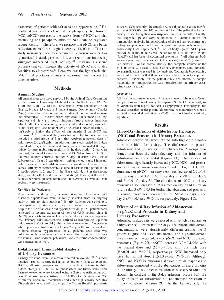
exosomes of patients with salt-sensitive hypertension.10 Re-cently, it has become clear that the phosphorylated form ofNCC (pNCC) represents the active form of NCC and thattrafficking and phosphorylation of NCC can be regulatedindependently.11 Therefore, we propose that pNCC is a betterreflection of NCC’s biological activity. ENaC is difficult tostudy in urinary exosomes because it is present in very lowquantities.4 Instead, prostasin has emerged as an interestingsurrogate marker of ENaC activity.12 Prostasin is a serineprotease that can increase the activity of ENaC and is alsosensitive to aldosterone.13 Here, we test the hypothesis thatpNCC and prostasin in urinary exosomes are markers foraldosteronism.
MethodsAnimal StudiesAll animal protocols were approved by the Animal Care Committeeof the Erasmus University Medical Center Rotterdam (EUR 127-11-01 and EUR 127-10-11). Three studies were conducted. In thefirst study, ten 15-week–old male Sprague-Dawley rats (CharlesRiver) were adrenalectomized via a bilateral lumbodorsal incisionand randomized to receive either high-dose aldosterone (100 �g/kg/d) or vehicle via osmotic minipump (subcutaneous insertion,Alzet). All rats also received glucocorticoid replacement (dexameth-asone, 5 �g/kg/d) and an angiotensin receptor blocker (losartan, 10mg/kg/d) to inhibit the effects of angiotensin II on pNCC andprostasin.14,15 The second study was similar to the first one but nowincluded a third group of 5 rats that received a normal dose ofaldosterone (50 �g/kg/d).16 In addition, this study lasted for 8 daysinstead of 3 days. In the second study, we also harvested the rightkidney for immunoblotting analysis. In the third study, 12 rats wererandomly assigned to receive a normal (0.5%) or low (0.001% to0.002%) sodium chloride diet for 8 days (Harlan diets, HarlanLaboratories). In all 3 experiments, animals were housed in meta-bolic cages to collect 24-hour urine for isolation of urinary exo-somes. Urinary exosomes were isolated at various time-points in the3 studies (days 1, 2, and 3 in the first study; day 8 in the secondstudy; and days 0, 4, and 8 in the third study). Finally, at the end ofeach experiment, plasma renin and aldosterone, as well as urinesodium, were measured.
Studies in PatientsFive patients with primary aldosteronism and 4 patients withessential hypertension were randomly selected from an ongoingstudy on primary aldosteronism.17 Briefly, patients were eligible toparticipate in this study when they had uncontrolled hypertensiondespite the use of at least 2 antihypertensive drugs. All patients weresubjected to volume expansion (2 liters of 0.9% sodium chloride[NaCl] during 4 hours) to analyze whether aldosterone was suppress-ible. Primary aldosteronism was defined as insuppressible plasmaaldosterone (�235 pmol/L) after volume expansion. The patientswhose posttest aldosterone was below 235 pmol/L were consideredto have essential hypertension. In all patients, spot urine wascollected under controlled circumstances for isolation of urinaryexosomes. Urine sodium, potassium, and creatinine concentrationswere measured as well.
Isolation and Immunoblot Analysisof Urinary ExosomesUrinary exosomes were isolated as reported previously4,18,19; a moredetailed protocol is provided as an online-only Data Supplement.Briefly, all urine samples were treated with a protease inhibitorbefore storage at �80°C; no phosphatase inhibitors were used.Urinary exosomes were isolated using a 2-step centrifugation pro-cess. First, urine was centrifuged at 17000�g for 15 minutes at 37°Cto remove whole cell membranes and other high density particles.Dithiothreitol was used to disrupt the Tamm-Horsfall polymeric
network. Subsequently, the samples were subjected to ultracentrifu-gation at 200000�g for 105 minutes at 25°C. The pellet that formedduring ultracentrifugation was suspended in isolation buffer. Finally,the suspended pellets were solubilized in Laemmli buffer forimmunoblot analysis. Immunoblotting of the urinary exosomes andkidney samples was performed as described previously (see alsoonline-only Data Supplement).8 The antibody against NCC phos-phorylated at threonine 58 was generated by 1 of the investigators(R.A.F.) and has been characterized previously.20 All other antibod-ies were purchased: prostasin (BD Biosciences) and NCC (StessmarqBiosciences). For the animal studies, the complete volume of the24-hour urine was used to isolate urinary exosomes, and, therefore,no normalization was used in the analysis. Coomassie blue stainingwas used to confirm that there were no differences in total proteincontents. Conversely, for the patient study, the amount of sampleloaded during immunoblotting was normalized by the urinary creat-inine concentration.1
StatisticsAll data are expressed as mean � standard error of the mean. Groupcomparisons were made using the unpaired Student t test or analysisof variances with a post hoc test, as appropriate. For analysis, thenatural logarithm of the plasma aldosterone concentration was usedto yield a normal distribution. P�0.05 was considered statisticallysignificant.
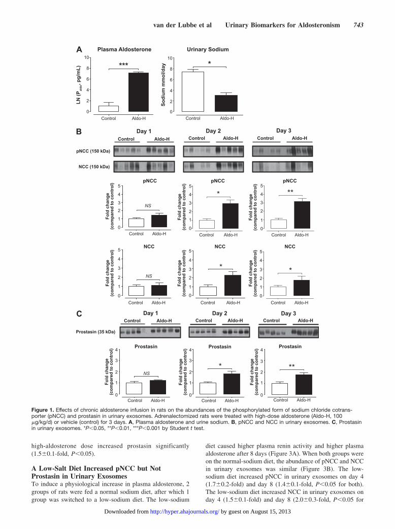
ResultsThree-Day Infusion of Aldosterone IncreasedpNCC and Prostasin in Urinary ExosomesAdrenalectomized rats were infused with high-dose aldoste-rone or vehicle for 3 days. The differences in plasmaaldosterone and urinary sodium between the 2 groups con-firmed that both the adrenalectomy and the infusion ofaldosterone were successful (Figure 1A). The infusion ofaldosterone significantly increased pNCC, NCC, and prosta-sin in urinary exosomes on days 2 and 3 (Figure 1B). Theabundance of pNCC in urinary exosomes increased 2.9�0.4-fold on day 2 and 3.2�0.3-fold on day 3 (P�0.05 for day 2and P�0.01 for day 3). The abundance of NCC in urinaryexosomes also increased 2.3�0.4-fold on day 2 and 1.8�0.4-fold on day 3 (P�0.05 for both). The abundance of prostasinin urinary exosomes increased 1.8�0.2-fold on days 2 andday 3 (P�0.05 and P�0.01, respectively, Figure 1C).
Effects of an 8-Day Infusion of Aldosteroneon pNCC and Prostasin in Kidney andUrinary ExosomesAdrenalectomized rats were infused with vehicle, a normal ora high dose of aldosterone for 8 days. The plasma aldosteroneconcentrations were significantly different among the 3groups (Figure 2A). Both the normal and high-aldosteronedose increased the abundance of pNCC and NCC in urinaryexosomes (Figure 2B). pNCC increased 3.0�0.4-fold withthe normal dose and 2.5�0.5-fold with the high dose(P�0.01 and P�0.05, respectively); NCC increased onlywith the normal dose (1.5�0.2-fold, P�0.05). AlthoughpNCC and NCC in exosomes showed similar responses toaldosterone compared with the abundances of these proteinsin the kidney,21 no direct correlation was observed (data notshown). In contrast to the 3-day infusion (Figure 1C), the8-day infusion of aldosterone did not increase prostasin inurinary exosomes (Figure 2C). In the kidney, only the
742 Hypertension September 2012
by guest on August 15, 2013http://hyper.ahajournals.org/Downloaded from
high-aldosterone dose increased prostasin significantly(1.5�0.1-fold, P�0.05).
A Low-Salt Diet Increased pNCC but NotProstasin in Urinary ExosomesTo induce a physiological increase in plasma aldosterone, 2groups of rats were fed a normal sodium diet, after which 1group was switched to a low-sodium diet. The low-sodium
diet caused higher plasma renin activity and higher plasmaaldosterone after 8 days (Figure 3A). When both groups wereon the normal-sodium diet, the abundance of pNCC and NCCin urinary exosomes was similar (Figure 3B). The low-sodium diet increased pNCC in urinary exosomes on day 4(1.7�0.2-fold) and day 8 (1.4�0.1-fold, P�0.05 for both).The low-sodium diet increased NCC in urinary exosomes onday 4 (1.5�0.1-fold) and day 8 (2.0�0.3-fold, P�0.05 for
A
pNCC (150 kDa)
Day 1 Day 2 Day 3 Control Aldo-H Control Aldo-H Control Aldo-H
B
Prostasin (35 kDa)
Control Aldo-H Control Aldo-H Control Aldo-HC
NCC (150 kDa)
Day 1 Day 2 Day 3
10
8
6
4
2
0
10
8
6
4
2
0Control Aldo-H Control Aldo-H
LN (P
aldo
, pg/
mL)
Sodi
um m
mol
/day*** *
Plasma Aldosterone Urinary Sodium
5
4
3
2
1
0Control Aldo-H
NS
Fold
cha
nge
(com
pare
d to
con
trol
) pNCC5
4
3
2
1
0
Fold
cha
nge
(com
pare
d to
con
trol
)
Control Aldo-H
pNCC
*5
4
3
2
1
0Control Aldo-H
Fold
cha
nge
(com
pare
d to
con
trol
)
**pNCC
NS
Control Aldo-H
5
4
3
2
1
0
Fold
cha
nge
(com
pare
d to
con
trol
) NCC
Control Aldo-H
5
4
3
2
1
0
Fold
cha
nge
(com
pare
d to
con
trol
) NCC
*
NS
Control Aldo-H
4
3
2
1
0
Fold
cha
nge
(com
pare
d to
con
trol
)
Control Aldo-H
4
3
2
1
0
Fold
cha
nge
(com
pare
d to
con
trol
)
Control Aldo-H
4
3
2
1
0
Fold
cha
nge
(com
pare
d to
con
trol
)Prostasin
* **
ProstasinProstasin
Fold
cha
nge
(com
pare
d to
con
trol
) 5
4
3
2
1
0
*
Control Aldo-H
NCC
Figure 1. Effects of chronic aldosterone infusion in rats on the abundances of the phosphorylated form of sodium chloride cotrans-porter (pNCC) and prostasin in urinary exosomes. Adrenalectomized rats were treated with high-dose aldosterone (Aldo-H, 100�g/kg/d) or vehicle (control) for 3 days. A, Plasma aldosterone and urine sodium. B, pNCC and NCC in urinary exosomes. C, Prostasinin urinary exosomes. *P�0.05, **P�0.01, ***P�0.001 by Student t test.
van der Lubbe et al Urinary Biomarkers for Aldosteronism 743
by guest on August 15, 2013http://hyper.ahajournals.org/Downloaded from

both). In contrast, it did not cause significant changes in theabundance of prostasin in urinary exosomes (Figure 3C).
pNCC and Prostasin Are Increased in UrinaryExosomes of Patients With Primary AldosteronismThe characteristics of 5 patients with primary aldosteronismand 4 patients with essential hypertension are shown inFigure 4. Both groups had a similar degree of hypertensionbut had significant differences regarding the plasma aldoste-rone-to–renin ratio and the urine sodium-to–potassium ratio.The abundance of pNCC in urinary exosomes of patients withprimary aldosteronism was higher in patients with essential
hypertension (2.6�0.3-fold, P�0.05, Figure 4). The abun-dance of prostasin in urinary exosomes showed a trendtoward being higher in the patients with primary aldosteron-ism (1.5�0.3-fold, P�0.07).
DiscussionIn this study, we asked whether NCC and prostasin in urinaryexosomes can be used as markers for primary and secondaryaldosteronism. In our hands, both total and phosphorylatedNCC were superior to prostasin as a marker for aldoste-ronism. In fact, the abundance of prostasin in urinaryexosomes only increased during high-dose, short-termtreatment with aldosterone (Figure 1C), whereas the abun-dance of pNCC in urinary exosomes was higher duringaldosteronism, regardless of the duration, dose, or stimulus(Figure 5). Because the changes in pNCC were more pro-nounced than for NCC, pNCC appears the better marker. Inaddition, pNCC was increased significantly in urinary exo-somes of patients with primary aldosteronism compared withpatients with essential hypertension (Figure 4). In contrast,the increase in the abundance of prostasin in urinary exo-somes of patients with primary aldosteronism was of border-line significance.
Several observations regarding the dynamics of pNCC andprostasin excretion in urinary exosomes during aldosteronismin this study merit discussion. For example, the results of theshort-term infusion of aldosterone in rats suggest that it takesat least 1 day for both pNCC and prostasin to increase inurinary exosomes (Figure 1). This may be related to the timeit takes aldosterone to increase transcription or posttransla-tional modification of these proteins.22 The long-term infu-sion of aldosterone in rats showed that a lower (physiologi-cal) dose of aldosterone was already sufficient to increasepNCC (Figure 2B). In fact, pNCC expression in urinaryexosomes was slightly lower with the high dose. Thissuggests that the stimulatory effect of aldosterone on pNCCin urinary exosomes either saturates or that aldosteroneescape has occurred, a defense mechanism known to reduceNCC in the kidney.23 Although the induction of secondaryaldosteronism by a low-sodium diet also increased pNCC inurinary exosomes (Figure 3B), the magnitude of this effectwas less than with aldosterone infusion (Figure 5). One couldhave predicted even higher pNCC in urinary exosomes duringa low-sodium diet because this maneuver is likely to increaseboth plasma angiotensin II and aldosterone. We recentlyshowed that angiotensin II can increase renal pNCC indepen-dently of aldosterone and that the combination of angiotensinII and aldosterone leads to an additive effect.15,21 Apparently,other factors limit pNCC excretion in urinary exosomesduring a low-sodium diet. Furthermore, differences in theplasma aldosterone concentrations in the control groupsshould be taken into consideration (virtually absent in adre-nalectomized rats versus �100 pg/mL in rats on a normal-sodium diet, Figures 1A and 3A). Of interest, the increase inNCC in urinary exosomes we observed in this study with alow-sodium diet was similar to the increase Esteva-Font et alfound for NCC in a separate study.10
Our study suggests that pNCC was better than prostasin asa marker of aldosteronism, which could be explained by
pNCC (150kDa)
Control Aldo Aldo-H
KidneyProstasin (35kDa)
Urine
Control Aldo Aldo-H
A
C
B
NCC (150kDa)
Urine
Urine
2.0
1.5
1.0
0.5
0.0
Fold
cha
nge
(com
pare
d to
con
trol
)
NS NS
*2.0
1.5
1.0
0.5
0.0
Fold
cha
nge
(com
pare
d to
con
trol
)
NS
Control Aldo Aldo-H
Prostasin Kidney Prostasin Urine
Control Aldo Aldo-H
Control Aldo Aldo-H Control Aldo Aldo-H
pNCC Urine NCC Urine
NS
*
***
5
4
3
2
1
0
Fold
cha
nge
(com
pare
d to
con
trol
) 5
4
3
2
1
0
Fold
cha
nge
(com
pare
d to
con
trol
)
Control Aldo Aldo-H
2500
2000
1500
1000
500
0
Ald
oste
rone
(pg/
mL)
Plasma Aldosterone
*
**
***
Figure 2. Effects of normal and high-aldosterone infusion in ratsfor 8 days on plasma aldosterone, phosphorylated form ofsodium chloride cotransporter (pNCC), and prostasin in kidneyand urinary exosomes. Adrenalectomized rats were infused withvehicle (control), normal (Aldo, 50 �g/kg/d), or high-dose(Aldo-H, 100 �g/kg/d) aldosterone for 8 days. A, Plasma aldo-sterone concentrations. B, pNCC and NCC in urinary exosomes.C, Prostasin abundance in kidney and urinary exosomes.*P�0.05, **P�0.01, ***P�0.001 by analysis of variance and posthoc test.
744 Hypertension September 2012
by guest on August 15, 2013http://hyper.ahajournals.org/Downloaded from

several factors. First, although prostasin is sensitive to aldo-sterone and can activate ENaC, this does not necessarilyrender it a direct marker of ENaC activity and, as such, it maynot be a direct marker of distal sodium reabsorption.12,13
Prostasin is only one of the proteins present in the complexsignaling cascade that regulates ENaC.24 Second, because
prostasin is also present in prostate epithelial cells, urinaryexosomes may also contain prostasin from this source.25 Thismay have limited the specificity of prostasin as a marker foraldosterone actions in the kidney, especially in males. Third,by using 2-dimensional electrophoresis, Olivieri and col-leagues have previously shown that several subunits of
A
pNCC (150 kDa)
Day 0 Day 4 Day 8 B
Prostasin (35 kDa)
C
NCC (150 kDa)
Day 0 Day 4 Day 8Normal sodium Normal sodium Normal sodium Low sodium Normal sodium Low sodium
Normal sodium Normal sodium Normal sodium Low sodium Normal sodium Low sodium
2.0
1.5
1.0
0.5
0.0
Fold
cha
nge
(com
pare
d to
con
trol
)
Plasma renin activity
NS
*
Prostasin
NS NS
2.0
1.5
1.0
0.5
0.0
Fold
cha
nge
(com
pare
d to
con
trol
) 2.0
1.5
1.0
0.5
0.0
Fold
cha
nge
(com
pare
d to
con
trol
)Prostasin Prostasin
Normal Normalsodium sodium
Normal Lowsodium sodium
Normal Lowsodium sodium
NCC
*
Normal Normalsodium sodium
Normal Lowsodium sodium
2.5
2.0
1.5
1.0
0.5
0.0
Fold
cha
nge
(com
pare
d to
con
trol
)
NS
NCC
*NCC
2.5
2.0
1.5
1.0
0.5
0.0
Fold
cha
nge
(com
pare
d to
con
trol
)
Normal Lowsodium sodium
2.5
2.0
1.5
1.0
0.5
0.0
Fold
cha
nge
(com
pare
d to
con
trol
)
pNCC pNCC pNCC
*
Normal Normalsodium sodium
Normal Lowsodium sodium
2.5
2.0
1.5
1.0
0.5
0.0
Fold
cha
nge
(com
pare
d to
con
trol
)
NS*
2.5
2.0
1.5
1.0
0.5
0.0
Fold
cha
nge
(com
pare
d to
con
trol
)
Normal Lowsodium sodium
2.5
2.0
1.5
1.0
0.5
0.0
Fold
cha
nge
(com
pare
d to
con
trol
)
20
15
10
5
0R
enin
act
ivity
(ng
AN
G I/
mL/
h)Normal Lowsodium sodium
Plasma aldosterone
*700600500400300200100
0
Ald
oste
ron
(pg/
mL)
Normal Lowsodium sodium
Figure 3. A low-salt diet increased phosphorylated form of sodium chloride cotransporter (pNCC) but not prostasin in urinary exo-somes of rats. A, Plasma renin activity and plasma aldosterone. B, pNCC and NCC in urinary exosomes. C, Prostasin in urinary exo-somes. *P�0.05 by Student t test.
van der Lubbe et al Urinary Biomarkers for Aldosteronism 745
by guest on August 15, 2013http://hyper.ahajournals.org/Downloaded from

prostasin exist, only some of which are aldosterone-sensitive.12 Our prostasin results differ from those reported byNarikiyo and coworkers.13 They found that rats continued toincrease urinary prostasin during 7 days of aldosteroneinfusion (from 1.5- to 4-fold), whereas we were unable todetect increased prostasin in urinary exosomes after 8 days oftreatment (Figure 2). This difference may be explained by theuse of a 3-fold higher aldosterone dose in the previous studyand that their analysis of prostasin was performed on wholeurine instead of urinary exosomes.
In recent years, the potential to use proteins in urinaryexosomes as markers of diseases affecting the kidney hasattracted much interest.1–3,26 Progress has been somewhathindered by technical and normalization issues, but theproposal of uniform protocols has been a step in the rightdirection.18,27 We believe that the strength of this study wasto combine a controlled experimental setting with a clinicalsetting for the analysis of urinary exosomes in aldosteronism.Because of the well-characterized actions of aldosterone on
distal tubular sodium transport, aldosteronism appears espe-cially suitable for analysis with urinary exosomes. Clinically,primary aldosteronism is important because recent studiessuggest it to be a common condition among patients withresistant hypertension that is often difficult to diagnose.28 Wedo emphasize, however, that the primary aim of this studywas to provide proof of principle that the abundance ofaldosterone-sensitive proteins in urinary exosomes is in-creased during aldosteronism. The question whether pNCCin urinary exosomes has diagnostic potential in patientswith primary aldosteronism remains to be determined. Thiswill require larger and well-characterized groups of pa-tients to be tested against a golden standard. In addition,the overall differences in NCC and pNCC between condi-tions were relatively mild even under controlled experi-mental conditions. Even if the sensitivity and specificity ofpNCC in urinary exosomes would outweigh existing testssuch as the ARR, the current method of exosome isolationis not suitable for clinical use.29 Instead, development of
A
B EH PA
pNCC (150kDa)
Prostasin (40kDa)
25
20
15
10
5
0
5
4
3
2
1
0
5
4
3
2
1
0
200
150
100
50
0
SBP
(mm
Hg)
AR
R
UN
a/UK
EH PA EH PA EH PA
NS***Aldosterone to renin ratio Urine sodium to potassium ratio Systolic blood pressure
Fold
cha
nge
(com
pare
d to
con
trol
)
Essential Hypertension (EH)Primary Aldosteronism (PA)
*NS
pNCC
Prostas
in
Figure 4. Patients with primary aldosteronism show increased abundance of phosphorylated form of sodium chloride cotransporter(pNCC) and prostasin in urinary exosomes. A, Characteristics of patients with essential hypertension (EH) or primary aldosteronism(PA). B, Patients with PA show increased abundance of pNCC and prostasin in urinary exosomes. *P�0.05 by Student t test orMann-Whitney.
Aldosterone infusion day 1day 2day 3
Aldosterone infusion day 8High-dose aldosterone infusion day 8
Low sodium diet day 4Low sodium diet day 8
Primary aldosteronism (patients)
**
* *
***
*
**
3 2 1 1 2 3
Prostasin pNCC
Figure 5. Summary of the performance of urinary exosomal prostasin and phosphorylated form of sodium chloride cotransporter(pNCC) as markers of aldosteronism. The densitometry values of the immunoblot analyses are shown. A value of 1 represents no differ-ence compared with the control group. *P�0.05, **P�0.01 vs control.
746 Hypertension September 2012
by guest on August 15, 2013http://hyper.ahajournals.org/Downloaded from
an enzyme-linked immunoassay for pNCC would be anattractive alternative.
This study suggests several directions for future research.One obvious next step will be to evaluate how pNCC andprostasin in patients with primary aldosteronism respond totreatment with either mineralocorticoid receptor antagonistsor adrenalectomy. Narikiyo et al showed that urinary prosta-sin decreased in 3 patients with primary aldosteronism whohad undergone adrenalectomy.13 Similarly, Olivieri et alshowed that prostasin decreased in normotensive subjectswith aldosteronism owing to a low-sodium diet who weresubsequently treated with spironolactone.12 It would also beinformative to know whether other commonly used antihy-pertensive drugs such as diuretics, angiotensin-convertingenzyme inhibitors, or angiotensin receptor blockers affecturinary exosome excretion. Because total NCC also in-creased, it would be important to known whether NCCmRNA is also present in urinary exosomes and if it behavessimilarly. If so, it could be a more sensitive marker becausethe signal can be amplified. In the kidney, however, previousstudies found that the protein and mRNA abundances of NCCdid not always correlate.21,23 Finally, given the recent insightsinto the effects of angiotensin II on distal tubular sodiumtransport,14,15 it would be important to compare urinaryexosomal pNCC and prostasin in patients with primary andsecondary aldosteronism.
In conclusion, in urinary exosomes of animals and patients,pNCC was superior to prostasin as a marker of aldosteronism.These results justify further evaluation of the applicability ofurinary exosomes as a diagnostic tool in primary aldosteron-ism and, possibly, other forms of hypertension. Furthermore,pNCC and, to a lesser extent, prostasin may be used experi-mentally as a noninvasive method to analyze the biologicalaction of aldosterone in the kidney.
PerspectivesIn hypertension, urinary exosomes could be applied as adiagnostic test for primary aldosteronism, to evaluate salt-sensitivity or the response to antihypertensive drugs acting onthe kidney. Our study can be regarded as proof of principlethat analysis of urinary exosomes can be applied to aldoste-ronism. Subsequent validation studies will be necessary todefine their use in experimental or clinical settings.30
Sources of FundingDr Hoorn is supported by grants from the Erasmus Medical Center,Dutch Kidney Foundation (KJPB 08.004), and Netherlands Organi-sation for Scientific Research (Veni). Dr van der Lubbe is supportedby The Netherlands Foundation for Cardiovascular Excellence. DrJansen is supported by a grant from the Dutch Kidney Foundation(C05-2151).
DisclosuresNone.
References1. Hoorn EJ, Pisitkun T, Zietse R, Gross P, Frokiaer J, Wang NS, Gonzales
PA, Star RA, Knepper MA. Prospects for urinary proteomics: exosomesas a source of urinary biomarkers. Nephrology (Carlton). 2005;10:283–290.
2. Goligorsky MS, Addabbo F, O’Riordan E. Diagnostic potential of urineproteome: a broken mirror of renal diseases. J Am Soc Nephrol. 2007;18:2233–2239.
3. van Balkom BW, Pisitkun T, Verhaar MC, Knepper MA. Exosomes andthe kidney: prospects for diagnosis and therapy of renal diseases. KidneyInt. 2011;80:1138–1145.
4. Pisitkun T, Shen RF, Knepper MA. Identification and proteomic profilingof exosomes in human urine. Proc Natl Acad Sci U S A. 2004;101:13368–13373.
5. Gonzales PA, Pisitkun T, Hoffert JD, Tchapyjnikov D, Star RA, Kleta R,Wang NS, Knepper MA. Large-scale proteomics and phosphoproteomicsof urinary exosomes. J Am Soc Nephrol. 2009;20:363–379.
6. Joo KW, Lee JW, Jang HR, Heo NJ, Jeon US, Oh YK, Lim CS, Na KY,Kim J, Cheong HI, Han JS. Reduced urinary excretion of thiazide-sensitive Na-Cl cotransporter in Gitelman syndrome: preliminary data.Am J Kidney Dis. 2007;50:765–773.
7. Mayan H, Attar-Herzberg D, Shaharabany M, Holtzman EJ, Farfel Z.Increased urinary Na-Cl cotransporter protein in familial hyperkalaemiaand hypertension. Nephrol Dial Transplant. 2008;23:492–496.
8. Kim GH, Masilamani S, Turner R, Mitchell C, Wade JB, Knepper MA.The thiazide-sensitive Na-Cl cotransporter is an aldosterone-inducedprotein. Proc Natl Acad Sci U S A. 1998;95:14552–14557.
9. Masilamani S, Kim GH, Mitchell C, Wade JB, Knepper MA.Aldosterone-mediated regulation of ENaC alpha, beta, and gammasubunit proteins in rat kidney. J Clin Invest. 1999;104:R19–R23.
10. Esteva-Font C, Wang X, Ars E, Guillen-Gomez E, Sans L, GonzalezSaavedra I, Torres F, Torra R, Masilamani S, Ballarin JA,Fernandez-Llama P. Are Sodium Transporters in Urinary ExosomesReliable Markers of Tubular Sodium Reabsorption in HypertensivePatients? Nephron Physiol. 2010;114:p25–p34.
11. Hoorn EJ, Nelson JH, McCormick JA, Ellison DH. The WNK kinasenetwork regulating sodium, potassium, and blood pressure. J Am SocNephrol. 2011;22:605–614.
12. Olivieri O, Castagna A, Guarini P, Chiecchi L, Sabaini G, Pizzolo F, Cor-rocher R, Righetti PG. Urinary prostasin: a candidate marker of epithelialsodium channel activation in humans. Hypertension. 2005;46:683–688.
13. Narikiyo T, Kitamura K, Adachi M, Miyoshi T, Iwashita K, Shiraishi N,Nonoguchi H, Chen LM, Chai KX, Chao J, Tomita K. Regulation ofprostasin by aldosterone in the kidney. J Clin Invest. 2002;109:401–408.
14. Sun P, Yue P, Wang WH. Angiotensin II stimulates epithelial sodiumchannels in the cortical collecting duct of the rat kidney. Am J PhysiolRenal Physiol. 2012;302:F679–F687.
15. van der Lubbe N, Lim CH, Fenton RA, Meima ME, Danser AHJ, ZietseR, Hoorn EJ. Angiotensin II induces phosphorylation of the thiazide-sensitive sodium chloride cotransporter independent of aldosterone.Kidney Int. 2011;79:66–76.
16. Velazquez H, Bartiss A, Bernstein P, Ellison DH. Adrenal steroidsstimulate thiazide-sensitive NaCl transport by rat renal distal tubules. AmJ Physiol. 1996;270:F211–F219.
17. Jansen PM, Boomsma F, van den Meiracker AH. Aldosterone-to–reninratio as a screening test for primary aldosteronism–the Dutch ARRATStudy. Neth J Med. 2008;66:220–228.
18. Fernandez-Llama P, Khositseth S, Gonzales PA, Star RA, Pisitkun T,Knepper MA. Tamm-Horsfall protein and urinary exosome isolation.Kidney Int. 2010;77:736–742.
19. Gonzales PA, Zhou H, Pisitkun T, Wang NS, Star RA, Knepper MA,Yuen PS. Isolation and purification of exosomes in urine. Methods MolBiol. 2010;641:89–99.
20. Pedersen NB, Hofmeister MV, Rosenbaek LL, Nielsen J, Fenton RA. Vaso-pressin induces phosphorylation of the thiazide-sensitive sodium chloridecotransporter in the distal convoluted tubule. Kidney Int. 2011;78:160–169.
21. van der Lubbe N, Lim CH, Meima ME, Veghel R, Lintoft Rosenbaek L,Mutig K, Danser AHJ, Fenton RA, Zietse R, Hoorn EJ. Aldosterone doesnot require angiotensin II to activate NCC through a WNK4-SPAK-dependent pathway. Pflugers Archiv. 2012;463:853–863.
22. Arroyo JP, Ronzaud C, Lagnaz D, Staub O, Gamba G. Aldosteroneparadox: differential regulation of ion transport in distal nephron. Phys-iology (Bethesda). 2011;26:115–123.
23. Wang XY, Masilamani S, Nielsen J, Kwon TH, Brooks HL, Nielsen S,Knepper MA. The renal thiazide-sensitive Na-Cl cotransporter as mediator ofthe aldosterone-escape phenomenon. J Clin Invest. 2001;108:215–222.
24. Soundararajan R, Melters D, Shih IC, Wang J, Pearce D. Epithelialsodium channel regulated by differential composition of a signalingcomplex. Proc Natl Acad Sci U S A. 2009;106:7804–7809.
van der Lubbe et al Urinary Biomarkers for Aldosteronism 747
by guest on August 15, 2013http://hyper.ahajournals.org/Downloaded from

25. Yu JX, Chao L, Chao J. Molecular cloning, tissue-specific expression,and cellular localization of human prostasin mRNA. J Biol Chem. 1995;270:13483–13489.
26. Dimov I, Jankovic Velickovic L, Stefanovic V. Urinary exosomes. SciWorld J. 2009;9:1107–1118.
27. Zhou H, Yuen PS, Pisitkun T, Gonzales PA, Yasuda H, Dear JW, GrossP, Knepper MA, Star RA. Collection, storage, preservation, and normal-ization of human urinary exosomes for biomarker discovery. Kidney Int.2006;69:1471–1476.
28. Douma S, Petidis K, Doumas M, Papaefthimiou P, Triantafyllou A,Kartali N, Papadopoulos N, Vogiatzis K, Zamboulis C. Prevalence ofprimary hyperaldosteronism in resistant hypertension: a retrospectiveobservational study. Lancet. 2008;371:1921–1926.
29. Rood IM, Deegens JK, Merchant ML, Tamboer WP, Wilkey DW, Wetzels JF,Klein JB. Comparison of three methods for isolation of urinary microvesicles toidentify biomarkers of nephrotic syndrome. Kidney Int. 2010;78:810–816.
30. Hewitt SM, Dear J, Star RA. Discovery of protein biomarkers for renaldiseases. J Am Soc Nephrol. 2004;15:1677–1689.
Novelty and Significance
What Is New?● The phosphorylated form of the sodium chloride cotransporter, a salt
transporter in the kidney, is increased in so-called urinary exosomes inexperimental models of aldosteronism and in patients with the hyper-tensive disorder primary aldosteronism.
What Is Relevant?● pNCC in urinary exosomes may be used as a noninvasive urinary
biomarker for aldosteronism and, potentially, as a diagnostic test inprimary aldosteronism.
Summary
Disease-associated proteins in the kidney are excreted in the urinein so-called exosomes. We tested 2 of these proteins in experi-mental and clinical aldosteronism. In urinary exosomes of animalsand patients, we found that the phosphorylated sodium chloridecotransporter was superior to prostasin as a marker ofaldosteronism.
748 Hypertension September 2012
by guest on August 15, 2013http://hyper.ahajournals.org/Downloaded from
ONLINE SUPPLEMENT
The phosphorylated sodium chloride cotransporter in urinary exosomes
is superior to prostasin as a marker for aldosteronism
Nils van der Lubbe1, Pieter M. Jansen1, Mahdi Salih1, Robert A. Fenton2, Anton H. van den
Meiracker1, Alexander H. Jan Danser1, Robert Zietse1, Ewout J. Hoorn1
1Department of Internal Medicine, Erasmus Medical Center, Rotterdam, The Netherlands
2Department of Anatomy, Water and Salt Research Institute,
Aarhus University, Aarhus, Denmark
by guest on August 15, 2013http://hyper.ahajournals.org/Downloaded from
Protocol for urinary exosome isolation Collection and storage of urine To collect 24 hour urine from rats, they were placed in metabolic cages and mineral oil
(Sigma-Aldrich, Zwijndrecht, The Netherlands) was added to the collection tubes to prevent evaporation.
The total 24 hour urine volume was used for isolation of urinary exosomes. The following protease inhibitor cocktail was added per 50 ml of urine (all purchased
from Sigma-Aldrich): o 1.67 ml 100mM NaN3 o 2.5 ml (2,75 mg/ml) 4-(2-Aminoethyl)benzenesulfonyl fluoride hydrochloride
(AEBSF) o 50 µl (1 mg/ml) Leupeptin
In patients, second morning urine samples were collected and a Complete Protease Inhibitor Tablet (Roche Diagnostics, Mannheim, Germany) was added. Twelve ml of urine was used for urinary exosome isolation.
Urine samples were stored immediately after collection in –80°C. Isolation of urinary exosomes The following isolation solution was used (all compounds were purchased from Sigma-
Aldrich): o 10 mM triethanolamine (0.5ml of 1M stock) o 250 mM sucrose(25ml of 0.5M Stock) o ddH20 was added to 45ml o pH was adjusted to 7.6 with 1M NaOH o ddH20 was added to 50 ml
To remove whole cell membranes and other high density particles, urine samples were centrifuged at 17,000 x g for 15 minutes at 24°C in 70 ml polycarbonate centrifuge bottles (Beckman Coulter, Woerden, The Netherlands) in an ultracentrifuge (Beckman L8-70M ultracentrifuge, Rotor 45 Ti).
The supernatant was stored at room temperature (Supernatant 1). The pellet was resuspended in 360 µl (dithiothreitol 500 mg/ml) and 450 µl isolation
solution and incubated for 5 minutes at 37°C and vortexed until the pellet was dissolved. The dissolved pellet was centrifuged again in at 17,000 x g for 15 minutes at 24°C in 10
ml polycarbonate tubes (Rotor 70.1 Ti) . This supernatant was pooled with Supernatant 1 and centrifuged in 70 ml polycarbonate
tubes at 200,000 x g for 1:54 minutes at 24°C. Th supernatant was discarded and the pellet was resuspended in 1.5 x Laemmli buffer and
stored at –20°C for immunoblotting. Immunoblotting Electrophoresis and immunoblotting were carried out using the Mini-PROTEAN Tetra
Electrophoresis system (Bio-Rad laboratories, Hercules, CA, USA) Samples were preheated at 60°C for 15 min. For the animal studies 10 µl of sample was used. In the patient study the samples were
adjusted for the urine creatinine concentration with 1 x Laemmli buffer to a final volume of 20µl.
SDS/PAGE was carried out on a 8% polyacrylamide gel, and proteins were transferred to Immobilon®-P membranes (Millipore, Amsterdam, The Netherlands) .
by guest on August 15, 2013http://hyper.ahajournals.org/Downloaded from
The membranes were blocked in 5% milk and were probed overnight at 4°C with the antibody. The antibody against NCC phosphorylated at threonine 58 (1:500 in 5% Bovine Serum Albumin) was generated by one of the investigators (dr. R.A. Fenton, University of Aarhus). The other antibodies were obtained: NCC (Stressmarq biosciences, Victoria, Canada, 1:500 in 5% Milk) and Prostasin (BD biosciences, Breda, the Netherlands 1:500 in 5% Milk).
After three washes (3 x 10 minutes in TBS-Tween 20), the membranes were incubated with the secondary antibody in 5% milk (Thermo Scientific, Rockford, IL USA, 1:3000) for 1 hour and washed again.
For visualization blots were exposed to Pierce® enchanced chemiluminescent substrate and exposed to X-ray film (Kodak Biomax MR Film, Rochester, NY, USA).
by guest on August 15, 2013http://hyper.ahajournals.org/Downloaded from