hype, hope and reality - european hospital
TRANSCRIPT
ISO 13485:2016
LATEX GLOVESMAKE GREAT BALLOONS BUT THEY MAKELOUSY PROBE COVERS.
AD 38-1 REV 3
Sure, exam gloves are “handy”, but using one as a probe cover isawkward, especially with a large3D/4D probe. It also allows for wasted ultrasound gel, can bemessy, and if the glove is latex,may cause an allergic reaction inpatient, clinician, or both. That’s why we developed the Eclipse® 3DProbe Cover, specially designedfor 3D/4D probes.
So you can save your examgloves for their intended use, or for decorating the next office party.
Like our original Eclipse® ProbeCover, Eclipse 3D is latex-free andconveniently pre-gelled with Aquasonic® 100, the world standardfor medical ultrasound procedures.
Visit us at Hall 9 Stand D41
© 2018 Parker Laboratories, Inc. The sound choice in patient care is atrademark of Parker Laboratories, Inc.
Eclipse_Cover_AD-38-1_REV_3_Euro-Hosp_Medica-2019-Thurs_10-4-19.qxp_Euro Hosp - MEDICA 2019 - Thurs 10/4/19 2:31 PM Page 1
Hall 10B60
Fujitsu IoT Connectivity Solutions for Hospitals
More info: www.fujitsu.com/IoT
FujitsuSmart HospitalSolutions
Asked which AI methods are cur-rently being applied in healthcare, the professor said: ‘The best known methods are machine learning meth-ods that detect patterns in existing data pools and can thus find solu-tions autonomously. One of these methods is Deep Learning, which uses neuronal networks. Today we use it in imaging, for example. More recently, boosting procedures such as XGBoost, which is also a machine learning approach, have been quickly gaining ground.
‘Moreover, AI is used to detect depression by analysing language and movement patterns. Other areas of application are production, selec-tion and dosage of pharmaceuticals, error detection in patient records and signal detection in ECG to recognise arrhythmias. These are diagnostic methods that have been approved and are commercially available. Additionally, AI methods are used in therapy, for example in triage, where they support decision-making processes.’
Which types of AI innovations will have the greatest impact on healthcare?‘Hard to tell. Any prognosis has to consider the hype curve. AI has reached peak hype in imaging. I’m sure that in other areas the hype is still to come. AI will sweep over us in waves at different points. The next focus will be on false diagnoses and treatments, which will generate con-siderable attention. These AI applica-tions will become part of medical practice sooner or later, without the patient ever noticing their existence, a bit like anti-lock brakes in the car.’
Which regulations must manufac-turers of AI healthcare applica-tions meet? ‘The legislator neither can, nor wants to, adopt a separate set of laws for each medical device category. The Medical Device Directive, in the near future the Medical Device Regulation,
defines the requirements. This legal framework has to be adjusted for the current AI procedures with a focus on verification and validation of the systems, stability and reproducibil-ity as well as fitness for use – issues that are already regulated. The law demands that benefits of the systems be proven quantitatively and that they outweigh the inherent risks of the systems.
‘This regulatory framework pro-vides good guidance. We are cur-rently working on specific stand-ards for AI. In cooperation with the notified bodies, such as TÜV, I drafted a guideline covering these requirements. This not only refers to the devices themselves, but also to organisations – inter alia they must define and prove staff competen-cies.’
What limitations does or should AI have? ‘AI should not cement bias – some-thing that happens quite eas-ily depending on the data pool the developers use to train their algo-rithms. Furthermore, AI methods must not be used when there is no clear evidence of their benefit and when the risks outweigh the ben-efits.
‘We also have to discuss whether we need to regulate the econom-ic framework together with the AI methods. For instance: Should health insurers be allowed to use AI for reimbursement decisions?’
Some people fear AI will replace physicians, particularly in imag-ing. Could this happen?‘There’s already a shortage of physi-cians and healthcare will not become easier in the future. I hope that AI will ease physicians’ workload – after all, these systems are programmed to support them in routine tasks. In some areas, AI is more powerful than the human brain, but that’s not a new experience. A truck, we all agree, is better at transporting
humans. AI will give physicians more time to actually deal with patients.
‘This means we need physicians who can use the information culled from AI in a meaningful way, in the diagnostic workup, or who can assess and implement therapy sug-gestions made by an AI system. These skills create an added value for human intelligence.’
things than a human. That’s how we should look at AI: it is a tool that can perform certain tasks better than we
WWW.HEALTHCARE-IN-EUROPE.COM
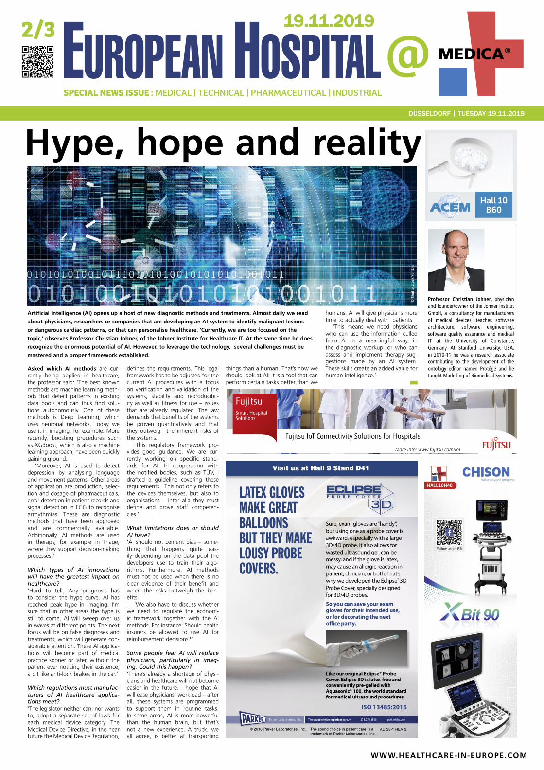
Artificial intelligence (AI) opens up a host of new diagnostic methods and treatments. Almost daily we read
about physicians, researchers or companies that are developing an AI system to identify malignant lesions
or dangerous cardiac patterns, or that can personalise healthcare. ‘Currently, we are too focused on the
topic,’ observes Professor Christian Johner, of the Johner Institute for Healthcare IT. At the same time he does
recognize the enormous potential of AI. However, to leverage the technology, several challenges must be
mastered and a proper framework established.
Hype, hope and reality
Professor Christian Johner, physician and founder/owner of the Johner Institut GmbH, a consultancy for manufacturers of medical devices, teaches software architecture, software engineering, software quality assurance and medical IT at the University of Constance, Germany. At Stanford University, USA, in 2010-11 he was a research associate contributing to the development of the ontology editor named Protégé and he taught Modelling of Biomedical Systems.
© S
hutt
erst
ock/
kent
oh
DÜSSELDORF | TUESDAY 19.11.2019
19.11.2019
www.hettichlab.comPlus d‘informations souswww.hettichlab.com
Transparency and traceability in the Blood Bank – learn more at Hettich booth B69 hall 3.
MKC-X800: NATIVE 4K MEDICAL GRADE CAMERA
MKC-X800 is the beginning of Ikegami‘s new
camera generation for 4K resolution. MKC-X800
is a 4K-native progressive-scan 1-CMOS model
with an ultra-compact head designed to be
operated from a remote CCU. Features include
ultra-high resolution, intuitive GUI and single
cable 4K output.
perience the new generation
Please visit us in Hall 10/B12
18 – 21 Nov 2019
www.ikegami.de . [email protected]
EH @ MEDICA No 2 2019
Transforming minuscule ‘swimmers’
Five Healthy Living finalists selected
Research: Microrobots to re-shape drug deliveryThe KUKA Innovation Award 2019
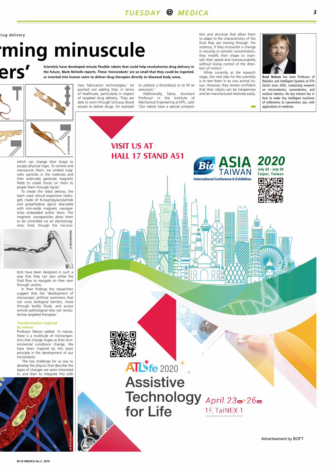
The microbot project is still very much at the research stage, but sci-entists masterminding the research at ETHZ (Swiss Federal Institute of Technology Zürich) and EPFL in Lausanne are confident it has enor-mous potential in the delivery of spe-cialised treatments. The highly-flex-ible biocompatible microrobots – or microswimmers – have been devel-oped by scientists led by Professor Brad Nelson at ETHZ and Professor Selman Sakar at EPFL.
With tiny magnets within the design, the machines – which vary in size from a few millimetres down to
less than a millimetre in total length – can swim through liquids and change shape as needed to move through narrow blood vessels and intricate systems.
Kirigami allows different shapes or stiffness Additionally, the microswimmers can be of different shapes or stiffness to reflect a specific task, with the study team following a variant of origami, called kirigami, to design and fold compliant 3-D microstructures from a thermo-responsive gel composite reinforced with micronanoparticles.
In the robot design, Nelson, who is Professor of Robotics and Intelligent Systems in the Department of Mechanical and Process Engineering at ETHZ, said: ‘We were inspired by tiny microorganisms, like bacteria,
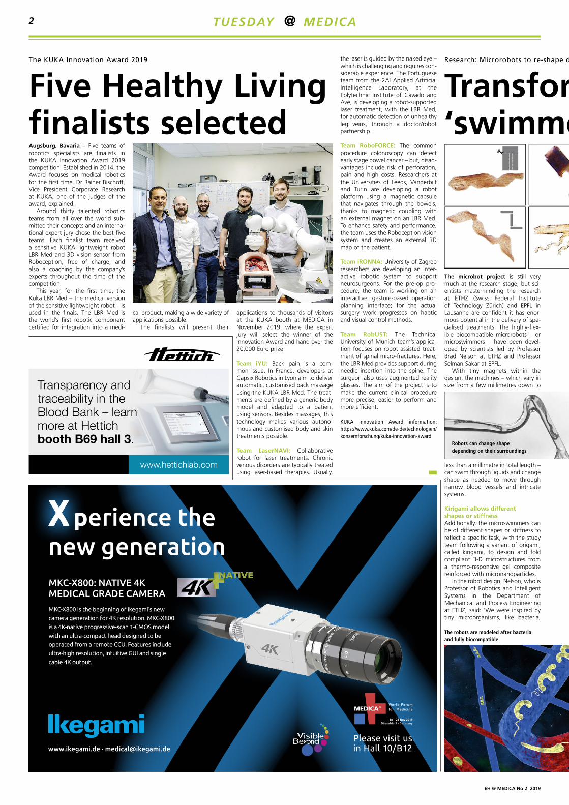
Augsburg, Bavaria – Five teams of robotics specialists are finalists in the KUKA Innovation Award 2019 competition. Established in 2014, the Award focuses on medical robotics for the first time, Dr Rainer Bischoff, Vice President Corporate Research at KUKA, one of the judges of the award, explained.
Around thirty talented robotics teams from all over the world sub-mitted their concepts and an interna-tional expert jury chose the best five teams. Each finalist team received a sensitive KUKA lightweight robot LBR Med and 3D vision sensor from Roboception, free of charge, and also a coaching by the company’s experts throughout the time of the competition.
This year, for the first time, the Kuka LBR Med – the medical version of the sensitive lightweight robot – is used in the finals. The LBR Med is the world’s first robotic component certified for integration into a medi-
cal product, making a wide variety of applications possible.
The finalists will present their
applications to thousands of visitors at the KUKA booth at MEDICA in November 2019, where the expert jury will select the winner of the Innovation Award and hand over the 20,000 Euro prize.
Team iYU: Back pain is a com-mon issue. In France, developers at Capsix Robotics in Lyon aim to deliver automatic, customised back massage using the KUKA LBR Med. The treat-ments are defined by a generic body model and adapted to a patient using sensors. Besides massages, this technology makes various autono-mous and customised body and skin treatments possible.
Team LaserNAVI: Collaborative robot for laser treatments: Chronic venous disorders are typically treated using laser-based therapies. Usually,
the laser is guided by the naked eye – which is challenging and requires con-siderable experience. The Portuguese team from the 2AI Applied Artificial Intelligence Laboratory, at the Polytechnic Institute of Cávado and Ave, is developing a robot-supported laser treatment, with the LBR Med, for automatic detection of unhealthy leg veins, through a doctor/robot partnership.
Team RoboFORCE: The common procedure colonoscopy can detect early stage bowel cancer – but, disad-vantages include risk of perforation, pain and high costs. Researchers at the Universities of Leeds, Vanderbilt and Turin are developing a robot platform using a magnetic capsule that navigates through the bowels, thanks to magnetic coupling with an external magnet on an LBR Med. To enhance safety and performance, the team uses the Roboception vision system and creates an external 3D map of the patient.
Team iRONNA: University of Zagreb researchers are developing an inter-active robotic system to support neurosurgeons. For the pre-op pro-cedure, the team is working on an interactive, gesture-based operation planning interface; for the actual surgery work progresses on haptic and visual control methods.
Team RobUST: The Technical University of Munich team’s applica-tion focuses on robot assisted treat-ment of spinal micro-fractures. Here, the LBR Med provides support during needle insertion into the spine. The surgeon also uses augmented reality glasses. The aim of the project is to make the current clinical procedure more precise, easier to perform and more efficient.
KUKA Innovation Award information: https://www.kuka.com/de-de/technologien/konzernforschung/kuka-innovation-award
The robots are modeled after bacteria and fully biocompatible
Robots can change shape depending on their surroundings
TUESDAY @ MEDICA2
EH @ MEDICA No 2 2019
Transforming minuscule ‘swimmers’
Research: Microrobots to re-shape drug delivery
Brad Nelson has been Professor of Robotics and Intelligent Systems at ETH Zürich since 2002, conducting research on microrobotics, nanorobotics, and medical robotics. His key interest lies in how to make tiny intelligent machines of millimetres to nanometres size, with applications in medicine.
which can change their shape to escape physical traps. To control and manoeuvre them, we embed mag-netic particles in the materials and then externally generate magnetic fields to create forces on them to propel them through liquid.’
To create the robot devices, the team used stimuli-responsive hydro-gels made of N-Isopropylacrylamide and polyethylene glycol diacrylate with iron-oxide magnetic nanopar-ticles embedded within them. The magnetic nanoparticles allow them to be controlled via an electromag-netic field, though the microro-
bots have been designed in such a way that they can also utilise the fluid flow to navigate on their own through cavities.
In their findings the researchers suggest that the ‘development of microscopic artificial swimmers that can cross biological barriers, move through bodily fluids, and access remote pathological sites can revolu-tionise targeted therapies.’
Transformation inspired by natureProfessor Nelson added: ‘In nature, there is a multitude of microorgan-isms that change shape as their envi-ronmental conditions change. We have been inspired by this basic principle in the development of our microrobots.
‘The key challenge for us was to develop the physics that describe the types of changes we were interested in, and then to integrate this with
new fabrication technologies,’ he pointed out adding that, in terms of healthcare, particularly in respect of targeted drug delivery, ‘They are able to swim through tortuous blood vessels to deliver drugs, for example
to unblock a thrombosis or to fill an aneurysm.’
Additionally, Sakar, Assistant Professor in the Institute of Mechanical Engineering at EPFL, said: ‘Our robots have a special composi-
tion and structure that allow them to adapt to the characteristics of the fluid they are moving through. For instance, if they encounter a change in viscosity or osmotic concentration, they modify their shape to main-tain their speed and manoeuvrability without losing control of the direc-tion of motion.’
While currently at the research stage, the next step for the scientists is to test them in ex vivo animal tis-sue. However, they remain confident that their robots can be inexpensive and be manufactured relatively easily.
© 2
019
EPFL
/ETH
Z©
201
9 EP
FL/E
THZ
© 2
019
EPFL
/ETH
ZScientists have developed minute flexible robots that could help revolutionise drug delivery in
the future, Mark Nicholls reports. These ‘microrobots’ are so small that they could be ingested,
or inserted into human veins to deliver drug therapies directly to diseased body areas.
TUESDAY @ MEDICA 3
-Adjustable 3D effect. -One processor, upgrade into 3D.
Connect and work with most of the 2D endoscope systems.
-3D Images, with depth perception. -Better accuracy and precision without nausea.
Experience at Hall 10 B03
���������������
����������������������������������� �������������������������������������������
3D 2D
KEEP 2D SEE 3DMonoStereo® Endoscopic Visualization System.
EH @ MEDICA No 2 2019
Report: Sascha Keutel
The American technology com-pany NVIDIA has contributed to healthcare for the last twelve years. In the first few of those, the firm concentrated on building technol-ogy for use in medical devices and life sciences applications, such as molecular dynamics simulation and drug discovery. Then, around 2012 a dataset called ‘ImageNet’ entered, which Powell dubbed the ‘Big Bang of Modern Artificial Intelligence’. Researchers discovered that, with the deep learning algorithms and big dataset, they could create a new computer vision algorithm that was approaching human accuracy. For Powell, ‘that’s what kicked off the modern AI movement.’
That kind of ‘ImageNet moment’ is happening right now for language, because researchers have made sig-nificant breakthroughs on natural lan-guage processing (NLP) and natural
language understanding. ‘There was a new critical deep learning architecture that was created, called ‘Transformer Architecture’, which has an uncanny ability to understand the structure of language,’ Powell explains.
There is a huge number of use cases for NLP. It can be used for the identification of medical imaging data, for labelling medical imaging data by reading the patient’s elec-tronic health records (EHR) and having
it understand what is pictured in the image and putting a circle around the lung nodules that is detected as cancer. ‘NLP technique will unlock the power of that data and will change our insights into the data we’ve been collecting over the last decade in healthcare records, published research and clinical trials.’
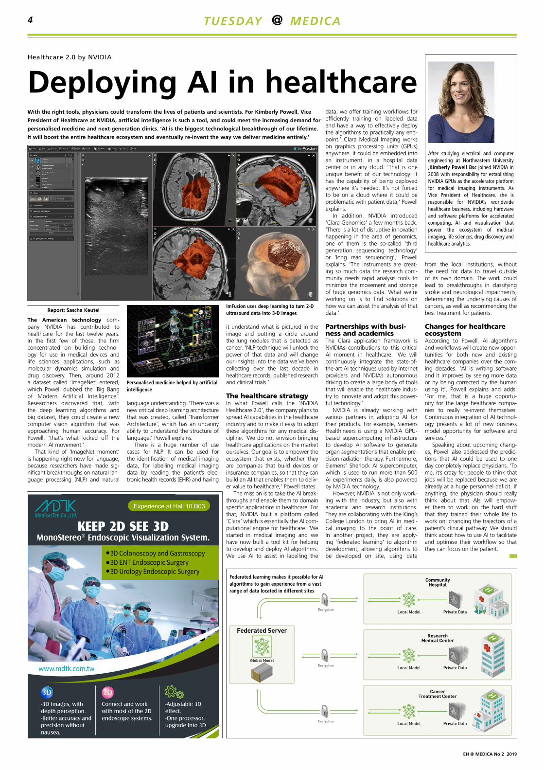
The healthcare strategyIn what Powell calls the ‘NVIDIA Healthcare 2.0’, the company plans to spread AI capabilities in the healthcare industry and to make it easy to adopt these algorithms for any medical dis-cipline. ‘We do not envision bringing healthcare applications on the market ourselves. Our goal is to empower the ecosystem that exists, whether they are companies that build devices or insurance companies, so that they can build an AI that enables them to deliv-er value to healthcare,’ Powell states.
The mission is to take the AI break-throughs and enable them to domain specific applications in healthcare. For that, NVIDIA built a platform called ‘Clara’ which is essentially the AI com-putational engine for healthcare. ‘We started in medical imaging and we have now built a tool kit for helping to develop and deploy AI algorithms. We use AI to assist in labelling the
data, we offer training workflows for efficiently training on labeled data and have a way to effectively deploy the algorithms to practically any end-point.’ Clara Medical Imaging works on graphics processing units (GPUs) anywhere. It could be embedded into an instrument, in a hospital data center or in any cloud. ‘That is one unique benefit of our technology: it has the capability of being deployed anywhere it’s needed. It’s not forced to be on a cloud where it could be problematic with patient data,’ Powell explains.
In addition, NVIDIA introduced ‘Clara Genomics’ a few months back. ‘There is a lot of disruptive innovation happening in the area of genomics, one of them is the so-called ‘third generation sequencing technology’ or ‘long read sequencing’,’ Powell explains. ‘The instruments are creat-ing so much data the research com-munity needs rapid analysis tools to minimize the movement and storage of huge genomics data. What we’re working on is to find solutions on how we can assist the analysis of that data.’
Partnerships with busi-ness and academicsThe Clara application framework is NVIDIAs contributions to this critical AI moment in healthcare. ‘We will continuously integrate the state-of-the-art AI techniques used by internet providers and NVIDIA’s autonomous driving to create a large body of tools that will enable the healthcare indus-try to innovate and adopt this power-ful technology.’
NVIDIA is already working with various partners in adopting AI for their products. For example, Siemens Healthineers is using a NVIDIA GPU-based supercomputing infrastructure to develop AI software to generate organ segmentations that enable pre-cision radiation therapy. Furthermore, Siemens’ Sherlock AI supercomputer, which is used to run more than 500 AI experiments daily, is also powered by NVIDIA technology.
However, NVIDIA is not only work-ing with the industry, but also with academic and research institutions. They are collaborating with the King’s College London to bring AI in medi-cal imaging to the point of care. In another project, they are apply-ing ‘federated learning’ to algorithm development, allowing algorithms to be developed on site, using data
from the local institutions, without the need for data to travel outside of its own domain. The work could lead to breakthroughs in classifying stroke and neurological impairments, determining the underlying causes of cancers, as well as recommending the best treatment for patients.
Changes for healthcare ecosystemAccording to Powell, AI algorithms and workflows will create new oppor-tunities for both new and existing healthcare companies over the com-ing decades. ‘AI is writing software and it improves by seeing more data or by being corrected by the human using it’, Powell explains and adds: ‘For me, that is a huge opportu-nity for the large healthcare compa-nies to really re-invent themselves. Continuous integration of AI technol-ogy presents a lot of new business model opportunity for software and services.’
Speaking about upcoming chang-es, Powell also addressed the predic-tions that AI could be used to one day completely replace physicians. ‘To me, it’s crazy for people to think that jobs will be replaced because we are already at a huge personnel deficit. If anything, the physician should really think about that AIs will empow-er them to work on the hard stuff that they trained their whole life to work on: changing the trajectory of a patient’s clinical pathway. We should think about how to use AI to facilitate and optimise their workflow so that they can focus on the patient.’
After studying electrical and computer engineering at Northeastern University ,Kimberly Powell Bsc joined NVIDIA in 2008 with responsibility for establishing NVIDIA GPUs as the accelerator platform for medical imaging instruments. As Vice President of Healthcare, she is responsible for NVIDIA’s worldwide healthcare business, including hardware and software platforms for accelerated computing, AI and visualisation that power the ecosystem of medical imaging, life sciences, drug discovery and healthcare analytics.
With the right tools, physicians could transform the lives of patients and scientists. For Kimberly Powell, Vice
President of Healthcare at NVIDIA, artificial intelligence is such a tool, and could meet the increasing demand for
personalised medicine and next-generation clinics. ‘AI is the biggest technological breakthrough of our lifetime.
It will boost the entire healthcare ecosystem and eventually re-invent the way we deliver medicine entirely.’
Federated learning makes it possible for AI algorithms to gain experience from a vast range of data located in different sites
ImFusion uses deep learning to turn 2-D ultrasound data into 3-D images
Personalised medicine helped by artificial intelligence
Deploying AI in healthcareHealthcare 2.0 by NVIDIA
TUESDAY @ MEDICA4
EH @ MEDICA No 2 2019
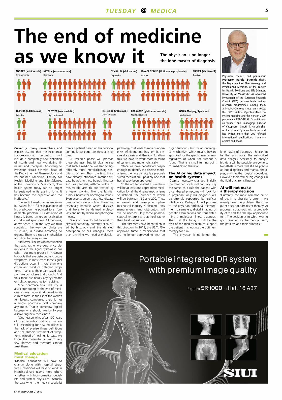
Currently, many researchers and experts assume that the next great socio-economic revolution will include a completely new definition of health and how we define ill-nesses and therapies. According to Professor Harald Schmidt, head of the Department of Pharmacology and Personalised Medicine, Faculty for Health, Medicine and Life Sciences at the University of Maastricht, ‘Our health system today can no longer be sustained in its existing form. It has become too expensive and too ineffective.’
The end of medicine, as we know it? Asked for a fuller explanation of his prediction, he pointed to a fun-damental problem. ‘Our definition of illness is based on organ localisation or individual symptoms. All medicine, as we teach it, in the way we train specialists, the way our clinics are structured, is divided according to organs. There is a specialist physician and clinic for every organ.
‘However, illnesses do not function that way, rather we experience dis-ruptions in the signal systems in our cells – put more precisely, in certain hotspots that are disturbed and cause symptoms. In most cases these signal disruptions occur in more than one organ and produce different symp-toms. Thanks to the organ-based divi-sion, we do not see that though. And thus there are hardly any systematic or holistic approaches to medicine.
‘The pharmaceutical industry is also contributing to the end of medi-cine as we know it, doomed in its current form. In the list of the world’s ten largest companies there is not a single pharmaceutical company any more. That is somehow logical because why should we be forever discovering new medicines?
‘One reason why, after 100 years of pharmaceutical industry, we are still researching for new medicines is the lack of precise illness definitions and the chronic treatment of symp-toms instead of healing. To date, we know the molecular causes of very few illnesses and therefore cannot treat them.’
Medical education must change‘Medical education will have to change along with hospital struc-tures. Physicians will have to work in interdisciplinary teams more often, together with bioinformatics special-ists and system physicians. Actually the days when the medical specialist
pathology that leads to molecular dis-ease definitions and thus permits pre-cise diagnosis and therapy. To attain this, we have to work more in terms of systems and more holistically.
‘Once we have penetrated deeply enough to identify the disease mech-anisms, then we can apply a precisely suited medication – possibly one that has already been approved.
‘In the not too distant future there will be at least one appropriate medi-cation for all the disease mechanisms so defined, the number of which will be between 160 and 200. Thus, a research and development phar-maceutical industry is obsolete; only manufacturers and distributors will still be needed. Only those pharma-ceutical enterprises that heal rather than treat will survive.
‘The first steps have been taken in this direction. In 2018, the USA’s FDA approved tumour medications that are no longer approved to treat an
treats a patient based on his personal current knowledge are now already over.
‘A research phase will precede these changes. But, it’s clear to see that such a medicine will lead to sig-nificantly more interdisciplinary hos-pital structures. Thus, the first clinics have already introduced immune dis-ease boards. In these boards, illnesses such as psoriasis, asthma, colitis or rheumatoid arthritis are treated by a team, working like the familiar tumour boards for oncological issues. Even experts agree that these disease designations are obsolete. These are essentially immune system diseases that have to be defined molecu-larly and not by clinical morphological means.
‘We also have to bid farewell to classical pathology, currently exhaust-ed by histology and the detailed description of cell changes. More than anything we need a molecular
organ tumour – but for an oncologi-cal mechanism, which means they are approved for the specific mechanism, regardless of where the tumour is found. That is a small turning point for medication therapy.’
The AI or big data impact on health systems‘Despite necessary changes, initially, the treatment cycle will naturally stay the same: as a rule the patient with organ-based symptoms will look for a physician, only his diagnosis will be strongly supported by artificial intelligence. Perhaps AI will propose to the physician additional measure-ment parameters, digital imaging or genetic examinations and thus deter-mine a molecular illness diagnosis. Then just like today it will be the task of the medical team to support the patient in choosing the optimum therapy for him.
‘The physician is no longer the
lone master of diagnosis – he cannot be that any more. The networked data analysis necessary to analyse big data will be possible everywhere. Nonetheless there will still be precise ad hoc diagnoses and manual thera-pies, such as the surgical specialties. However, there will be big changes in the field of chronic illnesses.’
AI will not make a therapy decision ‘Currently, the most common cause of death is physician’s error – we already have the problem. The com-puter does not administer therapy; AI proposes a diagnosis with a probabil-ity of x and the therapy appropriate to it. The decision as to which way to go is reserved for the medical team, the patients and their priorities.’
Physician, chemist and pharmacist Professor Harald Schmidt chairs the Department of Pharmacology and Personalised Medicine, at the Faculty for Health, Medicine and Life Sciences, University of Maastricht. As advanced investigator of the European Research Council (ERC) he also leads various research programmes, among them a Proof-of-Concept study on strokes; the COST Action OpenMultiMed on system medicine and the Horizon 2020 programme REPO-TRIAL. Schmidt was co-founder and managing director of Vasopharm GmbH, is co-publisher of the journal Systems Medicine and has written more than 200 refereed international publications, summary articles and books.
The end of medicine as we know it The physician is no longer
the lone master of diagnosis
TUESDAY @ MEDICA 5
More than just MRI accessories
www.allmri.com
• A new compact lightweight housing, specifically designed for mobile equipment.
• A low weight, less than 8.5 kg, combined with compact dimensions, 116 mm diameter and 342 mm length, allows significant reductions in the equipment supporting structures.
• A range of tube inserts up to 54 kW peak radiographic power at high rotation speed is available for this unit.
Visit IAE at MEDICA – Hall 10, Booth B 73 • www.iae.it
IAE – C20
EH @ MEDICA No 2 2019
4K medical cameras gain ISO 13485 Certification
On show at Medica is an expanded range of medical cameras with 4K enhanced-dynamic-range monitor displays. Their manufacturer, Ikegami, reports that its model MKC-750UHD delivers very high-quality imaging from an ultra-compact camera head measuring only 34 x 40 x 40 mm. ‘Based on a three-chip CMOS optical block with progressive scanning and an advanced digital processor, the camera accepts standard C-mount lenses,’ the manufacturer reports. ‘Display-related functions include mirror, flip, rotate, digital zoom and scene file storage.
‘The MKC-750UHD camera head connects via up to 15 metres of medical grade cable to a mains-powered control unit; both units have an anti-bacterial coating. The CCU is designed for easy setup by medical staff, including the ability to operate automatically,’ the firm adds. ‘Technical features include 1/100 to 10,000 shutter speeds, CCU-adjustable iris, auto gain, auto white and auto iris. Sensitivity is 2,000 lux at F8 at lighting levels of 3,200 K, or better. Simultaneous 4K and HD outputs are provided.
‘The camera can be locked to external HDTV tri-level sync and is rated for use across a temperature range of 0 to 40 degrees C. The MKC-750UHD head weighs less than 100g and the CCU less than 3kg.’
The MKC-704UHD is a Full-HD/4K
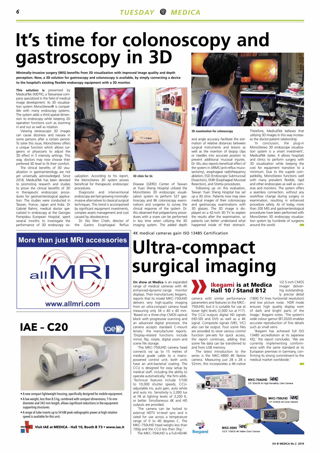
This solution is presented by MedicalTek (MDTK), a Taiwanese com-pany specialized in the field of medical image development. Its 3D visualiza-tion system MonoStereo® is compat-ible with many endoscopy systems. The system adds a third spatial dimen-sion to endoscopy while keeping 2D operation functions such as zooming in and out as well as rotation.
Viewing stereoscopic 3D images can cause dizziness and nausea in some persons after a certain period. To solve this issue, MonoStereo offers a unique function which allows sur-geons or physicians to adjust the 3D effect in 5 intensity settings. This way, doctors may now choose their preferred 3D level to fit their comfort.
The clinical benefits of 3D visu-alization in gastroenterology are not yet universally acknowledged. Since 2018, MedicalTek has been devoted to promoting research and studies to prove the clinical benefits of 3D in therapeutic endoscopic proce-dures for gastroenterological applica-tion. The studies were conducted in Taiwan, France, Japan and India. Dr Gabriel Rahmi, medical doctor spe-cialized in endoscopy at the Georges Pompidou European Hospital, spent several months to investigate the performance of 3D endoscopy vis-
camera with similar performance parameters and features to the MKC-750UHD, but it is suitable for use at lower light levels (2,000 lux at F17). The CCU outputs digital HD signals (HD-SDI and DVI) as well as a 4K signal. Composite signals (VBS, Y/C) also can be output. Four scene files are provided to store various control function pre-sets for quick access, the report continues, adding that scene file data can be transferred to and from USB memory.
The latest introduction to the series is the MKC-X800 4K Native camera. Measuring just 28 x 28 x 52mm, this incorporates a 4K-native
ualization. According to his report, the MonoStereo 3D system proves beneficial for therapeutic endoscopic procedures.
Diagnostic and interventional endoscopy are fast-growing minimally-invasive alternatives to classical surgical techniques. This trend is accompanied by significant equipment investments, complex assets management and cost caused by obsolescence.
Dr. Wu Wen Chieh, director of the Gastro Esophageal Reflux
1/2.5 inch CMOS imager deliver-ing outstanding-ly precise detail
(1800 TV lines horizontal resolution) and low picture noise. ‘HDR mode ensures high quality display even of dark and bright parts of the image,’ Ikegami notes. ‘The system’s wide colour gamut (BT.2020) enables accurate reproduction of fine details such as small veins.
‘Ikegami has achieved full ISO 13485 accreditation at its Japanese HQ,’ the report concludes. ‘We are currently implementing conform-ance with the same standard at its European premises in Germany, con-firming its strong commitment to the medical market worldwide.’
Disease (GERD) Center of Taiwan at Yuan Sheng Hospital utilized the MonoStereo 3D endoscopic visuali-zation system to perform 527 gas-troscopy and 86 colonoscopy exami-nations and surgeries to survey the clinical response of the system. Dr. Wu observed that polypectomy proce-dures with a snare can be performed in less time when utilizing the 3D imaging system. The added depth
and angle accuracy facilitate the esti-mation of relative distances between surgical instruments and lesions as well as the insertion of biopsy clips or needles into accurate position to prevent additional mucosal injuries. Dr. Wu also reports beneficial effect of the system in ARMS (anti-reflux muco-sectomy), esophageal radiofrequency ablation, ESD (Endoscopic Submucosal Dissection), EMR (Esophageal Mucosal Resection), and Stretta procedures.
Following up on this evaluation, Taiwan Yuan Sheng Hospital has set up a 3D clinic. Patients now may view medical images of their colonoscopy and gastroscopy examinations with 3D glasses. The 3D image is dis-played on a 42-inch 3D TV to explain the results after the examination, so patients can better understand what happened inside of their stomach.
Therefore, MedicalTek believes that utilizing 3D images in this way increas-es the doctor-patient relationship.
‘In conclusion, the plug-in MonoStereo 3D endoscope visualiza-tion system is a smart investment’, MedicalTek states. It allows hospitals and clinics to perform surgery with 3D visualization while keeping the cost for equipment transition to a minimum. Due to the superb com-patibility, MonoStereo functions well with many prevalent flexible, rigid and other endoscopes as well as cam-eras and monitors. The system offers a seamless connection, without any workflow change during surgery or examination, resulting in enhanced procedure safety. As of today, more than 200 MIS and gastroenterological procedures have been performed with MonoStereo 3D endoscopy visualiza-tion system by hundreds of surgeons around the world.
Ultra-compact surgical imaging
Ikegami is at Medica Hall 10 / Stand B12
It’s time for colonoscopy and gastroscopy in 3DMinimally-invasive surgery (MIS) benefits from 3D visualization with improved image quality and depth
perception. Now, a 3D solution for gastroscopy and colonoscopy is available, by simply connecting a device
to the hospital’s existing flexible endoscopy equipment with a 3D monitor.
3D examination for colonoscopy
3D clinic for GI.
TUESDAY @ MEDICA6
EH @ MEDICA No 2 2019
Trailblazers in medical solutions
From Augmented Reality to Robotics, and all exciting technol-ogies in between, the Taiwanese companies present at Medica always manage to impress visitors with health tech innovations. In co-opera-tion with the Bureau of Foreign Trade (BOFT) of the Taiwanese Ministry of Economics and the Taiwan External Trade Development Council (TAITRA), 20 elite healthcare companies put on display their achievements in Artificial Intelligence (AI), Virtual Reality (VR), 5G and more.
Growing economic importanceAt Medica, Taiwanese companies showcase their focus on innovative manufacturing and in-house ICT, which have increased the efficiency and quality of production. Today, Taiwan has become the market lead-er with its concept of ‘Asian Silicon Valley’. The government wants to further strengthen this key position by independently developing medi-
cal products that meet the high demands of international markets at attractive terms.
The future of healthcare relies on Big Data and its collection, transmis-sion and evaluation. The develop-ment and implementation of 5G net-works now enables the immediate transfer of large image and monitor-
ing data sets to specialists at remote locations. This reduces the amount of travel required for patient care. The AI supports the time-consum-ing and expensive data evaluation that is becoming increasingly impor-tant in the diagnostic process. The enormous potential of AR/VR offers advantages in the training of medical staff, the performance of complicat-ed operations and the treatment of mental disorders. Robotics has also
made the jump into the realm of healthcare and is now used for the benefit of patients and care-givers.
Medica visi-tors will have the opportunity
to meet – among others – the following
innovative Taiwanese manu-facturers and their products:
AmCad BioMedArtificial Intelligence innovation expertise company with professional clinical experience, AmCad BioMed has created an ultrasound-based obstructive sleep apnea (OSA) detec-tion system, AmCAD®-UO.
According to a recent survey, around one billion people in the
world suffer from OSA, 80% of them remain undiagnosed. The most com-mon method of diagnosis is a poly-somnography (PSG), which requires an overnight stay at a sleep center. However, the limited availability of sleep tests may lead to long waiting times.
While PSG helps diagnosing sleep disorders through physiological index overnight, AmCAD-UO provides effi-cient anatomical OSA detection for patients within 10 minutes – with-out the need for the patient to be asleep. With laser-guided position-ing, the device precisely scans the upper airway and analyzes the dif-ference between normal breathing and Müller’s Maneuver (mimicking snoring model). AmCAD-UO system standardizes ultrasound transducer scanning to make the assessment more consistent and avoid highly operator-dependent variations.
The president further pointed out that “through our patented laser-
guided system, intra-observer vari-ability is largely reduced.”
iHelperChiliGO AI Robot Kiosk and iCart from manufacturer iHelper offer many advantages and more useful features than other mobile kiosk robots. With its pleasant robot design and intuitive user-friendly interface, users of the ChiliGO will have a faster and more satisfying experience get-ting the information they need, while the iCart handling robot can be used in hospitals or all other fields where items need to be transported.
The ChiliGO uses facial recogni-tion technology to analyze user infor-mation like gender and age, which can be used to enhance a company’s marketing based on key consumer demographics. It also features AI voice interaction.
iCart operates independent of tracks, instead using opti-cal radar (LiDar) and ultra-sonic sensors with AI self-optimized path correction technology to achieve automatic navigation and smart obstacle avoidance, to
quickly complete equipment delivery tasks.
IEIIndustrial computer provider IEI Integration Corp., presents its solu-tions for AI in healthcare at Medica. These include a global platform for health management and real-time medical data collection to offer a flexible way and enhance work effi-ciency for daily hospital routine.
This year at Medica, IEI will show-case its latest AI ready hardware – a POCm Series Panel PC with hot-swapped batteries, a box PC solution for endoscopy and AI an all-in-one terminal. Further new prod-ucts include solutions for big data management, AI training (machine learning and accelerator cards) and AI decision support.
ConclusionTake advantage of this unique setting during the upcoming Medica to feel the pulse of these leaps forward in 5G, AI, VR/AR and robotics. Medica is a premier opportunity for Taiwanese companies to develop relationships throughout the year. Staff and rep-
resentatives of these innova-tive companies wel-
come the chance to engage, exchange
and demonstrate their advances in hall 17,
stand A40. For more
information, please visit: https://www.taiwanexcel-lence.org/de
Laboratory staff is permanently exposed to harmful formalin vapors. The GrossPath GP-1500 reduces this risk to a bare minimum as contami-nated air is being extracted immedi-ately downward and also backwards. Additionally, the tissue grossing sta-tion complies with MAC (maximum allowable concentration) values for formalin.
Convince yourself of the advantages of a GrossPathThe No. 1 solution for eco-friendly tissue grossing stations is coming to Düsseldorf. Visit our booth and let us convince you of all the advantages that our GrossPath has to offer.
The special worktable designed to ensure a pollution-free, laboratory-grade working environment for slicing and preparing histological slide prep-arations. Its compact design makes the GrossPath Special Worktable the perfect laboratory equipment, espe-cially for small facilities. As a product of our ECO-line, this worktable was designed with a special view to con-serving energy and resources while delivering outstanding air extraction results. Buy it, set it up and power
it on: thanks to its active-carbon air recirculation system, the GrossPath is ready for immediate use – no need for installing a costly, separate air ventilation system! As a result, a customer-supplied ventilation is not necessary, which can reduce invest-ment and even overheads by up to 30 %. A permanganate carbon filter intercepts and efficiently purifies contaminated air and directs the clean air back to the labora-tory. Thanks to the down-draft and additional backdraft system, formalin con-cen t r a t i on will even be signifi-cantly below the MAC value throughout the entire extraction zone. Additionally, the GrossPath has a soundproofed exhaust fan that substantially keeps the noise level to
a minimum. Thanks to its large per-manganate carbon filter, the table can be in use for up to 800 working hours*
* operating time varies between 600 and 800 working hours
Taiwanese excellence on display at Medica
See the GrossPath GP-1500 live in action!
More safety for blood banks
KUGEL medical is at Medica Hall 13 / Stand E09
Ensuring the quality of blood dona-tions is vital. Data collected dur-ing the various processes involved must be fully traceable and docu-mented, the company Andreas Hettich GmbH emphasises. Based in Tuttlingen, Germany, the com-pany has produced laboratory equip-ment for 115 years, today focusing on centrifuges and incubators and employing 420 people worldwide.
Its new HettInfo II documents all steps before and during centrifuga-tion. ‘The touch display guides the user step-by-step through the process, from scanning the blood bags to centrifu-gation,’ Hettich reports. ‘The blood bag ID, operator and program are entered using a barcode scanner. The running parameters are recorded in the background during centrifuga-tion and can later be exported as a CSV file to a USB stick or, alternatively, directly to the network. The export can take place cyclically after each run or time-controlled.’
iHelper ChiliGO has a faster and more satisfying experience getting the information they need, while the iCart handling robot can be used in hospitals or all other fields where items need to be transported.
Hettich provides transparency and traceability
Hettich is at Medica Hall 3 / Stand B69
Taitra is at Medica Taiwan Pavilion Hall 17 / A40
TUESDAY @ MEDICA 7
EH @ MEDICA No 2 2019
ACEMSO15F is a new focusable and flexible LED examination light designed by the Italian firm ACEM. The touch panel controls all lamp functions, including light intensity adjustment and beam focusing. The result is a uniform, homogeneous and shadow-less light. List of assets:
• Light intensity 110,000 lux • IR-free light beam • Light beam focusing • Colour temperature (CCT) of 4,500 K• Colour rendering index (CRI) of 97 • Low power consumption (25 W)
and long life.
The distribution of light is uniform and the beam can be focused with perfect illumination both on the sur-face and in depth, providing the oper-ator with the best working conditions, ACEM points out, adding that the easy-to-grip removable and sterilis-able handle makes the device suitable even for critical sanitary applications.
An optional rechargeable battery powered system, ABPS, supplies effi-cient light in emergencies, or situ-ations without electrical supply, or in sudden black-outs, humanitarian interventions, and in field hospitals without UPS or power generators.
The ABPS cover is made of resist-
NICO Corporation, a company based in Indianapolis, Indiana, is releas-ing its new Myriad Novus resection tool. The FDA cleared product com-bines xenon illumination with NICO’s proven Myriad neural tissue removal technology.
Xenon lights reproduce the visible spectrum of daylight much better than most other bulbs – although LED contenders are now available – and so are often the optimal choice when working in a confined environ-ment and where precision is key.
Microscope, surgical loupe or exoscopeThe Myriad Novus is intended to help with intracerebral hemorrhage clot evacuation and subcortical tumor resection, whether using a micro-scope, surgical loupe, or an exo-scope. The company claims that it provides, “for the first time ever, automated and intraoperative tissue harvesting for post-procedural analy-sis in the modern molecular era.”
The technology gives surgeons one solution for intracerebral hemor-rhage clot evacuation and subcorti-cal tumor resection whether using a microscope, surgical loupes or an exoscope. Its capabilities include illumination offering enhanced visu-alization with the surgeon’s preferred optical platform, improved in-situ tis-sue identification, non-ablative and non-thermal resection, automated
tissue harvesting with the ability to annotate by intratumoral location, and biological preservation of har-vested tissue for post-procedural analysis.
More versatility for surgeons“The Novus represents the next generation of NICO’s flagship product – the Myriad,” said Jim Pearson, President and CEO of NICO Corporation. “We are excited to give
surgeons more versatility across mini-mally invasive port access, endoscop-ic, and skull base surgical approaches while still addressing their needs sur-rounding procedural efficiencies and convenience.”
ant plastic and the lamp delivers the same performance with the battery as without.
The control panel enables manage-ment of the residual charge, type of power supply, recharge status and electrical power supply presence.
This lamp is available for mount-ing on wall, ceiling (single or double configuration), trolley (as above, ABPS battery on demand).
Acem is at Medica Hall 10 / Stand B60
Lighting the way forward
Xenon lighting for neurosurgery
A focusable LED exam and minor surgery lampNICO Myriad NOVUS resection tool
The FDA-cleared NICO Myriad Novus gives surgeons precise light exactly where they need it.
More dynamic, more digital, and more networked – the medical industry is striding into the future. Thus, it’s wise to keep a finger on the pulse, be informed and deepen com-munications with specialists.
At Medica, the presence of start-ups has grown annually, largely due to the special attention they receive. This year, 36 out of several hun-dred of these mostly young and digitally-driven firms can be visited in the Medica Start-up Park (Hall 13, Stand 13 D56-F56). There, they present expertise and exciting ideas – involving the Internet of Things (IoT),
smart solutions, artificial intelligence, the exciting virtual and augmented reality, and wearable solutions, lab-on-skin technology and biomarker analysis.
Those pioneering innova-tions include live presentations in the Medica Disrupt programme, held within the Medica Connected Healthcare Forum (same hall).
For the ‘11th Innovation World Cup’ for the best healthcare solu-tion, blocks of 10 finalists will be announced. Day one of Medica will see a focus on healthcare IoT solu-tions. On the second day, the aim is to select the world’s best health app.
place to ensure contact can be made between healthcare industrialists and
these young companies. In addition, ‘accelerator programs’ have been organised so that companies such as Merck, Bayer and Roche can intro-
duce themselves to the start-ups. Additionally, for biotech diagnostics start-ups, an investors’ gathering has been arranged at the nearby Maritim Hotel.
On 18-19 November, the finalists will be on stage to receive international recognition and their cash awards.
The Hall also provides a meeting
Look out for the Start-Up Park in Hall 13
Young and digitally driven inventors
MEET THE START-UPS
• In the Medica Start-Up Park in hall 13, stand 13 D56-F56 you will find innovative pro-ducts of 36 start-ups from the fields of diagnostics, Internet of Medical Things, smart solu-tions, Artificial Intelligence, Virtual and Augmented Reality, mHealth and wear-able solutions.
• In the Medica Connected Healthcare Forum in hall 13, stand 13 D45, around 100 start-ups will present their products and ideas in the sessions and Medica Disrupt Start-up Pitches. In the after-noon of November 19, health-care accelerator programs such as by Merck, Bayer G4A and Roche will introduce themselves to the start-ups via the Reverse Pitch Session.
• Start-ups can apply for the leading international com-petitions of the 8th Medica App Competition and the 11th Healthcare Innovation World Cup. The finalists will be on the MCHF stage in hall 13, stand 13 D45, on November 18 and 19, and can win valuable business contacts, internation-al recognition and cash prizes.
Medica Start-up Park Hall 13 / Stand 13 D56-F56
TUESDAY @ MEDICA8
EH @ MEDICA No 2 2019
AI? We shouldn’t worry about it – yet
Report: Mélisande Rouger
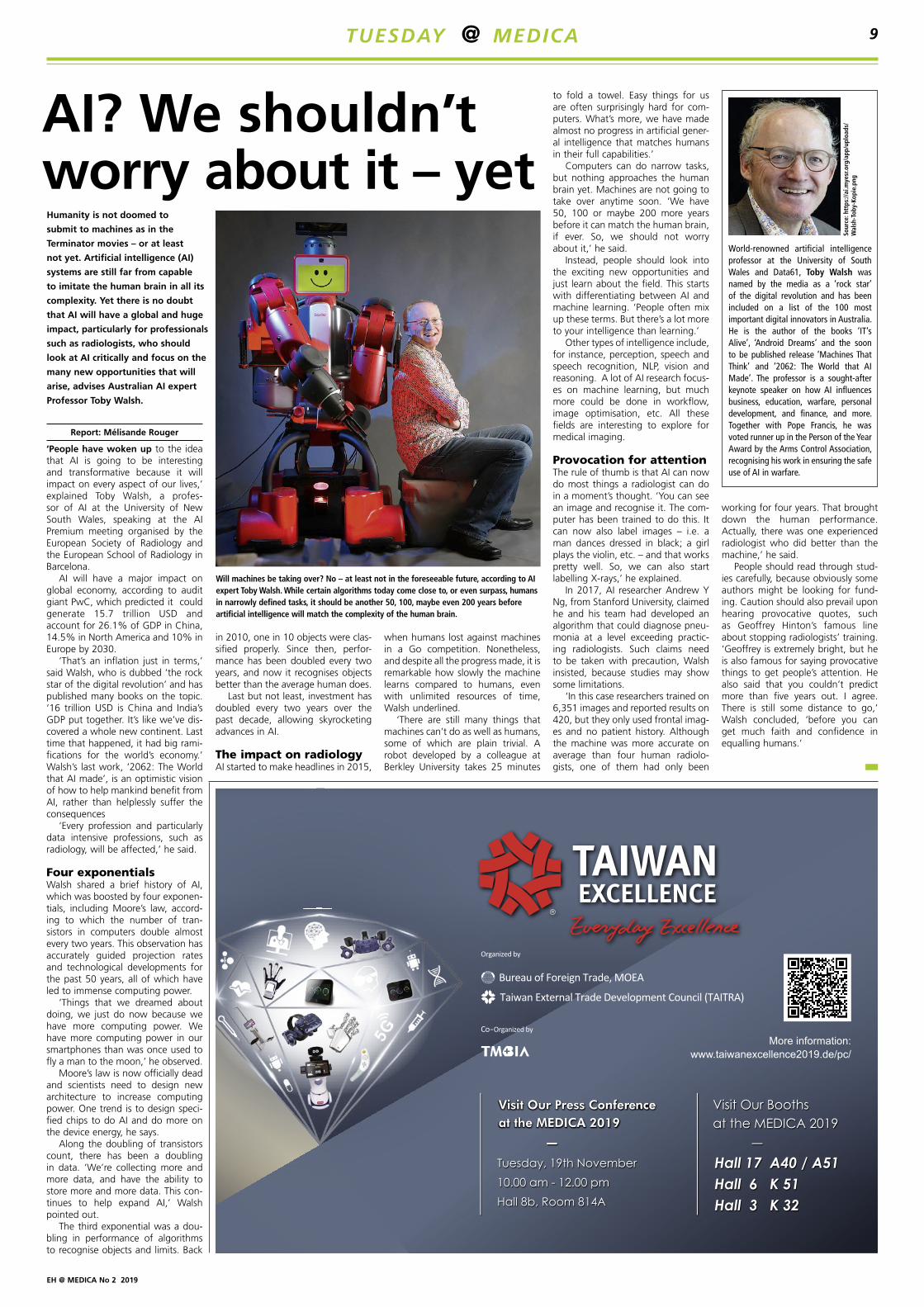
‘People have woken up to the idea that AI is going to be interesting and transformative because it will impact on every aspect of our lives,’ explained Toby Walsh, a profes-sor of AI at the University of New South Wales, speaking at the AI Premium meeting organised by the European Society of Radiology and the European School of Radiology in Barcelona.
AI will have a major impact on global economy, according to audit giant PwC, which predicted it could generate 15.7 trillion USD and account for 26.1% of GDP in China, 14.5% in North America and 10% in Europe by 2030.
‘That’s an inflation just in terms,’ said Walsh, who is dubbed ‘the rock star of the digital revolution’ and has published many books on the topic. ‘16 trillion USD is China and India’s GDP put together. It’s like we’ve dis-covered a whole new continent. Last time that happened, it had big rami-fications for the world’s economy.’ Walsh’s last work, ‘2062: The World that AI made’, is an optimistic vision of how to help mankind benefit from AI, rather than helplessly suffer the consequences
‘Every profession and particularly data intensive professions, such as radiology, will be affected,’ he said.
Four exponentials Walsh shared a brief history of AI, which was boosted by four exponen-tials, including Moore’s law, accord-ing to which the number of tran-sistors in computers double almost every two years. This observation has accurately guided projection rates and technological developments for the past 50 years, all of which have led to immense computing power.
‘Things that we dreamed about doing, we just do now because we have more computing power. We have more computing power in our smartphones than was once used to fly a man to the moon,’ he observed.
Moore’s law is now officially dead and scientists need to design new architecture to increase computing power. One trend is to design speci-fied chips to do AI and do more on the device energy, he says.
Along the doubling of transistors count, there has been a doubling in data. ‘We’re collecting more and more data, and have the ability to store more and more data. This con-tinues to help expand AI,’ Walsh pointed out.
The third exponential was a dou-bling in performance of algorithms to recognise objects and limits. Back
in 2010, one in 10 objects were clas-sified properly. Since then, perfor-mance has been doubled every two years, and now it recognises objects better than the average human does.
Last but not least, investment has doubled every two years over the past decade, allowing skyrocketing advances in AI.
The impact on radiologyAI started to make headlines in 2015,
when humans lost against machines in a Go competition. Nonetheless, and despite all the progress made, it is remarkable how slowly the machine learns compared to humans, even with unlimited resources of time, Walsh underlined.
‘There are still many things that machines can’t do as well as humans, some of which are plain trivial. A robot developed by a colleague at Berkley University takes 25 minutes
to fold a towel. Easy things for us are often surprisingly hard for com-puters. What’s more, we have made almost no progress in artificial gener-al intelligence that matches humans in their full capabilities.’
Computers can do narrow tasks, but nothing approaches the human brain yet. Machines are not going to take over anytime soon. ‘We have 50, 100 or maybe 200 more years before it can match the human brain, if ever. So, we should not worry about it,’ he said.
Instead, people should look into the exciting new opportunities and just learn about the field. This starts with differentiating between AI and machine learning. ‘People often mix up these terms. But there’s a lot more to your intelligence than learning.’
Other types of intelligence include, for instance, perception, speech and speech recognition, NLP, vision and reasoning. A lot of AI research focus-es on machine learning, but much more could be done in workflow, image optimisation, etc. All these fields are interesting to explore for medical imaging.
Provocation for attentionThe rule of thumb is that AI can now do most things a radiologist can do in a moment’s thought. ‘You can see an image and recognise it. The com-puter has been trained to do this. It can now also label images – i.e. a man dances dressed in black; a girl plays the violin, etc. – and that works pretty well. So, we can also start labelling X-rays,’ he explained.
In 2017, AI researcher Andrew Y Ng, from Stanford University, claimed he and his team had developed an algorithm that could diagnose pneu-monia at a level exceeding practic-ing radiologists. Such claims need to be taken with precaution, Walsh insisted, because studies may show some limitations.
‘In this case researchers trained on 6,351 images and reported results on 420, but they only used frontal imag-es and no patient history. Although the machine was more accurate on average than four human radiolo-gists, one of them had only been
working for four years. That brought down the human performance. Actually, there was one experienced radiologist who did better than the machine,’ he said.
People should read through stud-ies carefully, because obviously some authors might be looking for fund-ing. Caution should also prevail upon hearing provocative quotes, such as Geoffrey Hinton’s famous line about stopping radiologists’ training. ‘Geoffrey is extremely bright, but he is also famous for saying provocative things to get people’s attention. He also said that you couldn’t predict more than five years out. I agree. There is still some distance to go,’ Walsh concluded, ‘before you can get much faith and confidence in equalling humans.’
Humanity is not doomed to
submit to machines as in the
Terminator movies – or at least
not yet. Artificial intelligence (AI)
systems are still far from capable
to imitate the human brain in all its
complexity. Yet there is no doubt
that AI will have a global and huge
impact, particularly for professionals
such as radiologists, who should
look at AI critically and focus on the
many new opportunities that will
arise, advises Australian AI expert
Professor Toby Walsh.
World-renowned artificial intelligence professor at the University of South Wales and Data61, Toby Walsh was named by the media as a ‘rock star’ of the digital revolution and has been included on a list of the 100 most important digital innovators in Australia. He is the author of the books ‘IT’s Alive’, ‘Android Dreams’ and the soon to be published release ‘Machines That Think’ and ‘2062: The World that AI Made’. The professor is a sought-after keynote speaker on how AI influences business, education, warfare, personal development, and finance, and more. Together with Pope Francis, he was voted runner up in the Person of the Year Award by the Arms Control Association, recognising his work in ensuring the safe use of AI in warfare.
Sour
ce: h
ttps
://ai
.mye
sr.o
rg/a
pp/u
ploa
ds/
Wal
sh-T
oby-
Kopi
e.pn
g
Will machines be taking over? No – at least not in the foreseeable future, according to AI expert Toby Walsh. While certain algorithms today come close to, or even surpass, humans in narrowly defined tasks, it should be another 50, 100, maybe even 200 years before artificial intelligence will match the complexity of the human brain.
TUESDAY @ MEDICA 9
EH @ MEDICA No 2 2019
AI in diagnostics
Learn like a human, deduce like a machine
For some people, machine learning is the computer equivalent of intel-ligence, but Dr Martin Hirsch begs to differ. ‘For me, first and foremost, intelligence is the ability to generate sensible behaviour in an entirely new situation. An algorithm that iterates a procedure thousands of times and then deduces certain rules from these runs is not necessarily intelligent.’ In other words: intelligence is the ability to generate hypotheses from existing knowledge and, in a next step, to rec-ognise possible courses of action.
While a computer requires innumer-able data sets to master a certain pro-cedure reliably, humans in many cases need but a single illustrative example to recognise and understand complex relationships. ‘That’s due to our abil-ity to abstract,’ Hirsch explains. While building knowledge in humans may take much longer than feeding an algorithm with data sets, the brain of a physician, for example, will acquire a deep understanding that can offer a sound solution even in unfamiliar cases.
Pattern recognition – all the rage in medical applications – might be sufficient to provide a working hypoth-esis, but never a reliable diagnosis. A pattern recognised by the computer may indeed be due to patient physiol-ogy, but it may also be due to poor data, such as image artefacts. ‘When building a decision support system, therefore, we shouldn’t start from square one, but build upon validated
scientific knowledge,’ the neuroscien-tist underlines. Ideally, he says, we use existing knowledge and incorpo-rate insights from pattern recognition. This approach is rather similar to the “human model” where junior physi-cians first acquire knowledge during their medical school studies and later expand and adapt this knowledge with each day of practical experience.
‘How did you figure this out?’Ada, an AI-based platform developed at Hirsch’s eponymous company, works pretty much according to this pat-tern. It analyses certain parameters, weighs them and generates a hypoth-esis from these data, accompanied by a probability score for the validity of the hypothesis. ‘This includes collect-ing positive and negative evidence, very much like during a differential diagnosis. Ada tries to compare and contrast similar pathologies to differ-entiate and exclude.’ This procedure is meant to make the ‘thought process’ of the algorithm understandable for the human brain. ‘Transparency is cru-cial,’ according to Hirsch, because ‘AI systems will only be able to prevail in a hospital and healthcare system if the machine can explain why it arrived at a certain conclusion.’
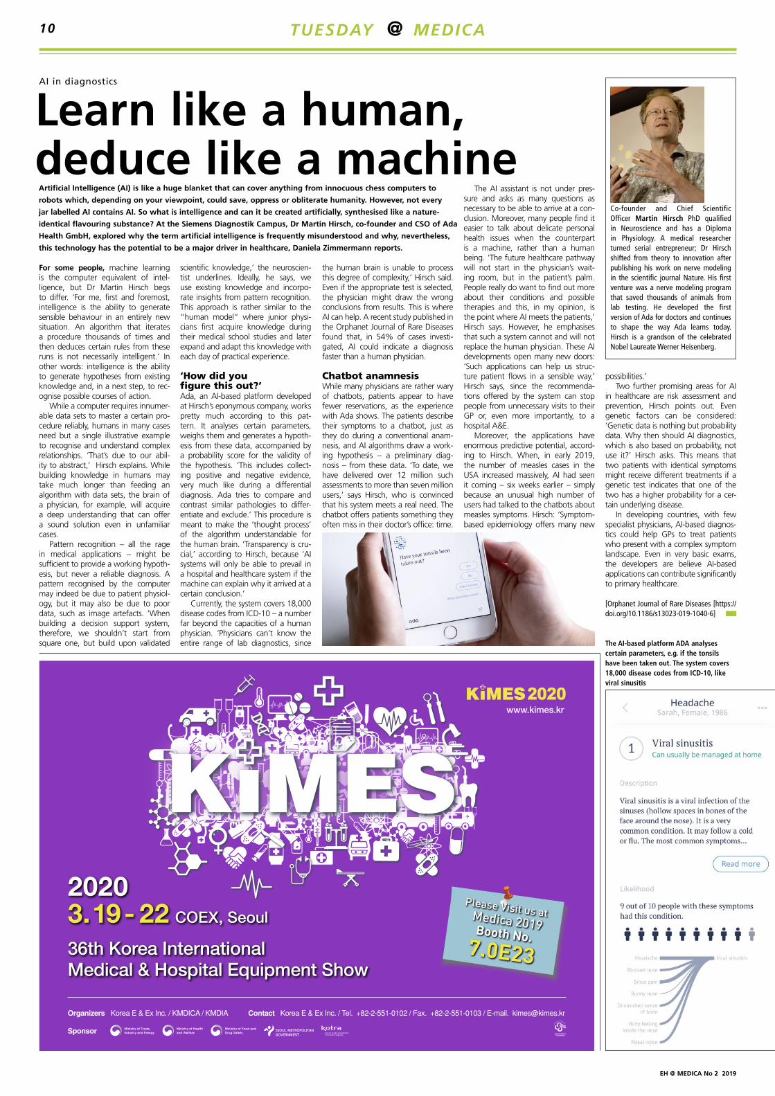
Currently, the system covers 18,000 disease codes from ICD-10 – a number far beyond the capacities of a human physician. ‘Physicians can’t know the entire range of lab diagnostics, since
the human brain is unable to process this degree of complexity,’ Hirsch said. Even if the appropriate test is selected, the physician might draw the wrong conclusions from results. This is where AI can help. A recent study published in the Orphanet Journal of Rare Diseases found that, in 54% of cases investi-gated, AI could indicate a diagnosis faster than a human physician.
Chatbot anamnesis While many physicians are rather wary of chatbots, patients appear to have fewer reservations, as the experience with Ada shows. The patients describe their symptoms to a chatbot, just as they do during a conventional anam-nesis, and AI algorithms draw a work-ing hypothesis – a preliminary diag-nosis – from these data. ‘To date, we have delivered over 12 million such assessments to more than seven million users,’ says Hirsch, who is convinced that his system meets a real need. The chatbot offers patients something they often miss in their doctor’s office: time.
The AI assistant is not under pres-sure and asks as many questions as necessary to be able to arrive at a con-clusion. Moreover, many people find it easier to talk about delicate personal health issues when the counterpart is a machine, rather than a human being. ‘The future healthcare pathway will not start in the physician’s wait-ing room, but in the patient’s palm. People really do want to find out more about their conditions and possible therapies and this, in my opinion, is the point where AI meets the patients,’ Hirsch says. However, he emphasises that such a system cannot and will not replace the human physician. These AI developments open many new doors: ‘Such applications can help us struc-ture patient flows in a sensible way,’ Hirsch says, since the recommenda-tions offered by the system can stop people from unnecessary visits to their GP or, even more importantly, to a hospital A&E.
Moreover, the applications have enormous predictive potential, accord-ing to Hirsch. When, in early 2019, the number of measles cases in the USA increased massively, AI had seen it coming – six weeks earlier – simply because an unusual high number of users had talked to the chatbots about measles symptoms. Hirsch: ‘Symptom-based epidemiology offers many new
possibilities.’Two further promising areas for AI
in healthcare are risk assessment and prevention, Hirsch points out. Even genetic factors can be considered: ‘Genetic data is nothing but probability data. Why then should AI diagnostics, which is also based on probability, not use it?’ Hirsch asks. This means that two patients with identical symptoms might receive different treatments if a genetic test indicates that one of the two has a higher probability for a cer-tain underlying disease.
In developing countries, with few specialist physicians, AI-based diagnos-tics could help GPs to treat patients who present with a complex symptom landscape. Even in very basic exams, the developers are believe AI-based applications can contribute significantly to primary healthcare.
[Orphanet Journal of Rare Diseases [https://doi.org/10.1186/s13023-019-1040-6]
Co-founder and Chief Scientific Officer Martin Hirsch PhD qualified in Neuroscience and has a Diploma in Physiology. A medical researcher turned serial entrepreneur; Dr Hirsch shifted from theory to innovation after publishing his work on nerve modeling in the scientific journal Nature. His first venture was a nerve modeling program that saved thousands of animals from lab testing. He developed the first version of Ada for doctors and continues to shape the way Ada learns today. Hirsch is a grandson of the celebrated Nobel Laureate Werner Heisenberg.
Artificial Intelligence (AI) is like a huge blanket that can cover anything from innocuous chess computers to
robots which, depending on your viewpoint, could save, oppress or obliterate humanity. However, not every
jar labelled AI contains AI. So what is intelligence and can it be created artificially, synthesised like a nature-
identical flavouring substance? At the Siemens Diagnostik Campus, Dr Martin Hirsch, co-founder and CSO of Ada
Health GmbH, explored why the term artificial intelligence is frequently misunderstood and why, nevertheless,
this technology has the potential to be a major driver in healthcare, Daniela Zimmermann reports.
The AI-based platform ADA analyses certain parameters, e.g. if the tonsils have been taken out. The system covers 18,000 disease codes from ICD-10, like viral sinusitis
TUESDAY @ MEDICA10
EH @ MEDICA No 2 2019
Lithuanian chemist advances cell research
Crafting new molecular tools
Imaging robot for spinal surgery
Put simply, viscosity sensors are small molecules used to measure surrounding viscosity; they are used as tools to study events inside a liv-ing cell. Various diseases, such as Alzheimer’s and diabetes, cause vis-cosity changes in cells. With sensors of improved sensitivity, it will be possible to improve determination of what adverse effects arrive with changed cell viscosity.
‘I call myself a craftsman of tools – molecular kind of tools,’ Vyšniauskas explains. ‘Personally, I will not cure Alzheimer’s, nor diabetes; but, if I’m successful, I will finally manage to create a tool that would help other scientists to treat these and other incurable diseases, popularly called civilizational epidemics.’
In early-stage diabetes, the viscos-ity of the membrane of liver cells increases. The receptors of insulin in the membrane of these cells con-tinually deliver signals and connect with one another, but the increase in viscosity leads to poor connections,
The Loop-X robotic architecture intro-duces a new standard in flexibility, add-ing additional degrees of freedom to any surgical procedure: By automating imaging workflow steps and roboti-cally moving with the procedure and on command, the system is in sync with other devices like robotic arms and with the surgeon and staff.
One interesting feature of the Loop-X is the non-isocentric move-ment of the X-ray source and detector. Unlike in other CT imagers, the two are not always directly across from each other, and the X-ray beam turns to always point toward the detector. This allows the device to better target the specific anatomy of interest while minimizing the delivered radiation dose. Additionally, the capability can be used to better view anatomy at a larger scale by widely sweeping the beam closer to
the outer edge of the scanner.The highly integrated device is
designed for 2D and 3D imaging, com-bining ultra-high resolution with extra-low doses, and proprietary technology to image with interlaced energies for soft-tissue visualization.
Loop-X imaging robot can be inte-grated with Brainlab technology as well as third party products through an open interface, maximizing inter-operability and data integration. “For Brainlab, Loop-X is a critical milestone in contributing disruptive innovations in spinal surgery,” said Stefan Vilsmeier, President and Chief Executive Officer, Brainlab. “It provides us with an even stronger foundation for leveraging emerging technologies such as AI, big data, cloud computing, augmented reality and spatial computing.”
the regular cell activity is disrupted, and the cells are no longer able to recognise insulin.
‘Up to this point, the most popu-lar viscosity sensor could measure cell viscosity from five to 1,500 vis-cosity units of measurement, or cP,’
Vyšniauskas adds. ‘We managed to increase this – our method can measure viscosity in the range of 0.5 to 50,000 cP. That’s a big accom-plishment. Imagine a thermometer that could only measure tempera-ture from 10 to 20 degrees Celsius.
Metaphorically speaking, we have created a new thermometer with a measurement range from -10 to 60 degrees Celsius.’
Currently, viscosity sensors are only used by scientists who study them, but this scientist has no doubt: once this method develops to become a reliable tool, it will be used by bio-chemists and cell biologists alike.
This breakthrough in viscosity measurement resulted from a collab-oration between many scientists from the Centre for Physical Sciences and Technology and Vilnius University, in Lithuania, where Vyšniauskas now works.
‘Since physical chemistry is an interdisciplinary field, we needed knowledge from various scientific fields to make this achievement pos-sible,’ he points out. ‘As a chemist, I need to consult with theoretical physicists who are specialists in theo-retical calculations, which are cru-cial to explain how viscosity sensors
work. I also cooperate with other chemists, particularly those who syn-thesise molecules. Biologists are also involved, because the implementa-tion of our research would be in their field, biology. The collaborative process is fascinating.’
The decision to head home after gaining his doctorate in the UK was pretty straightforward, he explains. ‘A couple of reasons were at play when deciding where to continue my career after the studies. For one, the Department of Molecular Compound Physics, where I now work, employs several excellent sci-entists who produce international-level results. Being a young scientist, it is a privilege to work hand in hand with these experienced researchers. Also, it’s a more stable environment for a scientist here than in the UK. Extremely high competition in the UK science community causes unneces-sary stress, while here, I can focus on my research and feel valued. Finally, we have state-of-the-art facilities and favourable all-round conditions.’
The scientific achievement of Vyšniauskas and colleagues is not an isolated case of success for Vilnius-based scientists. Just this year, Vilnius physicists have developed a laser that possibly could solve the nuclear waste problem, while other scientists from the Centre for Physical Sciences and Technology, the same institution in which Vyšniauskas works, have developed a cheaper and faster DNA testing method.
Recently, chemist Aurimas Vyšniauskas a 31-year-old Oxford and Imperial College London graduate, was
featured in the European journal Chemistry because he and his research team have changed the structure of
viscosity sensors and enhanced their viscosity-sensitivity range by a very significant margin.
Brainlab recently unveiled its flagship Loop-X mobile intraoperative imaging
robot that is specifically designed for spinal surgical procedures. Developed
by medPhoton, an Austrian company, the device is now the core of Brainlab’s
imaging offerings.
Loop-X-positioning
© S
hutt
erst
ock/
Ligh
tspr
ing
© S
hutt
erst
ock/
Prox
ima
Stud
io
TUESDAY @ MEDICA 11
Omnia Medical’s Boxcar is the first cervical VBR system to be made from PEEK-OPTIMA™ HA Enhanced, from Invibio. ©
JA
LEX
Med
ical
EH @ MEDICA No 2 2019
Editor-in-Chief: Brenda Marsh
Editorial team: Wolfgang Behrends, Sonja Buske
Senior Writer: John Brosky
Executive Director: Daniela Zimmermann
Founded by Heinz-Jürgen Witzke
ISSN 0942-9085
CorrespondentsAustria: Michael Krassnitzer, Christian Pruszinsky
China: Nat Whitney
France: Jane MacDougall
Germany: Anja Behringer, Annette Bus, Walter Depner, Cornelia Wels-Maug, Holger Zorn
Great Britain: Brenda Marsh, Mark Nicholls
France, Italy, Spain: Eric Jund Phone: +33 493 58 77 43 E-Mail: [email protected]
GB, Scandinavia, BeNeLux: Simon Kramer Phone: +31 180 6200 20 E-Mail: [email protected]
Israel: Hannah Wizer, International Media Dep. of El-Ron Adv. & PR Co., Ltd., Phone: +972-3-6 955 367 E-Mail: [email protected]
South Korea: Jane Park, MCI Phone: +82 2 730 1234 E-Mail: [email protected]
Taiwan: Charles Yang, Phone: +886 4 232 236 33 E-Mail: [email protected]
USA & Canada: Hanna Politis, Media International Phone: +1 301 869 66 10 E-Mail: [email protected]
All company, brand and product names in this publication are the property of their respective holders. Users must obtain permission from those holders before copying or using the owner’s trademarks, product and company names or logos.
Malta: Moira Mizzi
Spain: Mélisande Rouger, Eduardo de la Sota
The Netherlands: Madeleine van de Wouw
USA: Cynthia E. Keen, i.t. Communications, Lisa Chamoff
SubscriptionsDorothea Fleischer, Theodor-Althoff-Str. 45, 45133 Essen, Germany
Subscription rate6 issues: 42 Euro, Single copy: 7 Euro.
Send order and cheque to:
European Hospital Subscription Dept
Printed by: WVD, Möhrfelden, Germany
Publication frequency: bi-monthly
RepresentativesChina & Hongkong: Gavin Hua, Sun China Media Co, Ltd. Phone: +86-0755-81 324 036 E-Mail: [email protected]
Germany, Austria, Switzerland: Ralf Mateblowski Phone: +49 6735 912 993 E-Mail: [email protected]
TomTom Telematics delivers patients on time
‘Smart shirt’ to monitor lung disease
Help for cervical spine
A smart shirt, developed by Canadian startup Hexoskin, has been success-fully tested as a potential diagnos-tic modality for chronic obstructive pulmonary disease (COPD) at the Radboud University Medical Centre in The Netherlands. “COPD is a growing problem with around 64 million people suffering with the condition worldwide. When patients suffer an increase in their symptoms, such as coughing and breathlessness, they need to be monitored more closely,” said lead researcher Denise Mannée, a technical physician and PhD candidate at Radboud University Medical Centre in The Netherlands.
For their study, the researchers recruited 15 healthy volunteers and
Robert Gewirtz, MD - Neurosurgeon, Columbus, Ohio, collaborated on the development of the product. He says: “I see many patients that require a cervical corpectomy, and it’s exciting to have such an innovative option for these patients. Boxcar is very easy to use and provides intra-operative flexibility, allowing me to dial in a VBR construct to match my patient’s anatomy”.
The novel single-use device is available in two footprints and allows fine adjustments of height and lor-dosis using spacers and endplates. It features a hollow center and holes to
accommodate autograft or allograft, while encouraging the formation of new bone, and includes anti-migra-tion features.
The system uses the enhanced biomaterial hydroxyapatite (HA), which has been shown solid fusions with dense bone apposition at 6 months and beneficial clinical out-comes for patients, revealing specific improvements in overall pain and neurological function in early clinical results.
had them wear the shirt while doing everyday activities including walk-ing, climbing stairs, vacuuming, sit-ting, standing and even lying down. The smart shirt, called the Hexoskin, senses how the fabric stretches when the wearer’s chest expands and con-tracts and uses these measurements to gauge the volume of air inhaled and exhaled.
At the same time, the participants also wore the equipment tradition-ally used to measure breathing that includes a face mask and bulky back-pack. They repeated the tasks again wearing both pieces of equipment, to generate a second set of data.
In general, the researchers found that the measurements were very
similar. When lying, the difference between measurements taken by the two pieces of equipment was just 0.2% on average, which only represents a few millilitres of air. In more strenuous activities there were slightly greater differences, for exam-ple with vacuuming the difference was 3.1% on average, or around 40ml. “These results are important because they indicate that the smart shirt can be worn by patients while they go about their daily lives to accurately measure their lung func-tion,” explained Mannée.
The researchers now plan to repeat tests on the smart shirts with COPD patients, but they believe the technology might also help in
other respiratory conditions such as asthma, cystic fibrosis or after trans-plantation. “Ultimately, we want to improve patients’ quality of life. If we can accurately monitor patients’ symptoms while they go about their normal activities, we might be able to spot problems and treat them
sooner, and this in turn could mean less time in hospital,” Mannée con-cluded.
TomTom Telematics is helping Kent Central Ambulance Service to meet its NHS targets for patient deliv-ery 97% of the time, following the installation of the WEBFLEET fleet management solution in June 2018.
With a fleet of 28 specialised vehi-cles, Kent Central Ambulance Service provides non-emergency transport for high dependency patients attend-ing hospital for outpatient clinics, operations or life-saving treatments such as chemotherapy or renal dialy-sis.
The Webfleet system provides irrefutable evidence of journey times, with ‘on-time’ arrivals against NHS targets increasing from 68% in June 2018 to 97% in July 2019.
The Service chose Webfleet fol-lowing negative experiences with previous telematics suppliers. Nigel Patton, Managing Director of Kent Central Ambulance Service Ltd explains: “We had tried vehicle track-
ing before but it did not meet our expectations – it kept losing sig-nal and there were no live traffic updates. With the new Webfleet system, there are no connectivity issues so we always know where our vehicles are. The traffic alerts also enable our drivers to avoid conges-tion and deliver our VIP cargo as quickly as possible.
“There is always a lot of road building and development around Kent, but the live map updates keep our drivers ahead of the game and on time. It’s like having a new system every day.”
The Service is also using Webfleet’s integral OptiDrive 360 functionality to measure and monitor the efficien-cy and safety of employees’ driving style on the road. “We get immedi-ate notifications in the office if a driver is speeding, braking or corner-ing too harshly so we can address the issue straight away. We’ve seen
a definite reduction in speeding tick-ets for our fleet as a result,” says Nigel. “It’s made all our drivers think greener and safer.”
Beverley Wise, Director UK & Ireland for TomTom Telematics, added: “It’s great to see Webfleet being used to deliver clear benefits in such an important context, helping the NHS to provide the best support possible to patients and their carers.”
(c) D
enis
e M
anné
e
(c) D
enis
e M
anné
eA ‘smart shirt,’ which gauges the volume of air inhaled and exhaled based on how the fabric stretches when the wearer’s chest expands and contracts, may provide a simple way for physicians to monitor patients with pulmonary diseases, including COPD.
‘Smart shirt’ can accurately measure breathing and could be used to monitor lung disease.
Omnia Medical’s ‘Boxcar’ has been granted the first US FDA 510(k)
clearance for a cervical vertebral body replacement (VBR) system
manufactured from PEEK-OPTIMA HA Enhanced polymer. The system
has been designed for use in cervical-corpectomy procedures – the
replacement of a collapsed, damaged, or unstable vertebral body located
in the cervical spine.
Beverly Wise, Direktor UK & Ireland for TomTom Telematics
TUESDAY @ MEDICA12