hydrophobic interactions control the self-assembly of dna
TRANSCRIPT

Quarterly Reviews ofBiophysics
cambridge.org/qrb
Review
Cite this article: Lindman B, Medronho B,Alves L, Norgren M, Nordenskiöld L (2021).Hydrophobic interactions control theself-assembly of DNA and cellulose. QuarterlyReviews of Biophysics 54, e3, 1–22. https://doi.org/10.1017/S0033583521000019
Received: 8 October 2020Revised: 23 December 2020Accepted: 5 January 2021
Key words:Amphiphilic additives; cellulose; DNA;hydrogen bonding; hydrophobic interactions
Author for correspondence:Bruno Medronho, E-mail: [email protected]
© The Author(s), 2021. Published byCambridge University Press. This is an OpenAccess article, distributed under the terms ofthe Creative Commons Attribution licence(http://creativecommons.org/licenses/by/4.0/),which permits unrestricted re-use,distribution, and reproduction in any medium,provided the original work is properly cited.
Hydrophobic interactions control theself-assembly of DNA and cellulose
Björn Lindman1,2 , Bruno Medronho3,4 , Luís Alves5 , Magnus Norgren4
and Lars Nordenskiöld2
1Physical Chemistry, University of Lund, P.O. Box 124, S-221 00 Lund, Sweden; 2School of Biological Sciences,Nanyang Technology University, 60 Nanyang Drive, Singapore 637551, Singapore; 3MED–Mediterranean Institutefor Agriculture, Environment and Development, Universidade do Algarve, Faculdade de Ciências e Tecnologia,Campus de Gambelas, Ed. 8, 8005-139 Faro, Portugal; 4FSCN, Surface and Colloid Engineering, Mid SwedenUniversity, SE-851 70 Sundsvall, Sweden and 5Department of Chemical Engineering, University of Coimbra,CIEPQPF, Rua Sílvio Lima, Pólo II, PT-3030-790 Coimbra, Portugal
Abstract
Desoxyribosenucleic acid, DNA, and cellulose molecules self-assemble in aqueous systems.This aggregation is the basis of the important functions of these biological macromolecules.Both DNA and cellulose have significant polar and nonpolar parts and there is a delicate bal-ance between hydrophilic and hydrophobic interactions. The hydrophilic interactions relatedto net charges have been thoroughly studied and are well understood. On the other hand, thedetailed roles of hydrogen bonding and hydrophobic interactions have remained controversial.It is found that the contributions of hydrophobic interactions in driving important processes,like the double-helix formation of DNA and the aqueous dissolution of cellulose, are domi-nating whereas the net contribution from hydrogen bonding is small. In reviewing the roles ofdifferent interactions for DNA and cellulose it is useful to compare with the self-assembly fea-tures of surfactants, the simplest case of amphiphilic molecules. Pertinent information on theamphiphilic character of cellulose and DNA can be obtained from the association with sur-factants, as well as on modifying the hydrophobic interactions by additives.
Table of contents
Introduction 1
Amphiphilic molecules and the hydrophobic effect 2
Surfactant self-assembly: a good reference of hydrophobic interactions 4
Interaction of surfactants with cellulose and DNA in solution 5
Other aspects of DNA hydrophobicity 7
Charging up cellulose counteracts hydrophobic association andfacilitates dissolution 10
Cellulose amphiphilicity 11
Segregation between polar and nonpolar groups in cellulose 12
Additives may weaken the hydrophobic interactions in cellulose 12
Organic and inorganic counterions interact differently with cellulose 13
Cellulose regeneration is also controlled by hydrophobic interactions 14
Ionic liquids are good solvents for cellulose 15
Other manifestations of cellulose amphiphilicity: emulsion stabilization 16
Conclusions 16
Introduction
Despite the ever-increasing attention on research on DNA and cellulose (exemplified, forinstance, by the works of Kostag et al. (2018, 2019), Budtova and Navard (2016), Chen et al.(2019), Podgornik et al. (2016), Travers and Muskhelishvili (2015), Frank-Kamenetskii and
https://doi.org/10.1017/S0033583521000019Downloaded from https://www.cambridge.org/core, IP address: 65.21.228.167, on subject to the Cambridge Core terms of use, available at https://www.cambridge.org/core/terms.
Prakash (2014) or Peters and Maher (2010) dictated by the geneticrole and the role as a raw material in achieving a more sustainablesociety, respectively, aspects related to basic mechanisms continueto be controversial. In particular, the recent attempts to understandthe balance between hydrophilic and hydrophobic properties arenoted (Glasser et al., 2012; Vologodskii and Frank-Kamenetskii,2018; Feng et al., 2019).
For DNA, the self-assembly into the double helix is a most sig-nificant feature whereas for cellulose, since it cannot be processedvia melting, dissolution in aqueous media is a central issue. Aswill be discussed in this review, an understanding relates to thebalance between hydrogen bonding and hydrophobic interactions.Recent research has demonstrated that often the hydrophobicinteractions have been underestimated. Both DNA and cellulosehave parts that are distinctly nonpolar, and to understand thebehaviour of these macromolecules in an aqueous environment,they must be treated as amphiphilic (Fig. 1).
For many situations where hydrophobic interactions are thedriving force, hydrogen bonds are simultaneously established.Whereas hydrogen bonds do not drive association in an aqueousmedium, they may occur as molecules are transferred to a lesspolar environment; in nonpolar solvents, hydrogen bonding isan important driving force for the association. An illustrativeexample is benzoic acid; in water association by hydrogen bond-ing does not occur, but as it is transferred to a nonpolar environ-ment dimerization is induced due to hydrogen bonding (Nordén,1977). For more amphiphilic molecules there are numerous caseswhere self-assembly due to hydrophobic interactions create non-polar regions and drive hydrogen bonding to occur. One exampleis the peculiar behaviour of acid soaps in water (Ekwall, 1937).Here, hydrogen bonding between fatty acid molecules and soapanions accompanies a hydrophobically driven self-assembly.Another case is that of surfactants with amide groups; in thiscase, hydrogen bonding is induced on micelle formation butdoes not occur at concentrations below the critical micelle con-centration. Similarly, hydrogen bonding is induced between theamide surfactant molecules at the air−water interface and has alarge impact on foam stability (Stubenrauch et al., 2017; Preisiget al., 2019; Kanduč et al., 2021). That the same considerationsapply for DNA and cellulose has frequently been overlooked.As will be discussed in this review, two strands of DNA associatedue to hydrophobic interactions, the process not being driven byhydrogen bonding. In the nonpolar interior of the double helix,hydrogen bonding is naturally established and provides the specif-icity in base-pairing. In both native and regenerated cellulose,there are strong hydrophobic interactions between cellulose mol-ecules; in the nonpolar environment created on the association,hydrogen bonds between cellulose molecules are formed butagain they are not driving the association. Since for DNA and cel-lulose hydrogen bonds with water are as strong as solute−solutehydrogen bonds, hydrogen bonding cannot be the driving forcefor the association.
Surfactant and polar lipid micellization and other types of self-assembly are probably the most clear-cut and deeply studiedaspects of intermolecular interactions due to hydrophobic interac-tions. As we will see, some important points of association inDNA and cellulose systems have their parallels in surfactant self-assembly; in particular, we will note here the role of electrostaticinteractions due to net charges and the effect of polar additives onthe hydrophobic interactions. Sodium dodecyl sulphate, as themost studied surfactant, is taken as an example. As a result ofelectrostatic repulsions between the sulphate head-groups, notably
counterion entropy effects, self-assembly is relatively weak, with ahigh critical micelle concentration (CMC) compared to non-ionicsurfactants. On screening the electrostatic repulsions, by addingsalt, or an oppositely charged amphiphilic substance, the associa-tion becomes much stronger. This has parallels for both DNA andcellulose. Thus, the DNA double helix is less prone to form in theabsence of electrolyte; its formation is stabilized by the addition ofsalt as well as many cationic co-solutes, including surfactants.Cellulose dissolution in water only occurs if cellulose ischarged-up, either by protonation or by deprotonation; amphi-philic cosolutes can strongly affect solubility and regenerationand the same applies to electrolytes.
Polar additives, such as urea, dioxane and polyethylene glycolmarkedly reduce the tendency of surfactant self-assembly, as canbe learnt from a huge literature on additive effects on CMC.Interestingly, completely analogous effects are seen of such coso-lutes in base stacking in DNA and cellulose dissolution.
Since we feel a need to clarify the balance between differentinteractions in DNA and cellulose systems we here present anoverview of pertinent observations. In particular, we argue thatthe role of hydrogen bonding has often been overemphasizedwith respect to hydrophobic interactions. Whereas this reviewwill deal only with DNA and cellulose, it is our contention thatsimilar analyses would be valuable for many other biologicaland synthetic macromolecules.
Amphiphilic molecules and the hydrophobic effect
DNA and cellulose are macromolecules of widely different char-acter. Still, we will see that very much the same aspects applyregarding amphiphilicity and the balance between hydrophobicand hydrophilic interactions. Amphiphilic compounds, i.e. thosewhich have distinct hydrophilic and lipophilic parts, are used inmost branches of industry and are ubiquitous in biological sys-tems. They range from low molecular weight substances, like sur-factants and lipids, to macromolecules, comprising synthetic graftand block copolymers, and biomacromolecules, such as proteins,polysaccharides and nucleic acids (Alexandridis et al., 1998; Evansand Wennerström, 1999; Alexandridis and Lindman, 2000;Berezhnoy et al., 2012, 2014; Kronberg et al., 2014).
Amphiphilic molecules are characterized by an affinity for twodifferent types of environments. They self-organize both in bulksolution and at interfaces. Low molecular weight amphiphiliccompounds, mainly constituted by surfactants and polar lipids,have been thoroughly investigated for a long time and are wellunderstood both with respect to their bulk self-assembly andsurface-modifying ability. The large research efforts have beenstimulated by the numerous applications, ranging from soilremoval to pharmaceutical and other formulations, and the bio-logical implications, such as cell membranes and chromatin, giv-ing two important examples.
Surfactants constitute a simple example of molecules withamphiphilic properties. Many common surfactants are built upof an alkyl chain and a polar group, which can be ionic or non-ionic. Driven by the hydrophobic interactions, they display acooperative self-assembly in water, e.g. into micelles. Dependingon the balance between hydrophobic and hydrophilic properties,the onset of self-assembly (characterized by a critical micelle con-centration) and the aggregate type will be different. Increasing thehydrophobicity lowers the CMC and leads to larger aggregates.Reducing the opposing hydrophilic interactions gives the sametendency; a simple illustration is the addition of electrolyte to
2 Björn Lindman et al.
https://doi.org/10.1017/S0033583521000019Downloaded from https://www.cambridge.org/core, IP address: 65.21.228.167, on subject to the Cambridge Core terms of use, available at https://www.cambridge.org/core/terms.

ionic surfactant solutions, thus reducing the opposing electrostaticinteractions.
The study of high molecular weight amphiphilic molecules isof recent date. There are two reasons for this. Firstly, syntheticamphiphilic polymers, illustrated by block and graft copolymers,have become synthesized to any important extent only in thelast decades. Secondly, the recognition of amphiphilicity of bio-macromolecules has been very limited and, in our view, therehas been a neglect of considering the significance of hydrophobicinteractions. Recently, however, the importance of hydrophobicinteractions has received considerable attention in biology withinthe context of liquid−liquid phase separation (LLPS) (Alberti
et al., 2019). While proteins, where the secondary structure isdetermined by a balance between hydrophilic and hydrophobicinteractions, and lipopolysaccharides are obvious examples ofamphiphilic biological macromolecules, there are many caseswhere the role of amphiphilicity is not properly considered. Asa way of illustration of these aspects, we will here take two exam-ples, DNA and cellulose. The double-helix structure of DNA owesits stability to hydrophobic interactions and these are also behindthe insolubility of cellulose in water; whereas this has been wellrecognized by many it has also been disputed. Thus, often theassociation of DNA and cellulose is discussed in terms of hydro-gen bonding. However, as said above, it is our contention that
Fig. 1. Amphiphilic nature of DNA, cellulose andsurfactants.
Quarterly Reviews of Biophysics 3
https://doi.org/10.1017/S0033583521000019Downloaded from https://www.cambridge.org/core, IP address: 65.21.228.167, on subject to the Cambridge Core terms of use, available at https://www.cambridge.org/core/terms.

hydrogen bonding is generally not the driving force for associa-tion in the presence of an excess of water; the water itself has atoo strong hydrogen bonding ability.
Our understanding of the hydrophobic effect has beenreviewed recently by Kronberg (2016) emphasizing that it canbe described in terms of two contributions: one originatingfrom cavity formation and the other from water structuringaround a nonpolar solute. Cavity formation involves large energysince the water molecules are small and the hydrogen bondsbetween the water molecules are strong; consequently, the cohe-sive energy of water is high. As described by Kronberg (2016),in some early work the low solubility of hydrocarbons in waterwas referred to ‘iceberg formation’ (or ordering) of water mole-cules around the hydrocarbon. However, Shinoda (1977, 1992)presented an alternative explanation of the low solubility ofhydrocarbons in water. He showed that the formation of ‘icebergs’around a hydrocarbon moiety, i.e. water structuring, wouldincrease the solubility in water and hence the low solubilityneeded another explanation. His analysis suggests that it is thestrong water−water interactions that are the cause for the low sol-ubility of hydrocarbons in water. Shinoda showed that, whereasthe cavity contribution is dominating, the temperature depen-dence is entirely determined by the water structuring, or rear-rangement, in the vicinity of a hydrophobe.
It is emphasized that (i) the cause of the hydrophobic effect,e.g. the low solubility of a hydrocarbon in water, is to be foundin the high internal energy of water resulting in high energy to cre-ate a cavity to accommodate the hydrophobe, (ii) the ‘structuring’of water molecules around a hydrophobic compound increases thesolubility of the hydrophobe. This structuring effect increases atlower temperatures and explains the peculiar minimum in the sol-ubility of hydrocarbons as a function of temperature. It alsoexplains why the critical micelle concentration of many surfactantsdisplays a non-monotonic temperature dependence.
Kronberg has illustrated the combination in the hydrophobiceffect of cavity formation, to accommodate the nonpolar solute,and the water structuring around it, in Fig. 2 (Kronberg, 2016).
A large body of computer simulations have contributed sub-stantially to the understanding of the origin of hydrophobic inter-actions ranging from studies of the methane/water system (Despaand Berry, 2008) to micelle formation (Stephenson et al., 2007),peptide interactions (Stock et al., 2017) and DNA double-helixstability (Elder et al., 2015). A particularly interesting study inves-tigated the effect of salt in the methane/water system and con-cluded that ‘the number of broken H-bonds is significantlylarger in the presence of salt, and should contribute to an increasein the free energy of dissolution and hence to a lowering of the
solubility and an increase in the hydrophobic interaction’ (L.Mancera, 1998).
Surfactant self-assembly: a good reference ofhydrophobic interactions
As a basis for an analysis of the problem, we will consider the sim-plest case of amphiphile self-assembly, i.e. surfactants. For surfac-tants with one alkyl chain, self-assembly into spherical orelongated micelles starts as already mentioned at a well-definedconcentration, the CMC. In micelle formation there is a balancebetween polar and nonpolar interactions. The latter, due to thehydrophobic effect, are easily deduced from the solubility ofhydrocarbons in water. The polar interactions are very differentfor ionic and non-ionic surfactants (Wennerström andLindman, 1979; Lindman and Wennerström, 1980).
In understanding the driving forces of association and theresponse to additives in complex systems, such as DNA and cel-lulose, the characteristics of surfactant self-assembly form a suit-able basis. By examining the effects of additives on the CMC wecan infer information on the effects on the hydrophobic interac-tions. As we will see, it appears that additives that destabilize sur-factant micelles and thus increase the CMC, also weaken theassociation of bases in DNA and facilitate the aqueous dissolutionof cellulose.
In surfactant self-assembly and other types of association dueto hydrophobic interactions, the nonpolar groups are removedfrom the aqueous environment into a nonpolar surrounding.For typical surfactants, the CMC decreases with decreasing tem-perature until 20–30°C and then shows a shallow minimumand a slight increase; as described above, the later is a manifesta-tion of the role of water structuring at lower temperatures(Shinoda, 1977, 1992).
The driving force for surfactant self-assembly is thus hydro-phobic interactions and an important aspect in considering sur-factant systems is that the effect of additives can give clear-cutinformation on how they affect hydrophobic interactions; as itwill be discussed, this will have a bearing on DNA and cellulose.The effect of additives on the CMC has been well documented formany surfactants. There is an early very extensive compilation(Mukerjee and Mysels, 1971). Numerous studies of the effectsof polar cosolutes, such as urea and dioxane on surfactant andblock copolymer micellization continue to appear in the literature(Mukerjee and Ray, 1963; Emerson and Holtzer, 1967; Ruiz andSánchez, 1994; Alexandridis et al., 1995; Berberich andReinsborough, 1999; Ruiz, 1999; Jalali et al., 2000; El-Aila, 2005;Tiwari and Ghosh, 2008; Bharatiya et al., 2009; Hierrezuelo
Fig. 2. Schematic representation of the cavity formation and water structuring. Adapted from Kronberg (2016) with permission of Elsevier.
4 Björn Lindman et al.
https://doi.org/10.1017/S0033583521000019Downloaded from https://www.cambridge.org/core, IP address: 65.21.228.167, on subject to the Cambridge Core terms of use, available at https://www.cambridge.org/core/terms.

et al., 2009; Bianco et al., 2011; Broecker and Keller, 2013; Koyaet al., 2013; Thapa and Ismail, 2013; Das et al., 2014; Soodet al., 2016; Nishio, 2018; Velikov, 2018).
Of relevance for the discussion below on DNA and cellulose isthat the addition of slightly nonpolar water-miscible substances,such as urea, dioxane, and polyethylene glycol (PEG) raise theCMC, thus weaken the hydrophobic association. It is strikingthat the effect of additives to DNA, which was recently shownby Nordén and co-workers (Feng et al., 2019) leading to adecrease in the stability of the DNA double helix, also reducethe stability of surfactant micelles. As we will see below, thesame type of additives affect cellulose dissolution andself-assembly.
An important feature of the systems described is that theremay be a delicate balance between hydrophobic interactions andhydrogen bonding, so that the former dominate in an aqueousenvironment but as polarity is decreased hydrogen bonding isenhanced. As mentioned, a nice illustration of this effect is ben-zoic acid, which gives hydrogen-bonded dimers in a medium oflower polarity but not in water (Nordén, 1977). An analogousobservation has been made for surfactants with groups ofhydrogen-bonding ability in the nonpolar parts; see above regard-ing amide surfactants.
Interaction of surfactants with cellulose and DNAin solution
As said above, surfactants are excellent probes for nonpolargroups or surfaces. Surfactants associate broadly to macromole-cules but there are very different scenarios depending on the char-acteristics of the macromolecule, particularly if it is ionic ornon-ionic and if it contains distinct hydrophobic groups. Thebinding of a surfactant is typically a cooperative process, asdescribed by a generic binding isotherm (Fig. 3, insert); thebehaviour is often best described in terms of surfactant self-assembly induced by a polymer. The binding of the type described
in Fig. 3 (insert) is found for ionic surfactants associating withnon-ionic homopolymers, like poly(ethylene glycol).
For cellulose, it is difficult to examine the situation directlysince cellulose is not soluble in water (unless under extreme pHconditions). However, essentially any chemical modification ofcellulose makes it water soluble and cellulose derivatives arevery suitable systems to study. The reason for the stronglyincreased solubility of cellulose on substitution is related to thelow energy of the solid state due to favourable packing but isnot yet completely understood. Many cellulose derivatives areavailable; anionic, cationic as well as non-ionic and their interac-tions with surfactants have been extensively described in the liter-ature. In analysing the data, it is important to distinguish betweensurfactant interactions with the substituents and with the cellulosebackbone; interactions with the substituents can occur if those areamphiphilic/hydrophobic or charged.
We first consider non-ionic cellulose derivatives with polarsubstituents so that they are more polar than cellulose itself.Good examples are hydroxyethyl cellulose (HEC) and ethylhydroxyethyl cellulose (EHEC), which have rather hydrophilicsubstituents. These cellulose derivatives are well known to bindboth anionic and cationic surfactants (Carlsson et al., 1988,1989; Zana et al., 1992; Kamenka et al., 1994; Joabsson et al.,2001), a binding that is thus attributed to hydrophobic interac-tions of the surfactants with the cellulose backbone. Binding iso-therms are of the type given in Fig. 3 (see insert).
This type of binding isotherm is also found for oppositelycharged surfactants mixed with polyelectrolytes. A simple exam-ple is the binding of cationic surfactants to sodium polyacrylate(Hansson and Almgren, 1994). In such oppositely charged sys-tems, there is typically a precipitation around charge stoichiome-try; the precipitate can have different character depending on thesystem, but it is common to display liquid crystalline structures ofthe type displayed by surfactants alone (Fig. 4).
Similarly, there is binding of anionic surfactants to cationiccellulose derivatives and binding of cationic surfactants toDNA. However, the binding is here more complex and follows
Fig. 3. Surfactant binding to polymers. For the bindingof ionic surfactant to an oppositely charged polymerwith some hydrophobic character, there is a two-stepbinding, involving electrostatic and hydrophobic interac-tions. Important features are phase separation andredissolution. The redissolution is related to surfactantbinding over charge stoichiometry, thus to a chargereversal of the polymer−surfactant complex. The simple(one-step) cooperative binding behaviour shown in theinset is characteristic for surfactant binding to non-ionicpolymers and ionic surfactant binding to oppositelycharged polymers without hydrophobic character.Adapted from Svensson et al. (2009) with permissionof ACS.
Quarterly Reviews of Biophysics 5
https://doi.org/10.1017/S0033583521000019Downloaded from https://www.cambridge.org/core, IP address: 65.21.228.167, on subject to the Cambridge Core terms of use, available at https://www.cambridge.org/core/terms.

the isotherm shown in Fig. 3. The reason is that cellulose deriva-tives and DNA not only interact via electrostatic, like the verypolar polyacrylate, but also by hydrophobic interactions.
If only electrostatic interactions were at play, binding up to acharge stoichiometry of one or close to that would be expected.However, interestingly binding can occur over that so that thecharge reversal of the complex occurs. Piculell and co-workers(Piculell, 2013) introduced this idea, which can be exemplifiedby the system cationic hydroxyethyl cellulose (cat-HEC)- sodiumdodecyl sulphate (SDS). Addition of SDS to solutions of cat-HECleads to the onset of binding at a concentration denoted as CAC(critical association concentration), which can be regarded as theCMC in the presence of the polymer. At a somewhat higher SDSconcentration, there is phase separation with the formation of aphase concentrated in polyions and surfactant ions. At still higherconcentration re-dissolution occurs. This can be referred to anexcess binding of DS− ions to cat-HEC so that a soluble negativelycharged complex forms; this is also confirmed in direct bindingstudies. The fact that the DS− ions continue to associate to the sim-ilarly charged complex can only be explained by hydrophobic inter-actions between the surfactant ions and the cellulose backbone.
Cationic surfactants and lipids are well known to bind cooper-atively to DNA in solution, which has been studied in detail andwill be discussed below. This has consequences for phase separa-tion and DNA conformational change (condensation). Here,there was pioneering work by Yoshikawa and coworkers by fluo-rescence microscopy (Minagawa et al., 1991; Mel’nikov et al.,1995). As illustrated in Fig. 5, it can also be demonstrated bylight scattering (Dias et al., 2005).
The compaction has been mainly attributed to electrostaticinteractions and a robust demonstration of DNA-surfactanthydrophobic interactions was not evident in early work (Dias
et al., 2000, 2008; Rosa et al., 2005). In early work on mixed solu-tions of DNA and cationic surfactants, cooperative binding, aswell as associative phase separation, as illustrated in Fig. 6 (Diaset al., 2000, 2002; Rosa et al., 2005), was reported. Important fea-tures of the phase diagrams were that the extent of phase separa-tion increases with increasing surfactant chain length. This isexpected since long-chain surfactants form larger aggregates andthus have a higher charge number. However, in contrast to theassociation of ionic surfactants with ionic polymers, which arehydrophilic, not only electrostatics are significant. This can belearnt from parallel studies with single-stranded (ss) DNAwhich, despite a lower linear charge density, gives a stronger asso-ciation; this can be seen inter alia from larger extensions of thephase separation regions. In ss-DNA, the hydrophobic groupsof the bases are more accessible for interaction with cosolutesand this observation demonstrates the role of hydrophobic inter-actions in the association process; the hydrophobic interactionsmore than compensate for the higher linear charge density ofds-DNA than of ss-DNA.
Following the observations for other polymers (see above),because of the inferred hydrophobic character of DNA, bindingof surfactants above charge equivalence would be expectable tooccur and lead to complexes with a net positive charge and alsoto re-dissolution, as excess surfactant is added. However, thiswas not observed in earlier work (Rosa et al., 2005).
The surfactant−DNA precipitate would also be expected to dis-solve with the addition of electrolyte, but by another mechanism,the screening of the electrostatic attraction. Such an effect has beenobserved for a large number of polyelectrolyte−surfactant systems(Thalberg et al., 1991). However, long-term observations using alarge range of electrolytes and electrolyte concentrations did notindicate any dissolution; the lack of dissolution was referred to
Fig. 4. Typical structures formed in mixed systems of apolyelectrolyte and an oppositely charged surfactant.Adapted from Krivtsov et al. (2012) with permission ofACS.
6 Björn Lindman et al.
https://doi.org/10.1017/S0033583521000019Downloaded from https://www.cambridge.org/core, IP address: 65.21.228.167, on subject to the Cambridge Core terms of use, available at https://www.cambridge.org/core/terms.

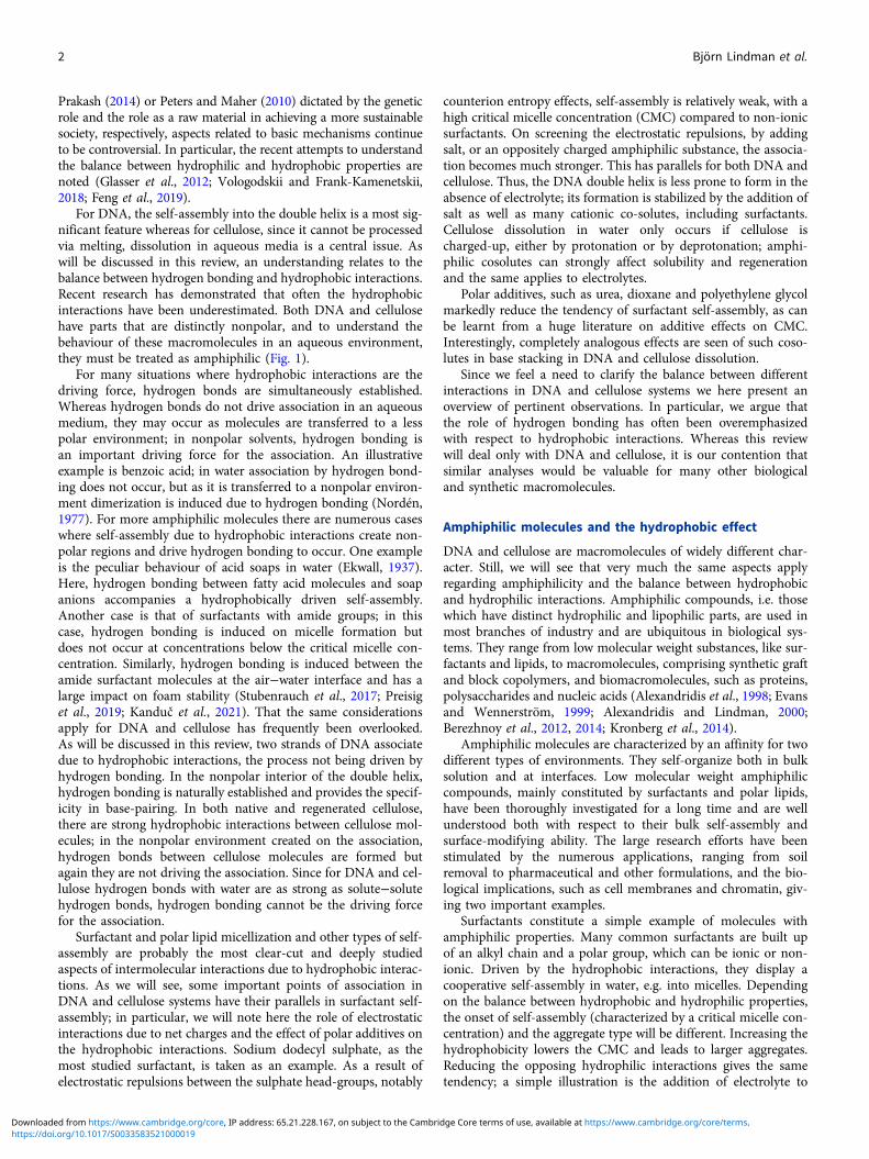
as kinetic trapping (not uncommon for large polymers). Carlstedtand Dias (Carlstedt et al., 2012) solved this puzzling observationby changing the design of the experiments and making directobservations of the charge of the aggregates by electrophoresis.Strikingly, it was observed that there is a large concentrationrange, with an excess of surfactant, without any sign of phase sep-aration. Furthermore, it was observed that in this region of excesssurfactant, there are aggregates with net positive charge, thus withmore surfactant molecules than charges of DNA; the aggregatesare negatively charged below charge stoichiometry, as shown
from electrophoretic measurements (Fig. 7). This provides directevidence for the role of hydrophobic interactions in the associationand that DNA must be considered as an amphiphilic polymer.
Other aspects of DNA hydrophobicity
As expected, and as has been documented for some time, manycationic cosolutes, polymers, multivalent metal ions, proteins,and surfactants/lipids, associate with DNA. This is driven byentropic electrostatic interactions and the association is expected
Fig. 5. Intensity weighted distribution functions of 0.5 μM T2DNAsolution in the absence (upper curve) and the presence of CTAB.The concentrations of the cationic surfactant are from top tobottom: 0 (only DNA), 1.0, 2.0, 4.0, 6.0, 10.0, and 30.0 μM.Scattering angle (θ) = 90° and T = 27°C. Adapted from Diaset al. (2005) with permission of ACS.
Fig. 6. Visually determined phase map of the DNA−CTAB system presented as a function of (a) CTAB concentration and (b) CTAB:DNA molar ratio, in terms ofcharges, at four different DNA concentrations (in nucleotides, indicated to the right). Open circles correspond to clear solutions whereas filled circles correspondto turbid or macroscopically phase-separated samples. Adapted from Carlstedt et al. (2012) with permission of ACS.
Quarterly Reviews of Biophysics 7
https://doi.org/10.1017/S0033583521000019Downloaded from https://www.cambridge.org/core, IP address: 65.21.228.167, on subject to the Cambridge Core terms of use, available at https://www.cambridge.org/core/terms.
to be stronger with increasing charge number and charge densityof both cosolutes. For ds-DNA, all these additives typically inducetwo effects: intermolecular association leading to phase separationand, for dilute solutions, monomolecular compaction. Note thatthe compaction is a cooperative process which can be viewed asa phase separation on individual DNA molecules.
The association of two DNA strands into the double helix isdriven by the hydrophobic interactions between the bases. Polarinteractions, associated with the phosphate and carbohydrategroups, counteract the association. Hydrogen bonding and spe-cific packing of the bases control the details of the double-helixstructure (Dias and Lindman, 2008).
The electrostatic interactions of DNA have been analysed indetail, as reviewed by different authors in Dias and Lindman(2008). The hydrophobic interactions have been much less dis-cussed; in particular, the balance between the polar and nonpolarinteractions have a deep impact into how DNA interacts withcosolutes, including electrolytes, nonpolar molecules, surfactants,lipids and macromolecules, as well as with interfaces (Dias andLindman, 2008).
Some additional brief comments on the amphiphilic nature ofDNA and its consequences for the solution behaviour are next pro-vided. DNA is clearly different from both block and graft copoly-mers, but closer to the graft copolymer situation, with hydrophobicgrafts on a hydrophilic backbone. However, the segregationbetween hydrophilic and lipophilic parts is less pronounced inDNA and the force opposing self-assembly stronger, due to ahigh charge density and a large persistence length. While thedetailed structure of the double helix has been extensively investi-gated, we note that the balance between the hydrophobic force,driving self-assembly, and the opposing force, is very subtle. Twoconsequences arise: Firstly, the stability of the double helix(ds-DNA) is critically dependent on the electrolyte concentration.In the absence of electrolyte, the opposing force dominates, and theds-DNA dissociation into ss-DNA may occur (depending on DNAconcentration) (Korolev et al., 1994). Small amounts of electrolyte,or essentially any cationic cosolute, overcome the electrostaticrepulsion and stabilize ds-DNA. Secondly, if the driving force ischanged, for example by changing the base composition, there isa significant change in the stability of the double helix.
Manifestations of the hydrophobic interactions include:
– Solubilization of hydrophobic molecules (Gaugain et al., 1978;Howe-Grant and Lippard, 1979; Kapuscinski, 1995; Rye and
Glazer, 1995; Spielmann et al., 1995; Brabec and Nováková,2006; Irena, 2006; Uma Maheswari et al., 2006; Richards andRodger, 2007): This area can be illustrated by the so-called‘intercalating agents’. Ethidium bromide is a well-known fluo-rescent dye commonly used to study the interaction betweenDNA and cosolutes due to its displacement when other mole-cules bind to DNA. Other dyes binding to DNA are not solublein water. Recent work has focused on the role of the ligandhydrophobicity on DNA binding and it was found, not surpris-ingly, that the most hydrophobic compounds have a higherbinding affinity to DNA. In this case, however, the ligandsdid not interact with DNA by intercalation but by hydrophobicinteractions with the surface of the DNA, that is, the pockets ofthe groves. This sort of interaction is common for some fluo-rescent dyes, such as DAPI and in protein−DNA interactions.
– Adsorption on hydrophobic surfaces: It was observed by ellips-ometry that, whereas both ds- and ss-DNA molecules adsorbon hydrophobic surfaces, ss-DNA generally adsorbs more pref-erentially than ds-DNA (Eskilsson et al., 2001; Cárdenas et al.,2003). Also, while ds-DNA molecules form a very thick anddiffuse layer on the surface, the ss-DNA molecules adsorb ina thin layer of ca. 20 Å indicating that the molecules are parallelto the surface (Cárdenas et al., 2003). This is naturally due tothe larger hydrophobicity of the ss-DNA, as each base willserve as an attachment point to the surface overcoming theentropy loss of the adsorption; ss-DNA is much more flexiblethan ds-DNA. In fact, the bases were shown to have differentadsorption properties depending on their hydrophobicity.The purine bases, more hydrophobic due to the two aromaticrings, present larger adsorption than the pyrimidine bases(Sowerby et al., 2001; Chiorcea Paquim et al., 2006).
– Effects of hydrophobic cosolutes on DNA melting: The interac-tions between DNA and alkyltrimethylammonium bromidesalts with short hydrophobic chains and the influence of thechain length on the melting have been previously addressed(Orosz and Wetmur, 1977). It was observed that the meltingtemperature of DNA decreases linearly with the increase ofthe hydrophobic group up to the pentyl substitution.Short-chain alcohols showed the same behaviour. The meltingtemperature of DNA was found to decrease in water/methanolsolutions (Geiduschek and Herskovits, 1961). Furthermore, themidpoint of the solvent denaturation decreased in the order:methanol, ethanol, propanol; i.e. the secondary structure (inter-actions between bases) stability was lowered as the length of thealiphatic chain was increased (Geiduschek and Herskovits,1961).
For alkyltrimethylammonium salts there is a striking nonmo-notonic variation of the melting point with alkyl chain length,illustrating the balance between interactions. For short alkylchains the cosolute competes with the association betweenbases, whereas with longer alkyl chains there is a self-assemblyinto highly charged aggregates, which interact electrostaticallywith DNA; with a longer alkyl chain the CAC is lower and themicelles larger.
– Differences in interactions of cationic surfactants between ss- andds-DNA: One other indication that points to the importance ofthe hydrophobic moieties of DNA on the interaction withcosolutes is the difference in interactions of ss- and ds-DNAwith cationic surfactants. It was observed that the precipitationbehaviour for DNA – dodecyltrimethylammonium bromide
Fig. 7. Dynamic light scattering (RH app) and electrophoretic mobility (μe) data foraqueous mixtures of DNA and CTAB, with a constant DNA concentration of 120 μMin nucleotides (40 μg ml−1) and varying CTAB concentration. Adapted fromCarlstedt et al. (2012) with permission of ACS.
8 Björn Lindman et al.
https://doi.org/10.1017/S0033583521000019Downloaded from https://www.cambridge.org/core, IP address: 65.21.228.167, on subject to the Cambridge Core terms of use, available at https://www.cambridge.org/core/terms.
(C12TAB) is different when DNA is in the denaturated or in thedouble-helix conformation (Rosa et al., 2005). In this case, theDNA conformation was controlled by the temperature. Thefact that C12TAB interacts preferentially with ss-DNA, forlow concentrations of surfactant, signifies that the melting tem-perature of DNA will be shifted to a lower temperature (Rosaet al., 2005). Other illustrations on the role of hydrophobicinteractions in DNA self-assembly (Dias and Lindman, 2008)relate to DNA−protein interactions (Härd and Lundbäck,1996; Jen-Jacobson et al., 2000; West and Wilson, 2002), thedependence of DNA melting on the base sequence and thepreparation of DNA chemical and physical gels. Regardingchemical gels (Costa et al., 2007), covalently cross-linked ss-and ds-DNA interact differently with cationic surfactants. Onthe other hand, it is notable that DNA can form physicalgels in combination with hydrophobically modified cationicpolymers (Costa et al., 2006). The effects of hydrophobic inter-actions on DNA condensation has been discussed in severalarticles (Patel and Anchordoquy, 2005; Sumi et al., 2009;Filippov et al., 2010; Zhou et al., 2013; Xiao et al., 2020).Another particularly relevant example of considerable biologi-cal significance deserves some attention; DNA, in the eukary-otic cells, exists in the form of the histone−DNA complexchromatin. Although electrostatic interactions between nega-tively charged DNA and cationic histones make a decisive
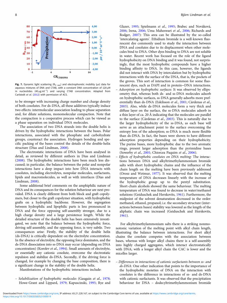
contribution to chromatin formation and stability, hydropho-bic interactions within the histone octamer are critical for theestablishment of specific structure of the universal basic unitof chromatin, the nucleosome core particle (NCP) (Fig. 8).
This importance of hydrophobic forces was revealed in exper-imental studies of systems that included the NCP or model nucle-osome arrays (in vitro reconstituted chromatin fibres) incombination with lipids. The addition of lipids resulted in ashift of the delicate balance between hydrophobic and electrostaticforces, dissociation of the NCP and model chromatin, which wasaccompanied by the transfer of histones from DNA to the lipidsand formation of various lamellar lipid−DNA structures(Lundberg et al., 2010; Berezhnoy et al., 2012, 2014).
Another example of how hydrophobic interactions controlDNA self-assembly was recently revealed by Wong andco-workers, studying antimicrobial peptides (AMPs) that areamphiphilic ɑ-helices (Lee et al., 2019). This example is of consid-erable medical importance for understanding activation inimmune cells by the proinflammatory activity of AMPs.Comparing the three AMPs melittin, LL37, and buforin andusing simulations and synchrotron X-ray diffraction, they showedhow the AMP hydrophobicity controlled the peptide’s ability tofunction as subunits that assemble into superhelical protofibrils
Fig. 8. Illustrations of (a) an NCP, (b) the core histones, and (c)the nucleosome array. (a) Two projections of the NCP whereDNA is shown as a surface with electrostatic potential (positivein red and negative in blue) and the histone octamer with sche-matic secondary structure (each of the eight histones is coloureddifferently). Approximate dimensions of the NCP are indicated.The core histones shown in panel b illustrate the folded domainsof each histone shown with the surface coloured according to itselectrostatic potential (positive in blue) and hydrophobicity(orange). In panel c, a nucleosome array comprising 12 nucleo-somes formed by the wrapping of 147 bp DNA around the his-tone octamer, is schematically shown. DNA is red and thehistone octamer core light grey with histone tails in blue.Reproduced from Berezhnoy et al. (2012) with permission fromACS.
Quarterly Reviews of Biophysics 9
https://doi.org/10.1017/S0033583521000019Downloaded from https://www.cambridge.org/core, IP address: 65.21.228.167, on subject to the Cambridge Core terms of use, available at https://www.cambridge.org/core/terms.

in the presence of DNA by forming columnar protofibril−DNAnanocrystals (Fig. 9).
The topic of the role of hydrophobic interactions for the stabil-ity of ds-DNA has received much-renewed interest through recentstudies by Nordén et al. (Feng et al., 2019) using a novel perspec-tive. These authors investigated the effect of slightly nonpolarcosolutes on the association between the bases in DNA. Theyfound that additives, such as short-chain PEG, diglyme and diox-ane significantly reduced the association. Interestingly, this type ofsubstances reduces the association between surfactant molecules,thus causing demicellization. As an example, the CMC ofCTAB was found to increase with increasing PEG concentration,as well as its molar mass (Manna and Panda, 2011).
The addition of water-soluble polymers to DNA solution canlead to DNA compaction by a ‘crowding effect’. Here, there is alarge difference between dextran and PEG; as argued by Nordénet al. (Feng et al., 2019) this fits very well with the amphiphilicproperties of DNA since dextran is strongly polar whereas PEGis much less polar. A similar difference in the crowding effectwas observed for the compaction of the chromatin (Zinchenkoet al., 2020) where PEG results in the complete compaction ofT4 DNA reconstituted chromatin fibres into globules while thestrongly polar dextran causes only slight chromatin compaction.The compaction of T4 DNA alone occurs in the presence ofPEG (Zinchenko et al., 2018) but not in concentrated solutionsof dextran (Zinchenko et al., 2020).
Important contributions regarding the balance betweenhydrogen-bonding and hydrophobic interactions are due toFrank-Kamenetskii and co-workers (Protozanova et al., 2004;Yakovchuk et al., 2006; Vologodskii and Frank-Kamenetskii,2018). The most recent one is also the most comprehensive,including an extensive analysis of the large body of earlierworks. A major point is that they found stacking enthalpy inthe range of −8.2 to −10.2 kcal mol−1 (depending on the bptype). However, they obtained estimates for the base pairing of
ΔHbpA⋅T = 0.9 ± 1 kcal mol−1 (for the AT bp) and ΔHbp
G⋅C = 0.6 ± 1kcal mol−1 (for the GC bp), concluding that the contribution todouble-helix formation from base pairing has an enthalpy valuethat is insignificant compared to the stacking enthalpy.
Charging up cellulose counteracts hydrophobic associationand facilitates dissolution
From a theoretical perspective, the dissolution of cellulose inaqueous media has been shown to be energetically unfavourablebecause the entropy loss due to water–cellulose interactions isnot balanced by the related entropy gain from the increasedchain conformations upon dissolution (Bergenstråhle et al.,2010; Parthasarathi et al., 2011; Bao et al., 2015).
This is verified in practice since cellulose is not soluble inwater; thus, the cellulose behaviour in solution is mostly analysedin solvent systems of a rather complex composition (i.e. concen-trated salt solutions, ionic liquids, organic/salt mixtures, etc.)(Medronho and Lindman, 2014, 2015). As mentioned, cellulosesolubility in aqueous solutions is typically observed at extremepHs and literature has largely been attributing this phenomenonon breaking hydrogen bonds. Instead, it has been argued thatsuch enhanced solubility at extreme pH values is a clear manifes-tation of a polyelectrolyte-like behaviour where cellulose mole-cules attain net charge due to protonation/deprotonation.
If the polymer is ionized (for instance, by adding base), theenergy balance changes since the counterions and Coulombicinteractions contribute largely to the entropy gain (Schneiderand Linse, 2002, 2003). Consequently, polymers that are chargedare generally soluble in water, even if they are not markedly polar(Lindman et al., 2017). The polyelectrolyte nature of cellulose hasbeen subjected of numerous investigations in the past, mainlyregarding the controversy as to whether the reaction with alkaliyields a true alcoholate or an addition compound without ionizationof the cellulose (Pennings and Prins, 1962). It was found that theosmotic pressure data could be well described considering celluloseas a weak polyacid. The ionization of the hydroxyls of glucose arewell known and easily admitted to other related biomacromolecules,such as amylose (Bertoft, 2017). Citing Bertoft, ‘…at pH > 13 thehydroxyl groups on the glucose residues become negatively chargedand the molecule expands to its largest volume.’ Surprisingly, thisbehaviour has been underrated for cellulose.
One possible explanation relies on the fact that, classically, cel-lulose solubility/swelling studies have been performed by plottingdata as a function of the NaOH concentration (in %), and not as afunction of pH as typically done in other systems, such as pro-teins. This may have contributed to not giving adequate relevanceto the effect of pH on ionization.
The finding of maxima for the solubility/swelling of celluloseon increasing the NaOH concentration could be interpreted asa maximum produced by a Donnan effect (e.g. as made byNeale (Kasbekar and Neale, 1947)), similar to that already seenin other charged polymers, such as collagen when going intoextreme pHs (Bowes and Kenten, 1948).
In the late 1990s, cellulose ionization was inferred from NMRdata by considering that not all OH groups are required to be fullyionized, but form transient dissociated structures of relativelyshort duration (Isogai, 1997).
This hypothesis found support in recent electrophoretic NMRstudies where it was shown that cellobiose can act as a weak acidundergoing two base-independent (KOH and NaOH) dissocia-tion states at pHs of 12 and 13.5 (Bialik et al., 2016) (Fig. 10).
Fig. 9. Structure of melittin-double-stranded DNA (dsDNA) three-dimensional (3D)tetragonal lattice from molecular simulations verified by X-ray diffraction. View of amelittin-dsDNA square lattice. Columnar dsDNA is coloured red, and the melittin pro-tofibril is coloured green (N to C terminus polarity) and teal (C to N terminus polar-ity). Reproduced from Lee et al. (2019) with permission from Nature.
10 Björn Lindman et al.
https://doi.org/10.1017/S0033583521000019Downloaded from https://www.cambridge.org/core, IP address: 65.21.228.167, on subject to the Cambridge Core terms of use, available at https://www.cambridge.org/core/terms.

Additional MD results further confirmed this ionization effectshowing that charging up cellulose prevents its aggregation.
We also note that cellulose dissolution in systems based onaqueous metal complexes (Burchard et al., 1994; Klüfers andSchuhmacher, 1994; Saalwächter et al., 2000) or zinc chloride(Letters, 1932; Xu and Chen, 1999) requires ionization ofOH-groups. This provided further evidence to the deprotonationof cellulose in basic media. In some cases, cellulose decreases itssolubility in basic solution upon addition of ionic or non-ionicadditives and this can be understood from the overall decreasein entropy of the system (Medronho et al., 2016; Alves et al.,2016a, 2016b).
Cellulose amphiphilicity
It is striking that, through the years, cellulose insolubility in waterhas been attributed to strong cellulose–cellulose hydrogen bonds.This view has been rooted in the cellulose community for decades,but clearly conflicts with our fundamental understanding of wateras a solvent. It is important to realize that during the dissolutionprocess, intermolecular interactions in the solute have to be bro-ken, such as the hydrogen bonds between cellulose molecules,which are unfavourable for dissolution. However, new interactionsbetween the solute and the solvent molecules are established and
it is the final balance of all different interactions that govern theoutcome of the dissolution process. When considering cellulosein water, not only hydrogen bonding among cellulose−celluloseis important but also between cellulose and water and amongwater molecules. It appears that these different hydrogen bondsare not markedly different in magnitude and thus aqueous insol-ubility cannot be attributed to hydrogen bonding. The energyneeded to break hydrogen bonding represents a fraction of thetotal free energy required to dissolve cellulose. This has been val-idated in a detailed analysis of the balance of interactions byBergenstråhle et al. and in other related reports (Bergenstråhleet al., 2010; Parthasarathi et al., 2011; Bao et al., 2015).
An analogous conclusion can be reached if we compare a sim-ilar number of glucose units, but distributed in different blocklengths (Fig. 11).
Although the number of established hydrogen bonds is prettymuch the same, shorter chains remain in solution while the lon-ger ones do aggregate. Since insolubility cannot be attributed tohydrogen bonding other causes must be identified. As earlier dis-cussed for DNA, the cause of insolubility of the longer-chainpolymers is related to entropy; the longer polymer chains self-aggregate and release their bound solvent molecules thus maxi-mizing the entropy of the system. On the other hand, the shorterchains still have a fair degree of movement and random positions,
Fig. 10. Left: Effective charge of cellobiose as a function of the pH of the solution either using KOH (filled circles) or NaOH (empty circles). Right: Cellulose con-figurations in the last frame of a 1 μs simulation for (a) neutral and (b) deprotonated cellodecaose. Taken from Bialik et al. (2016) with permission of ACS.
Fig. 11. Schematic representation of glucose-based oligomers with different degrees of polymerization.
Quarterly Reviews of Biophysics 11
https://doi.org/10.1017/S0033583521000019Downloaded from https://www.cambridge.org/core, IP address: 65.21.228.167, on subject to the Cambridge Core terms of use, available at https://www.cambridge.org/core/terms.

contributing to the total entropy of the system, and can remain insolution.
For the last decade, it has been argued that cellulose is strik-ingly amphiphilic and that its aqueous insolubility should havea significant contribution from hydrophobic interactions(Lindman et al., 2010; Medronho et al., 2012). For instance,returning to the dissolution process at high pH, we have observedthat cellulose dissolution becomes more favourable if the disrup-tion of hydrophobic interactions accompanies the ionization. Thiscan be done by using organic hydroxides, such as tetrabutylam-monium hydroxide (TBAH), which are more efficient thantheir inorganic counterparts (NaOH) because the cations of theformer are capable of weakening the hydrophobic interactionswhile the inorganic cations are not (Alves et al., 2015; Gubitosiet al., 2016).
Unsurprisingly, these claims on the role of hydrophobic inter-actions on cellulose solubility are far from being original and sim-ilar conclusions have been drawn in much earlier publications(French et al., 1993, 1996; Cousins and Brown, 1995; Nishiyamaet al., 2002). However, such contributions have been mostlyneglected and instead there has been a massive flood of publica-tions claiming hydrogen bonding as the principal cause of cellu-lose insolubility in water. The complex interplay betweenH-bonding, ionization effects, and hydrophobic interactions iscrucial to control dissolution, regeneration, gelation, and relatedphenomena (Lindman et al., 2017).
Segregation between polar and nonpolar groupsin cellulose
Several observations stand out regarding the fundamental impor-tance of cellulose amphiphilicity and concomitant role of hydro-phobic interactions in the behaviour of cellulose in aqueoussystems (Medronho et al., 2015). Looking at the cellulose molec-ular structure, cellulose chains consist of D-pyranose rings con-nected by β-1,4 glycosidic bonds where the polar hydroxylgroups render cellulose hydrophilic, while the nonpolar back-bones of carbon rings make it hydrophobic (Biermann et al.,2001; Yamane et al., 2006; Diddens et al., 2008; Miyamotoet al., 2009; Youssefian and Rahbar, 2015).
The distinction between the hydrophilic lateral rim and thehydrophobic top and bottom of a cellulose molecule renders cel-lulose clear amphiphilicity. This has been very early recognized,for example in the work of Hermans (1949) (Fig. 12).
Quoting Hermans: ‘…the atoms of the (cellulose) ring can bedistributed over two parallel planes, above and below these plansare the hydrogen atoms and laterally we have the hydroxyl groups.
The cellulose chain exhibits two hydrophobic and two hydrophilicboundary surfaces.’
Due to the hydrophobic properties of the glucopyranose plane,the cellulose chains can stack via hydrophobic interactions andcan form a sheet-like structure that should be disrupted for disso-lution to occur. This was already documented by Sponsler’s dif-fraction work (Sponsler, 1931) and later by Warwicker andWright (Warwicker and Wright, 1967).
Additives may weaken the hydrophobic interactionsin cellulose
The addition of specific additives, such as urea, thiourea, guani-dine and their derivatives weakens hydrophobic interactionsthus facilitating cellulose dissolution (Lilienfeld, 1924, 1927;Zhou and Zhang, 2000; Cai and Zhang, 2005; Cai et al., 2006,2007, 2008; Egal et al., 2008; Qi et al., 2008; Ruan et al., 2008;Liu and Zhang, 2009). In aqueous solutions, these additivescause, inter alia, protein denaturation and demicellization of sur-factant aggregates (Tanford, 1964; Piercy et al., 1971; Brigantiet al., 1991; Zangi et al., 2009). In the case of cellulose dissolution,it was recently shown by a set of unusual techniques (cryo-transmission electronic microscopy, diffusion wave spectroscopyand solid-state nuclear magnetic resonance) that cellulose solubil-ity in aqueous alkali is significantly improved by urea, as seen inFig. 13 (Alves et al., 2018).
Besides, this additive remarkably affects the solution stability,preventing thermal gelation in certain conditions (Swensson et al.,2020a). Interestingly, urea has been shown to concentrate on cellu-lose surfaces in solutions of aqueous urea (Bergenstråhle-Wohlertet al., 2012; Chen et al., 2017; Walters et al., 2020).
This is also in agreement with the recent work of Swenssonet al. where the authors show that adding urea to solutions ofNaOH and/or tetramethylammonium hydroxide (TMAH)improves dissolution and can successfully be used together withthese bases (Swensson et al., 2020b). Interestingly, adding ureato benzyltrimethylammonium hydroxide (known as Triton B)solutions did not seem to have a significant effect on cellulose dis-solution, and the solvatochromic probes indicated that urea mightbe excluded from interacting with cellulose in the presence ofTriton B. Both urea and Triton B are believed to weaken thehydrophobic effect by replacing water around the pyranose ringbut, most likely, this effect is more pronounced for Triton Bthan for urea (Swensson et al., 2020b). In a related work, Weiet al. argue that the role of urea in tetrabutylammonium hydrox-ide (TBAH)/urea aqueous solvents can be regarded as a hydro-phobic contributor, where the amphiphilic properties of the
Fig. 12. The nuclear frame of a cellobiose residue. The centres ofgravity of the atoms of the ring are distributed over two parallelplanes. The hydrophobic H atoms are above and below theseplanes and laterally there are the hydrophilic OH groups.Taken from Hermans (1949) with the permission of John Wileyand Sons, Inc.
12 Björn Lindman et al.
https://doi.org/10.1017/S0033583521000019Downloaded from https://www.cambridge.org/core, IP address: 65.21.228.167, on subject to the Cambridge Core terms of use, available at https://www.cambridge.org/core/terms.

solvent system can be tuned. The authors suggest that for a suit-able amphiphilicity similar to that of the crystal surface of pristinecellulose, the interfacial resistance between the solvent and thecrystal surface can be reduced so that the crystalline areas of cel-lulose can be effectively infiltrated and subsequently dissolved bythe solvent (Wei et al., 2017). This same conclusion was alsoreached regarding the role of urea in enhancing the solubility inaqueous solutions of amino acids and proteins (Whitney andTanford, 1962; Nozaki and Tanford, 1963; Zangi et al., 2009),and also of chitin chains (which are structurally very similar tocellulose) (Huang et al., 2020). In this latter case, the presenceof urea changes the chemical shifts very little in the α-chitin/KOH/water system, confirming that urea solubilized chitin chainsby preferentially solvating the hydrophobic parts of the chitinbackbone without interfering with the chitin chain conformationin the aqueous KOH/solution.
Similarly to urea, the addition of a zwitterionic surfactant tocellulose dissolved in an alkali-based solvent can prevent the gela-tion of the dope. After dissolution, as temperature increases, gela-tion of the cellulose dope is observed; the gelation temperature,Tg, can be estimated from the crossover of the storage (G′) andloss (G′′) moduli. The addition of the zwitterionic surfactant tothe cold alkali solvent system led to a shift in gelation temperatureof ca. 10°C (Fig. 14). The same behaviour was observed using aconcentrated zinc chloride aqueous solution (Medronho et al.,2015).
MD simulation studies of the interaction of cellulose with dif-ferent compounds in aqueous media have confirmed that, in cel-lulose crystals, apart from hydrogen bonding, hydrophobicinteractions also play a substantial role (Alqus et al., 2015; Chen
et al., 2017). The same was concluded regarding the absorptionof a soluble hEGF protein to the cellulose surface. Interestingly,it was found that the hEGF protein binds to both the (010) and(100) cellulose surfaces through different regions of the proteinwhich contain both polar and apolar residues. In the case of the(010) surface, the amino acids involved in adsorption are mostlypolar whereas in the case of adsorption at the (100) surface theamino acids involved are preferentially apolar. This result suggeststhat the simultaneous hydrophilic and hydrophobic character ofcellulose induces a substantial interaction of cellulose with pro-teins (which are also amphiphilic) (Malaspina and Faraudo,2019).
Organic and inorganic counterions interact differentlywith cellulose
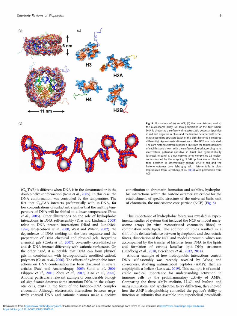
Solutions of organic acids or bases are superior solvents to thoseof inorganic ones. For instance, cellulose dissolution in TBAH isobserved to proceed down to the molecular level (Gubitosi et al.,2016), while NaOH does not dissolve cellulose molecularly (Alveset al., 2015); it rather leaves aggregates of high crystallinity stablein the cellulose dope (Pereira et al., 2018) (Fig. 15).
It was inferred that 1.2 TBA+ ions bind per anhydroglucoseunit and this was later supported by detailed scattering studieswhere the SAXS data suggest the presence of a solvation shellenriched in TBA+ ions around the cellulose molecules. TheTBA+ cation is suggested to interact by electrostatic interactionswith the (partially) deprotonated hydroxyl groups of cellulose,in addition to hydrophobic interactions due to its amphiphilicity(Gentile and Olsson, 2016). Furthermore, cellulose solubility has
Fig. 13. Cryo-transmission electronic microscopy(cryo-TEM) images of 0.5 wt % MCC dissolved in(a) 8 wt % NaOH(aq.) solution and (b) in 8 wt %NaOH(aq.)/12 wt % urea system. Scale bars correspondto 100 nm.
Fig. 14. Elastic modulus, G′ (filled symbols), and viscousmodulus, G′ ′ (open symbols) as a function of tempera-ture for 3.5 wt% microcrystalline cellulose samples dis-solved in a 10 wt% NaOH/H2O solvent system: (a),without cocamidopropylbetaine and (b) with cocamido-propylbetaine. Constant heating rate of 1°C min−1 at 0.5Hz. The temperature of gelation (G′ = G′ ′) is increased byca. 10°C in the presence of the amphiphilic additive. Thevertical dashed grey line indicates the transition region.Taken from Medronho et al. (2015) with the permissionof De Gruyter.
Quarterly Reviews of Biophysics 13
https://doi.org/10.1017/S0033583521000019Downloaded from https://www.cambridge.org/core, IP address: 65.21.228.167, on subject to the Cambridge Core terms of use, available at https://www.cambridge.org/core/terms.
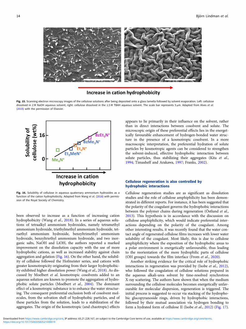
been observed to increase as a function of increasing cationhydrophobicity (Wang et al., 2018). In a series of aqueous solu-tions of tetraalkyl ammonium hydroxides, namely tetramethylammonium hydroxide, triethylmethyl ammonium hydroxide, tet-raethyl ammonium hydroxide, benzyltrimethyl ammoniumhydroxide, benzyltriethyl ammonium hydroxide, and two inor-ganic salts, NaOH and LiOH, the authors reported a markedimprovement on the dissolution capacity with the use of morehydrophobic cations, as well as increased stability against chainaggregation and gelation (Fig. 16). On the other hand, the solubil-ity of cellulose followed the Hofmeister series, and cations withgreater kosmotropicity originating from their larger hydrophobic-ity exhibited higher dissolution power (Wang et al., 2018). As dis-cussed by Moelbert et al. kosmotropic cosolvents added to anaqueous solution are known to promote the aggregation of hydro-phobic solute particles (Moelbert et al., 2004). The dominanteffect of a kosmotropic substance is to enhance the water structur-ing. The consequent preferential exclusion both of cosolvent mol-ecules, from the solvation shell of hydrophobic particles, and ofthese particles from the solution, leads to a stabilization of theaggregates. The origin of the kosmotropic (and chaotropic) effects
appears to lie primarily in their influence on the solvent, ratherthan in direct interactions between cosolvent and solute. Themicroscopic origin of these preferential effects lies in the energet-ically favourable enhancement of hydrogen-bonded water struc-ture in the presence of a kosmotropic cosolvent. In a moremacroscopic interpretation, the preferential hydration of soluteparticles by kosmotropic agents can be considered to strengthenthe solvent-induced, effective hydrophobic interaction betweensolute particles, thus stabilizing their aggregates (Kita et al.,1994; Timasheff and Arakawa, 1997; Franks, 2002).
Cellulose regeneration is also controlled byhydrophobic interactions
Cellulose regeneration studies are as significant as dissolutionstudies and the role of cellulose amphiphilicity has been demon-strated in different reports. For instance, it has been suggested thatthe polarity of the coagulant governs the hydrophobic interactionsbetween the polymer chains during regeneration (Östlund et al.,2013). This hypothesis is in accordance with the discussion oncellulose amphiphilicity, which would indicate preferential inter-actions depending on the polarity of the coagulant. Amongother interesting results, it was recently found that the water con-tact angle of regenerated cellulose films increases with lower watersolubility of the coagulant. Most likely, this is due to celluloseamphiphilicity where the exposition of the hydrophobic areas toa polar environment is energetically unfavourable, thus leadingto the reorientation of the more hydrophilic parts of cellulose(OH groups) towards the film interface (From et al., 2020).
Another striking evidence for the critical role of hydrophobicinteractions in regeneration was provided by (Isobe et al., 2012),who followed the coagulation of cellulose solutions prepared inthe aqueous alkali-urea solvent by time-resolved synchrotronX-ray scattering. The authors have shown that when the mediumsurrounding the cellulose molecules becomes energetically unfav-ourable for molecular dispersion, regeneration is triggered. Theinitial process is suggested to occur via stacking of the hydropho-bic glucopyranoside rings, driven by hydrophobic interactions,followed by their mutual association via hydrogen bonding toform a hydrated form of cellulose II (Isobe et al., 2012) (Fig. 17).
Fig. 15. Scanning electron microscopy images of the cellulose solutions after being deposited onto a glass lamella followed by solvent evaporation. Left: cellulosedissolved in 2 M NaOH aqueous solvent; right: cellulose dissolved in the 1.5 M TBAH aqueous solvent. The scale bar represents 5 μm. Adapted from Alves et al.(2015) with the permission of Elsevier.
Fig. 16. Solubility of cellulose in aqueous quaternary ammonium hydroxides as afunction of the cation hydrophobicity. Adapted from Wang et al. (2018) with permis-sion of the Royal Society of Chemistry.
14 Björn Lindman et al.
https://doi.org/10.1017/S0033583521000019Downloaded from https://www.cambridge.org/core, IP address: 65.21.228.167, on subject to the Cambridge Core terms of use, available at https://www.cambridge.org/core/terms.
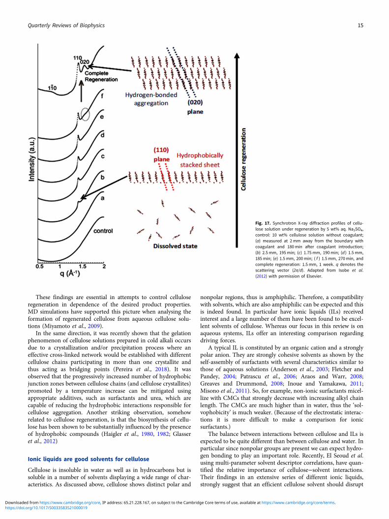
These findings are essential in attempts to control celluloseregeneration in dependence of the desired product properties.MD simulations have supported this picture when analysing theformation of regenerated cellulose from aqueous cellulose solu-tions (Miyamoto et al., 2009).
In the same direction, it was recently shown that the gelationphenomenon of cellulose solutions prepared in cold alkali occursdue to a crystallization and/or precipitation process where aneffective cross-linked network would be established with differentcellulose chains participating in more than one crystallite andthus acting as bridging points (Pereira et al., 2018). It wasobserved that the progressively increased number of hydrophobicjunction zones between cellulose chains (and cellulose crystallites)promoted by a temperature increase can be mitigated usingappropriate additives, such as surfactants and urea, which arecapable of reducing the hydrophobic interactions responsible forcellulose aggregation. Another striking observation, somehowrelated to cellulose regeneration, is that the biosynthesis of cellu-lose has been shown to be substantially influenced by the presenceof hydrophobic compounds (Haigler et al., 1980, 1982; Glasseret al., 2012)
Ionic liquids are good solvents for cellulose
Cellulose is insoluble in water as well as in hydrocarbons but issoluble in a number of solvents displaying a wide range of char-acteristics. As discussed above, cellulose shows distinct polar and
nonpolar regions, thus is amphiphilic. Therefore, a compatibilitywith solvents, which are also amphiphilic can be expected and thisis indeed found. In particular have ionic liquids (ILs) receivedinterest and a large number of them have been found to be excel-lent solvents of cellulose. Whereas our focus in this review is onaqueous systems, ILs offer an interesting comparison regardingdriving forces.
A typical IL is constituted by an organic cation and a stronglypolar anion. They are strongly cohesive solvents as shown by theself-assembly of surfactants with several characteristics similar tothose of aqueous solutions (Anderson et al., 2003; Fletcher andPandey, 2004; Patrascu et al., 2006; Araos and Warr, 2008;Greaves and Drummond, 2008; Inoue and Yamakawa, 2011;Misono et al., 2011). So, for example, non-ionic surfactants micel-lize with CMCs that strongly decrease with increasing alkyl chainlength. The CMCs are much higher than in water, thus the ‘sol-vophobicity’ is much weaker. (Because of the electrostatic interac-tions it is more difficult to make a comparison for ionicsurfactants.)
The balance between interactions between cellulose and ILs isexpected to be quite different than between cellulose and water. Inparticular since nonpolar groups are present we can expect hydro-gen bonding to play an important role. Recently, El Seoud et al.using multi-parameter solvent descriptor correlations, have quan-tified the relative importance of cellulose−solvent interactions.Their findings in an extensive series of different ionic liquids,strongly suggest that an efficient cellulose solvent should disrupt
Fig. 17. Synchrotron X-ray diffraction profiles of cellu-lose solution under regeneration by 5 wt% aq. Na2SO4,control: 10 wt% cellulose solution without coagulant;(a) measured at 2 mm away from the boundary withcoagulant and 180 min after coagulant introduction;(b) 2.5 mm, 195 min; (c) 1.75 mm, 190 min; (d ) 1.5 mm,185 min; (e) 1.5 mm, 200 min; ( f ) 1.5 mm, 270 min, andcomplete regeneration: 1.5 mm, 1 week. q denotes thescattering vector (2π/d). Adapted from Isobe et al.(2012) with permission of Elsevier.
Quarterly Reviews of Biophysics 15
https://doi.org/10.1017/S0033583521000019Downloaded from https://www.cambridge.org/core, IP address: 65.21.228.167, on subject to the Cambridge Core terms of use, available at https://www.cambridge.org/core/terms.

both the inter- and intramolecular hydrogen bonding in celluloseand the solvophobic interactions arising from the marked amphi-philic character of the biopolymer (El Seoud et al., 2021).
As discovered some time ago, ILs are among the most efficientdissolution systems for cellulose (Swatloski et al., 2002; Zhu et al.,2006). Their superior dissolution performance has been ascribedto their ability to disrupt the extensive H-bond network and sol-vophobic interactions present in cellulose. From a structural pointof view, ILs are strongly amphiphilic and can be regarded as weaksurfactants. As an example, in Fig. 18, the molecular structure of1-ethyl-3-methylimidazolium acetate, [EMIM][Ac], is shownemphasizing the asymmetry in ion size and charge distribution.This is a powerful ionic liquid solvent for cellulose (and lignocel-lulose biomass) dissolution (Sun et al., 2009).
Therefore, ILs fit well into the picture of solvophobic interac-tions in cellulose dissolution. This has been experimentally vali-dated, for instance, in the work of Xu et al. where the cationstructure was systematically modified while the anion was keptconstant. In the binary solvent mixture Cx MeIm AcOs/dimethylsulphoxide (DMSO), where x, Me, Im, AcO refer to the numberof carbon atoms in the IL side chain, methyl, imidazolium andacetate, respectively, it was found that the dissolution efficiencyincreases from C2 MeIm AcO to C4 MeIm AcO and thendecreases for C8 MeIm AcO (Xu et al., 2015). A related workby Kostag et al. reported similar results for quaternary ammo-nium ILs (Kostag and El Seoud, 2019). The negative enthalpywas taken as support for the formation of hydrogen bondsbetween acetate and OH-groups and cation-cellulose solvophobicinteractions (Kostag et al., 2020). Theoretical work has been grow-ing fast in this area and data support the critical role of solvopho-bic interactions. For instance, Mostofian et al. conducted all-atomMD simulations of a 36-chain cellulose microfibril in the ionicliquid [Bmim][Cl] and water for 100 ns. The authors found that[Cl]– interacted preferentially with hydroxyl groups in differentcellulose layers while the [Bmim]+ stacked on the nonpolar cellu-lose surface, stabilizing the detached cellulose chains (Li et al.,2018). Similar conclusions have been reached by Ishida using[C2MIm][OAc] as the solvent system (Ishida, 2020). Otherrelated MD studies have generally concluded that the nonpolarcations interact via van der Waals forces with the nonpolar back-bone of cellulose while the anion, which is typically polar, formsstrong hydrogen bonds with cellulose’s hydroxyl groups (Liuet al., 2010; Gross et al., 2011; Rabideau et al., 2014; Xionget al., 2014; Wang et al., 2017; Walters et al., 2020). Neitheranions nor cations alone cover all of the strongly attractive inter-actions within glucose residues; their coupled actions are neces-sary. Overall, the charge density (hardness and volume of theanion), and the volume, rigidity, Lewis acidity, and nonpolarcharacter of the cation are now regarded as determinant for cellu-lose dissolution (Kostag et al., 2019; El Seoud et al., 2020).
The amphiphilicity of ILs finds parallelism in other good sol-vents for cellulose, such as N-methylmorpholine N-oxide(Perepelkin, 2007) or the above-mentioned aqueous alkylammo-nium hydroxides (Abe et al., 2015).
Other manifestations of cellulose amphiphilicity:emulsion stabilization
It is generally assumed that natural starch and cellulose cannotstabilize emulsions. However, since cellulose behaves as anamphiphilic polymer, its adsorption onto the oil−water interfacesis indeed expected to occur. Recent studies have found that cellu-lose nanocrystals display amphiphilic properties and can be usedin the formation of stable emulsions (Paximada et al., 2016;Vasconcelos et al., 2017). In the work of (Kalashnikova et al.,2012) it was further suggested that crystals with low surfacecharge favour the stability of emulsions suggesting that theamphiphilic nature of cellulose is the main driving force for thestabilization. Although less common, emulsion formation hasbeen observed not only using cellulose crystals (stabilized by aPickering-like mechanism) but also using molecularly dissolvedcellulose where the interfacial tension between oil and the aque-ous medium is found to be lowered by the molecularly dissolvedcellulose (Costa et al., 2019). This decrease in the interfacial ten-sion is similar in magnitude to that displayed by non-ionic cellu-lose derivatives.
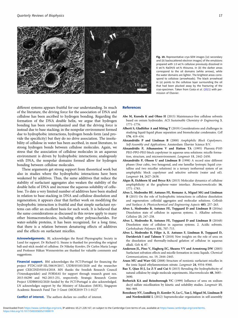
Molecularly dissolved cellulose is expected to behave close totypical cellulose derivatives, but it has been much less exploreddue to the well-known cellulose dissolution limitations. Recentmolecular dynamics simulations have shown that molecularly dis-persed cellulose gradually assembles, eventually surrounding theoil droplet and stabilizing the formed emulsion (Miyamotoet al., 2017). This has been recently observed in practice bycryo-SEM (Costa et al., 2021), (Fig. 19).
The interfacial activity of cellulose indicates a significantamphiphilic character and that the interfacial activity of cellulosederivatives is not only related to the derivatization but inherent inthe cellulose backbone. This finding suggests that cellulose wouldhave the ability to stabilize dispersions, like oil-in-water emulsionsin a similar way as a large number of cellulose derivatives, such asmethylcellulose, hydroxyethylcellulose, hydroxypropyl methylcel-lulose and ethyl hydroxyethyl cellulose (Sheth et al., 1962;Yonekura et al., 1998; Schulz and Daniels, 2000; Melzer et al.,2003; Costa et al., 2019).
Conclusions
Amphiphilic molecules have a large tendency to self-assemble inwater. For simple amphiphiles with well-separated polar and non-polar parts, like in surfactants and block copolymers, simple well-defined structures with extensive nonpolar regions intermixedwith water regions are formed. DNA and cellulose also self-assemble in water but because of the distribution of nonpolargroups, special structures form. For DNA, the double helix isthe most common structure. For cellulose, it is more unclearand it appears that the state of cellulose in a homogeneous solu-tion can be different for different solvents; in many cases, cellu-lose molecules are aggregated and even show signs ofcrystallinity, in others non-aggregated molecules appear.
DNA, cellulose and surfactants are very different, but it isstriking that the balance between hydrophilic and hydrophobicinteractions has common features; a comparison between the
Fig. 18. Structure of 1-ethyl-3-methylimidazolium acetate.
16 Björn Lindman et al.
https://doi.org/10.1017/S0033583521000019Downloaded from https://www.cambridge.org/core, IP address: 65.21.228.167, on subject to the Cambridge Core terms of use, available at https://www.cambridge.org/core/terms.
different systems appears fruitful for our understanding. In muchof the literature, the driving force for the association of DNA andcellulose has been ascribed to hydrogen bonding. Regarding theformation of the DNA double helix, we argue that hydrogenbonding has been overemphasized and that the driving force isinstead due to base stacking; in the nonpolar environment formeddue to hydrophobic interactions, hydrogen bonds form (and pro-vide the specificity) but they do no drive association. The insolu-bility of cellulose in water has been ascribed, in most literature, tostrong hydrogen bonds between cellulose molecules. Again, westress that the association of cellulose molecules in an aqueousenvironment is driven by hydrophobic interactions; analogouslywith DNA, the nonpolar domains formed allow for hydrogenbonding between cellulose molecules.
These arguments get strong support from theoretical work butalso in studies where the hydrophobic interactions have beenweakened by additives. Thus, the same additives that reduce thestability of surfactant aggregates also weaken the stability of thedouble helix of DNA and increase the aqueous solubility of cellu-lose. To date a very limited number of additives have been studiedin relation to base-stacking in DNA and cellulose dissolution andregeneration; it appears clear that further work on modifying thehydrophobic interaction is fruitful and that simple surfactant sys-tems can offer an excellent basis for such work. It is believed thatthe same considerations as discussed in this review apply to manyother biomacromolecules, including other polysaccharides. Forwater-soluble proteins, it has been recognized, for a long time,that there is a relation between denaturing effects of additivesand the effects on surfactant micelles.
Acknowledgements. BL acknowledges the Royal Physiographic Society inLund for support. Dr Richard G. Steane is thanked for providing the originalball-and-stick model of cellulose. Dr Nikolay Korolev, Dr Carlos Maria Longoand Professor Håkan Wennerström are thanked for valuable comments andsuggestions.
Financial support. BM acknowledges the FCT/Portugal for financing theproject PTDC/ASP-SIL/30619/2017, UIDB/05183/2020 and the researchergrant CEECIND/01014/2018. MN thanks the Swedish Research Council(Vetenskapsrådet) and FORMAS for support through research grant nos.2015-04290 and 942-2015-251, respectively. Strategic Research CentreProject UIDB00102/2020 funded by the FCT/Portugal is also acknowledged.LN acknowledges support by the Ministry of Education (MOE), SingaporeAcademic Research Fund Tier 3 Grant (MOE2019-T3-1-012)”
Conflict of interest. The authors declare no conflict of interest.
References
Abe M, Kuroda K and Ohno H (2015) Maintenance-free cellulose solventsbased on onium hydroxides. ACS Sustainable Chemistry & Engineering 3,1771–1776.
Alberti S, Gladfelter A and Mittag T (2019) Considerations and challenges instudying liquid-liquid phase separation and biomolecular condensates. Cell176, 419–434.
Alexandridis P and Lindman B (2000) Amphiphilic Block Copolymers.Self-Assembly and Applications. Amsterdam: Elsevier Science B.V.
Alexandridis P, Athanassiou V and Hatton TA (1995) Pluronic-P105PEO-PPO-PEO block copolymer in aqueous urea solutions: micelle forma-tion, structure, and microenvironment. Langmuir 11, 2442–2450.
Alexandridis P, Olsson U and Lindman B (1998) A record nine differentphases (four cubic, two hexagonal, and one lamellar lyotropic liquid crys-talline and two micellar solutions) in a ternary isothermal system of anamphiphilic block copolymer and selective solvents (water and oil).Langmuir 14, 2627–2638.
Alqus R, Eichhorn SJ and Bryce RA (2015) Molecular dynamics of celluloseamphiphilicity at the graphene–water interface. Biomacromolecules 16,1771–1783.
Alves L, Medronho BF, Antunes FE, Romano A, Miguel MG and LindmanB (2015) On the role of hydrophobic interactions in cellulose dissolutionand regeneration: colloidal aggregates and molecular solutions. Colloidsand Surfaces A: Physicochemical and Engineering Aspects 483, 257–263.
Alves L, Medronho B, Antunes FE, Topgaard D and Lindman B (2016a)Dissolution state of cellulose in aqueous systems. 1. Alkaline solvents.Cellulose 23, 247–258.
Alves L, Medronho B, Antunes FE, Topgaard D and Lindman B (2016b)Dissolution state of cellulose in aqueous systems. 2. Acidic solvents.Carbohydrate Polymers 151, 707–715.
Alves L, Medronho B, Filipe A, E. Antunes F, Lindman B, Topgaard D,Davidovich I and Talmon Y (2018) New insights on the role of urea onthe dissolution and thermally-induced gelation of cellulose in aqueousalkali. Gels 4, 87.
Anderson JL, Pino V, Hagberg EC, Sheares VV and Armstrong DW (2003)Surfactant solvation effects and micelle formation in ionic liquids. ChemicalCommunications, no. 19, 2444–2445.
Araos MU and Warr GG (2008) Structure of nonionic surfactant micelles inthe ionic liquid ethylammonium nitrate. Langmuir 24, 9354–9360.
Bao Y, Qian H-J, Lu Z-Y and Cui S (2015) Revealing the hydrophobicity ofnatural cellulose by single-molecule experiments.Macromolecules 48, 3685–3690.
Berberich KA and Reinsborough VC (1999) Influence of urea on sodiumdecyl sulfate micellization by kinetic and solubility studies. Langmuir 15,966–969.
Berezhnoy NV, Lundberg D, Korolev N, Lu C, Yan J, Miguel M, Lindman Band Nordenskiöld L (2012) Supramolecular organization in self-assembly
Fig. 19. Representative cryo-SEM images [(a) secondaryand (b) backscattered electron images] of the emulsionsprepared with 1.0 wt.% cellulose previously dissolved in8 wt.% NaOH/6 wt.% thiourea. In (b) the darker areascorrespond to the oil domains (white arrows), whilethe water domains are lighter. The brightest areas corre-spond to cellulose (arrowheads). The black arrowheadin (a) points to the cellulose layer surrounding the oilthat had been plucked away by the fracturing of thecryo-specimen. Taken from Costa et al. (2021) with per-mission of Elsevier.
Quarterly Reviews of Biophysics 17
https://doi.org/10.1017/S0033583521000019Downloaded from https://www.cambridge.org/core, IP address: 65.21.228.167, on subject to the Cambridge Core terms of use, available at https://www.cambridge.org/core/terms.

of chromatin and cationic lipid bilayers is controlled by membrane chargedensity. Biomacromolecules 13, 4146–4157.
Berezhnoy NV, Korolev N and Nordenskiöld L (2014) Principles of electro-static interactions and self-assembly in lipid/peptide/DNA systems: applica-tions to gene delivery. Advances in Colloid and Interface Science 205, 221–229.
Bergenstråhle-Wohlert M, Berglund LA, Brady JW, Larsson PT, WestlundP-O and Wohlert J (2012) Concentration enrichment of urea at cellulosesurfaces: results from molecular dynamics simulations and NMR spectro-scopy. Cellulose 19, 1–12.
Bergenstråhle M, Wohlert J, Himmel ME and Brady JW (2010) Simulationstudies of the insolubility of cellulose. Carbohydrate Research 345, 2060–2066.
Bertoft E (2017) Understanding starch structure: recent progress. Agronomy 7, 56.Bharatiya B, Guo C, Ma JH, Kubota O, Nakashima K and Bahadur P (2009)
Urea-induced demicellization of Pluronic L64 in water. Colloid and PolymerScience 287, 63–71.
Bialik E, Stenqvist B, Fang Y, Östlund Å, Furó I, Lindman B, Lund M andBernin D (2016) Ionization of cellobiose in aqueous alkali and the mech-anism of cellulose dissolution. The Journal of Physical Chemistry Letters7, 5044–5048.
Bianco CL, Schneider CS, Santonicola M, Lenhoff AM and Kaler EW (2011)Effects of urea on the microstructure and phase behavior of aqueous solu-tions of poly(oxyethylene) surfactants. Industrial & Engineering ChemistryResearch 50, 85–96.
Biermann O, Hädicke E, Koltzenburg S and Müller-Plathe F (2001)Hydrophilicity and lipophilicity of cellulose crystal surfaces. AngewandteChemie International Edition 40, 3822–3825.
Bowes JH and Kenten RH (1948) The effect of alkalis on collagen. TheBiochemical Journal 43, 365–372.
Brabec V and Nováková O (2006) DNA binding mode of ruthenium com-plexes and relationship to tumor cell toxicity. Drug Resistance Updates 9,111–122.
Briganti G, Puvvada S and Blankschtein D (1991) Effect of urea on micellarproperties of aqueous solutions of nonionic surfactants. The Journal ofPhysical Chemistry 95, 8989–8995.
Broecker J and Keller S (2013) Impact of urea on detergent micelle properties.Langmuir 29, 8502–8510.
Budtova T and Navard P (2016) Cellulose in NaOH–water based solvents: areview. Cellulose 23, 5–55.
Burchard W, Habermann N, Klüfers P, Seger B and Wilhelm U (1994)Cellulose in Schweizer’s reagent: a stable, polymeric metal complex withhigh chain stiffness. Angewandte Chemie International Edition in English33, 884–887.
Cai J and Zhang L (2005) Rapid dissolution of cellulose in LiOH/urea andNaOH/urea aqueous solutions. Macromolecular Bioscience 5, 539–548.
Cai J, Liu Y and Zhang L (2006) Dilute solution properties of cellulose inLiOH/urea aqueous system. Journal of Polymer Science Part B: PolymerPhysics 44, 3093–3101.
Cai J, Zhang L, Chang C, Cheng G, Chen X and Chu B (2007)Hydrogen-bond-induced inclusion complex in aqueous cellulose/LiOH/urea solution at low temperature. Chemphyschem 8, 1572–1579.
Cai J, Zhang L, Liu S, Liu Y, Xu X, Chen X, Chu B, Guo X, Xu J, Cheng H,Han CC and Kuga S (2008) Dynamic self-assembly induced rapid dissolu-tion of cellulose at low temperatures. Macromolecules 41, 9345–9351.
Cárdenas M, Braem A, Nylander T and Lindman B (2003) DNACompaction at hydrophobic surfaces induced by a cationic amphiphile.Langmuir 19, 7712–7718.
Carlsson A, Karlström G, Lindman B and Stenberg O (1988) Interactionbetween ethyl(hydroxyethyl)cellulose and sodium dodecyl sulphate in aque-ous solution. Colloid and Polymer Science 266, 1031–1036.
Carlsson A, Lindman B, Watanabe T and Shirahama K (1989)Polymer-surfactant interactions. Binding of N-tetradecylpyridinium bro-mide to ethyl (hydroxyethyl) cellulose. Langmuir 5, 1250–1252.
Carlstedt J, Lundberg D, Dias RS and Lindman B (2012) Condensation anddecondensation of DNA by cationic surfactant, spermine, or cationic sur-factant–cyclodextrin mixtures: macroscopic phase behavior, aggregate prop-erties, and dissolution mechanisms. Langmuir 28, 7976–7989.
Chen P, Nishiyama Y, Wohlert J, Lu A, Mazeau K and Ismail AE (2017)Translational entropy and dispersion energy jointly drive the adsorptionof urea to cellulose. The Journal of Physical Chemistry B 121, 2244–2251.
Chen Y-L, Zhang X, You T-T and Xu F (2019) Deep eutectic solvents (DESs)for cellulose dissolution: a mini-review. Cellulose 26, 205–213.
Chiorcea Paquim A-M, Oretskaya TS and Oliveira Brett AM (2006)Adsorption of synthetic homo- and hetero-oligodeoxynucleotides ontohighly oriented pyrolytic graphite: atomic force microscopy characteriza-tion. Biophysical Chemistry 121, 131–141.
Costa D, Santos SD, Antunes FE, Miguel MG and Lindman B (2006) Somenovel aspects of DNA physical and chemical gels. Arkivoc 2006, 161–172.
Costa D, Miguel MG and Lindman B (2007) Responsive polymer gels:double-stranded versus single-stranded DNA. The Journal of PhysicalChemistry B 111, 10886–10896.
Costa C, Mira I, Benjamins J-W, Lindman B, Edlund H and Norgren M(2019) Interfacial activity and emulsion stabilization of dissolved cellulose.Journal of Molecular Liquids 292, 111325.
Costa C, Rosa P, Filipe A, Medronho B, Romano A, Liberman L, Talmon Yand Norgren M (2021) Cellulose-stabilized oil-in-water emulsions: struc-tural features, microrheology, and stability. Carbohydrate Polymers 252,117092.
Cousins SK and Brown RM (1995) Cellulose I microfibril assembly: compu-tational molecular mechanics energy analysis favours bonding by van derWaals forces as the initial step in crystallization. Polymer 36, 3885–3888.
Das S, Naskar B and Ghosh S (2014) Influence of temperature and organicsolvents (isopropanol and 1,4-dioxane) on the micellization of cationicgemini surfactant (14-4-14). Soft Matter 10, 2863–2875.
Despa F and Berry RS (2008) Hydrophobe-water interactions: methane as amodel. Biophysical Journal 95, 4241–4245.
Dias R, Mel’nikov S, Lindman B and Miguel MG (2000) DNA phase behaviorin the presence of oppositely charged surfactants. Langmuir 16, 9577–9583.
Dias RS, Lindman B and Miguel MG (2002) Compaction and decompactionof DNA in the presence of catanionic amphiphile mixtures. The Journal ofPhysical Chemistry B 106, 12608–12612.
Dias RS, Innerlohinger J, Glatter O, Miguel MG and Lindman B (2005)Coil−globule transition of DNA molecules induced by cationic surfactants:a dynamic light scattering study. The Journal of Physical Chemistry B 109,10458–10463.
Dias RS, Miguel MG and Lindman B (2008) DNA as an amphiphilic poly-mer. In Dias R and Lindman B (eds), DNA Interactions with Polymersand Surfactants. Hoboken, NJ: John Wiley & Sons, Inc, pp. 367–376.
Diddens I, Murphy B, Krisch M and Müller M (2008) Anisotropic elasticproperties of cellulose measured using inelastic X-ray scattering.Macromolecules 41, 9755–9759.
Egal M, Budtova T and Navard P (2008) The dissolution of microcrystallinecellulose in sodium hydroxide-urea aqueous solutions. Cellulose 15, 361–370.
Ekwall P (1937) Über die Existenz und die Natur der sauren Seifen.Kolloid-Zeitschrift 80, 77–100.
El-Aila HJY (2005) Effect of urea and salt on micelle formation of zwitterionicsurfactants. Journal of Surfactants and Detergents 8, 165–168.
Elder RM, Pfaendtner J and Jayaraman A (2015) Effect of hydrophobic andhydrophilic surfaces on the stability of double-stranded DNA.Biomacromolecules 16, 1862–1869.
El Seoud OA, Kostag M, Jedvert K and Malek NI (2020) Cellulose regenerationand chemical recycling: closing the “Cellulose Gap” using environmentallybenign solvents. Macromolecular Materials and Engineering 305, 1900832.
El Seoud OA, Bioni TA and Dignani MT (2021) Understanding cellulose dis-solution in ionic liquid-dimethyl sulfoxide binary mixtures: quantificationof the relative importance of hydrogen bonding and hydrophobic interac-tions. Journal of Molecular Liquids 322, 114848.
Emerson MF and Holtzer A (1967) Hydrophobic bond in micellar systems.Effects of various additives on the stability of micelles of sodium dodecylsulfate and of n-dodecyltrimethylammonium bromide. The Journal ofPhysical Chemistry 71, 3320–3330.
Eskilsson K, Leal C, Lindman B, Miguel M and Nylander T (2001) DNA−surfactant complexes at solid surfaces. Langmuir 17, 1666–1669.
Evans DF and Wennerström H (1999) The Colloidal Domain: Where Physics,Chemistry, Biology, and Technology Meet, 2nd Edn. New York: Wiley-VCH.
18 Björn Lindman et al.
https://doi.org/10.1017/S0033583521000019Downloaded from https://www.cambridge.org/core, IP address: 65.21.228.167, on subject to the Cambridge Core terms of use, available at https://www.cambridge.org/core/terms.
Feng B, Sosa RP, Mårtensson AKF, Jiang K, Tong A, Dorfman KD,Takahashi M, Lincoln P, Bustamante CJ, Westerlund F and Nordén B(2019) Hydrophobic catalysis and a potential biological role of DNAunstacking induced by environment effects. Proceedings of the NationalAcademy of Sciences 116, 17169.
Filippov SK, Koňák Č, Kopečková P, Starovoytova L, Špírková M andŠtěpánek P (2010) Effect of hydrophobic interactions on properties andstability of DNA−polyelectrolyte complexes. Langmuir 26, 4999–5006.
Fletcher KA and Pandey S (2004) Surfactant aggregation within room-temperature ionic liquid 1-ethyl-3-methylimidazolium Bis(trifluoromethyl-sulfonyl)imide. Langmuir 20, 33–36.
Frank-Kamenetskii MD and Prakash S (2014) Fluctuations in the DNA dou-ble helix: a critical review. Physics of Life Reviews 11, 153–170.
Franks F (2002) Protein stability: the value of ‘old literature’. BiophysicalChemistry 96, 117–127.
French AD, Miller DP and Aabloo A (1993) Miniature crystal models of cel-lulose polymorphs and other carbohydrates. International Journal ofBiological Macromolecules 15, 30–36.
French AD, Dowd MK, Cousins SK, Brown RM and Miller DP (1996)Molecular deformations and lattice energies of models of solid saccharides.In Saddler JN and Penner MH (eds). Enzymatic Degradation of InsolubleCarbohydrates, vol. 618. American Chemical Society, Washington, DC,pp. 13–37.
From M, Larsson PT, Andreasson B, Medronho B, Svanedal I, Edlund Hand Norgren M (2020) Tuning the properties of regenerated cellulose:effects of polarity and water solubility of the coagulation medium.Carbohydrate Polymers 236, 116068.
Gaugain B, Barbet J, Capelle N, Roques BP, Le Pecq JB and Le Bret M(1978) DNA bifunctional intercalators. 2. Fluorescence properties andDNA binding interaction of an ethidium homodimer and an acridine ethid-ium heterodimer. Appendix: numerical solution of McGhee and von Hippelequations for competing ligands. Biochemistry 17, 5078–5088.
Geiduschek EP and Herskovits TT (1961) Nonaqueous solutions of DNA.Reversible and irreversible denaturation in methanol. Archives ofBiochemistry and Biophysics 95, 114–129.
Gentile L and Olsson U (2016) Cellulose–solvent interactions from self-diffusion NMR. Cellulose 23, 2753–2758.
Glasser WG, Atalla RH, Blackwell J, Malcolm Brown R, Burchard W,French AD, Klemm DO and Nishiyama Y (2012) About the structureof cellulose: debating the Lindman hypothesis. Cellulose 19, 589–598.
Greaves TL and Drummond CJ (2008) Protic ionic liquids: properties andapplications. Chemical Reviews 108, 206–237.
Gross AS, Bell AT and Chu J-W (2011) Thermodynamics of cellulose solva-tion in water and the ionic liquid 1-butyl-3-methylimidazolim chloride. TheJournal of Physical Chemistry B 115, 13433–13440.
Gubitosi M, Duarte H, Gentile L, Olsson U and Medronho B (2016) On cel-lulose dissolution and aggregation in aqueous tetrabutylammoniumhydroxide. Biomacromolecules 17, 2873–2881.
Haigler C, Brown R and Benziman M (1980) Calcofluor white ST alters thein vivo assembly of cellulose microfibrils. Science (New York, N.Y.) 210,903–906.
Haigler CH, White AR, Brown RM JR and Cooper KM (1982) Alteration ofin vivo cellulose ribbon assembly by carboxymethylcellulose and other cel-lulose derivatives. The Journal of Cell Biology 94, 64–69.
Hansson P and Almgren M (1994) Interaction of alkyltrimethylammoniumsurfactants with polyacrylate and poly(styrenesulfonate) in aqueous solu-tion: phase behavior and surfactant aggregation numbers. Langmuir 10,2115–2124.
Härd T and Lundbäck T (1996) Thermodynamics of sequence-specificprotein-DNA interactions. Biophysical Chemistry 62, 121–139.
Hermans, P. (1949). Physics and Chemistry of Cellulose Fibres with ParticularReference to Rayon. New York: Elsevier Pub Co, pp. 11–12.
Hierrezuelo JM, Molina-Bolívar JA and Carnero Ruiz C (2009) On the ureaaction mechanism: a comparative study on the self-assembly of two sugar-based surfactants. The Journal of Physical Chemistry B 113, 7178–7187.
Howe-Grant M and Lippard SJ (1979) Binding of platinum(II) intercalationreagents to deoxyribonucleic acid. Dependence on base-pair composition,nature of the intercalator, and ionic strength. Biochemistry 18, 5762–5769.
Huang J, Zhong Y, Zhang L and Cai J (2020) Distinctive viewpoint on therapid dissolution mechanism of α-chitin in aqueous potassium hydrox-ide–urea solution at low temperatures. Macromolecules 53, 5588–5598.
Inoue T and Yamakawa H (2011) Micelle formation of nonionic surfactantsin a room temperature ionic liquid, 1-butyl-3-methylimidazolium tetra-fluoroborate: surfactant chain length dependence of the critical micelle con-centration. Journal of Colloid and Interface Science 356, 798–802.
Irena K (2006) Ruthenium complexes as anticancer agents. Current MedicinalChemistry 13, 1085–1107.
Ishida T (2020) Theoretical investigation of dissolution and decompositionmechanisms of a cellulose fiber in ionic liquids. The Journal of PhysicalChemistry B 124, 3090–3102.
Isobe N, Kimura S, Wada M and Kuga S (2012) Mechanism of cellulose gela-tion from aqueous alkali-urea solution. Carbohydrate Polymers 89, 1298–1300.
Isogai A (1997) NMR analysis of cellulose dissolved in aqueous NaOH solu-tions. Cellulose 4, 99–107.
Jalali F, Shamsipur M and Alizadeh N (2000) Conductance study of the ther-modynamics of micellization of 1-hexadecylpyridinium bromide in (water+ cosolvent). The Journal of Chemical Thermodynamics 32, 755–765.
Jen-Jacobson L, Engler LE and Jacobson LA (2000) Structural and thermo-dynamic strategies for site-specific DNA binding proteins. Structure(London, England: 1993) 8, 1015–1023.
Joabsson F, Thuresson K and Lindman B (2001) Interfacial interactionbetween cellulose derivatives and surfactants at solid surfaces. An ellipsom-etry study. Langmuir 17, 1499–1505.
Kalashnikova I, Bizot H, Cathala B and Capron I (2012) Modulation of cel-lulose nanocrystals amphiphilic properties to stabilize oil/water interface.Biomacromolecules 13, 267–275.
Kamenka N, Burgaud I, Zana R and Lindman B (1994) Electrical conductiv-ity, self-diffusion, and fluorescence probe investigations of the interactionbetween sodium dodecyl sulfate and ethyl(hydroxyethyl)cellulose. TheJournal of Physical Chemistry 98, 6785–6789.
Kanduč M, Schneck E and Stubenrauch C (2021) Intersurfactant H-bondsbetween head groups of n-dodecyl-β-d-maltoside at the air-water interface.Journal of Colloid and Interface Science 586, 588–595.
Kapuscinski J (1995) DAPI: a DNA-specific fluorescent probe. Biotechnic &Histochemistry 70, 220–233.
Kasbekar GS and Neale SM (1947) The swelling of cellulose in aqueous solu-tions of certain acids and salts with measurements of the vapour pressures,densities and viscosities of these solutions. Transactions of the FaradaySociety 43, 517–528.
Kita Y, Arakawa T, Lin T-Y and Timasheff SN (1994) Contribution of thesurface free energy perturbation to protein-solvent interactions.Biochemistry 33, 15178–15189.
Klüfers P and Schuhmacher J (1994) Linear coordination polymers of copper(II) and fourfold deprotonated sugar alcohols. Angewandte ChemieInternational Edition in English 33, 1742–1744.
Korolev NI, Vlasov AP and Kuznetsov IA (1994) Thermal denaturation ofNa- and Li-DNA in salt-free solutions. Biopolymers 34, 1275–1290.
Kostag M and El Seoud OA (2019) Dependence of cellulose dissolution inquaternary ammonium-based ionic liquids/DMSO on the molecular struc-ture of the electrolyte. Carbohydrate Polymers 205, 524–532.
Kostag M, Jedvert K, Achtel C, Heinze T and El Seoud OA (2018) Recentadvances in solvents for the dissolution, shaping and derivatization of cel-lulose: quaternary ammonium electrolytes and their solutions in water andmolecular solvents. Molecules (Basel, Switzerland) 23, 511.
Kostag M, Gericke M, Heinze T and El Seoud OA (2019) Twenty-five yearsof cellulose chemistry: innovations in the dissolution of the biopolymer andits transformation into esters and ethers. Cellulose 26, 139–184.
Kostag M, Pires PAR and El Seoud OA (2020) Dependence of cellulose dis-solution in quaternary ammonium acetates/DMSO on the molecular struc-ture of the electrolyte: use of solvatochromism, micro-calorimetry, andmolecular dynamics simulations. Cellulose 27, 3565–3580.
Koya PA, Ismail K, Kabir Ud D and Wagay TA (2013) Influence of1,4-dioxane on the aggregation of bis(tetradecyldimethylammonium)butane dibromide gemini surfactant. Journal of Molecular Liquids 177,288–294.
Quarterly Reviews of Biophysics 19
https://doi.org/10.1017/S0033583521000019Downloaded from https://www.cambridge.org/core, IP address: 65.21.228.167, on subject to the Cambridge Core terms of use, available at https://www.cambridge.org/core/terms.
Krivtsov A, Bilalov A, Olsson U and Lindman B (2012) DNA with double-chained amphiphilic counterions and its interaction with lecithin. Langmuir28, 13698–13704.
Kronberg B (2016) The hydrophobic effect. Current Opinion in Colloid &Interface Science 22, 14–22.
Kronberg B, Holmberg K and Lindman B (2014) Surface Chemistry ofSurfactants and Polymers. West Sussex, United Kingdom: John Wiley &Sons, Ltd.
Lee EY, Zhang C, Di Domizio J, Jin F, Connell W, Hung M, Malkoff N,Veksler V, Gilliet M, Ren P and Wong GCL (2019) Helical antimicrobialpeptides assemble into protofibril scaffolds that present ordered dsDNA toTLR9. Nature Communications 10, 1012.
Letters K (1932) Viskosimetrische Untersuchungen über die Reaktion vonZellulose mit konzentrierten Chlorzinklösungen. Kolloid-Zeitschrift 58,229–239.
Li Y, Wang J, Liu X and Zhang S (2018) Towards a molecular understandingof cellulose dissolution in ionic liquids: anion/cation effect, synergisticmechanism and physicochemical aspects. Chemical Science 9, 4027–4043.
Lilienfeld L (1924) Manufacture of cellulose solutions, vol. GB PatentApplication 216475 A.
Lilienfeld L (1927) Verfahren zur Herstellung von Loesungen der Celluloseund ihrer Umwandlungsprodukte sowie alkaliloeslicher Cellulosederivate,vol. Germany Patent Application No. 443095 C.
Lindman B and Wennerström H (1980) Miceles. Amphiphile aggregation inaqueous solution. Topics in Current Chemistry 87, 1–87.
Lindman B, Karlström G and Stigsson L (2010) On the mechanism of dis-solution of cellulose. Journal of Molecular Liquids 156, 76–81.
Lindman B, Medronho B, Alves L, Costa C, Edlund H and Norgren M(2017) The relevance of structural features of cellulose and its interactionsto dissolution, regeneration, gelation and plasticization phenomena.Physical Chemistry Chemical Physics 19, 23704–23718.
Liu S and Zhang L (2009) Effects of polymer concentration and coagulationtemperature on the properties of regenerated cellulose films prepared fromLiOH/urea solution. Cellulose 16, 189–198.
Liu H, Sale KL, Holmes BM, Simmons BA and Singh S (2010)Understanding the interactions of cellulose with ionic liquids: a moleculardynamics study. The Journal of Physical Chemistry. B 114, 4293–4301.
L. Mancera R (1998) Computer simulation of the effect of salt on the hydro-phobic effect. Journal of the Chemical Society, Faraday Transactions 94,3549–3559.
Lundberg D, Berezhnoy NV, Lu C, Korolev N, Su C-J, Alfredsson V, MiguelMDG, Lindman B and Nordenskiöld L (2010) Interactions between cat-ionic lipid bilayers and model chromatin. Langmuir 26, 12488–12492.
Malaspina DC and Faraudo J (2019) Molecular insight into the wettingbehavior and amphiphilic character of cellulose nanocrystals. Advances inColloid and Interface Science 267, 15–25.
Manna K and Panda AK (2011) Physicochemical studies on the interfacialand micellization behavior of CTAB in aqueous polyethylene glycolmedia. Journal of Surfactants and Detergents 14, 563–576.
Medronho B and Lindman B (2014) Competing forces during cellulose dis-solution: from solvents to mechanisms. Current Opinion in Colloid &Interface Science 19, 32–40.
Medronho B and Lindman B (2015) Brief overview on cellulose dissolution/regeneration interactions and mechanisms. Advances in Colloid andInterface Science 222, 502–508.
Medronho B, Romano A, Miguel MG, Stigsson L and Lindman B (2012)Rationalizing cellulose (in)solubility: reviewing basic physicochemicalaspects and role of hydrophobic interactions. Cellulose 19, 581–587.
Medronho B, Duarte H, Alves L, Antunes F, Romano A and Lindman B(2015) Probing cellulose amphiphilicity. Nordic Pulp & Paper ResearchJournal 30, 58–66.
Medronho B, Duarte H, Alves L, Antunes FE, Romano A and Valente AJM(2016) The role of cyclodextrin-tetrabutylammonium complexation on thecellulose dissolution. Carbohydrate Polymers 140, 136–143.
Mel’nikov SM, Sergeyev VG and Yoshikawa K (1995) Discrete coil-globuletransition of large DNA induced by cationic surfactant. Journal of theAmerican Chemical Society 117, 2401–2408.
Melzer E, Kreuter J and Daniels R (2003) Ethylcellulose: a new type of emul-sion stabilizer. European Journal of Pharmaceutics and Biopharmaceutics56, 23–27.
Minagawa K, Matsuzawa Y, Yoshikawa K, Matsumoto M and Doi M (1991)Direct observation of the biphasic conformational change of DNA inducedby cationic polymers. FEBS Letters 295, 67–69.
Misono T, Sakai H, Sakai K, Abe M and Inoue T (2011) Surface adsorptionand aggregate formation of nonionic surfactants in a room temperatureionic liquid, 1-butyl-3-methylimidazolium hexafluorophosphate (bmimPF6).Journal of Colloid and Interface Science 358, 527–533.
Miyamoto H, Umemura M, Aoyagi T, Yamane C, Ueda K and Takahashi K(2009) Structural reorganization of molecular sheets derived from cellulose IIby molecular dynamics simulations. Carbohydrate Research 344, 1085–1094.
Miyamoto H, Rein DM, Ueda K, Yamane C and Cohen Y (2017) Moleculardynamics simulation of cellulose-coated oil-in-water emulsions. Cellulose24, 2699–2711.
Moelbert S, Normand B and De Los Rios P (2004) Kosmotropes and chaot-ropes: modelling preferential exclusion, binding and aggregate stability.Biophysical Chemistry 112, 45–57.
Mukerjee P and Mysels KJ (1971) Critical micelle concentrations of aqueoussurfactant systems (NSRDS-NBS 36). US Government Printing Office,Washington, DC.
Mukerjee P and Ray A (1963) The effect of urea on micelle formation andhydrophobic bonding. The Journal of Physical Chemistry 67, 190–192.
Nishio M (2018) Myths in modern science: the hydrogen bond and its sur-roundings part 2. The hydrophobic-bond-myth. Chem-Bio InformaticsJournal 18, 10–20.
Nishiyama Y, Langan P and Chanzy H (2002) Crystal structure and hydrogen-bonding system in cellulose Iβ from synchrotron X-ray and neutron fiber dif-fraction. Journal of the American Chemical Society 124, 9074–9082.
Nordén B (1977) General aspects on linear dichroism spectroscopy and itsapplication. Spectroscopy Letters 10, 381–400.
Nozaki Y and Tanford C (1963) The solubility of amino acids and relatedcompounds in aqueous urea solutions. Journal of Biological Chemistry238, 4074–4081.
Orosz JM and Wetmur JG (1977) DNA melting temperatures and renatur-ation rates in concentrated alkylammonium salt solutions. Biopolymers16, 1183–1199.
Östlund Å, Idström A, Olsson C, Larsson PT and Nordstierna L (2013)Modification of crystallinity and pore size distribution in coagulated cellu-lose films. Cellulose 20, 1657–1667.
Parthasarathi R, Bellesia G, Chundawat SPS, Dale BE, Langan P andGnanakaran S (2011) Insights into hydrogen bonding and stacking interac-tions in cellulose. The Journal of Physical Chemistry A 115, 14191–14202.
Patel MM and Anchordoquy TJ (2005) Contribution of hydrophobicity tothermodynamics of ligand-DNA binding and DNA collapse. BiophysicalJournal 88, 2089–2103.
Patrascu C, Gauffre F, Nallet F, Bordes R, Oberdisse J, De Lauth-Viguerie Nand Mingotaud C (2006) Micelles in ionic liquids: aggregation behavior ofalkyl poly(ethyleneglycol)-ethers in 1-butyl-3-methyl-imidazolium typeionic liquids. Chemphyschem 7, 99–101.
Paximada P, Tsouko E, Kopsahelis N, Koutinas AA and Mandala I (2016)Bacterial cellulose as stabilizer of o/w emulsions. Food Hydrocolloids 53,225–232.
Pennings AJ and Prins W (1962) The polyelectrolytic nature of cellulose gelsin alkali solutions. Journal of Polymer Science 58, 229–248.
Pereira A, Duarte H, Nosrati P, Gubitosi M, Gentile L, Romano A,Medronho B and Olsson U (2018) Cellulose gelation in NaOH solutionsis due to cellulose crystallization. Cellulose 25, 3205–3210.
Perepelkin KE (2007) Lyocell fibres based on direct dissolution of cellulose inN-methylmorpholine N-oxide: development and prospects. Fibre Chemistry39, 163–172.
Peters JP III and Maher LJ (2010) DNA curvature and flexibility in vitro andin vivo. Quarterly Reviews of Biophysics 43, 23–63.
Piculell L (2013) Understanding and exploiting the phase behavior of mixturesof oppositely charged polymers and surfactants in water. Langmuir 29,10313–10329.
20 Björn Lindman et al.
https://doi.org/10.1017/S0033583521000019Downloaded from https://www.cambridge.org/core, IP address: 65.21.228.167, on subject to the Cambridge Core terms of use, available at https://www.cambridge.org/core/terms.
Piercy J, Jones MN and Ibbotson G (1971) Some effects of urea on surfactantmicellization as a function of temperature. Journal of Colloid and InterfaceScience 37, 165–170.
Podgornik R, Aksoyoglu MA, Yasar S, Svenšek D and Parsegian VA (2016)DNA equation of state: in vitro vs in viro. The Journal of Physical ChemistryB 120, 6051–6060.
Preisig N, Schad T, Jacomine L, Bordes R and Stubenrauch C (2019) Howpromoting and breaking intersurfactant H-bonds impact foam stability.Langmuir 35, 14999–15008.
Protozanova E, Yakovchuk P and Frank-Kamenetskii MD (2004) Stacked–unstacked equilibrium at the nick site of DNA. Journal of MolecularBiology 342, 775–785.
Qi H, Chang C and Zhang L (2008) Effects of temperature and molecularweight on dissolution of cellulose in NaOH/urea aqueous solution.Cellulose 15, 779–787.
Rabideau BD, Agarwal A and Ismail AE (2014) The role of the cation in thesolvation of cellulose by imidazolium-based ionic liquids. The Journal ofPhysical Chemistry B 118, 1621–1629.
Richards AD and Rodger A (2007) Synthetic metallomolecules as agents forthe control of DNA structure. Chemical Society Reviews 36, 471–483.
Rosa M, Dias R, Da Graça Miguel M and Lindman B (2005) DNA−cationicsurfactant interactions are different for double- and single-stranded DNA.Biomacromolecules 6, 2164–2171.
Ruan D, Lue A and Zhang L (2008) Gelation behaviors of cellulose solution dis-solved in aqueous NaOH/thiourea at low temperature. Polymer 49, 1027–1036.
Ruiz CC (1999) Micelle formation and microenvironmental properties ofsodium dodecyl sulfate in aqueous urea solutions. Colloids and SurfacesA: Physicochemical and Engineering Aspects 147, 349–357.
Ruiz CC and Sánchez FGA (1994) Effect of urea on aggregation behavior oftriton X-100 micellar solutions: a photophysical study. Journal of Colloidand Interface Science 165, 110–115.
Rye HS and Glazer AN (1995) Interaction of dimeric intercalating dyes withsingle-stranded DNA. Nucleic Acids Research 23, 1215–1222.
Saalwächter K, Burchard W, Klüfers P, Kettenbach G, Mayer P, Klemm Dand Dugarmaa S (2000) Cellulose solutions in water containing metal com-plexes. Macromolecules 33, 4094–4107.
Schneider S and Linse P (2002) Swelling of cross-linked polyelectrolyte gels.The European Physical Journal E 8, 457–460.
Schneider S and Linse P (2003) Monte Carlo simulation of defect-free cross-linked polyelectrolyte gels. The Journal of Physical Chemistry B 107, 8030–8040.
Schulz MB and Daniels R (2000) Hydroxypropylmethylcellulose (HPMC) asemulsifier for submicron emulsions: influence of molecular weight and sub-stitution type on the droplet size after high-pressure homogenization.European Journal of Pharmaceutics and Biopharmaceutics 49, 231–236.
Sheth BB, Mcvean DE and Mattocks AM (1962) Rheological properties ofcorn oil emulsions with methylcellulose. Journal of PharmaceuticalSciences 51, 265–269.
Shinoda K (1977) “Iceberg” formation and solubility. The Journal of PhysicalChemistry 81, 1300–1302.
Shinoda K (1992) Characteristic property in aqueous solutions: effect of ice-berg formation of water surrounding solute on the solubility (or cmc)and its peculiar temperature dependence. Advances in Colloid andInterface Science 41, 81–100.
Sood AK, Kaur R and Banipal TS (2016) Influence of organic solvents, head-groups and temperature on the micellization behavior of some cationic sur-factants. Indian Journal of Chemistry Section A-Inorganic Bio-InorganicPhysical Theoretical & Analytical Chemistry 55, 34–43.
Sowerby SJ, Cohn CA, Heckl WM and Holm NG (2001) Differential adsorp-tion of nucleic acid bases: relevance to the origin of life. Proceedings of theNational Academy of Sciences 98, 820–822.
Spielmann HP, Wemmer DE and Jacobsen JP (1995) Solution structure of aDNA complex with the fluorescent bis-intercalator TOTO determined byNMR spectroscopy. Biochemistry 34, 8542–8553.
Sponsler OL (1931) Orientation of cellulose space lattice in the cell wall.Additional X-ray data from valonia cell-wall. Protoplasma 12, 241–254.
Stephenson BC, Goldsipe A, Beers KJ and Blankschtein D (2007) Quantifyingthe hydrophobic effect. 1. A computer simulation−molecular-thermodynamic
model for the self-assembly of hydrophobic and amphiphilic solutes in aque-ous solution. The Journal of Physical Chemistry B 111, 1025–1044.
Stock P, Monroe JI, Utzig T, Smith DJ, Shell MS and Valtiner M (2017)Unraveling hydrophobic interactions at the molecular scale using force spec-troscopy and molecular dynamics simulations. ACS Nano 11, 2586–2597.
Stubenrauch C, Hamann M, Preisig N, Chauhan V and Bordes R (2017) Onhow hydrogen bonds affect foam stability. Advances in Colloid and InterfaceScience 247, 435–443.
Sumi T, Suzuki C and Sekino H (2009) Hydrophobic effects on multivalent-salt-induced self-condensation of DNA. The Journal of Chemical Physics131, 161103.
Sun N, Rahman M, Qin Y, Maxim ML, Rodríguez H and Rogers RD (2009)Complete dissolution and partial delignification of wood in the ionic liquid1-ethyl-3-methylimidazolium acetate. Green Chemistry 11, 646–655.
Svensson AV, Huang L, Johnson ES, Nylander T and Piculell L (2009)Surface deposition and phase behavior of oppositely charged polyion/sur-factant ion complexes. 1. Cationic guar versus cationic hydroxyethylcellu-lose in mixtures with anionic surfactants. ACS Applied Materials &Interfaces 1, 2431–2442.
Swatloski RP, Spear SK, Holbrey JD and Rogers RD (2002) Dissolution ofcellose with ionic liquids. Journal of the American Chemical Society 124,4974–4975.
Swensson B, Larsson A and Hasani M (2020a) Dissolution of cellulose using acombination of hydroxide bases in aqueous solution. Cellulose 27, 101–112.
Swensson B, Larsson A and Hasani M (2020b) Probing interactions in com-bined hydroxide base solvents for improving dissolution of cellulose.Polymers 12, 1310.
Tanford C (1964) Isothermal unfolding of globular proteins in aqueous ureasolutions. Journal of the American Chemical Society 86, 2050–2059.
Thalberg K, Lindman B and Karlstroem G (1991) Phase behavior of a systemof cationic surfactant and anionic polyelectrolyte: the effect of salt. TheJournal of Physical Chemistry 95, 6004–6011.
Thapa U and Ismail K (2013) Urea effect on aggregation and adsorption ofsodium dioctylsulfosuccinate in water. Journal of Colloid and InterfaceScience 406, 172–177.
Timasheff SN and Arakawa T (1997) Stabilization of protein structure by sol-vents. In Creighton TE (ed), Protein Structure: A Practical Approach.Oxford: IRL Press, p. 331.
Tiwari S and Ghosh KK (2008) Micellization of cetyltributylphosphoniumbromide in some binary aqueous solvents mixtures. Tenside SurfactantsDetergents 45, 263–267.
Travers A and Muskhelishvili G (2015) DNA structure and function. TheFEBS Journal 282, 2279–2295.
Uma Maheswari P, Rajendiran V, Palaniandavar M, Thomas R andKulkarni GU (2006) Mixed ligand ruthenium(II) complexes of5,6-dimethyl-1,10-phenanthroline: the role of ligand hydrophobicity onDNA binding of the complexes. Inorganica Chimica Acta 359, 4601–4612.
Vasconcelos NF, Feitosa JPA, Da Gama FMP, Morais JPS, Andrade FK, DeSouza Filho MDSM and Rosa MDF (2017) Bacterial cellulose nanocrystalsproduced under different hydrolysis conditions: properties and morpholog-ical features. Carbohydrate Polymers 155, 425–431.
Velikov AA (2018) Effect of urea on the thermodynamics of hexadecyltrime-thylammonium bromide micelle formation in aqueous solutions. RussianJournal of Physical Chemistry A 92, 392–394.
VologodskII A and Frank-Kamenetskii MD (2018) DNA melting and ener-getics of the double helix. Physics of Life Reviews 25, 1–21.
Walters MG, Mando AD, Matthew Reichert W, West CW, West KN andRabideau BD (2020) The role of urea in the solubility of cellulose in aque-ous quaternary ammonium hydroxide. RSC Advances 10, 5919–5929.
Wang S, Lyu K, Sun P, Lu A, Liu M, Zhuang L and Zhang L (2017)Influence of cation on the cellulose dissolution investigated by MD simula-tion and experiments. Cellulose 24, 4641–4651.
Wang Y, Liu L, Chen P, Zhang L and Lu A (2018) Cationic hydrophobicitypromotes dissolution of cellulose in aqueous basic solution by freezing–thawing. Physical Chemistry Chemical Physics 20, 14223–14233.
Warwicker JO and Wright AC (1967) Function of sheets of cellulose chainsin swelling reactions on cellulose. Journal of Applied Polymer Science 11,659–671.
Quarterly Reviews of Biophysics 21
https://doi.org/10.1017/S0033583521000019Downloaded from https://www.cambridge.org/core, IP address: 65.21.228.167, on subject to the Cambridge Core terms of use, available at https://www.cambridge.org/core/terms.

Wei W, Meng F, Cui Y, Jiang M and Zhou Z (2017) Room temperaturedissolution of cellulose in tetra-butylammonium hydroxide aqueous solventthrough adjustment of solvent amphiphilicity. Cellulose 24, 49–59.
Wennerström H and Lindman B (1979) Micelles. Physical chemistry ofsurfactant association. Physics Reports 52, 1–86.
West M and Wilson VG (2002) Hydrophobic residue contributions tosequence-specific DNA binding by the bovine papillomavirus helicase E1.Virology 296, 52–61.
Whitney PL and Tanford C (1962) Solubility of amino acids in aqueous ureasolutions and its implications for the denaturation of proteins by urea.Journal of Biological Chemistry 237, 1735–1737.
Xiao F, Chen Z, Wei Z and Tian L (2020) Hydrophobic interaction: a prom-ising driving force for the biomedical applications of nucleic acids.Advanced Science 7, 2001048.
Xiong B, Zhao P, Hu K, Zhang L and Cheng G (2014) Dissolution ofcellulose in aqueous NaOH/urea solution: role of urea. Cellulose 21,1183–1192.
Xu Q and Chen L-F (1999) Ultraviolet spectra and structure of zinc–cellulosecomplexes in zinc chloride solution. Journal of Applied Polymer Science 71,1441–1446.
Xu A, Cao L, Wang B and Ma J (2015) Dissolution behavior of cellulose inIL + DMSO solvent: effect of alkyl length in imidazolium cation oncellulose dissolution. Advances in Materials Science and Engineering 2015,406470.
Yakovchuk P, Protozanova E and Frank-Kamenetskii MD (2006)Base-stacking and base-pairing contributions into thermal stability of theDNA double helix. Nucleic Acids Research 34, 564–574.
Yamane C, Aoyagi T, Ago M, Sato K, Okajima K and Takahashi T (2006)Two different surface properties of regenerated cellulose due to structuralanisotropy. Polymer Journal 38, 819–826.
Yonekura K, Hayakawa K, Kawaguchi M and Kato T (1998) Preparation ofstable silicone oil emulsions in the presence of hydroxypropyl methyl cellu-lose. Langmuir 14, 3145–3148.
Youssefian S and Rahbar N (2015) Molecular origin of strength and stiffnessin bamboo fibrils. Scientific Reports 5, 11116.
Zana R, Binana-Limbele W, Kamenka N and Lindman B (1992) Ethyl(hydroxyethyl)cellulose-cationic surfactant interactions: electrical conduc-tivity, self-diffusion and time-resolved fluorescence quenching investiga-tions. The Journal of Physical Chemistry 96, 5461–5465.
Zangi R, Zhou R and Berne BJ (2009) Urea’s action on hydrophobic interac-tions. Journal of the American Chemical Society 131, 1535–1541.
Zhou J and Zhang L (2000) Solubility of cellulose in NaOH/urea aqueous sol-ution. Polymer Journal 32, 866–870.
Zhou T, Llizo A, Wang C, Xu G and Yang Y (2013) Nanostructure-inducedDNA condensation. Nanoscale 5, 8288–8306.
Zhu S, Wu Y, Chen Q, Yu Z, Wang C, Jin S, Ding Y and Wu G (2006)Dissolution of cellulose with ionic liquids and its application: a mini-review.Green Chemistry 8, 325–327.
Zinchenko A, Berezhnoy NV, Chen Q and Nordenskiöld L (2018)Compaction of single-molecule megabase-long chromatin under the influ-ence of macromolecular crowding. Biophysical Journal 114, 2326–2335.
Zinchenko A, Chen Q, Berezhnoy NV, Wang S and Nordenskiöld L (2020)Compaction and self-association of megabase-sized chromatin are inducedby anionic protein crowding. Soft Matter 16, 4366–4372.
22 Björn Lindman et al.
https://doi.org/10.1017/S0033583521000019Downloaded from https://www.cambridge.org/core, IP address: 65.21.228.167, on subject to the Cambridge Core terms of use, available at https://www.cambridge.org/core/terms.