hunters or farmers? microbiome characteristics help ... file* correspondence:...
TRANSCRIPT

Zurich Open Repository andArchiveUniversity of ZurichMain LibraryStrickhofstrasse 39CH-8057 Zurichwww.zora.uzh.ch
Year: 2018
Hunters or farmers? Microbiome characteristics help elucidate the dietcomposition in an aquatic carnivorous plant
Sirová, Dagmara ; Bárta, Jiří ; Šimek, Karel ; Posch, Thomas ; Pech, Jiří ; Stone, James ; Borovec,Jakub ; Adamec, Lubomír ; Vrba, Jaroslav
Abstract: Background Utricularia are rootless aquatic carnivorous plants which have recently attractedthe attention of researchers due to the peculiarities of their miniaturized genomes. Here, we focus on anovel aspect of Utricularia ecophysiology—the interactions with and within the complex communities ofmicroorganisms colonizing their traps and external surfaces. Results Bacteria, fungi, algae, and protozoainhabit the miniature ecosystem of the Utricularia trap lumen and are involved in the regenerationof nutrients from complex organic matter. By combining molecular methods, microscopy, and otherapproaches to assess the trap-associated microbial community structure, diversity, function, as well as thenutrient turn-over potential of bacterivory, we gained insight into the nutrient acquisition strategies of theUtricularia hosts. Conclusions We conclude that Utricularia traps can, in terms of their ecophysiologicalfunction, be compared to microbial cultivators or farms, which center around complex microbial consortiaacting synergistically to convert complex organic matter, often of algal origin, into a source of utilizablenutrients for the plants.
DOI: https://doi.org/10.1186/s40168-018-0600-7
Posted at the Zurich Open Repository and Archive, University of ZurichZORA URL: https://doi.org/10.5167/uzh-163331Journal ArticlePublished Version
The following work is licensed under a Creative Commons: Attribution 4.0 International (CC BY 4.0)License.
Originally published at:Sirová, Dagmara; Bárta, Jiří; Šimek, Karel; Posch, Thomas; Pech, Jiří; Stone, James; Borovec, Jakub;Adamec, Lubomír; Vrba, Jaroslav (2018). Hunters or farmers? Microbiome characteristics help elucidatethe diet composition in an aquatic carnivorous plant. Microbiome, 6(1):225.DOI: https://doi.org/10.1186/s40168-018-0600-7

RESEARCH Open Access
Hunters or farmers? Microbiomecharacteristics help elucidate the dietcomposition in an aquatic carnivorousplantDagmara Sirová1,2*† , Jiří Bárta2†, Karel Šimek1,2, Thomas Posch3, Jiří Pech2, James Stone4,5, Jakub Borovec1,Lubomír Adamec6 and Jaroslav Vrba1,2
Abstract
Background: Utricularia are rootless aquatic carnivorous plants which have recently attracted the attention ofresearchers due to the peculiarities of their miniaturized genomes. Here, we focus on a novel aspect of Utriculariaecophysiology—the interactions with and within the complex communities of microorganisms colonizing theirtraps and external surfaces.
Results: Bacteria, fungi, algae, and protozoa inhabit the miniature ecosystem of the Utricularia trap lumen and areinvolved in the regeneration of nutrients from complex organic matter. By combining molecular methods,microscopy, and other approaches to assess the trap-associated microbial community structure, diversity, function,as well as the nutrient turn-over potential of bacterivory, we gained insight into the nutrient acquisition strategiesof the Utricularia hosts.
Conclusions: We conclude that Utricularia traps can, in terms of their ecophysiological function, be compared tomicrobial cultivators or farms, which center around complex microbial consortia acting synergistically to convertcomplex organic matter, often of algal origin, into a source of utilizable nutrients for the plants.
Keywords: Algae, Bacteria, Ciliate bacterivory, Digestive mutualism, Fungi, Herbivory, Nutrient turnover, Plant–microbe interactions, Protists, Utricularia traps
BackgroundPlant-associated microorganisms have long beenrecognized as key partners in enhancing plant nutrientacquisition, mitigating plant stress, promoting growth,or facilitating successful defense mechanisms againstpathogens or grazers [1]. Apart from the well-studiedand close symbioses such as mycorrhizal and rhizobialinteractions, there is a large pool of diverse microor-ganisms in varying degrees of association with differentplant surfaces and tissues. These often highly complex
microbial communities clearly play a significant role inplant ecophysiology, but many of the underlying mech-anisms governing these looser associations still remainunexplored [2].One example of such associations is that between
rootless aquatic carnivorous plants from the genusUtricularia and the complex microbial communitiesactively colonizing their traps [3–5] and external leafsurfaces [6]. The exudation of large amounts of bio-available photosynthates into Utricularia traps andtheir subsequent rapid utilization by the microorgan-isms present has been experimentally confirmed andrepresents a direct link between the plant host andassociated microbiota [7, 8]. Utricularia are among themost numerous and cosmopolitan genera of carnivor-ous plants, attractive to researchers, due to their
* Correspondence: [email protected]†Dagmara Sirová and Jiří Bárta contributed equally to this work.1Biology Centre CAS, Institute of Hydrobiology, Na Sádkách 7, CZ-37005České Budějovice, Czech Republic2Faculty of Science, University of South Bohemia, Branišovská 1760, CZ-37005České Budějovice, Czech RepublicFull list of author information is available at the end of the article
© The Author(s). 2018, corrected publication January 2019. Open Access This article is distributed under the terms of theCreative Commons Attribution 4.0 International License (http://creativecommons.org/licenses/by/4.0/), which permitsunrestricted use, distribution, and reproduction in any medium, provided you give appropriate credit to the original author(s)and the source, provide a link to the Creative Commons license, and indicate if changes were made. The Creative CommonsPublic Domain Dedication waiver (http://creativecommons.org/publicdomain/zero/1.0/) applies to the data made available inthis article, unless otherwise stated.
Sirová et al. Microbiome (2018) 6:225 https://doi.org/10.1186/s40168-018-0600-7

extremely small and unusual genomes [9–11]. Depend-ing on the species and growth conditions, a single Utri-cularia plant may bear hundreds to thousands of traps,usually on highly segmented leaves (Additional file 1:Figure S1). These are tiny (1–5 mm long) liquid-filledbladders, whose lumen is completely isolated from theenvironment by a two-cell thick trap wall [12]. Due tohigh respiration rates of both plant tissues and microor-ganisms present, traps become deeply anoxic at night orduring intensive organic matter digestion. Short bursts ofoxygenated water from the outside transiently improvethe oxygen conditions each time the trap fires [13].Utricularia were long thought to be classical exam-
ples of the carnivorous habit (Darwin, 1875). However,their nutrient acquisition strategy is the subject of de-bate and the importance of carnivory in their nutritionhas been questioned [14, 15]. During trap lifespan, onlya minority of traps capture a macroscopic prey, whileall of them contain communities of microbial com-mensals [14, 16]. It has therefore been proposed thatalgae, frequently observed and abundant in both theplant periphyton and traps [15, 17], rather than meta-zoan plankton, are the main source of nutrients for theplants [18, 19]. Over a hundred different species fromvirtually all major freshwater algal groups were de-tected inside the traps of Utricularia species at a par-ticular location, with large differences among plantspecies or sampling locations (for review, see [13]).However, only a few of the genera, mainly species cap-able of osmotrophy (Euglena spp., Phacus spp., Scene-desmus spp.), are able to survive and propagate in thetraps. The rest of the algal cells die and decay, repre-senting a potentially abundant source of nutrients forthe plant-microbe system. According to previouslypublished research, shoots of aquatic Utricularia serveas a substrate for rich periphytic communities and areoften colonized significantly more abundantly thanother aquatic macrophytes at the same location [16].Published data suggest that it is the periphyton itself,not the surrounding water, which is the main source ofalgal cells found inside of the traps [3].It has been well established in both vertebrates and
invertebrates that microbial community compositionand diversity in the digestive tract reflects closely thecomposition of the prevailing food source [20–22].Looking through this perspective, the traps of Utricu-laria species also function as sophisticated digestivesystems, and the associated microbiome should reflectthe main nutrient source, which has so far remainedambiguous and its direct verification under in situ con-ditions technically challenging [23]. Based on the ob-servations of trap-associated microbial communitystructure and behavior and reports from literature, wehypothesize here that aquatic Utricularia traps are
structures that are able to successfully digest andutilize organic material of algal (and/or plant) origin.Taking advantage of the knowledge base from studiesfocusing on the animal digestive system-associatedmicrobiomes, we designed an experimental setup totest this hypothesis by assessing the following:
1. Microbial community structure and diversity. Wepresent the results of the trap microbiome(amplicon and metatranscriptome) sequencing,distinguishing between the inner and periphyticcommunities associated with two Utriculariaspecies—U. australis and U. vulgaris.
2. The nutrient recycling potential by trap-inhabitingbacterivorous protists as well as their importancefor plant growth in a third Utricularia species,U. reflexa.
3. Functional capabilities of trap-associatedmicroorganisms with respect to gene expression.
4. In addition, we compare the microbial communitystructure with similar datasets from pitcher traps ofother carnivorous plants, gut microflora of variousvertebrates and invertebrates, including carnivoresand detritivores, the rumen microbiota of variousherbivores, and environmental samples from thesoil, rhizosphere, and freshwater.
By combining molecular methods, microscopy, andmeta-analysis, we were able to shed light onto the ecol-ogy of this highly specific microbial niche which repre-sents a unique system in the study of plant–microbeinteractions.
Results and discussionWhen considering the overall activity of U. vulgaristrap-associated microbiota, inferred from the metatran-scriptome analysis, our results confirm highly dynamiccommunities of both prokaryotes and eukaryotes(Table 1). Expression profiles indicate rapid growth, in-tensive protein metabolism, respiration, DNA synthesis,and motility. By combining the above metatranscrip-tomic data with results from amplicon sequencing, wewere able to determine whether the taxa present arealso participating in the metabolic activities of themicrobiome.
Unique trap-associated prokaryotic communitiesOur results show that Utricularia plants harbor surpris-ingly diverse microbial communities: we have identifiedover 4500 distinct prokaryotic OTUs in the traps andperiphyton (see the complete OTU list in Add-itional file 2: Table S5a). Prokaryotic alpha diversity asso-ciated with U. vulgaris and U. australis was significantlyhigher than that found associated with other carnivorous
Sirová et al. Microbiome (2018) 6:225 Page 2 of 13

Table 1 The 40 most expressed prokaryotic (a) and eukaryotic (b) genes found in U. vulgaris traps, grouped as functional KEGGmodules (level 3). Functional assignments were done in MEGAN 6 using the KEGG database. Abundances were estimated in Trinityby mapping raw reads to assembled contigs using the bowtie2 algorithm. For complete metatranscriptomic data, please refer toAdditional file 3: Table S6 or the website http://utricularia.prf.jcu.cz/
aFPKM normalized transcript abundances. Averages were calculated from three biological metatranscriptome replicates (Additional file 3: Table S6)
Sirová et al. Microbiome (2018) 6:225 Page 3 of 13

plants and, in the case of U. australis traps, was compar-able to that observed in another high-diversity system—the rhizosphere (Additional file 1: Table S2). A compari-son with datasets from different environments (Fig. 1a)revealed that the U. australis and U. vulgaris trap micro-biomes, although species-specific, are highly similar toeach other in terms of composition. Out of the differentenvironments selected, Utricularia microbiomes mostclosely resembled microbial communities inhabiting thepitchers of a terrestrial carnivorous plant, Sarracenia.Both of these ecosystems are known to harbor micro-biota that is able to effectively process complex materialof plant origin and thus supply nutrients to the host [24,25]. When comparing the metatranscriptomic data from
the traps of U. vulgaris with other systems, the highest(in this case more function-related) similarity was, un-surprisingly, found in the freshwater microbial commu-nities (Fig. 1b).Comparisons of the trap and periphytic communities
associated with the same Utricularia species revealedthat the bacterial communities in both environmentswere dominated by Proteobacteria (58% and 54% ofassigned sequences in periphyton and trap, respect-ively). Over half of the taxa were shared even at thegenus level (Additional file 1: Figure S2). Actinobacteriawere significantly more abundant in the U. australistraps and periphyton while Proteobacteria, Acidobac-teria, Firmicutes, Chloroflexi, Cyanobacteria, and
Fig. 1 a Metanalyses comparing 4221 samples representing ten distinct microbiomes (including both Utricularia species). Data (results of 16SrRNA gene sequencing) were downloaded from the Qiita database (https://qiita.ucsd.edu/) and reanalyzed together with Utricularia microbiomes.Microbiomes were compared by non-metric multidimensional scaling (NMDS, stress 0.231). Both U. vulgaris and U. australis microbiomes cluster closelywith those of the terrestrial pitcher plants of the genera Sarracenia and Nephentes. They are positioned between the freshwater and rhizospheremicrobiomes, which reflects both the growing environment and the close relationship with their hosts. b Comparison of 43 samples representingseven distinct active microbiomes (i.e., transcribed genes), including those associated with U. vulgaris. Six metagenomes were obtained from MG-RAST(for details please see the “Methods” section). Neighbor-joining tree was chosen for the visualization. Active microbiome of U.vulgaris clustered againclosely with the freshwater and phyllosphere microbiomes
Sirová et al. Microbiome (2018) 6:225 Page 4 of 13

Verrucomicrobia were significantly enriched in U. vul-garis trap and periphyton, respectively (Fig. 2a, b). Theclose similarity of periphytic and trap communities sug-gests strong links between Utricularia external surfacesand the internal trap environment, with periphyton be-ing the most likely source of inoculum for microbialcolonization of traps as well as the primary source ofalgae as potential nutrients [8, 15]. Looking more spe-cifically at the composition of trap lumen bacterial
communities, several taxa stand out. We found thefamily Comamonadaceae to be the most abundantgroup, constituting > 10% of the total community.Members of this family are strong competitors withflexible metabolism and are considered essential for thedigestion of nutritionally poor diet of animal hosts [26,27]. Another group of Bacteria, which was significantlymore abundant in the Utricularia trap environment,was the family Peptostreptococcaceae (order
a
b
Fig. 2 Compositional overlap in Utricularia-associated prokaryotic microbiomes a Comparison between U. australis and U. vulgaris trap microbiomesand b between the U. australis and U. vulgaris periphytic communities. The cladograms are the result of the linear discriminant effect size analysis(LEfSe) and show significantly differentially abundant taxa (> 3-fold change of relative OTU abundance) and taxonomy (i.e., going from the centralcircle in the following order: phylum-class-order). The circle color shows which branch of the phylogenetic tree more significantly represents one ofthe two studied Utricularia hosts (red or green), while circle size corresponds to the relative abundance of the taxon. Yellow color indicates nostatistically significant difference between the compared microbiomes
Sirová et al. Microbiome (2018) 6:225 Page 5 of 13

Clostridiales, Additional file 2: Table S5a). In traps, thisfamily represented approximately 2% while in periph-yton only < 0.001%. The group represents anaerobic fer-menters often associated with habitats such as animalguts and oral cavities, manure, soil, and sediments [28].In the rumen ecosystem, Peptostreptococcus spp. pro-duce high amounts of ammonia but are not able tohydrolyze intact proteins and do not use carbohydratesas a carbon source. Thus, they occupy a niche of pep-tide- and amino-acid-degrading microorganisms anddepend on proteolytic Bacteria [29]. The genesexpressed in the traps that were assigned to this family,such as pyrroline-5-carboxylate reductase [EC:1.5.1.2]or threonine aldolase [EC:4.1.2.5] (Additional file 3:Table S6), are all involved in the metabolism of aminoacids. The trap lumen contains very high concentra-tions of ammonium (2.0–4.3 mg l−1 NH4-N) [30], andmany bacteria that are able to hydrolyze proteins arepresent (Additional file 3: Table S6); it is thereforelikely that a process analogous to that involving Peptos-treptococcaceae in the rumen is taking place therein.We have also detected the family Bacteriovoracaceae
in the lumen, whose members are known for being ob-ligatory predators of other, especially Gram-negative,bacteria. There have been few studies of the ecologicalroles of predatory bacteria. They are, however, presentin diverse habitats, which indicates that, like viruses,they are important determinants of microbial communitydynamics and functioning [31]. In the Utricularia traps,Bacteriovoraceae together with Myxococcales, anotherpotentially predatory bacterial group, represent almost 5%(Additional file 2: Table S5a); both groups are metabolic-ally active (Additional file 3: Table S6) and, therefore,likely to selectively influence the trap microbial commu-nity dynamics through enhanced mortality rates of par-ticular bacterial species in this isolated environment [32].Deeper analyses of our sequencing data revealed
other interesting microbial functional guilds in thetraps. These included the cellulolytic species capable ofdegrading complex organic material of plant (algal)origin (Additional file 2: Table S5a), for example, Clos-tridium, Ruminococcus, Caldicellulosiruptor, Butyrivi-brio, Acetivibrio, Cellulomonas, Erwinia, Thermobifida,Fibrobacter, Cytophaga, and Sporocytophaga. Also not-able is the significant presence of active myxobacteria(Cystobacter spp.), which are known to include cellulo-lytic species and are frequently isolated from systemswith high decomposing plant material content [33].Overall the cellulolytic bacteria represented approx.10% of the total bacterial community (Table 3,Additional file 2: Table S5a). The transcriptomic ana-lysis offers several clues indicating the ability of thesemicrobes to degrade complex organic material of algalorigin. Algal cell walls and other cellular structures are
composed of various monosaccharides, derived fromglucose, linked into polymers (cellulose and hemicellu-lose). These monosaccharides also include D-galactose[34]. The α- and β-galactosidases catalyze the cleavageof the terminal D-galactosyl residues of plant and algalhemicelluloses and their activity is often associatedwith herbivore digestive systems [35, 36]. These wereamong the most expressed prokaryotic genes in Utri-cularia traps (Additional file 4: Table S7), together withUDP-glucose 4-epimerase, which performs the finalstep in the Leloir pathway catalyzing the reversibleconversion of UDP-galactose to UDP-glucose. Thehigh expression levels of these enzymes underscorethe importance of microbial galactose metabolism inthe traps and are a further indication that the trap mi-crobes, especially bacteria, actively degrade complex algal/plant material. Other enzymes, such as those belonging tothe families of glycoside hydrolases, cellulases, peroxi-dases, and xylanases, were also found to be expressed inthe trap lumen (Additional file 4: Table S7).Looking at Utricularia-associated microbiome struc-
ture, despite of the high similarity between the traplumen and the periphyton in terms of prokaryoticcommunity composition, when we compare the micro-bial co-occurrence analyses, we see two strikingly dif-ferent systems with distinct potential “keystone” taxa(Additional file 1: Table S4) and a distinct degree ofinterconnectedness (Additional file 1: Figures S3 andS4) in each of the two environments. While the peri-phytic communities show a co-occurrence pattern typ-ically observed in highly spatially and functionallyinterconnected microbial biofilms (Additional file 1:Figure S3), the co-occurrence pattern of the trap com-munity consists of several smaller, mutually discon-nected microbial networks and implies a much moreheterogeneous and fragmented environment inside thetrap lumen (Additional file 1: Figure S4). This result isconsistent with previously published observationsshowing progressing degree of microbial aggregationinto flocks and multispecies biofilms with progressingUtricularia trap development [15]. Further support isprovided by the metatranscriptomic data. The high ex-pression of bacterial UDP-glucose 6-dehydrogenase,which has been linked to the environmentally regu-lated biosynthesis of exopolysaccharides [37], or thatof transaldolase, which is also one of the highlyexpressed proteins in the trap fluid and has beenshown to be an important colonization factor favoringthe establishment of bacteria in the gut [38], providesfurther support for bacterial aggregation and attach-ment to organic particles in the trap lumen. This activ-ity is typical for the gut environment of herbivores andsuggests that metabolically related organisms in theUtricularia trap lumen associate with their preferred
Sirová et al. Microbiome (2018) 6:225 Page 6 of 13

substrates and produce the myriad of enzymes neces-sary for the digestion of chemically and structurallycomplex particles, hence creating a system of mutuallydisconnected micro-environments.
Traps as methane sources?Herbivore gut ecosystems generally tend to producecopious amounts of methane as a result of the anaer-obic respiration activity by the strictly anaerobic meth-anogenic Archaea [39]. Using gas chromatography, wehave not detected the release of methane gas from theUtricularia traps (data not shown), and methanogenswere not detected in our trap fluid samples using theqPCR assay (Additional file 1: Table S3). However, sig-nificant amounts of diverse methanotrophs were foundin the traps, constituting up to 40% of the totalprokaryotic community (Additional file 1: Table S3,Additional file 2: Table S5b. These included active obli-gate methanotrophs, for example, from the genusMethylococcus (Gammaproteobacteria, Additional file 2:Table S5). Moreover, methane metabolism was alsofound to be one of the most expressed prokaryoticmodules (KEGG) in the trap fluid metatranscriptomefrom U. vulgaris (Table 1). This raises a question ofwhere the methanotrophs acquire methane sincethere are no active methanogens. These somewhatparadoxical results may be explained by a recent dis-covery [40] that the degradation of mainly polysac-charides and their derivates in the aquaticenvironment by commonly occurring bacteria, e.g.,Pseudomonas spp., can, even in the presence of oxy-gen, result in the release of methane, ethylene, andpropylene.We speculate that this process can explain the pres-
ence and activity of methanotrophs in the Utriculariatraps, which may metabolize all of the produced me-thane, thus preventing its detection. This hypothesis,however, needs to be experimentally verified.
Trap-associated eukaryotic communitiesCompared to the prokaryotes, the eukaryotic communitieswere relatively poor in diversity. The most diverse andabundant group were the Algae, whose species compos-ition and richness was described in detail elsewhere [18,24]. However, we also found the Fungi to be present in thetraps as a significant proportion of the total microbialcommunity, as quantified by qPCR (Additional file 1:Table S3). Many fungal taxa, whose presence inside of theUtricularia traps was determined by SSU rRNA sequen-cing (e.g., Chrysomphalina sp., Agaricales, Basidiomycota,Additional file 2: Table S5c), were most probably en-trapped as spores from the ambient environment and donot represent trap-associated microbes as such, but rathera potential source of nutrients. Others, most notably the
saprotrophic Basidiobolus sp. (Basidiobolales, Zygomycota)abundant in the traps of all ages (up to 45% of totalOTUs), or species belonging to the Chytridiomycota(Additional file 2: Table S5c), found to be actively grow-ing in the traps (Additional file 3: Table S6), are likely acomponent of the trap microbial network and contributeto the nutrient release and assimilation by the plant host.Protists are another group of eukaryotes living inside
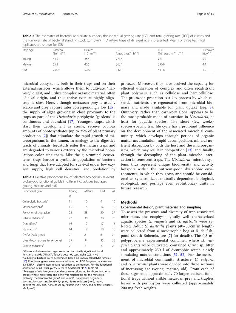
of traps. Although low in diversity, they are key playersin the trap microbial food webs. Their numbers in thestudied U. reflexa traps are immense, rising steadily withincreasing trap age, up to approximately 50,000 cells ofciliates alone per millilitre of trap fluid in the oldesttraps. This means that a single trap, depending on age,harbored tens to hundreds of individuals (Table 2). Suchhigh population densities are unheard of in natural envi-ronments and are comparable only to those found in themammalian rumen or in the activated sludge systems[41, 42]. The protist community consisted of variouseuglenid species and a monoculture of a conspicuouszoochlorellae-bearing ciliate (Additional file 1: FigureS5). This organism has recently been described as a newspecies—Tetrahymena utriculariae—and has not yetbeen found in any other environment [43, 44]. Ciliatesare important bacterial predators, mainly innutrient-rich freshwater environments [45–47], and aregenerally considered one of the key agents ensuring nu-trient recycling and transfer to other trophic levels (e.g.,[48]). We have confirmed and quantified bacterivory inT. utriculariae, and found the grazing rates to be com-parable to literature reports [49]. Due to their abun-dance, the ciliates were able to turn over the entirebacterial standing stock in the trap fluid extremely fast:four to five times in 24 h in the younger traps (Table 3).Turnover time increased markedly in the old traps, dueto the large increase in bacterial numbers. Obviously,the turnover of microbial mass in the U. reflexa trapsand the release of soluble mineral nutrients from micro-bial cells with their subsequent uptake by the plant fromthe trap fluid are, to a large extent, facilitated by protistpredation on bacteria. Although T. utriculariae has onlybeen found in the U. reflexa species, other species ofbacterivorous protozoa in high numbers have been ob-served in the traps of all aquatic Utricularia traps stud-ied by us so far. It can therefore be concluded thatUtricularia plants apparently depend on microbial activ-ity for the supply of nutrients and thus the amount ofbacterial mass produced and turned over in the trapslikely is as important to the plant host as the amount oforganic matter digested.
ConclusionsWe conclude that the aquatic Utricularia plants sup-port the development of diverse and sophisticated
Sirová et al. Microbiome (2018) 6:225 Page 7 of 13
microbial ecosystems, both in their traps and on theirexternal surfaces, which allows them to cultivate, “har-vest,” digest, and utilize complex organic material, oftenof algal origin, and thus thrive even at highly oligo-trophic sites. Here, although metazoan prey is usuallyscarce and prey capture rates correspondingly low [15],the supply of algae growing in close proximity to thetraps as part of the Utricularia periphytic “gardens” iscontinuous and abundant [17]. Youngest traps, whichstart their development as sterile, receive copiousamounts of photosynthates (up to 25% of plant primaryproduction [7]) that stimulate the rapid growth of mi-croorganisms in the lumen. In analogy to the digestivetracts of animals, feedstuffs enter the mature traps andare degraded to various extents by the microbial popu-lations colonizing them. Like the gut/ruminal ecosys-tems, traps harbor a symbiotic population of bacteriaand fungi that have adapted for survival under low oxy-gen supply, high cell densities, and predation by
protozoa. Moreover, they have evolved the capacity forefficient utilization of complex and often recalcitrantplant polymers, such as cellulose and hemicellulose.The protozoan predation is a key process by which es-sential nutrients are regenerated from microbial bio-mass and made available for plant uptake (Fig. 3).Omnivory, rather than carnivory alone, appears to bethe most probable mode of nutrition in Utricularia, atleast for aquatic species. The short (few weeks)species-specific trap life cycle has a profound influenceon the development of the associated microbial com-munity, which develops through periods of organicmatter accumulation, rapid decomposition, mineral nu-trient absorption by both the host and the microorgan-isms, which may result in competition [13], and, finally,through the decoupling of the plant–microbe inter-action in senescent traps. The Utricularia–microbe sys-tems thus represent unique biodiversity and activityhotspots within the nutrient-poor, dystrophic envi-ronments, in which they grow, and should be consid-ered as synchronized, mutually dependent biological,ecological, and perhaps even evolutionary units infuture research.
MethodsExperimental design, plant material, and samplingTo assess the presence and diversity of trap associatedmicrobiota, the ecophysiologically well characterizedaquatic species U. vulgaris and U. australis were se-lected. Adult U. australis plants (40–50 cm in length)were collected from a mesotrophic bog at Ruda fish-pond (South Bohemia, see [7] for details). The 0.8 m2
polypropylene experimental container, where U. vul-garis plants were cultivated, contained Carex sp. litterand approximately 250 l of dystrophic water, closelysimulating natural conditions [51, 52]. For the assess-ment of microbial community structure, U. vulgarisand U. australis plants were divided into three sectionsof increasing age (young, mature, old). From each ofthese segments, approximately 70 larger, excised, func-tional traps without visible metazoan prey and traplessleaves with periphyton were collected (approximately200 mg fresh weight).
Table 2 The estimates of bacterial and ciliate numbers, the individual grazing rate (IGR) and total grazing rate (TGR) of ciliates andthe turnover rate of bacterial standing stock (turnover) in U. reflexa traps of different age is presented. Means of three technicalreplicates are shown for IGR
Trap age Bacteria[106 ml−1]
Ciliates[103 ml−1]
IGR[bact. prot.− 1 h− 1]
TGR[106 bact. ml−1 d− 1]
Turnover[day−1]
Young 44.5 35.4 273.4 223.1 5.0
Mature 65.3 46.5 263.1 290.0 4.4
Old 266.9 50.8 342.1 411.8 1.5
Table 3 Relative proportions (%) of selected ecologically relevantprokaryotic functional guilds in different U. vulgaris trap ages(young, mature, and old)
Functional guild Young Mature Old All
%
Cellulolytic bacteria* 11 10 9 10
Methanotrophs‡ 15 15 14 15
Polyphenol degraders‡ 25 28 29 27
Nitrate reducers‡ 27 30 28 28
Denitrifiers‡ 16 17 17 16
N2 fixators‡ 14 17 18 16
DNRA (nrfA gene) 7 8 6 7
Urea decomposers (ureA gene) 29 34 35 33
Sulfate reducers‡ 2 2 2 2
Differences between trap ages were not statistically significant for allfunctional guilds (ANOVA, Tukey’s post hoc test, alpha 0.05, n = 3)*Cellulolytic bacteria were determined based on known cellulolytic families[50]. Functional genes were annotated based on RDP Fungene database ver.8.3; DNRA—dissimilatory nitrate reduction to ammonium. For the functionalannotation of all OTUs, please refer to Additional file 5: Table S8‡Averages of relative gene abundance were calculated for those functionalgroups where more than one gene was responsible for the metabolicpathway; methanotrophs (pmoA and mmoX), polyphenol degraders(laccase_Asco, laccase_Basidio, lip, ppo), nitrate reducers (narG, napA),denitrifiers (nirS, nirK, norB, nosZ), N2 fixators (nifH, nifD), and sulfate reducers(dsrA, dsrB)
Sirová et al. Microbiome (2018) 6:225 Page 8 of 13

To assess the actively transcribed microbial genepool, excised U. vulgaris traps from the entire shootwithout visible metazoan prey were collected randomly(approximately 250 mg fresh weight), with pooled trapsfrom a single plant considered a replicate; three bio-logical replicates were collected in total. All collectedplant material was immediately placed into liquid N2 andsamples were stored at −80 °C until further processing.For the estimation of the protozoan grazing rates, a dif-
ferent species with larger traps—the tropical U. reflexa—was selected, because relatively large volumes of trap fluidare needed for this analysis. The plants were cultivated in-doors in 3-l aquaria, in dystrophic cultivation water closelysimulating natural conditions (see [12]).
DNA extraction from Utricularia trap fluid and thetaxonomical evaluation of the Utricularia-associatedmicroorganismsNucleic acid extractions were conducted according to amodified bead-beating protocol [53]. Approximately500 μl of pooled trap fluid were extracted for each sample.Total DNA was quantified fluorometrically usingSybrGreen and StepOne (Applied Biosystems, USA)
instrument in “fluorescence reading mode” [54]. The PCRprimers (515F/806R) targeted the V4 region of the SSUrRNA, previously shown to yield accurate phylogenetic in-formation and to have only a few biases against any bac-terial taxa [55–57]. Each sample was amplified intriplicate, combined, and quantified using Invitrogen Pico-Green and a plate reader, and equal amounts of DNAfrom each amplicon were pooled into a single 1.5-mlmicrocentrifuge tube. Once pooled, amplicons werecleaned up using the UltraClean PCR Clean-up kit (MOBIO Laboratories). Amplicons were sequenced on the Illu-mina MiSeq platform at the Institute of Genomics andSystems Biology, Argonne National Laboratory, Argonne(Chicago, USA). Paired-end reads were joined using Perlscripts yielding approximately 253 bp amplicons. Approxi-mately 1.8 million paired-end reads were obtained withaverage 66.000 reads per sample. Quality filtering of readsand chimera check (UCHIME algorithm in de novomode) was applied as described previously [58]. Readswere assigned to operational taxonomic units (OTUs, cut-off 97% sequence identity) using an open-reference OTUpicking protocol with QIIME implemented scripts [58].Reads were taxonomically assigned using Green Genes
Fig. 3 Conceptual representation of the Utricularia trap ecophysiology: main microbe–microbe and plant–microbe interactions are shown
Sirová et al. Microbiome (2018) 6:225 Page 9 of 13

database, release 13.08 as reference. Those reads whichwere assigned as “chloroplast” and “mitochondrion” wereexcluded from further analyses. For the estimation ofunique microbial taxa in U. australis and U. vulgaris trapsand periphyton, the OTUs were grouped at the genuslevel. Genera presented only in U. australis or U. vulgarisand respective trap or periphyton samples were classifiedas unique for each microbiome. Differences between thevarious prokaryotic communities were tested with PER-MANOVA and the nonparametric method adonis inQIIME 1.9.0.
Comparative meta-analyses of prokaryotic communitiesfrom different habitatsTo compare the composition of Utricularia trap-associatedmicrobial communities, data (OTU tables in biom format)from nine relevant studies representing different habitatswere analyzed. Seven of the studies were obtained fromQiita (https://qiita.ucsd.edu/) database (Additional file 1:Table S1). The remaining two studies were obtainedfrom NCBI Genebank: the 16S rRNA sequences of theNepenthes pitcher microbiome from the SRA archive(project ID PRJNA225539) and Sarracenia pitcher se-quences from the Genebank database (accession num-bers JF745346–JF745532 and JN368236–JN368422)(Additional file 1: Table S1). Altogether, 4221 sampleswere included in the meta-analysis. The Qiita databaseworks with the closed-reference OTU picking algo-rithm; we have therefore processed our sequences andalso the sequences from Nepenthes and Sarraceniapitcher microbiomes in the same way as the Qiita pipe-line, to ensure comparability with Qiita OTU tables. AllOTU tables were then merged together using Qiimescripts and analyzed as one dataset. Non-metric multi-dimensional scaling (NMDS) using Bray-Curtis dissimi-latory metrics was used for computing distancesbetween samples (Fig. 1a). LDA Effect Size (LEfSe)based on the relative abundances of the microbial taxawas calculated to identify the corresponding taxa withhigher abundance in U.australis and U.vulgaris samples[59]. Analysis of LEfSe was performed according to theinstructions on the website (http://huttenhower.sph.-harvard.edu/galaxy).To obtain a more function-relatedpoint of view, three Utricularia vulgaris metatranscrip-tomes were compared to 43 metagenomes and/ormetatranscriptomes available from six different habi-tats (Fig. 1b). The sequences from these studies wereobtained from the MG-RAST server (Additional file 1:Table S1).
RNA-seq analysis for functional profiling of the U. vulgaristrap-associated microbiomeTo assess the actively transcribed microbial gene pool,total RNA was extracted from the U. vulgaris trap
samples (n = 3), using the protocol identical to that de-scribed in detail previously [30]. Briefly, DNA was re-moved from the extracts and two transcriptomiclibraries, eukaryotic and prokaryotic, were created atthe Institute of Genomics and Systems Biology, Ar-gonne National Laboratory, Argonne (Chicago, USA)using standard Illumina TruSeq RNA library prepar-ation kits. The ribosomal RNA as well as eukaryotic(plant) RNA fraction was removed in order to enrichprokaryotic transcripts, and, vice versa, eukaryotic tran-script enrichment was performed in parallel, to capturetranscripts from fungi, protists, and other eukaryoticmicroorganisms. Enriched mRNA from both librarieswas reverse transcribed to create metatranscriptomic li-braries and sequenced using Illumina HiSeq platform(100 × 100 cycle paired-end run). We obtained approxi-mately 40 million paired-end reads per sample. Readswere quality checked; low quality reads and reads withambiguous bases were filtered out. Reads from all threereplicates (approx. 120 million sequences) were then as-sembled with Velvet Optimizer [60] which resulted in ap-proximately 500,000 contigs. In this step, we filtered outthe potential Utricularia transcripts by blasting contigsagainst our Utricularia reference transcriptome [61]. Allcontigs which gave significant hit (e value < 0.0001, minscore 80) were excluded from further analyses and consid-ered as Utricularia transcripts. Contigs were also blastedagainst the SILVA database (release 111) to identify ribo-somal RNAs (rRNAs). Those sequences that gave BLASTbit score greater than 80 were marked as rRNAs and ex-tracted from the dataset. Reads were then mapped backonto the remaining contigs using Trinity package (bowtiealgorithm with default parameters [62] with FPKM genetranscript abundance normalization). To identify potentialprokaryotic functional gene transcripts, the remainingcontigs without rRNAs were blasted against the nrdatabase with e value of 0.001 using diamond algorithm[63]. Annotation was done in MEGAN 6 software [64].Genes below hit score 50 were manually excluded fromthe analyses (Additional file 4: Table S7).The lists of bacterial and archaeal species for each
ecologically relevant functional gene was downloadedfrom the FunGene database [65] from which a localdatabase was created (Additional file 5: Table S8). TheOTUs which were taxonomically annotated to genuslevel were scanned against this database for the pres-ence of the specific functional genes (Additional file 5:Table S8).
Microbial network analyses in U. vulgaris and U. australistraps and periphytonThe relative abundances of OTUs were square-roottransformed [66] (Additional file 6). To avoid
Sirová et al. Microbiome (2018) 6:225 Page 10 of 13

spurious correlations caused by the presence of rareOTUs, we chose only those OTUs which were foundin at least five out of nine traps and five out of eightU.vulgaris periphyton samples, and their sum ofabundance was at least 20 and 16 sequences out of1000, respectively. The resulting OTU tables, separ-ately for the trap (15 samples) and the periphyton (16samples), were used for microbial network analyses[67]. We performed the recommended calculations(neff, sparsity) regarding the composition of prokary-otic filtered OTU tables. Based on the sparsity of fil-tered OTU tables, we chose the CoNet networkalgorithm as the relevant calculation method [68, 69].The parameters and settings for network analyses in theCoNET application were as follow: -parent_child_exclu-sion, -row_minocc 5, -correlations (Spearman, Pearson,mutual information, Bray-Curtis and Kullback-Leibler dis-similatory). The threshold for edge selection was set to1000 top and bottom. During randomization, 100 itera-tions were calculated for edge scores. In the followingbootstrap step, 100 iterations were calculated, and un-stable edges were filtered out (p-level threshold of 0.05).The Brown method was chosen as the P value mergingmethod, and the Benjamini–Hochberg procedure was se-lected for multiple test correction. The resulting networkwas visualized and analyzed (i.e., degree of nodes, be-tweenness centrality, closeness centrality) in Cytoscape3.0.2. Potential keystone OTUs were identified [66].
Sequence depositionRaw sequences of 16S rRNA, ITS1 amplicons, and rawsequences of all three metatranscriptomes were depos-ited in European Nucleotide Archive (ENA) understudy ID PRJEB25993. Annotated metatranscriptomicsequences were deposited at the following website:http://utricularia.prf.jcu.cz/
Quantification of trap-associated bacterial, fungal,methanogenic, and methanotrophic communities in U.vulgaris and U. australisFor quantification of bacterial, fungal, methanogenic andmethanotrophic communities, the quantitative PCR(qPCR) of targeted 16S rRNA, 18S rRNA, mcrA, andpmoA gene was used, respectively. For detail description,please see Additional files 1, 2, 3, 4, 5, and 6.
Bacterial and protozoan enumeration and the estimationof protozoan grazing rates in U. reflexaTen U. reflexa plants were divided into three seg-ments of increasing age (young, mature, old). Eachsegment contained six leaf whorls. Trap fluid was col-lected from the traps in each segment (see [3]), and apooled sample (~ 750 μl for each age category) fromall ten plants was made. Bacterial and protozoan
counts in the trap fluid samples were estimated usingepifluorescence microscopy, according to methods de-scribed previously [45].The protist grazing rates were estimated using fluo-
rescently labeled bacteria (FLB) as a tracer. The FLBwere prepared from the strain Limnohabitans plank-tonicus, as detailed in [44]. Cell sizes of the strain arecomparable to that of bacterial cells commonly occur-ring within the U. reflexa traps. The FLB uptake rateswere determined in short-term triplicate experiments,where tracer FLB were added to the trap-fluid sam-ples to constitute 6–8% of the total bacterial concen-tration. For further details on sample fixation, protiststaining and enumeration, and tracer ingestion deter-minations, see [44]. At least 45 ciliates were inspectedfor FLB ingestion in each replicate sample. To esti-mate total protist grazing, we multiplied average up-take rates of protozoa by their in situ abundances aspreviously described [45].
Additional files
Additional file 1: Table S1. Source and identification of studies usedfor comparative meta-analyses in Fig. 1a, b and Additional file 1 FigureS2. Table S2. Comparison of selected alpha diversity indexes for various16S datasets from different habitats (N = number of samples in study). Alldatasets were subsampled to 2000 sequences prior to analyses, moreinformation on the data used can be found in Additional file 1: Table S1.Table S3. Comparison of the abundance of total bacteria, methanotrophs,methanogens, fungi, and fungal to bacterial ratio (F/B) in the trap andperiphyton of Utricularia species (data are averages from U. australisand U. vulgaris samples). Quantification of bacteria, fungi, methanotrophsand methanogens was done using the 16SrDNA, 18SrDNA, pmoA, andmcrA gene copy numbers, respectively. Quantity was normalized to totalamount of DNA. Table S4. The 5 most important potential keystone taxa inthe Utricularia-associated microbiomes, based on network analyses.Figure S1. Experimental Utricularia vulgaris shoot on a Petri dish. Segmentedleaves bearing traps and the growth tip are visible. Figure S2. Compositionaloverlap in Utricularia-associated prokaryotic microbiomes at the genus level.(a) Comparison between U. australis and U. vulgaris microbiomes and (b)between the U. australis and U. vulgaris periphyton and trap environments.Figure S3. Co-occurrence network for the prokaryotic community in theperiphyton of Utricularia vulgaris, constructed from QIIME 16S data. FigureS4. Co-occurrence network for the prokaryotic community in the trap fluid ofUtricularia vulgaris, constructed from QIIME 16S data. Figure S5. Tetrahymenautriculariae under the epifluorescence microscope. Zoochlorellae are visible inpurple, the nucleus is stained blue, and fluorescently labeled bacteria in foodvacuoles show green fluorescence. (DOCX 5108 kb)
Additional file 2: Table S5. a Prokaryotic OTUs distribution in samples.b Methanotropic OTUs distribution in samples (external file). c FungalOTUs distribution in samples. (XLSX 930 kb)
Additional file 3: Table S6. Summary of metatranscriptomic analyses.(XLSX 1461 kb)
Additional file 4: Table S7. Summary of selected protein families(Pfam) based on rpstblastx algorithm. (PDF 410 kb)
Additional file 5: Table S8. Functional annotation of OTUs (only forthose with genus assigment) genes. Annotation was based on RDPFungene database ver. 8.3. (PDF 673 kb)
Additional file 6: Table S9. Results of the adonis analysis for U.australis and U.vulgaris prokaryotic community based on 16S rDNA genesequencing. (PDF 459 kb)
Sirová et al. Microbiome (2018) 6:225 Page 11 of 13
AcknowledgementsWe would like to acknowledge Dr. Helena Štorchová (Institute of ExperimentalBotany CAS) for her help and enthusiastic support of the project, as well asHana Petrásková for her invaluable help in the laboratory.
FundingThis study was funded by the Czech Research Projects CSF P504/11/0783 (toLA) and CSF P504-17-10493S (to DS), as well as the Long-term researchdevelopment project No. RVO 67985939 (to LA) and BC CAS, SoWa (MEYSprojects LM2015075 and EF16_013/0001782 - SoWa Ecosystems Research).
Availability of data and materialsAll genetic raw data generated during this study have been uploaded tothe European Nucleotide Archive (ENA) under the study ID PRJEB25993.Metatranscriptomic and amplicon data were also deposited on thefollowing web page: http://utricularia.prf.jcu.cz within the section “Other/Download”. The datasets downloaded from the Qiita (https://qiita.ucsd.edu/)database or the MG-RAST server (https://www.mg-rast.org/) and used insupporting the conclusions are referenced in this article and itssupplementary material.
Authors’ contributionsDS, JB, LA, and JV designed the research. DS, LA, and JV collected thesamples. JB supervised the laboratory molecular work and bioinformaticsanalyses. JB, JS, and JP performed the bioinformatic analyses. JakBor supervisedand performed the chemical analyses. KŠ and TP performed the ciliate-relatedanalyses. DS and JB drafted the manuscript. All authors commented on previousversions of the manuscript and approved the final version.
Ethics approval and consent to participateNot applicable
Competing interestsThe authors declare that they have no competing interests.
Publisher’s NoteSpringer Nature remains neutral with regard to jurisdictional claims in publishedmaps and institutional affiliations.
Author details1Biology Centre CAS, Institute of Hydrobiology, Na Sádkách 7, CZ-37005České Budějovice, Czech Republic. 2Faculty of Science, University of SouthBohemia, Branišovská 1760, CZ-37005 České Budějovice, Czech Republic.3Limnological Station, Department of Plant and Microbial Biology, Universityof Zurich, CH-8802 Kilchberg, Switzerland. 4Department of Biology andWildlife, University of Alaska Fairbanks, Fairbanks AK-99775, USA. 5Institute ofExperimental Botany CAS, Rozvojová 263, CZ-16502 Praha 6-Lysolaje, CzechRepublic. 6Institute of Botany CAS, Dukelská 135, CZ-37982 Třeboň, CzechRepublic.
Received: 24 July 2018 Accepted: 18 November 2018
References1. Berg G, Grube M, Schloter M, Smalla K. Unraveling the plant microbiome:
looking back and future perspectives. Front Microbiol. 2014;5:148.2. Rinke C, Schwientek P, Sczyrba A, Ivanova NN, Anderson IJ, Cheng J-F, et al.
Insights into the phylogeny and coding potential of microbial dark matter.Nature. 2013;499:431–7.
3. Sirová D, Borovec J, Černá B, Rejmánková E, Adamec L, Vrba J. Microbialcommunity development in the traps of aquatic Utricularia species. AquatBot. 2009;90:129–36.
4. Caravieri FA, Ferreira AJ, Ferreira A, Clivati D, de Miranda VFO, Araújo WL.Bacterial community associated with traps of the carnivorous plantsUtricularia hydrocarpa and Genlisea filiformis. Aquat Bot. 2014;116:8–12.
5. Alcaraz LD, Martínez-Sánchez S, Torres I, Ibarra-Laclette E, Herrera-Estrella L.The metagenome of Utricularia gibba’s traps: into the microbial input to acarnivorous plant. PLoS One. 2016;11:e0148979.
6. Díaz-Olarte J, Valoyes-Valois V, Guisande C, Torres NN, González-BermúdezA, Sanabria-Aranda L, et al. Periphyton and phytoplankton associated withthe tropical carnivorous plant Utricularia foliosa. Aquat Bot. 2007;87:285–91.
7. Sirová D, Borovec J, Šantrůčková H, Šantrůček J, Vrba J, Adamec L. Utriculariacarnivory revisited: plants supply photosynthetic carbon to traps. J Exp Bot.2010;61:99–103.
8. Sirová D, Borovec J, Picek T, Adamec L, Nedbalová L, Vrba J. Ecologicalimplications of organic carbon dynamics in the traps of aquatic carnivorousUtricularia plants. Funct Plant Biol. 2011;38:583–93.
9. Carretero-Paulet L, Chang TH, Librado P, Ibarra-Laclette E, Herrera-Estrella L,Rozas J, et al. Genome-wide analysis of adaptive molecular evolution in thecarnivorous plant Utricularia gibba. Genome Biol Evol. 2015;7:444–56.
10. Carretero-Paulet L, Librado P, Chang TH, Ibarra-Laclette E, Herrera-Estrella L,Rozas J, et al. High gene family turnover rates and gene space adaptation inthe compact genome of the carnivorous plant Utricularia gibba. Mol BiolEvol. 2015;32:1284–95.
11. Silva SR, Alvarenga DO, Aranguren Y, Penha HA, Fernandes CC, Pinheiro DG,et al. The mitochondrial genome of the terrestrial carnivorous plantUtricularia reniformis (Lentibulariaceae): structure, comparative analysis andevolutionary landmarks. PLoS One. 2017;12:e0180484.
12. Adamec L. Oxygen concentrations inside the traps of the carnivorous plantsUtricularia and Genlisea (Lentibulariaceae). Ann Bot. 2007;100:849–56.
13. Sirová D, Bárta J, Borovec J, Vrba J. The Utricularia-associatedmicrobiome: composition, function, and ecology. Carnivorous PlantsPhysiology, Ecology, and Evolution. Oxford University Press. 2018;Chapter 25: 349–58.
14. Darwin C. Insectivorous plants. New York: D. Appleton and Company; 1875.15. Richards JH. Bladder function in Utricularia purpurea (Lentibulariaceae): is
carnivory important? Am J Bot. 2001;88:170–6.16. Friday LE. Rapid turnover of traps in Utricularia vulgaris L. Oecologia.
1989;80:272–7.17. Płachno BJ, Łukaszek M, Wołowski K, Adamec L, Stolarczyk P. Aging of
Utricularia traps and variability of microorganisms associated with thatmicrohabitat. Aquat Bot. 2012;97:44–7.
18. Peroutka M, Adlassnig W, Volgger M, Lendl T, Url WG, Lichtscheidl IK.Utricularia: a vegetarian carnivorous plant? Agae as prey of bladderwort inoligotrophic bogs. Plant Ecol. 2008;199:153–62.
19. Alkhalaf IA, Hübener T, Porembski S. Prey spectra of aquatic Utriculariaspecies (Lentibulariaceae) in northeastern Germany: the role of planktonicalgae. Flora Morphol Distrib Funct Ecol Plants. 2009;204:700–8.
20. Nalepa C a, Bignell DE, Bandi C. Detritivory, coprophagy, and the evolutionof digestive mutualisms in Dictyoptera. Insect Soc. 2001;48:194–201.
21. Ley RE, Hamady M, Lozupone C, Turnbaugh PJ, Ramey RR, Bircher JS, et al.Evolution of mammals and their gut microbes. Science. 2008;320:1647–51.
22. Barboza PS, Bennett A, Lignot J-H, Mackie RI, McWhorter TJ, Secor SM, et al.Digestive challenges for vertebrate animals: microbial diversity,cardiorespiratory coupling, and dietary specialization. Physiol Biochem Zool.2010;83:764–74.
23. Adamec L. Functional characteristics of traps of aquatic carnivorousUtricularia species. Aquat Bot. 2011;3:226–33.
24. Adlassnig W, Peroutka M, Lendl T. Traps of carnivorous pitcher plants as ahabitat: composition of the fluid, biodiversity and mutualistic activities. AnnBot. 2011;107:181–94.
25. Troyer K. Diet selection and digestion in Iguana iguana: the importance ofage and nutrient requirements. Oecologia. 1984;61:201–7.
26. Berg M, Zhou XY, Shapira M. Host-specific functional significance ofCaenorhabditis gut commensals. Front Microbiol. 2016. https://doi.org/10.3389/fmicb.2016.01622.
27. Willems A. The family Comamonadaceae. The prokaryotes: alphaproteobacteriaand Betaproteobacteria. In: Rosenberg E, DeLong EF, Lory S, Stackebrandt E,Thompson F, editors. The Prokaryotes. Berlin: Springer; 2014.
28. Slobodkin A. The family peptostreptococcaceae. The prokaryotes: firmicutesand tenericutes. In: Rosenberg E, DeLong EF, Lory S, Stackebrandt E,Thompson F, editors. The Prokaryotes. Berlin: Springer; 2014.
29. Attwood GT, Klieve AV, Ouwerkerk D, Patel BKC. Ammonia-hyperproducing bacteria from New Zealand ruminants. Appl EnvironMicrobiol. 1998;64:1796–804.
30. Sirová D, Šantrůček J, Adamec L, Bárta J, Borovec J, Pech J, et al. Dinitrogenfixation associated with shoots of aquatic carnivorous plants: is itecologically important? Ann Bot. 2014;114:125–33.
31. Velicer GJ, Mendes-Soares H. Bacterial predators. Curr Biol. 2009;19:R55–6.32. Chauhan A, Cherrier J, Williams HN. Impact of sideways and bottom-up
control factors on bacterial community succession over a tidal cycle. PNAS.2009;106:4301–6.
Sirová et al. Microbiome (2018) 6:225 Page 12 of 13

33. Dawid W. Biology and global distribution of myxobacteria in soils. FEMSMicrobiol Rev. 2000;24:403–27.
34. Popper ZA, Michel G, Hervé C, Domozych DS, Willats WGT, Tuohy MG, et al.Evolution and diversity of plant cell walls: from algae to flowering plants.Annu Rev Plant Biol. 2011;62:567–90.
35. Yapi Assoi Yapi D, Gnakri D, Lamine Niamke S, Patrice Kouame L.Purification and biochemical characterization of a specific beta-glucosidasefrom the digestive fluid of larvae of the palm weevil, Rhynchophoruspalmarum. J Insect Sci. 2009;9:4.
36. Chen B, Teh BS, Sun C, Hu S, Lu X, Boland W, et al. Biodiversity and activityof the gut microbiota across the life history of the insect herbivoreSpodoptera littoralis. Sci Rep. 2016;6:29505.
37. Petit C, Rigg GP, Pazzani C, Smith A, Sieberth V, Stevens M, et al. Region 2of the Escherichia coli K5 capsule gene cluster encoding proteins for thebiosynthesis of the K5 polysaccharide. Mol Microbiol. 1995;17:611–20.
38. González-Rodríguez I, Sánchez B, Ruiz L, Turroni F, Ventura M, Ruas-Madiedo P, et al. Role of extracellular transaldolase from Bifidobacteriumbifidum in mucin adhesion and aggregation. Appl Environ Microbiol.2012;78:3992–8.
39. Hackstein JHP, Van Alen TA. Fecal methanogens and vertebrate evolution.Evolution. 2016;50:559–72.
40. Repeta DJ, Ferrón S, Sosa OA, Johnson CG, Repeta LD, Acker M, et al.Marine methane paradox explained by bacterial degradation of dissolvedorganic matter. Nat Geosci. 2016;9:884–7.
41. Towne G, Nagaraja TG, Brandt RT, Kemp KE. Dynamics of ruminal ciliatedprotozoa in feedlot cattle. Appl Environ Microbiol. 1990;56:3174–8.
42. Abraham JV, Butler RD, Sigee DC. Ciliate populations and metals in anactivated-sludge plant. Water Res. 1997;31:1103–11.
43. Pitsch G, Adamec L, Dirren S, Nitsche F, Šimek K, Sirová D, et al. The greenTetrahymena utriculariae n. sp. (Ciliophora, Oligohymenophorea) with itsendosymbiotic algae (Micractinium sp.), living in traps of a carnivorousaquatic plant. J Eukaryot Microbiol. 2017;64:322–35.
44. Šimek K, Pitsch G, Salcher MM, Sirová D, Shabarova T, Adamec L, et al.Ecological traits of the algae-bearing Tetrahymena utriculariae (Ciliophora)from traps of the aquatic carnivorous plant Utricularia reflexa. J EukaryotMicrobiol. 2017;64:336–48.
45. Šimek K, Jürgens K, Nedoma J, Comerma M, Armengol J. Ecological role andbacterial grazing of Halteria spp.: small freshwater oligotrichs as dominantpelagic ciliate bacterivores. Aquat Microb Ecol. 2000;22:43–56.
46. Simek K, Armengol J, Comerma M, Garcia JC, Chrzanowski TH, Macek M, etal. Characteristics of protistan control of bacterial production in threereservoirs of different trophy. Int Rev Hydrobiol. 1998;83:485–94.
47. Thouvenot A. Bacterivory of metazooplankton, ciliates and flagellates in anewly flooded reservoir. J Plankton Res. 1999;21:1659–79.
48. Laybourn-Parry J. Protozoan plankton ecology. London: Chapman andHall; 1992.
49. Neuer S, Cowles TJ. Comparative size-specific grazing rates in field populationsof ciliates and dinoflagellates. Mar Ecol Prog Ser. 1995;61:99–103.
50. Koeck DE, Pechtl A, Zerlov VV, et al. Genomics of cellulolytic bacteria. CurrOpin Biotech. 2014;29:171–83.
51. Adamec L, Sirová D, Vrba J. Contrasting growth effects of prey capture intwo aquatic carnivorous plant species. Fundam Appl Limnol / Arch fürHydrobiol. 2010;176:153–60.
52. Borovec J, Sirová D, Adamec L. Light as a factor affecting the concentrationof simple organics in the traps of aquatic carnivorous Utricularia species.Fundam Appl Limnol / Arch für Hydrobiol. 2012;181:159–66.
53. Urich T, Lanzen A, Qi J, Huson DH, Schleper C, Schuster SC. Simultaneousassessment of soil microbial community structure and function throughanalysis of the meta-transcriptome. PLoS One. 2008;3:e2527.
54. Leininger S, Urich T, Schloter M, Schwark L, Qi J, Nicol GW, et al.Archaea predominate among ammonia-oxidizing prokaryotes in soils.Nature. 2006;442:806–9.
55. Liu Z, Lozupone C, Hamady M, Bushman FD, Knight R. Shortpyrosequencing reads suffice for accurate microbial community analysis.Nucleic Acids Res. 2007;35:e120.
56. Bates ST, Berg-Lyons D, Caporaso JG, Walters WA, Knight R, Fierer N.Examining the global distribution of dominant archaeal populations in soil.ISME J. 2011;5:908–17.
57. Bergmann GT, Bates ST, Eilers KG, Lauber CL, Caporaso JG, Walters WA, et al.The under-recognized dominance of Verrucomicrobia in soil bacterialcommunities. Soil Biol Biochem. 2011;43:1450–5.
58. Caporaso JG, Kuczynski J, Stombaugh J, Bittinger K, Bushman FD, CostelloEK, et al. QIIME allows analysis of high-throughput community sequencingdata. Nat Methods. 2010;7:335–6.
59. Segata N, Izard J, Walron L, Gevers D, Miropolsky L, Garrett W,Huttenhower C. Metagenomic biomarker discovery and explanation.Gen Biol. 2011;12:R60.
60. Zerbino DR, Birney E. Velvet: algorithms for de novo short read assemblyusing de Bruijn graphs. Genome Res. 2008;15:78.
61. Bárta J, Stone JD, Pech J, Sirová D, Adamec L, Campbell MA, et al. Thetranscriptome of Utricularia vulgaris, a rootless plant with minimalistgenome, reveals extreme alternative splicing and only moderate sequencesimilarity with Utricularia gibba. BMC Plant Biol. 2015;15:78.
62. Haas BJ, Papanicolaou A, Yassour M, Grabherr M, Blood PD, Bowden J, et al.De novo transcript sequence reconstruction from RNA-seq using the Trinityplatform for reference generation and analysis. Nat Protoc. 2013;8:1494–512.
63. Buchfink B, Xie C, Huson DH. Fast and sensitive protein alignment usingDIAMOND. Nat Methods. 2015;12:59–60.
64. Huson DH, Beier S, Flade I, Górska A, El-Hadidi M, Mitra S, et al. MEGANCommunity Edition - interactive exploration and analysis of large-scalemicrobiome sequencing data. PLoS Comput Biol. 2016;12:e1004957.
65. Fish JA, Chai B, Wang Q, Sun Y, Brown CT, Tiedje JM, et al. FunGene: thefunctional gene pipeline and repository. Front Microbiol. 2013;4:291.
66. Berry D, Widder S. Deciphering microbial interactions and detectingkeystone species with co-occurrence networks. Front Microbiol. 2014;5:219.
67. Weiss S, Van Treuren W, Lozupone C, Faust K, Friedman J, Deng Y, et al.Correlation detection strategies in microbial data sets vary widely insensitivity and precision. ISME J. 2016;10:1669–81.
68. Shannon P, Markiel A, Ozier O, Baliga NS, Wang JT, Ramage D, et al.Cytoscape: a software environment for integrated models of biomolecularinteraction networks. Genome Res. 2003;13:2498–504.
69. Faust K, Sathirapongsasuti JF, Izard J, Segata N, Gevers D, Raes J, et al.Microbial co-occurrence relationships in the human microbiome. PLoSComput Biol. 2012;8:e1002606.
Sirová et al. Microbiome (2018) 6:225 Page 13 of 13