human platelet lipids and their relationship to blood coagulation
TRANSCRIPT

500 T H E J O U R N A L OF T H E A M E R I C A N O I L C H E ~ £ I S T S ~ S O C I E T Y VOL. 42
REFERENCES
1. Falk, F., Biochem. Z. 13, 153 (1908). 2. Randall, L. 0., J. Biol. Chem, 125, 723 (1938). 3. Schmidt, G., J, Benotti, B, Hershman and S. 5. Thannhauser, J.
Biol. Chem. 166, 505 (1946). 4. Johnson, A. C., A. R. MeNabb and R. J. Rossiter, Biochem. J.
43, 578-580 (1948). 5. Brante, G., Acta I?hysiol. Seand. 18, Suppl. 63 (1949). 6. McCaman, R. E., and E. Robins, J. Neurochem. 5, 18 (1959). 7. Webster, G. R., Bioehim. Biophys. Acta 44, 109-116 (1960). 8. Webster, G. R., and R. t t . S. Thompson, Biochim. Biophys. Acta
63, 38 (1962). 9. Johnson, A. C., A. R. )/IeNabb and R. J. Rossiter, Biochem. J.
45, 500--508 (1949) . 10. Burr, N. S., A. R. MeNabb and R. J. Rossiter, Biochem. J. 47,
318-323 (1950) . 11. Magee, W. L., 5. F. Berry, ~¢L 1VIagee and R. 5. Rossiter, J.
Neurochem. 3, 333 (1958). 12. Dawson, I~. Iv[. C., Biochim. Biophys. Aeta 14, 374-379 (1954). 13. )[iani, N., 5. Neurochem. 9, 525-536 (4962). 14. Dawson, R. )¢I. C., Biochem. 5. 75, 45-53 (1960), 15. gossiter, R. 5., in "Neurochemistry," edited by Elliott, K. A. C.,
I. If . Page and J. n . Quastel, Charles C Thomas, Springfield, IlL, 1962, p. 33.
16. Kline, D., V~ r. L. ~¢Iagee, E. T. Pri¢chard and R. J. Rossiter, J . Neurochem. 3, 52-58 (1958).
17. Pritchard, E. T., and R. J. I¢ossiter, J. Neurochem. 3, 341--346 (1958) .
18. )~[ajno, G., and ~,~. L. Karnovsky, J. Exp. Med, 108, 197-214 (1958).
19. Baker, ~ . W. R,, Biochem. J. 79, 642 ( t 961 ) . 20. Bartley, \V., R. van :tteyningen, B. :hi. Norton and A. Renshaw,
Biochem. 5. 85, 332 (1962). 21. Gerstl, B., P~. B. Hayman, M. Tavast ierna and J. K. Smith, Ex-
perientia 18, 131 (1962). 22. Cevallos, W. It., and J. F. Berry, Fed. Prec. 20, 271 (1961). 23. Rouser, G., G. Kritchevsky, D. I~Ieller and E. Lieber, JAOCS
40, 425-454 (1963). 24. Folch, J., ~ . Lees and G, M. Sloane-StanleT, J. Biol. Chem. 226,
497-509 (1957). 25. Entenman, C., JAOCS 88, 534-538 (1961). 26. Trams, E. G., and C. J. Lauter, Biochim. Biophys. Acta 60,
350 (1962). 27. Rouser, G., A. J. Bauman, G. Kritchevsky, D. J. n en e r and
J. S. O'Brien, JAOCS 38, 544-555 (1961).
28. /-torning, M. G., E. A. Williams and E. C. Horning, J. Lipid Res. 1, 482 (1960).
29. Hirsch, J., and E. H. Ahrens, Jr., J. Biol. Chem. 233, 311-320 (1958).
30. Kishimoto, Y., and N. S. Radin, J. Lipid Res. v, 72 (1960). 31. ttaneI, I~[. K., and I t . Dam, Acta Chem. Scand. 9, 677 (1955). 32. Van ttandel, E., and D. B. Zilversmit, J. Lab. and Clin. Med.
50, 152 (1957), 33. Snyder, F., and N. Stephens, Biochim. Biophys. Acta 34, 244
(1959). 34. Bartlett, G. R., J. Biol. Chem. 234, 466 (1959). 35. Rouser, G., J. S. 0 'Br ien and D. tteller, JAOCS 35, 14-19
(1961) . 36. Gray, G. !Yi., and 1El. G. ~¢[cFarlane, Biochem. J. 70, 409 (1958). 37. Siakotos, A. N., personal communication to the author. 38. Rouser~ G,, A. J. Bauman, N. Nicolaides and D. Heller, 5AOOS
38, 565-581 (1961) . 39. Blank, M. L., 5. A. Schmit and O. S. Privett, JAOCS 41, 371-
376 (1964). 40. Farquhar, J. W,, W. Insull P. Rosen W. Stoffel and E. H.
Ahrens, Nutr. Revs 17, Suppl. 1 i1959) ' 41. Farquhar, J. W., 5. Lipid Res. 3, 2 1 - 3 0 (1962). 42. Horwitt, M. K., C. C. Harvey, B. Century and L. A. Witting,
5. Amer. Dietetic Assoc. 38, 231--235 (1961) . 43. :k[arco, G. 5., L. J. Machlin, E. Emery and R. S. Gordon, Arch.
Biechem. Biophys. 94, 115-120 (1961). 44. 5~ohrhauer, ]L, and R. T. Holman, 5. Neurochem. 10, 527
(1963) . 45. Silver, A., J. Physiol. 148, 66P (1959) . 46. Johnson, A. C., A. R. McNabb and R. J. Rossiter, Canad. J.
Res. 27, 63--71 (1949). 47. McCoI1, J. D., and J. K. Weston, Rev. Canad. Biol. 12, 68-76
(1953). 48. :~¢[c~array, W. C., J. NeurochenL 11, 287 (1964). 49. Guyon, L., Compt. Rend. Soc. Biol. 109, 1101 (1932). 50. Klenk, E , :H. Debuch, and H. Daun, Z. Physiol. Chem. 292,
24 (1953). 51. Dawson, R, ~[, C,, Biochem. J. 56, 621 (1954). 52. Weiss, B , J. Biol. Chem. 223, 523 (1956). 53, Edgar, G, W. F., and G. Smits, J. Neurochem. 3, 316 (1959). 54, Ansell, G. B,, and S. Spanner Biochem. J. 79, 176 (1961). 55, Carter, I'I. E,, B, B. Smith and D. N. Jones, J. Biol. Chem.
232, 681 (1958). 56. Berry, J. F., B. Kaye and K. Chang, in preparation. 57. O'Brien, J., and G. Rouser, Fed. Prec. 21, 284 (1962). 58. Carroll, K, K., 5. Lipid Res. 3, 263 (1962). 59. Kennedy, E, P., Fed. Prec. 20, 934-940 (1961).
Human Platelet Lipids and Their Blood Coagulation
Relationship to
AARON J. MARCUS and DOROTHEA ZUCKER-FRANKLIN, Veterans' Administration Hospital, New York; Cornell University Medical College, and New York Univ ersity School of Medicine
Abstract There is now reasonable agreement on the se-
quence of physiological and biochemical events leading to fibrin formation, and phospholipids are an impor tant par t of this process. The phos- phatides are ordinari ly provided by platelets, and it appears that a l ipoprotein complex is respon- sible for this activity. The anatomic site of this complex is not known, but evidence is presented that it may be a proper ty of the platelet mem- brane. Methods for the s tudy of platelet lipids including fa t ty acids and aldehydes are described, and include silicic acid column and paper chro- matography, as well as thin-layer and gas-liquid chromatographic procedures. These are also be- ing utilized in studies of subcellular platelet par- ticles, where only limited amts of biological ma- terial are available for study. I t is stressed that experimental results obtained from studies on iso- lated tipids should be interpreted with a certain degree of caution. I t is unlikely that they are available as such in in r ive coag~ulation, and the drastic procedures used for their extraction and isolation may alter their basic physiological properties.
I N r~ECE'~,~T YEARS, the role of Iipids in blood coagula- tion has been better understood, and there appear
to be two main reasons for this increase in our know- ledge. First , the sequence of protein interactions lead- ing to the formation of a fibrin clot has been fur ther elucidated (1). Secondly, advances in research have
enabled us to characterize the specific lipids involved in the coagulation mechanism. Our thinking has prob- ably been fu r the r clarified by the realization that there are three physiological events that should be dealt with separa te ly- -a t least for the time being. They are: a) blood coagulation; b) hemostasis; c) thrombosis. The tendency in the past to think of these as a single ent i ty has led to confusion. For example, an increase in plasma lipids or postprandial lipemia was equated with a "hypercoagu lab le" state. This was presumed to be directly related to thrombosis. Arterial thrombi histologically resemble hemostatic platelet plugs in that there is a white " h e a d " consist- ing of relatively intact platelets. Venous thrombi, Oil the other hand, more closely resemble clots as formed in the test tube. That is, they consist of a mixture of fibrin, entrapped red cells and leukocytes, as well as platelets. These are the so-called " r e d " thrombi. Thus, it is inlportant to realize that lipids may not play a role in thrombosis and hemostasis, but may only be important for coagulation. These concepts have been discussed in detail in recent re- views of the subject (2-4) .
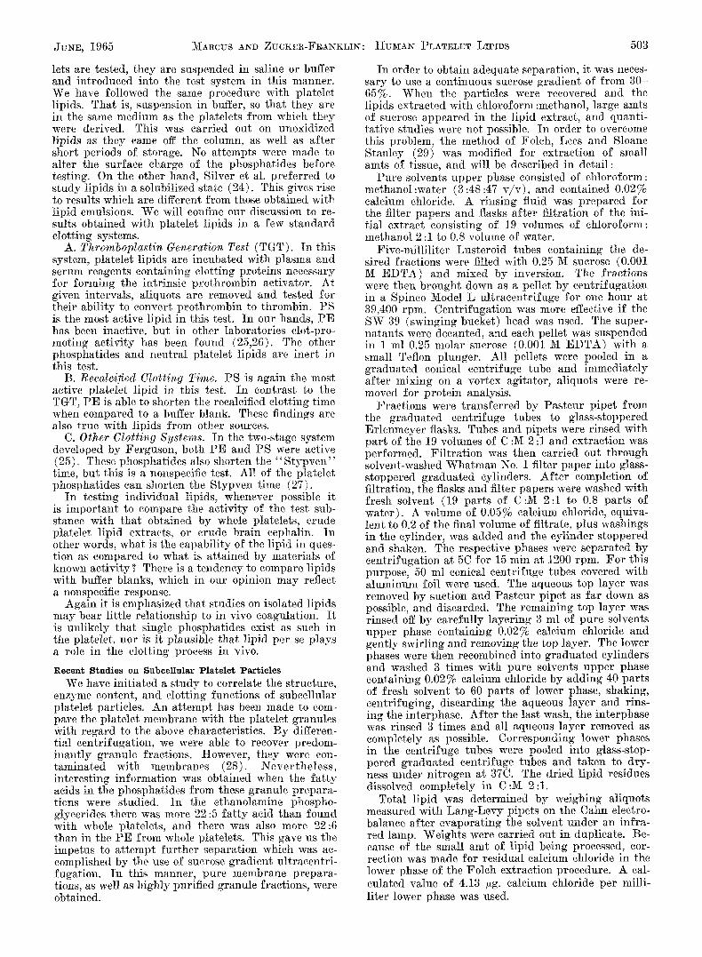
Current Theory of Coagulation Figure I shows the sequence of events leading to
the formation of fibrin. Contact with a fore ign sur- face, such as damaged endotheIium or the cut edge of a blood vessel appears to activate Hageman factor. This in tu rn activates PTA and a pa t tern of biochem- ical t ransformations appears to ensue, which involves the activation of a previously inactive coagulation

JUNE, 1965 2tlARcUS AND ZUCKER-FRANKLIN:
protein (1,31). Note tha t the lipid contribution ap- pears relat ively late in the reactions of the intrinsic prothrombin act ivator system. Current thinking is that the lipid is derived f rom platelets which have already ar r ived at the site of blood vessel damage because of their role in hemostasis. Classical theories of platelet involvement in clotting propose tha t gran- ules are discharged into the surrounding medium, which make their l ipid available to the coagulation process. Recent data in our own laboratory indicate that the platelet membrane has somewhat better clot- promoting capabilities than do the granules. We have proposed tha t the platelet granules are "storehouses" for metabolic substances concerned with other platelet functions, and the membrane l ipid acts as a surface catalyst for coagulation. I t is emphasized tha t these are merely theoretical proposals, and fu r the r investi- gation will be necessary before they can be considered unequivocal (5).
Conclusions drawn f rom studies on isolated platelet lipids must be considered in the light of their in vi tro act ivi ty only. Fo r example, phospholipids, especially of the ethanolamine and serine groups, are the most active in the test tube. However, in in vivo coagula- tion, all the platelet ]ipids are probably available as a l ipoprotein complex. In addition, when platelet homogenates are made, a soluble l ipoprotein appears which possesses clot-promoting properties. I t is not known whether this represents an ar tefact of the dis- rupt ion procedure or whether the platelet actually contains soluble l ipoprotein in an unbound form.
With these pre l iminary comments in mind, we would like to describe some of the methods utilized in our laboratory for the past ten years in the investiga- tion of human platelet lipids.
M e t h o d s a n d M a t e r i a l s
Methods of Collection and Lipid Extract ion of P late le ts
Platelets were isolated f rom freshly collected blood (6). The appearance of platelets as seen by electron microscopy is i l lustrated in Fig. 2. They were washed and frozen within a period of 5 hr. Ordinari ly, they were mainta ined at - 2 0 C for periods up to 3 months pr ior to extraction. The subcellular platelet particles had a tendency to deteriorate at - 20C , as evidenced by discoloration and putrefact ion. Therefore, the sub- cellular materials are now being stored at - 8 5 C. Following venisection, all operations involved in platelet processing were carr ied out in a cold room at 4C. Two main extraction procedures have been used. The first was based on the procedure of Bell and Alton (7). This involves suspension in four times the original volume of acetone for 30 rain, fol- lowing which the acetone was removed by centrifu- gation. This was repeated 4-6 times. The acetone- insoluble mater ia l was then extracted with ten times its volume of chloroform, and this was repeated twice, the th i rd extract ion being allowed to take place overnight. The yields appeared to be the same whether the procedure was carried out at 4C or at room temp. This extract ion procedure yielded less of the neutra l l ipid fract ion and is stitl our procedure of choice for prepara t ion of platelet or brain "eephalin" for use in blood coagulation tests as platelet substitutes.
In la ter studies, chloroform :methanol 2:1 was used. The procedures were ahvays carried out under highly purified nitrogen, and the solvents were deoxygenated immediately before use (8). In this method, extrac- tion of 1 g (wet weight) of platelet mater ia l yielded about 31 nag of crude lipid. The s ta r t ing mater ia l
HU]YIAN PLATELET LIPIDS 501
((FOREIGN" SURFACE CONTACT~HAGEMAN (XII)
ACTIVE XIIyPTA (XI)
ACTIVE XI.~.(PTC (IX)
ACTIVE IX~AHG (VIII)
ACTIVE VIII~/STUART (X)
V VII PLATELET PHOSPHOLIpID
Ca++ Ca++
EXTRINSIC PROTHROMBIN ACTIVATOR~PROTHROMBIN (II)~--INTRINSIC PROTHROMBIN ACTIVATOR
THROMB lN~f F LB KINOGE N (D
FZBR[N~-FIBRIN S T A B I L I Z I N G FACTOR XIII
FIG. I. Diagrammatic scheme of blood coagulation mecha- nism as currently accepted by most investigators.
for each column separat ion was approx imate ly 400 mg of lipid. A p p r o x 13 g (wet weight) of plate- lets was homogenized in 150 ml chloroform-methanol in a War ing Blendor under an atmosphere of nitro- gen. The insoluble mater ia l was removed by filtration through a sintered glass filter and finally the filtrate was passed ti lrough fat-free sharkskin filter paper. The filtrate was then dried under reduced pressure and nitrogen in an Er lenmeyer flask at 30C. I t was found useful to add aldehyde-free absolute ethanol to the flask, which formed a water-ethanol azeotrope, thus aiding in the removal of last traces of water (9). The lipid mater ial was stored under nitrogen in C :M 2:1 for a max of 12 hr at - 2 0 C pr ior to column chro- matography.
Column Chromatography
In earlier work (6,10), the procedures of Lea, Rhodes and Sto]l (11) were used, as well as those of Hirseh and Ahrens (9). Al though the methods them- selves were excellent, they did not serve our purpose as well as the procedures la ter developed by Rouser and associates. The reason for this was tha t there was an unfor tuna te overlapping between the ethanolamine and serine phosphoglycerides, and only small amts of serine phosphoglycerides could be obtained. The clot- promot ing propert ies of almost any combination of P E and PS were quite impressive, but we were mainly interested in the act ivi ty of individual phosphatides. Thus, we tu rned our at tent ion to the techniques of Rouser (12,13) in our later studies. Although we have tr ied others, Mallinckrodt silicie acid 100 mesh has always appeared to be the most suitable. Usually 60 g of the nmterial was washed as described by Rouser (12). The washed silieic acid was heated for 12-15 hr at 120C under negative pressure and nitrogen in a
FIG. 2. Electron micrograph of human blood platelets. G: granule ; G1 : glycogen particles ; M : mitochondrion. Magnifi- catlon: x 32,000.

502 THE JOURNAL OF TI:IE AMERICAN OIL CHEJ/IISTS' SOCIETY VOL. 42
1,000 ml three-necked flask. This was allowed to cool, and 200 ml chloroform:methanol 4:1 was added to make a slurry. The s lurry was poured into a glass column 2.5 × 40 cm. In such a column, 60 gm of silicic acid extended to a height of 20 cm af ter the application of nitrogen pressure. The platelet lipid was applied to the column in a small volume of chloro- form :methanol 4:1, and the chromatography was car- ried out at 10C by allowing cold water to be pumped through a jacket surrounding the column. Fract ions of 10 ml were collected by hand and examined by means of Rouser 's rapid n inhydr in test (12). About every tenth tube was studied in greater detail by thin- layer and silicic acid paper chromatography.
A rough quanti tat ive index was also obtained by adding 1 ml of eluate to 1 ml 5 N sulfuric acid in a 30 ml Kjeldahl flask and heating a~ 320C for 5-7 rain. The intensity of charr ing was an index of the amount of organic material present.
In the first silicic acid column separation, the elut- ing solvent was chloroform:methanol 4:1 until the fractions became negative to the ninhydrin reagent. Before storing the samples, 0.1 ml coned ammonia was added to each tube, which retarded plasmalogen break- down. Af ter the ninhydrin-positive fractions from the first run were verified as a mixture of P E and PS by thin-layer and silicic acid paper chromatography, they were all pooled and evaporated down to 5 ml. Twelve hours later, the P E and PS were separated from each other on a silicie acid-silicate-water column, which was prepared by allowing a mixture of chloro- form :aqueous ammonia to pass through the previously prepared silicic acid column. As Rouser has clearly shown, the PS remains bound to the column while P E is eluted first with chloroform :methanol 4:1. The PS is finally eluted with methanol.
Paper Chromatography We still use paper chromatographic procedures to
supplenlent our thin-layer techniques. The systems devised by Marinett i et al. have been most useful (14). Examinat ion of the phosphatides under ultraviolet light is occasionally more profitable than the identifi- cation procedures for thin-layer chromatography (TLC).
Thin-Layer Chromatography This technique has been most useful to us for iden-
t i fying lipids in individual fractions. I t has been less valuable for total lipid extracts, mainly because we have been interested in complete separation of PE from PS and PI. In systems current ly available, such as those of Skipski and associates (15-17), P I and PS run very close to each other. Thus, for thin-layer examination of column fract ion we have used a solvent system consisting of chloroform-methanol :water :coned ammonia 75:25:4:1 (18). The chromatographic plates were dried in air and sprayed with 40% sulfuric acid followed by charring on a hot plate. I f the separated phosphatides were oxidized or broken down, these phenomena could also be detected on thin-layer plates (8). Gas-Liquid Chromatography
For this we have employed a Barber-Colman model 10 apparatus, equipped with two strontium 9° detector cells. U-shaped glass eolmnns, 6 f t in overall length, were most f requent ly used. I t was important to cheek the l ineari ty of detector response to mass with the fa t ty acid standards providecl by the N a t i ~ a ! H e a r t Institute. The range of error using all of the mix- tures, A through D, was never more than 3%. Methyl esters and dimethyl aeetMs were prepared by the
method of Stoffel, Chu and Ahrens (19). I t was found that sublimation following esterifieation was not necessary.
A. Nonpolar Columns. Gas-Chrom P (Applied Science Laboratories, State College, Pa.) and Ana- krom AB (Analabs, Hamden, Conn.) were found to be satisfactory s tat ionary supports. Usually the sta- t ionary supports were washed with acid and alkali, and then brought back to neutral i ty. However, if this was done, precise quantification of the dimethyl ace- tals was not possible. Af ter the alkali wash it was necessary to leave the support material in an alkaline state. The manner in which this was done is as fol- lows: The support was washed in 1% methanolic po- tassium hydroxide which was removed by filtration through a Buchner funnel. Af ter drying in a con- vection oven for 12 hr at 120C, the p H was tested by suspending 1 mg of powder in 1 ml of distilled water. The p H of the support material was 7.4 I t was then coated with 10-15% Apiezon L or M (20). This ma- terial is now commercially available from Applied Science Laboratories, State College, Pa. Before use, the columns were conditioned for 12~-2~ hr at temps slightly above that used for normal operation. I t was important not to connect the columns to the detector cells during the conditioning period (21). During this time, large loads of mixed methyl esters were applied to the column for a " so a k i n g " effect (22). Detector responses were most linear if they were operated at a voltage of 350. The amplifier setting was usually 3 × 10-Sa. Column temp was 197C, the detector was maintained at, 220C, and flash heater at 197C.
B. Polar Columns. Stat ionary support was the same as used for nonpolar columns and pretreatment with alkali was helpful. The " s o a k i n g " procedure was also used here. Supports were coated with 10- 15% ethylene glycol adipate (EGA) . The cell voltage was about 400, amplifier setting 3 × 10 Sa, column temp 173C, detector 220C, and flash beater 173C.
We have found it advantageous to compare quanti- tative and qualitative results with another laboratory engaged in GLC. Aliquots of methyl esters prepared in our laboratory were analyzed by Dr. J. W. Fa rquha r at the Rockefeller Insti tute, New York. Our results were in excellent agreement.
C. Fatty Acids of PE and PS. The ethanolamine phosphoglycerides in human platelets had stearic acid as the main saturated fa t ty acid. The principle un- saturated acid was arachidonic. As in the red cell (18), the f a t ty aldehydes were confined mainly to PE. Almost all were of the saturated straight-chain 16:0 and 18:0 type. In PS, the main saturated fa t ty acid was also stearic, but the principle unsaturated acids were oleie and araehidonic. Only a small amt of plas- malogen was noted in PS. Platelet lecithin contained large amts of palmitic acid. The major unsaturated fa t ty acid was oleic. The plasmalogen content of PE was also measured by iodine addition (23) and was found to be 65%, which was in close agreement with the finding by GLC, which was 66%. I f we assume that the aldehydogenie group and the saturated fa t ty chains are linked to the alpha carbon atom, and the unsaturated f a t ty acids are linked to the beta carbon as acyl esters, then the ratio of saturated to unsatu- rated fa t ty acids in the plate]et diacyl phosphatides is approx 1.
Methods for Testing Lipids in in Vitro Coagulation Systems Many of t~e di~crenees found in vm-ioas labora-
tories working in the field are attr ibutable to a lack of agreement on which coagulation system to use for testing lipids they have isolated. When whole plate-
JUNE, 1965 ~/~ARCUS AND ZUCKER-FRANKLIN: HU3cIAN PLATELET LIPIDS 50'3
lets are tested, they are suspended in saline Or buffer and introduced into the test system in this manner. We have followed the same procedure with platelet lipids. That is, suspension in buffer, so that they arc in the same medium as the platelets from which they were derived. This was carried out on unoxidized lipids as they came off the column, as well as af ter short periods of storage. No attempts were made to alter the surface charge of the phosphatides before testing. On the other hand, Silver et al. prefer red to s tudy ]ipids in a solubilized state (24). This gives rise to results which are different f rom those obtained with lipid emulsions. We will confine our discussion to re- sults obtMned with platelet lipids in a few standard clotting systems.
A. Thromboplastin Generation Test (TGT) . In this system, platelet lipids are incubated with plasma and serum reagents containing clotting proteins necessary for forming the intrinsic prothrombin activator. At given intervMs, aliquots are removed and tested for their ability to convert prothrombin to thrombin. PS is the most active lipid in this test. In our hands, P E has been inactive, but in other laboratories clot-pro- moting activity has been found (25,26). The other phosphatides and neutral platelet lipids are inert in this test.
B. Recalcified Clotting Time. PS is again the most active platelet lipid in this test. In contrast to the TGT, P E is able to shorten the recalcified clotting time when compared to a buffer blank. These findings arc also true with ]ipids from other sources.
C. Other Clotting Systems. In the two-stage system developed by Ferguson, both PE and PS were active (25). These phosphatides also shorten the "Stypven" time, but this is a nonspeeific test. All of the platelet phosphatides can shorten the Stypven time (27).
In testing individual lipids, whenever possibIe it is impor tant to compare the activity of the test sub- stance with that obtained by whole platelets, crude platelet lipid extracts, or crude brain cephalin. In other words, what is the capability of the lipid in ques- tion as compared to what is attained by materiMs of known activity ? There is a tendency to compare lipids with buffer blanks, which in our opinion may reflect a nonspeeific response.
Again it is emphasized that studies on isolated lipids may bear little relationship to in vivo coagulation. I t is unlikely that single phosphatides exist as such in the platelet, nor is it plausible that lipid per se plays a role in the clotting process in vivo.
R e c e n t S t u d i e s on S u b c e l l u l a r P l a t e l e t P a r t i c l e s
We have initiated a s tudy to correlate the structure, enzyme content, and clotting functions of subcellular platelet particles. An at tempt has been made to com- pare the platelet membrane with the platelet granules with regard to the above characteristics. By differen- tiaI centrifugation, we were able to recover predom- inant ly granule fractions. However, they were con- taminated with membranes (28). Never the less , interesting information was obtained when the fa t ty acids in the phosphatides from these granule prepara- tions were studied. In the ethanolamine phospho- glycerides there was more 22:5 fa t ty acid than found with whole platelets, and there was also more 22:6 than in the P E from whole platelets. This gave us the impetus to a t tempt fur ther separation which was ac- complished by the use of sucrose gradient ultracentri- fugation. In this manner, pure membrane prepara- tions, as well as highly purified granule fractions, were obtained.
In order to obtain adequate separation, it was neces- sary to use a continuous sucrose gradient of from 30- 65%. When the particles were recovered and the ]ipids extracted with chloroform :methanol, large amts of sucrose appeared in the lipid extract, and quanti- tative studies were not possible. In order to overcome this problem, the method of Foleh, Lees and Sloane Stanley (29) was modified for extraction of small amts of tissue, and will be described in detail:
Pure solvents upper phase consisted of chloroform: methanol :water (3:48:47 v /v ) , and contained 0.02% calcium chloride. A rinsing fluid was prepared for the filter papers and flasks af ter filtration of the ini- t ial extract consisting of 19 volumes of chloroform: methanol 2:1 to 0.8 volume of water.
Five-milliliter Lusteroid tubes containing the de- sired fractions were filled with 0.25 M sucrose (0.001 hf E D T A ) and mixed by inversion. The fractions were then brought down as a pellet by centr ifugation in a Spineo Model L ul t racentr i fuge for one hour at 39,400 rpm. Centrifugation was more effective if the SW 39 (swinging bucket) head was used. The super- natants were decanted, and each pellet was suspended in I ml 0.25 molar sucrose (0.001 M E D T A ) with a small Teflon plunger. All pellets were pooled in a graduated conical centrifuge tube and immediately af ter mixing on a vortex agitator, aliquots were re- moved for protein analysis.
Fractions were t ransfer red by Pasteur pipet from the graduated centrifuge tubes to glass-stoppered Erlenmeyer flasks. Tubes and pipets were rinsed with par t of the 19 volumes of C :lV[ 2:1 and extraction was performed. Fi l t ra t ion was then carried out through solvent-washed Whatman No. 1 filter paper into glass- stoppered graduated cylinders. Af ter completion of filtration, the flasks and filter papers were washed with fresh solvent (19 parts of C :~ 2:1 to 0.8 par ts of water) . A volume of 0.05% calcium chloride, equiva- lent to 0.2 of the final volmne of filtrate, plus washings in the cylinder, was added and the cylinder stoppered and shaken. The respective phases were separated by centrifugation at 5C for 15 min at 1200 rpm. For this purpose, 50 ml conical centrifuge tubes covered with aluminum foil were used. The aqueous top layer was removed by suction and Pasteur pipet as far down as possible, and discarded. The remaining top layer was rinsed off by carefully layering 3 ml of pure solvents upper phase containing 0.02% calcium ehIoride and gently swirling and removing the top layer. The lower phases were then recombined into graduated cylinders and washed 3 times with pure solvents upper phase containing 0.02% calcium chloride by adding 40 parts of fresh solvent to 60 parts of lower phase, shaking, centrifuging, discarding the aqueous layer and rins- ing the interphase. Af ter the last wash, the interphase was rinsed 3 times and all aqueous layer removed as completely as possible. Corresponding lower phases in the centrifuge tubes were pooled into glass-stop- pered graduated centrifuge tubes and taken to dry- ness under nitrogen at 37C. The dried lipid residues dissolved completely in C : ~ 2:1.
Total lipid was determined by weighing aliquots measured with Lang-Levy pipets on the Cahn electro- balance af ter evaporating the solvent under an infra- red lamp. Weights were carried out in duplicate. Be- cause of the small amt of l ipid being processed, cor- rection was made for residual calcium chloride in the lower phase of the Folch extraction procedure. A cal- culated value of 4.13 ~g. calcium chloride per milli- liter lower phase was used.

504 T H E J O U R N A L OF T H E A M E R I C A N 0 I L C H E M : I S T S ' S O C I E T Y
The amt of phospholipid lost in the initial aqueous phase of the Foleh wash was also investigated (29). First , some visible remaining drops of lower phase at the bottom of the flasks were removed by Pasteur pipet. The aqueous layers were taken to dryness under vacuum at 37C. The residues were dissolved in 10 ml distilled water and dialysed in the cold over- night against 2 liters of distilled water. Dialysis was continued for about 31/2 hr longer with 2 liters of fresh distilled water. The material remaining in the dialysis bag was taken to dryness, the residue dis- solved and washed into tubes for phosphorus assay with about 3 ml C :M 2:1. Less than 1% of the lipid phosphorus recovered from the entire extraction was found in the undialysable portion of the first, aqueous phase.
Qualitatively, by TLC, the lipids in the granules and membranes were identical, bnt when the particles as such were tested in clotting systems, the membranes were more active than the granules. On the other hand, lipids extracted from both the granules and membranes behaved similarly in various clotting sys- tems. I t appeared that the lipid (or l ipoprotein) in the platelet membrane was more "available" for inter- action with the plasma clotting factors than was the lipid from the granules. In addition, the granules ap- peared to be the major site of acid phosphatase, beta- glucuronidase, and cathepsin activity, which was con- sistent with the hypothesis that they may be lyso- somes. Some of the granules are mitoehondria. As shown by Fleischer (30), these are rich in phospho- lipid, but we would suggest that such phospholipid does not ordinari ly play a role in blood coagulation.
An approach to the s tudy of platelet lipids and their role in in vitro coagulation has been described. I t is stressed that correlations with in vivo coagula- tion mechanisms be made with the realization that these situations may not be comparable.
Vob. 42
ACKNOWLEDG1VIENT
Supported by Grant HE09070 of the National Hear t Institute, NIH, USP/-IS, the New York l=Ieart Association, and USPHS 2G-297, A. )¢I. 01431.
REFERENCES
1. Macfarlane, R. G., Nature ~02, 498 (1964). 2. Spaet, T. 1[., Lipids, in "Progress in the Chemistry of Fats and
Other Lipids," R. T. Holman et aL, eds., Vol. VI, Pergamon Press, New York, 1963, p. 171.
3. Mustard, J. t s E. A. ~[urphy, H. C. Rowsell and H. G. nownie, J. Atheroscler. Res. 4, 1 (1964).
4, Merskey, C., and ~Ylarcus, A. ~., Ann. Rev. Med. 14, 323 (1963). 5. Marcus, A. J., and Zucker-Franklin, D., J. Clin. Invest. 43, 1241
(1964) (Abstract) . 6. ~g[a~rcus, A. J., and T. H. Spaet, J. Clin. Invest. 8'7, 1836
(1958). 7. Bell, W. N., and t-I. G. Alton, Nature 174, 880 (1954). 8. Marcus, A. J., H. L. Ullman, L. n . Sailer and H. S. Ballard,
J. Clin. Invest. 41, 2198 (1962). 9. Iiirsch, J., and E. H. Ahrens, Jr. , d. Biol. Chem. 233, 311
(1958). 10. Marcus, A. J., H. L. Ullman and M. Wolfman, J. Lipid Res.
1, 179 (1960). 11. Lea, C. 1[., n . N. Rhodes and R. D. Stoll, Biochem. J. 60, 353
0955). 12. Rouser, G., J. O'Brien and D. Heller, JAOCS 38, 14 (1961). 13. Rouser, G., A. J. Bauman, G. Kritchevsky and D. Heller, JAOCS
38, 544 (1961). 14. l~arinetti, G. V., g. Lipid Res, 3, 1 (1962). 15. Skipski, V. P., R. F. Peterson and M. Barclay, J. Lipid Res.
8, 467 (1962). 16. Skipski, V. P., R. F. Peterson and M. Barclay, J. Lipid Res.
4, 227 (1963). 17. Skipski, V. P., R. F. Peterson and B. Barclay, Biochem. g. 90,
374 (1964). chl 18. Farquhar, J. W., Bio "m. Biophys. Acta 60, 80 (1962). 19. StoffeI, W., F. Chu and E. n . Ahrens, Jr., Anal. Chem. 31, 397
(1959). 20. Farquha.r, g. W., W. InsuI1, Jr., P. Rosen, W. Stoffel and E. H.
Ahrens, Nutr. Rev, 17, Suppl. 1 (1959). 21. Lipsky, S. R., and Landowne, R. A., in Colowick, S. P., and
Kaplan, N. 0., eds.: Methods in Enzymology, Vol. VI. Academic Press, New York, 1963, p. 513.
22. James, A. T., in Glick, n. , ed.: Methods of Biochemical Analysis, vol. VIII, Interscience, New York, 1960, p. 1.
23. Zilversmit, R. D,, A. J. Marcus and t[ . L. Ullman, J. Biol. Chem. 236, 47 (1961).
24. Silver, IVl. 5 , D. L. Turner , t . ]~odalewiez, N. Girodano, R. Holburn, S. F. Herb and F. E. Luddy, Thromb. Diath. Haemorrh. 10, 164 (1963).
25. Ferguson, J. H , A. J. )£arcus and A. J, Robinson, Blood 22, 19 (1963).
26. Woodside, E. E., n. G. Therriault and ~L Koelmlaty, Blood 24, 76 (1964).
27. Marcus, A. a., unpublished observations. 28. Marcus, A. g., and Zucker-Franklin, D , Blood 23, 389 (1964). 29. Folch, J., M. Lees and G. 1[. Sloane-Stanley, J. Biol. Chem.
2~6, 497 (1957). 30. Fleiseher, S., G. Briefly, n . Klouwen and D. Ig. Slauttm-back,
g. Biol. Chem. 237, 3264 (1964). 31. Davie, E. W., and Ratnoff, O. D. Science 145, 1310 (1964).
Arachidonic, 5, 11, 14, 17-Eicosatetraenoic in Plants-Identification of Unsaturated Fatty HERMANN SCHLENK and JOANNE L. GELLERMAN, The Hormel Institute, University of Minnesota, Austin, Minnesota
Abstract Arachidonic and related fa t ty acids which
normally are found only in animals or micro- organisms have been isolated and identified from several mosses and ferns. F a t t y acids with a double bond in position 5, separated by more than one methylene group frost other double bonds, have been found in Ginkgo biloba and Equisetum. Analyses of fa t ty acids from numer- ous plants, in par t icular their chlorophyll con- taining parts, are listed according to components.
The experimental par t gives details on struc- ture determination of the usual methylene-inter- rupted fa t ty acids by ozonization-hydrogenation- GLC. Alkaline isomerization cmubined with these procedures was applied to determine the unusual double bond structures. The nlethod permits positional identification of an internal double bond.
Introduction
T ENTATIVE IDENTIFICATION'S o f unsaturated fa t ty acid methyl esters are often made by comparison of
retention times in gas-liquid chromatography (GLC)
and Related Acids Acids
of the unknown with an authentic ester. Preferably, their equivalent chain lengths (ECL) (38,63) in GLC over different phases are compared. Interpolation of ECL may be helphfl in characterizing a sanlple when an authentic ester of the expected s tructure is not accessible but when similar type esters of dif- ferent chain length are available. Obviously, esters of different chain length cannot have identical double bond structures. Systematic studies have been re- ported by Ackman (1-5) where relative retention times are correlated with the location of methylene- in ter rupted double bond systems. As such data are accumulated, they contribute fu r ther to tentative identifications of fa t ty esters with a minimum of effort and substance.
The composition of mixtures may be such that resolution by GLC is unsatisfactory, that coordination of peaks from different phases is uncertain, or that ECL ' s merely indicate the presence of esters which are different from any reference compound. Another reason for isolation and investigation of s tructure by chemical means is to ver i fy the tentative identification of an ester tha t is well known but is unexpected in