platelet review july 2012 - johns hopkins hospital course...platelet review july 2012 ... extrinsic...
TRANSCRIPT

Platelet ReviewJuly 2012
Thomas S. Kickler M.D.Johns Hopkins University
School of Medicine

Hemostasis
• Hemostasis is the process that leads to the stopping of bleeding
• Hemostasis involves blood vessels, platelets, plasma clotting proteins
• Primary Hemostasis is the initial response to injury to a blood vessel involving platelets
• Secondary Hemostasis occurs to fortify primary hemostasis thru the activation of clotting proteins to form a insoluble deposition of fibrin in and around platelets

Bleeding – Cut a Blood Vessel – What Happens ?
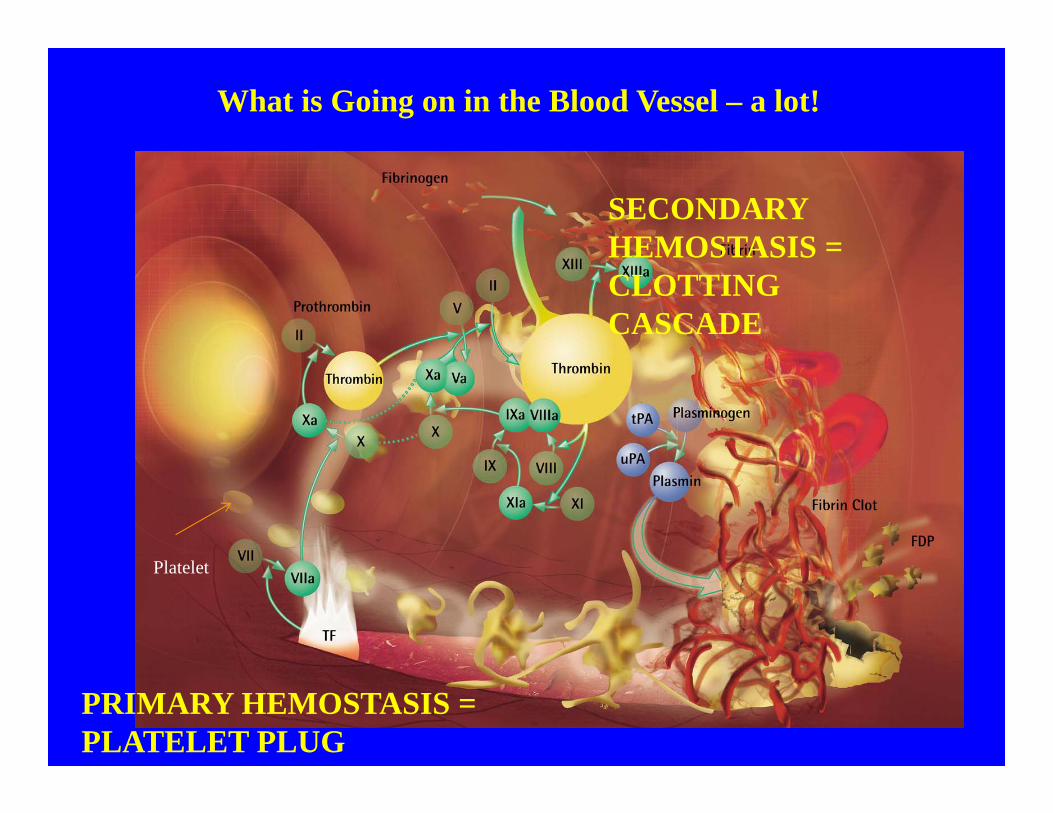
What is Going on in the Blood Vessel – a lot!
Platelet
PRIMARY HEMOSTASIS =PLATELET PLUG
SECONDARY HEMOSTASIS = CLOTTING CASCADE
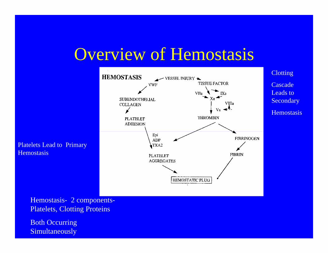
Overview of Hemostasis
Hemostasis- 2 components-Platelets, Clotting Proteins
Both Occurring Simultaneously
Platelets Lead to Primary Hemostasis
Clotting
Cascade Leads to Secondary
Hemostasis

Hemostasis• Intricate system maintaining blood in fluid state
– Reacts to vascular injury to stop blood loss and seal vessel wall
• Involves platelets, clotting factors, endothelium, and inhibitory/control mechanisms– Highly developed system of checks and balances
Bleeding Thrombosis
Normal HemostasisAbsence of overt bleeding/thrombosis
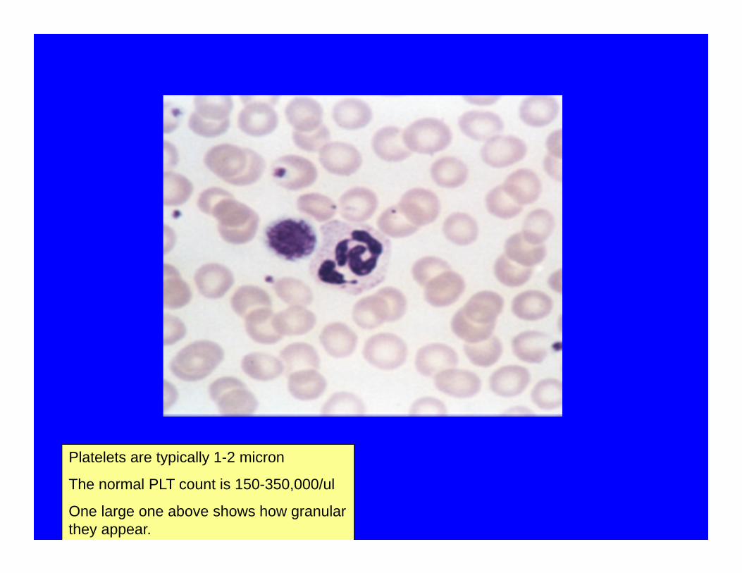
Platelets are typically 1-2 micron
The normal PLT count is 150-350,000/ul
One large one above shows how granular they appear.

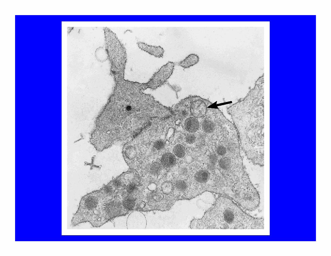
A scanning electron micrograph of normal platelets
Really are fragments of megakaryocyte cytoplasm

Platelet- Number, Lifespan and Kinetics
• Normal platelet concentration is 150,000-350,000/ ul
• Platelets are produced in the bone marrow by megakaryocytes and released into the circulation
• They circulate in the blood for about 10 days after release from the marrow
• About 1/3 of all the body’s platelet mass is stored in the spleen
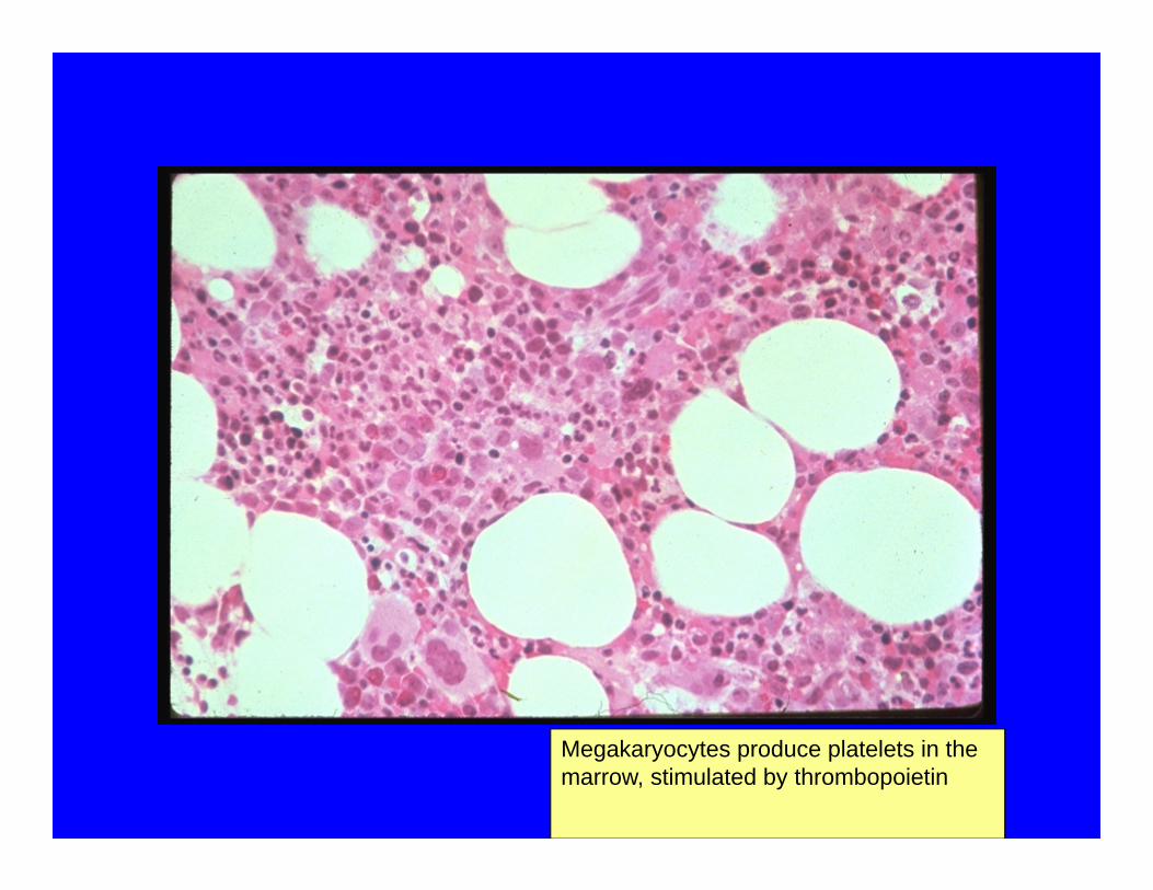
Megakaryocytes produce platelets in the marrow, stimulated by thrombopoietin

Normal Megakaryocyte
Platelets are released from megakaryocytes , this shows this process in vitro culture
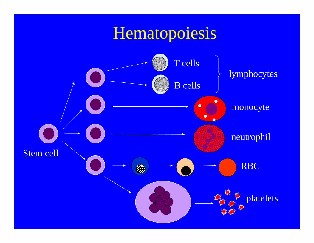
Hematopoiesis
Stem cell
T cells
B cellslymphocytes
neutrophil
RBC
monocyte
platelets
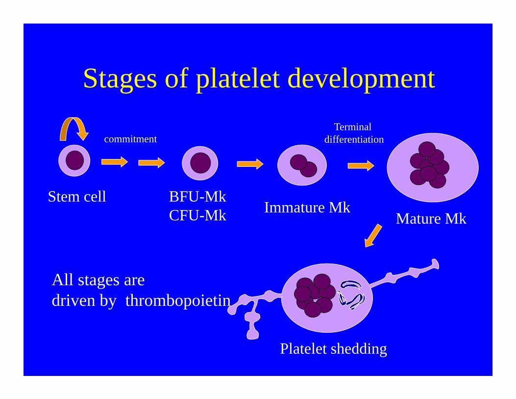
Stages of platelet development
Stem cell BFU-MkCFU-Mk Immature Mk
Mature Mk
Platelet shedding
commitmentTerminal
differentiation
All stages aredriven by thrombopoietin

Thrombopoietin (TPO)
• Growth factor produced in liver• Increases production of megakaryocytes• Essential for stem cells
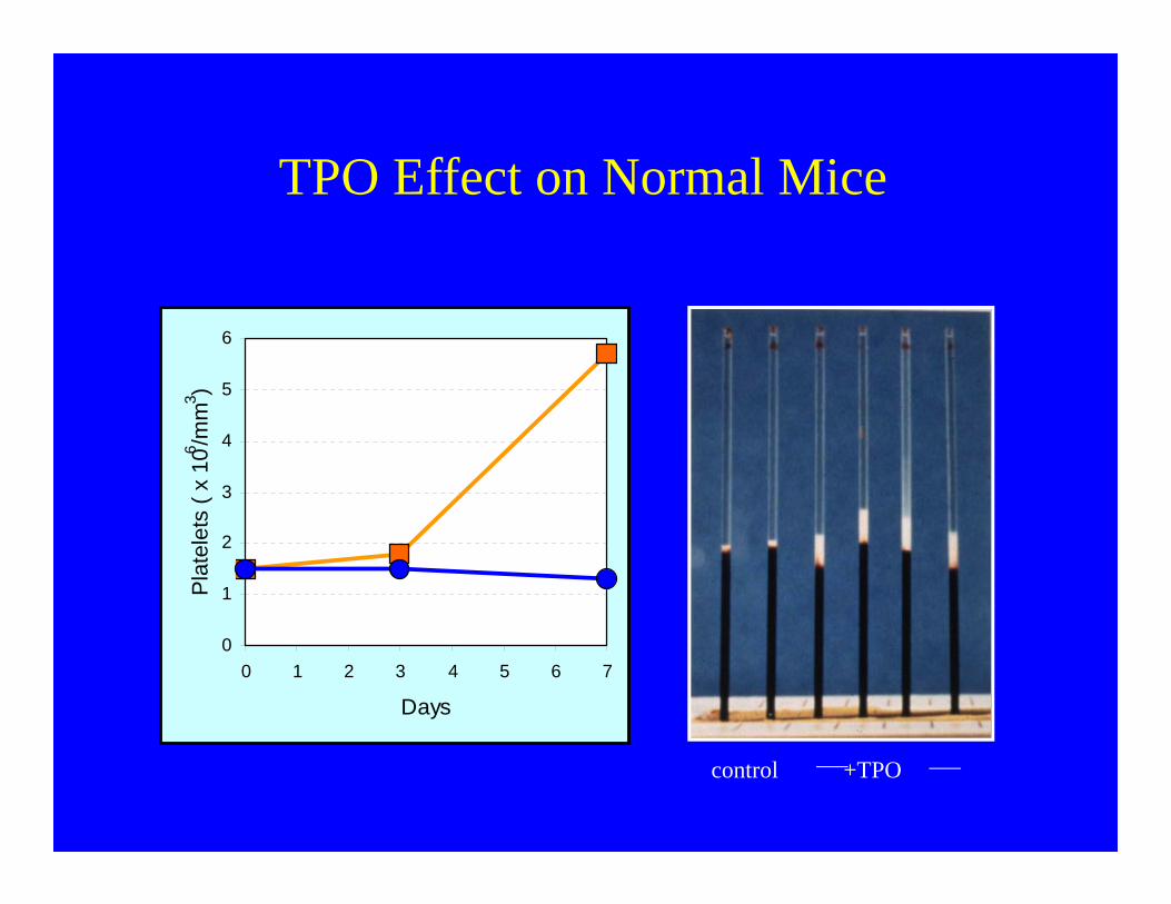
control +TPO
0
1
2
3
4
5
6
0 1 2 3 4 5 6 7
Days
Pla
tele
ts (
x 10
6 /mm
3 )TPO Effect on Normal Mice
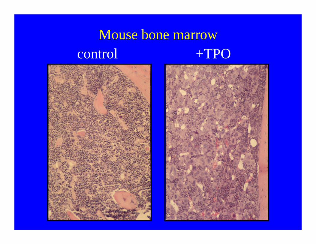
control +TPOMouse bone marrow
TPO regulation
• Constitutive (constant) production• Level depends on binding sites on platelets and
megakaryocytes

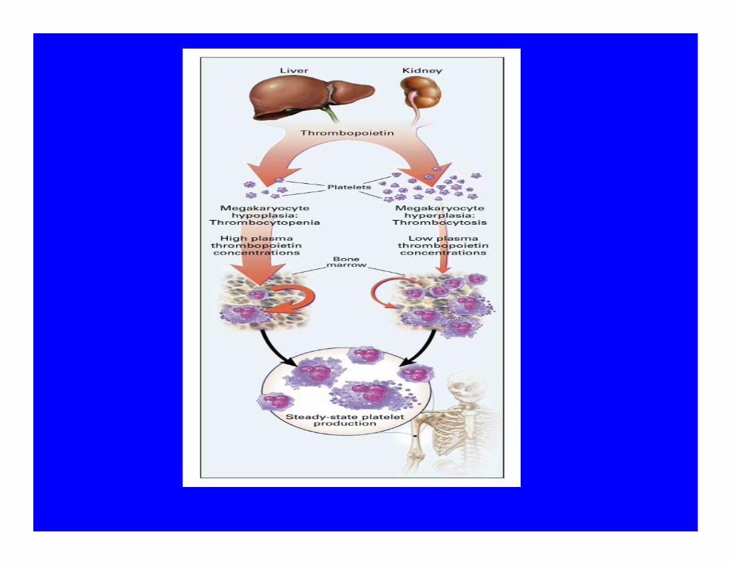
Thrombopoietin Regulation (Sponge theory)
PLT
TPO
MPL
PLT
As Platelet Count Increases, serve as a sponge , having less available to stimulate Megakaryocytes
Primary Hemostasis
AdhesionAggregation
Secretion

Adhesion occurs within 1-3 seconds after injury
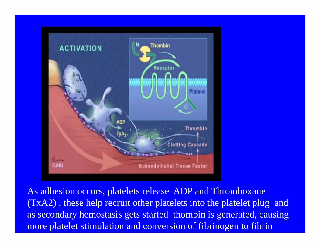
As adhesion occurs, platelets release ADP and Thromboxane (TxA2) , these help recruit other platelets into the platelet plug and as secondary hemostasis gets started thombin is generated, causing more platelet stimulation and conversion of fibrinogen to fibrin

3-7 minutes for entire process to occur-”The Bleeding Time”

Activated platelets
Note pseudopodia and how platelets aggregating to each other

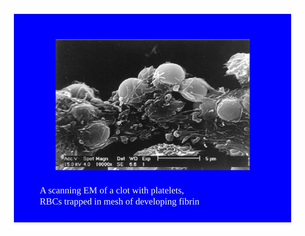
A scanning EM of a clot with platelets, RBCs trapped in mesh of developing fibrin

Fibrinogen
Fibrin –polymerized remains of fibrinogen
Think of fibrin as strands of protein that holds the platelets together
Thrombin transforms fibrinogen to a mesh of fibrin strands
EM of fibrinogen that has been treated with Thombin
Remember Platelets act in Concert with Fibrin Formation to Form a Firm Clot

Summary of Platelet Processess

Bleeding Time
Normal 3-7 minutes
Prolonged in platelet function abnormalities
Testing for Abnormal Platelet Function
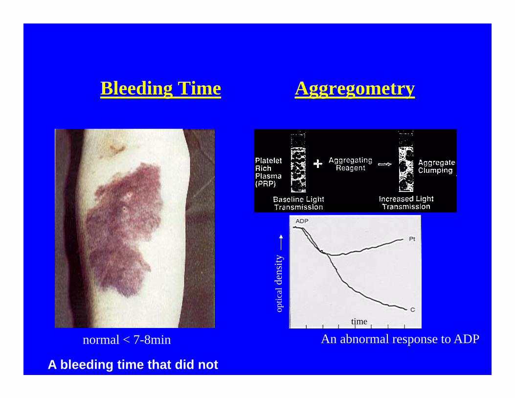
Bleeding Time Aggregometry
normal < 7-8min An abnormal response to ADP
optic
alde
nsity
time
A bleeding time that did not

Bleeding – Cut a Blood Vessel – What Happens ?

The endothelium is “antagonistic” to platelets under normal conditions
The Endothelium Prevents Excess Platelet Function In Vivo

Vascular Endothelium Function
Tissue factor pathway inhibitor
Thrombomodulin
Tissue plasminogen activator
Heparan sulfate proteoglycans
Tissue factor
Anticoagulant- Inhibits coagulation extrinsic pathway
Anticoagulant- Inhibits coagulation by activating protein C system
Anticoagulant- Inhibits coagulation by activating fibrinolysis
Anticoagulant- Inhibits coagulation by activating antithrombin
Procoagulant- Inflammatory cytokines (IL-1, TNF) induce expression

Vascular Endothelium Function
Prostacyclin
Thromboxane A2
ELAMs, ICAMs
von Willebrand factor
Vasodilation, inhibition of platelet aggregation
From platelets, muscular arteries constrict
Cytokines induce synthesis to promote leukocyte adhesion
Promote platelet-collagen adhesion to exposed sub-endothelium

Thombocytopenia
• > 100,000/ul no excessive bleeding, even with major surgery
• 50-100,000 may bleed longer than normal with severe trauma
• 20-50,000 bleed with minor trauma• < 20,000 may have spontaneous
hemorrhage

Petechiae- subcutaneous bleeding develops when the platelet counts falls below 20- 50, 000/ul
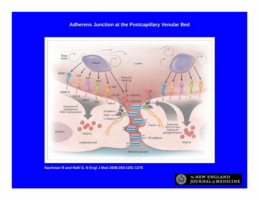
Nachman R and Rafii S. N Engl J Med 2008;359:1261-1270
Adherens Junction at the Postcapillary Venular Bed

Nachman R and Rafii S. N Engl J Med 2008;359:1261-1270
Bleeding in Patients with Thrombocytopenia through Disassembly of the Adherens Junction
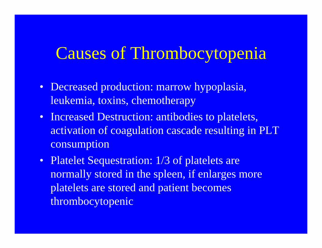
Causes of Thrombocytopenia
• Decreased production: marrow hypoplasia, leukemia, toxins, chemotherapy
• Increased Destruction: antibodies to platelets, activation of coagulation cascade resulting in PLT consumption
• Platelet Sequestration: 1/3 of platelets are normally stored in the spleen, if enlarges more platelets are stored and patient becomes thrombocytopenic

Decreased Production
Decreased production: marrow hypoplasia, leukemia, toxins, chemotherapy
No straightforward method to assess platelet production , unlike RBCs & Retic Count

Severe thrombocytopenia in
Autoimmune thrombocytopenia
Blood smear shows no platelets
Isotope labeled platelets are destroyed in the spleen, in presence of antibody

Pathophysiology of AutoimmuneThrombocytopenia
An example of a common consumptive thrombocytopenia
Autoantibodies are formed against the platelet glycoprotein receptor IIb-IIIa, and are destroyed in the Reticuloendothelial system

Panel A , patient without increased Megakaryocytes, versus patients with increased megakaryocytes

IPF(%)Ref: 3%
H-IPF(%)Ref: 1%
Fluorescence intensity
(Linear )
FSC
(Cel
l siz
e)
(Log
arith
m )
IPF#(x109/L)
PLT-X(ch)
Overview of Immature Platelet Fraction Percentage (IPF%) Measurement & Some Examples
normal ITP
Nadir after Chemo
Recovery from Chemo

Thrombocytosis
Seen in myeloproliferative disorders, chronic infection, iron deficiencies, malignant tumors

Platelets Role in Thrombosis
• Coronary or cerebrovascular thrombosis is multifactorial
• Genes – lipids• Society – diet,
exercise, smoking
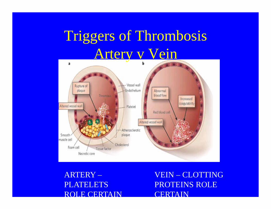
Triggers of ThrombosisArtery v Vein
VEIN – CLOTTING PROTEINS ROLE CERTAIN
ARTERY –PLATELETS ROLE CERTAIN

Summary
• Describe the major physiologic functions of platelets
• Describe the major platelet agonists• Describe the ligands responsible for
adhesion and aggregation• Describe the pathophysiology of
thrombocytopenia

Hemostasis- Summary