human papillomavirus infection and cervical
TRANSCRIPT

RESEARCH ARTICLE Open Access
Human papillomavirus infection andcervical intraepithelial neoplasiaprogression are associated with increasedvaginal microbiome diversity in a ChinesecohortYulian Chen1,2,3†, Xingdi Qiu1,2,3†, Wenjing Wang1,2,3, Dong Li1,2,3, Anyue Wu1,2,3, Zubei Hong1,2,3, Wen Di1,2,3* andLihua Qiu1,2,3*
Abstract
Background: In this study, the association between human papillomavirus (HPV) infection and related cervicalintraepithelial neoplasia (CIN) or cervical cancer and vaginal microbiome was evaluated in Chinese cohorts.
Methods: The vaginal bacterial composition of five groups, HPV-infected women without CINs (HPV, n = 78), womenwith low-grade squamous intraepithelial lesions (LSIL, n = 51), women with high-grade squamous intraepithelial lesions(HSIL, n = 23), women with invasive cervical cancer (Cancer, n = 9) and healthy women without HPV infection (Normal,n = 68), was characterized by deep sequencing of barcoded 16S rRNA gene fragments (V3–4) using Illumina MiSeq.
Results: HPV infection increased vaginal bacterial richness and diversity regardless of the status of CINs. The vaginalbacterial richness and diversity were further augmented in women with cervical cancer. Lactobacillus was the mostabundant genus in all groups. HPV infection had a negative influence on the abundances of Lactobacillus, Gardnerellaand Atopobium. Accordingly, HPV infection increased the relative abundance of Prevotella, Bacillus, Anaerococcus,Sneathia, Megasphaera, Streptococcus and Anaerococcus. The increased proportions of Bacillus, Anaerococcus and thereduced abundance of Gradnerella vaginalis were probably related with the progression of CINs severity. HPV infectionwithout CINs or cancerous lesions was strongly associated with Megasphaera. The most abundant bacterium in theLSIL group was Prevotella amnii. However, Prevotella timonensis, Shuttleworthia and Streptococcaceae at the family levelwere three taxa related to HSIL. Furthermore, more taxa were associated with the Cancer group including Bacillus,Sneathia, Acidovorax, Oceanobacillus profundus, Fusobacterium, Veillonellaceae at the family level, Anaerococcus andPorphyromonas uenonis. Samples in the Normal group were mostly assigned to CST III. HPV infection converted thevaginal bacterial community structure from CST III to CST IV. Furthermore, the proportions of CST IV were graduallyaugmented with the progression of the severity of CINs.
(Continued on next page)
© The Author(s). 2020 Open Access This article is licensed under a Creative Commons Attribution 4.0 International License,which permits use, sharing, adaptation, distribution and reproduction in any medium or format, as long as you giveappropriate credit to the original author(s) and the source, provide a link to the Creative Commons licence, and indicate ifchanges were made. The images or other third party material in this article are included in the article's Creative Commonslicence, unless indicated otherwise in a credit line to the material. If material is not included in the article's Creative Commonslicence and your intended use is not permitted by statutory regulation or exceeds the permitted use, you will need to obtainpermission directly from the copyright holder. To view a copy of this licence, visit http://creativecommons.org/licenses/by/4.0/.The Creative Commons Public Domain Dedication waiver (http://creativecommons.org/publicdomain/zero/1.0/) applies to thedata made available in this article, unless otherwise stated in a credit line to the data.
* Correspondence: [email protected]; [email protected]†Yulian Chen and Xingdi Qiu contributed equally to this work.1Department of Gynecology and Obstetrics, Ren ji Hospital, School ofMedicine, Shanghai Jiao Tong University, Shanghai, ChinaFull list of author information is available at the end of the article
Chen et al. BMC Infectious Diseases (2020) 20:629 https://doi.org/10.1186/s12879-020-05324-9

(Continued from previous page)
Conclusions: This work interpreted the differential vaginal bacteria under HPV infection and various precancerous orcancerous lesions in a Chinese cohort. We distinguished the specific microbes and the vaginal bacterial structure thatwere related with the progression of CINs severity in Chinese women.
Keywords: Vaginal microbiome, Human papillomavirus, Cervical intraepithelial neoplasia, Cervical cancer
BackgroundHuman papillomavirus (HPV) is regarded as one of themost common sexually transmitted agents in cervicalintraepithelial neoplasia (CIN) and cervical cancer [1].High-risk subtypes of HPV contribute to 99% of cervicalneoplasia [1]. However, it is known that high-risk HPV in-fection is necessary but not sufficient for the developmentof CINs or cervical cancer [2–4]. Many other events, suchas multiple sexual partners, early initiation of sexual activ-ity and co-infection with other sexually transmitted infec-tions, have been associated with higher risk of HPVinfection in the genital tract [5–7].In the female genital tract, a healthy vaginal status is
commonly associated with low microbial diversity andprevalence of Lactobacillus. Lactobacillus preventscolonization of other bacterial pathogens through pro-duction of lactic acid, hydrogen peroxide (H2O2) andbacteriocin in the vagina, and therefore keeps integrityof mucosal barriers against virus and opportunistic bac-teria [4, 8, 9]. The vaginal microbial profile of womencould by classified into five community state types(CSTs) by hierarchical taxonomic clustering, in whichCSTs I, II III and V are predominated by L. crispatus, L.iners, L. jensenii and L. gasseri respectively, while CSTIV is depleted of Lactobacillus and enriched with anaer-obic bacteria like Gardnerella, Megasphera, Sneathia,Prevotella, etc. [10]. Commensal vaginal Lactobacillusspecies are thought to defend against many pathogens,such as Candida infection [11, 12], sexually transmitteddiseases [13], urinary system infections [14, 15] and hu-man immunodeficiency virus (HIV) infection [16]. How-ever, L. iners has many properties different from otherLactobacillus spp., for example unable to produce H2O2,
and it often predominates in the presence of HPV infec-tion [4, 17, 18] and CIN [18, 19].Bacterial vaginosis (BV) is a cluster of microbial disor-
ders characterized by a decrease in Lactobacillus and theirreplacement by high concentrations of other anaerobicbacteria, with a microbial community structure in accord-ance with CST IV [9]. BV is associated with a higher riskof miscarriage, preterm premature rupture of membranesand a higher susceptibility to sexually transmitted infec-tions, as HPV infection [20–22]. Some studies to date havereported that vaginal microbiome (VM) plays an import-ant role in the persistence of the HPV infection and thesubsequent development of cervical precancerous or
cancerous lesions [2, 4, 8, 17–19, 23–28]. Increasing VMdiversity is associated with advancing CINs severity andviral persistence [26]. The potential mechanisms could belinked to less production of protective lactic acid, H2O2
and bactriocin by Lactobacillus, disruption of mucosal in-tegrity which may aid viral entry, higher levels of oxidativestress induced by dybiosis [8]. Particular species likeSneathia spp. have a probable pathological role in HPVacquisition and persistence through cellular targets suchas expression of immunosuppressive cytokines [29].Therefore, it is considerable to take vaginal microbiome asa promising marker not only for HPV infection but alsofor cervical precancerous lesions.Nevertheless, the vaginal communities could be influ-
enced by many other factors, including ethnicity, personalhygiene, sexual behaviors and hormonal levels [10, 30].Ethnicity is key to shape vaginal bacterial communities [4,10, 31]. Caucasian and Asian women display a significantlygreater prevalence of Lactobacillus in the vagina com-pared to Hispanic and Black women [8, 10, 31]. To ourknowledge, data with regard to the vaginal bacterial com-position of Chinese populations are inadequate [8, 18, 32].The analysis, which is performed in a large cohort ofwomen living in a different country and with supposeddifferent hygiene habits [10], is helpful to reinforce theunderlying associations. Furthermore, there are few stud-ies about the association between VM and HPV infectionand related CIN diseases in Chinese cohorts using highthroughput sequencing method [8, 18, 32].Hence, the objective of this research is to study the
role of VM on the HPV infection and the progression ofCIN diseases in Chinese populations. We try to identifythe microbiological markers related with HPV infectionand CINs severity in these cohorts.
MethodsStudy population and sample collectionWe included 229 non-pregnant women, 25–69 years ofage, who attended gynecological clinics at the Depart-ment of Gynecology, Renji Hospital of Shanghai, JiaoTong University School of Medicine, between May 2016and November 2016. Non-pregnant women were in-cluded irrespective of their phase in their cycle (exceptfor the menstrual period), parity, personal hygiene,smoking habits. No previous medical histories of CINdiseases or cervical cancer and other serious medical
Chen et al. BMC Infectious Diseases (2020) 20:629 Page 2 of 12

problems, such as hepatitis B/C, diabetes, autoimmunediseases, sexually transmitted diseases (chlamydia, gon-orrhea, trichomoniasis, genital herpes), HIV or othermalignant tumors, were declared. Participants who hadvaginal intercourse or vaginal douching within last 3days of sampling, abnormal metrorrhagia in the previousweeks, or used probiotics, antibiotics or immunosup-pressive drugs in the preceding 14 days were excluded.HPV genotyping test and ThinPrep cytology test
(TCT) were carried out in all enrolled patients, using acommercial HPV genotyping kit as previously described[33]. TCT results were interpreted on the basis of Be-thesda System criteria [34]. Women with HPV positiveand/or TCT ≥ASCUS accepted the biopsy under thecolposcopy examination by two gynecologists. Based ontheir histopathology, all participants were assigned intofive groups as follow (Fig. S1): HPV-infected womenwithout CINs (HPV, n = 78), women with low-gradesquamous intraepithelial lesions (LSIL, n = 51), womenwith high-grade squamous intraepithelial lesions (HSIL,n = 23), women with invasive cervical cancer (Cancer,n = 9) and healthy women without HPV infection (Nor-mal, n = 68).Sterile swab samples for 16S rRNA sequencing were
taken from the lateral and posterior fornix using a sterilespeculum as previously described [33].
Total bacterial genomic DNA extraction and MiSeqsequencingPrimers 338F (ACTCCTACGGGAGGCAGCA) and 806R(GGACTACHVGGGTWTCTAAT) were used to amplifythe V3–4 hypervariable fragments of the 16S rRNA geneby PCR as previously described [33]. All sequencing wasconducted using the Illumina MiSeq platform at MajorbioBiopharm Technology Company (Shanghai).
Sequence analysisSequence reads were quality checked by Trimmomatic[35]. OTUs were generated by QIIME and taxonomieswere classified using the Ribosomal Database Project(RDP) classifier script (version 2.2) as previously de-scribed [33].As previously described [33], alpha (Chao and
Shannon index) and beta indices (unweighted UniFracdistances in Principal coordinates analysis (PCoA)) werecalculated by mothur (version v.1.30.1) [36] and veganpackage in R [37]. We compared the differences of alphaand beta estimators between two groups by Student’s t-test and ANOSIM test respectively. Heat maps of rela-tive abundance for different taxa were generated usingR. The relative abundances of different taxa at differentlevels between the five groups were calculated by non-parametric Wilcoxon test. Linear discriminant analysis(LDA) effect size (LEfSe) algorithm [38] was used to
characterize the potential microbial markers with spe-cific disease phenotypes. Hierarchical clustering analysiswas used to classify different vaginal community statetypes (CSTs) as previously published [31, 39]. Q-values(p-value adjusted by false discovery rate (FDR)) and p-values < 0.05 were considered significant.
ResultsCharacteristics of the study populationThe average ages of each group (HPV, LSIL, HSIL,Cancer, Normal) were 47.78 ± 9.63, 46.00 ± 10.19,43.70 ± 10.74, 56.11 ± 9.02 and 43.00 ± 8.69 years, re-spectively. The proportions of 16/18 HPV subtypes inHPV positive groups were as follow: HPV (13/78), LSIL(10/51), HSIL (6/23), Cancer (4/9) (Table S1). Thedemographic characteristics of each group are presentedin the supplementary data (Table S2).
Sequencing resultsAfter filtering low-quality reads, 6,585,141 assembledclean reads were obtained from 229 samples, with anaverage number of reads per sample of 28,755.64 ±5389.65 and a mean read length of 444.90 ± 5.89 bp. Fornormalization, the reads in each sample were randomlysubsampled to the lowest number of 20,098 in sample318_LCQ (HPV group). After removing singletons (theOTUs contained less than 2 reads), 1878 OTUs wereidentified, ranging from 10 OTUs in sample 4391 (Nor-mal group) to 782 OTUs in sample 152_GPZ (HPVgroup) (Table S3). The average number of OTUs inHPV-negative group (Normal group: 46) was lower thanthat in HPV-positive groups (HSIL group: 116; LSILgroup: 145; HPV group: 157). We found more OTUs inthe samples of Cancer group (Mean = 256.70 ± 174.78),ranging from 51 to 599 (Table S3).
Vaginal microbiota richness and diversityAt the OTU level, microbial richness and diversity wereestimated using Chao and Shannon indices, respectively,as shown in Fig. 1. These two indices revealed that HPVinfection increased vaginal bacterial richness and diversity.The means of Chao and Shannon indices were muchlower in groups HPV-negative (Normal group: 84.02 ±73.88 and 0.94 ± 0.95, respectively) than in groups HPV-positive (HPV group: 272.26 ± 191.62 and 1.49 ± 1.01, q <0.01; LSIL group: 231.84 ± 195.39, q < 0.001 and 1.30 ±0.93, q = 0.052; HSIL group: 185.74 ± 162.62, q < 0.001 and1.51 ± 0.98, q = 0.02; Cancer group: 367.76 ± 208.63 and2.47 ± 0.98, q < 0.001, respectively). The means of Chaoand Shannon indices were the highest in group Cancerand were significantly higher than in groups HSIL (Chao:367.76 ± 208.63 vs. 185.74 ± 162.62, q = 0.02, Shannon:2.47 ± 0.98 vs. 1.51 ± 0.98, q = 0.02), LSIL (Shannon:2.47 ± 0.98 vs. 1.30 ± 0.93, q = 0.003) and HPV (Shannon:
Chen et al. BMC Infectious Diseases (2020) 20:629 Page 3 of 12

2.47 ± 0.98 vs. 1.49 ± 1.01, q = 0.02). However, there wereno differences found among groups HPV, LSIL and HSIL.
Vaginal bacterial structure and beta-diversity in differentgroupsIn PCoA, the first two principal components explained21.18 and 8.83%, respectively, of the variance along thefirst and second axes, with the Cancer, HSIL, LSIL andHPV samples visually separated from the Normal sample(Fig. 2). Comparison between two groups based on theANOSIM test revealed that the bacterial structure ofgroups Cancer, LSIL and HPV were significantly differentfrom that of group Normal (Table S4). Meanwhile, the
bacterial structure of groups HSIL and LSIL were also dif-ferent from that of group HPV (Table S4).
Taxonomy of the vaginal microbiota in different groupsOverall, 37 bacterial phyla were recovered across allsamples (Fig. S2), and All of the samples were domi-nated by Firmicutes, Actinobacteria, Bacteroidetes, Fuso-bacteria and Proteobacteria (Fig. S2). Firmicutes was themost abundant phylum, accounting for 73.15, 68.91,69.70, 70.43 and 47.47% of the Normal, HPV, LSIL,HSIL and Cancer groups, respectively (Fig. 3a). HPV in-fection tended to lower the proportion of Firmicutes(Cancer < Normal, q < 0.05; HPV <Normal, q < 0.05).
Fig. 1 Vaginal bacterial richness and diversity in five groups. a: Chao index; b: Shannon index; Student’s t-test was used to compare differencesbetween two groups; data are presented as the mean ± SD; ***: q≤ 0.001; **: 0.001 < q < 0.01; *: q < 0.05
Chen et al. BMC Infectious Diseases (2020) 20:629 Page 4 of 12

However, there were no differences among other HPVpositive groups. Accordingly, the proportions of Bacteroi-detes, Fusobacteria and Proteobacteria were significantlyincreased in the HPV positive groups as compared to theNormal group (Bacacteroidetes: Cancer 15.72% >HSIL13.1% >HPV 8.28% >Normal 6.94%, Fig. 3b; Fusobacteria:Cancer 15.84% > LSIL 3.04% >HSIL 2.62% >HPV 2.64% >Normal 2.54%, Fig. 3d; Proteobacteria: Cancer 10.39% >HPV 5.36% > LSIL 3.59% >Normal 1.18%, Fig. 3c). Therewere no differences among the different groups with re-gard to the proportion of Actinobacteria.At the genus level, a total of 749 taxa were found across
all samples (Fig. S3), with Lactobacillus being the mostdominant genus overall. The proportion of Lactobacilluswas reduced in the HPV positive groups (Cancer: 19.72%,q < 0.05; HSIL: 42.99%, q = 0.098; LSIL: 53.71%, q = 0.051;HPV: 49.20%, q < 0.01, respectively) compared to the Nor-mal group (64.93%) (Fig. 4a). However, there were no sig-nificant differences among the groups HPV, LSIL, HSILand Cancer with regard to the relative abundance ofLactobacillus. There were no differences among the differ-ent groups with regard to the abundance of Gardnerella(Fig. 4b). In addition, HPV infection had also a negativeeffect on the abundance of Atopobium, which sharply de-creased from 3.12% in the Normal group to 2.07% in theHPV group (q < 0.01), 2.72% in the LSIL group (q < 0.05)(Fig. 4e). Accordingly, HPV infection increased the relativeabundance of Prevotella (Normal: 5.91% <HPV: 7.18%,q < 0.05, Fig. 4c), Bacillus (Normal: 0.34% < LSIL: 3.30% <HPV: 5.77% <HSIL: 5.87% < Cancer: 9.18%, q < 0.001,Fig. 4d), Sneathia (Normal: 2.39% <HPV: 2.61% < LSIL:3.02% < Cancer: 10.03%, q < 0.05, Fig. 4f), Megasphaera(Normal: 1.01% < LSIL: 2.13% < Cancer: 2.30% <HSIL:
2.57% <HPV: 9.18%, q < 0.05, Fig. 4g), Streptococcus (Nor-mal: 1.03% < Cancer: 1.29% < LSIL: 1.35% <HPV: 1.93%,q < 0.05, Fig. 4h) and Anaerococcus (Normal: 1.22% < Can-cer: 3.83%, q < 0.05, Fig. 4i). The precancerous lesions in-creased the proportion of Bacillus, with a greater bacterialabundance in LSIL and HSIL groups than in HPV group(q < 0.05) (Fig. 4d). A higher proportion of Anaerococcuswas also found in the group Cancer compared to the pre-cancerous groups (HPV, LSIL and HSIL, q < 0.05) (Fig. 4i).Two taxa, Bacillus and Anaerococcus, were probably re-lated with the progression of CINs severity.
Identification of vaginal microbiological markers indifferent groupsLEfSe modeling was employed to identify microbio-logical markers related to HPV infection and CINs se-verity (Fig. 5). The threshold for the logarithmic LDAmodel score for discriminative features in this study was4.0 (p < 0.05). The most abundant genus in the Normalgroup was Lactobacillus. In addition, other two taxawere also more abundant in the Normal group, Bacilli atthe class level and Atopobium vaginae. HPV infectionwithout CIN or cancerous lesions (HPV group) wasstrongly associated with Megasphaera. The most abun-dant bacterium in the LSIL group was Prevotella amnii.However, Prevotella timonensis, Shuttleworthia andStreptococcaceae at the family level were three taxa re-lated to HSIL. Furthermore, more taxa were associatedwith the Cancer group including Bacillus, Sneathia,Acidovorax, Oceanobacillus profundus, Fusobacterium,Veillonellaceae at the family level, Anaerococcus andPorphyromonas uenonis.
Fig. 2 Unweighted UniFrac principal coordinates analysis (PCoA) plot comparing sample distribution for the different groups
Chen et al. BMC Infectious Diseases (2020) 20:629 Page 5 of 12

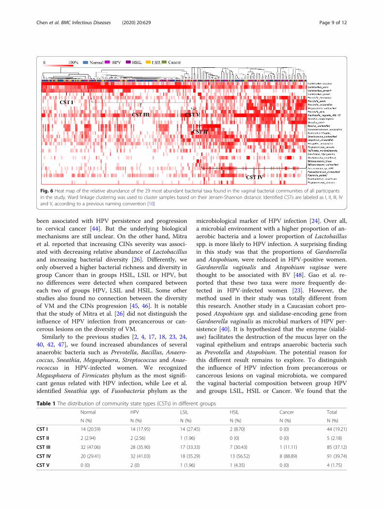
Characteristics of vaginal community state types (CSTs)for different groupsThe vaginal bacterial CST analysis visualized by hierarchicalclustering revealed that all samples clustered into five majorgroups: CST I, CST II, CSTII, CST IV and CST V (Fig. 6).The most commonly observed community was CST IV (91/229, 39.74%), followed by CST III (85/229, 37.12%), CST I(44/229, 19.21%), CST II (5/229, 2.18%) and CST V (4/229,1.75%) as shown in Table 1. The proportions of CSTs in dif-ferent groups were shown in Table 1. Samples in the Normalgroup were mostly assigned to CST III (32/68, 47.96%). HPVinfection converted the vaginal bacterial community struc-ture from CST III to CST IV, as all of the HPV positivegroups were dominated by CST IV (HPV: 32/78, 41.03%;LSIL: 18/51, 35.29%; HSIL: 13/23, 56.52%; Cancer: 8/9,88.89%, respectively) and presented less CST III (HPV: 28/78, 35.90%; LSIL: 17/51, 33.33%; HSIL: 7/23, 30.43%; Cancer:1/9, 11.11%, respectively). Furthermore, the proportions ofCST IV were gradually augmented with the progression of
the severity of CINs (Cancer: 88.89% >HSIL: 56.52%> LSIL:35.29%).We identified the most abundant species in each sam-
ple in Fig. 6, and the distributions of the 29 most pre-dominant species in five groups were documented inTable 2. Seventy-eight samples (34.1%) were predomi-nated by Lactobacillus iners, followed by Lactobacilluscrispatus (48 samples, 21%), Gardnerella vaginalis (26samples, 11.4%), Bacillus unclassified (16 samples, 7%),Sneathia amnii (8 samples, 3.5%) and Prevotella amnii(7 samples, 3.1%). HPV infection was related with thedecreased abundance of Lactobacillus iners, Lactobacil-lus crispatus, Gardnerella vaginalis, Lactobacillus gas-seri, Anaerococcus spp., Atopobium unclassified andPorphyromonas somerae as compared to the Normalgroup (Table 2). In addition, HPV infection was associ-ated with the increased abundance of Bacillus unclassi-fied, Escherichia Shigella, Megasphaera unclassified,Streptococcus unclassified, Lactobacillus jensenii,
Fig. 3 Relative abundance counts of Firmicutes (a), Bacteroidetes (b), Proteobacteria (c) and Fusobacteria (d), which were found to be the mostabundant phyla across all samples. The Wilcoxon test was used to compare differences in the abundance of each phylum between two groups.***: q≤ 0.001; **: 0.001 < q < 0.01; *: q < 0.05
Chen et al. BMC Infectious Diseases (2020) 20:629 Page 6 of 12

Sneathia sanguinegens, Bifidobacterium unclassified,Candidatus Mycoplasma, Comamonadaceae unclassi-fied, Veillonella montpellierensis, Faecalibacteriumunclassified, Finegoldia unclassified, Fusobacterium mor-tiferum, Porphyromonas uenonis and Ralstonia pickettii(Table 2). Furthermore, the abundance of Gradnerellavaginalis was gradually reduced with the progression ofCINs severity (Cancer 0% < HSIL 4.3% < LSIL 11.8% <HPV 12.8% < Normal 13.2% as shown in Table 2).
DiscussionOur study addressed a not well-elucidated topic about theassociation between HPV infection and related CINs orcervical cancer and vaginal microbiome in Chinese co-horts. We observed that HPV infection increased vaginalbacterial richness and diversity regardless of the status ofCINs. The vaginal bacterial richness and diversity werefurther augmented in the women with cervical cancer.Lactobacillus was the most abundant genus in all groups.
HPV infection had a negative influence on the abundancesof Lactobacillus, Gardnerella and Atopobium. Accord-ingly, HPV infection increased the relative abundance ofPrevotella, Bacillus, Anaerococcus, Sneathia, Megasphaera,Streptococcus and Anaerococcus. The increased propor-tions of Bacillus, Anaerococcus and the reduced abun-dance of Gradnerella vaginalis were probably related withthe progression of CINs severity. HPV infection withoutCINs or cancerous lesions was strongly associated withMegasphaera. The most abundant bacterium in the LSILgroup was Prevotella amnii. However, Prevotella timonen-sis, Shuttleworthia and Streptococcaceae at the family levelwere three taxa related to HSIL. Furthermore, more taxawere associated with the Cancer group including Bacillus,Sneathia, Acidovorax, Oceanobacillus profundus, Fusobac-terium, Veillonellaceae at the family level, Anaerococcusand Porphyromonas uenonis. Samples in the Normalgroup were mostly assigned to CST III. HPV infectionconverted the vaginal bacterial community structure from
Fig. 4 Relative abundance counts of Lactobacillus (a), Gardnerella (b), Prevotella (c), Bacillius (d), Atopobium (e), Sneathia (f), Megasphaera (g),Streptococcus (h) and Anaerococcus (i), which were found to be the most abundant genera across all samples. The Wilcoxon test was used tocompare differences in the abundance of each phylum between two groups. ***: q≤ 0.001; **: 0.001 < q < 0.01; *: q < 0.05
Chen et al. BMC Infectious Diseases (2020) 20:629 Page 7 of 12

CST III to CST IV. Furthermore, the proportions of CSTIV were gradually augmented with the progression of theseverity of CINs.Most of the studies have proven that HPV infection
can increase vaginal bacterial richness and diversity andlower the percentage of Lactobacillus [4, 8, 17, 18, 23,24, 40, 41], and our results are in agreement with theseprevious studies. However, a few studies found no
difference between HPV positive and negative groups[25, 42]. HPV infection is thought to alter the acidic en-vironment of the vagina, which might promote out-breaks of bacteria [24]. In addition, HPV infection mightlead to changes in the vaginal microbiota by inducinghost mucosal immune response and genital infalmma-tion [41, 43]. High genital inflammation with elevatedvaginal PH and non-Lactobacillus-dominant VM have
Fig. 5 The unique taxa and microbiomarkers for different groups. Shown is a histogram of LDA scores computed for features differentially abundantin the five groups
Chen et al. BMC Infectious Diseases (2020) 20:629 Page 8 of 12
been associated with HPV persistence and progressionto cervical cancer [44]. But the underlying biologicalmechanisms are still unclear. On the other hand, Mitraet al. reported that increasing CINs severity was associ-ated with decreasing relative abundance of Lactobacillusand increasing bacterial diversity [26]. Differently, weonly observed a higher bacterial richness and diversity ingroup Cancer than in groups HSIL, LSIL or HPV, butno differences were detected when compared betweeneach two of groups HPV, LSIL and HSIL. Some otherstudies also found no connection between the diversityof VM and the CINs progression [45, 46]. It is notablethat the study of Mitra et al. [26] did not distinguish theinfluence of HPV infection from precancerous or can-cerous lesions on the diversity of VM.Similarly to the previous studies [2, 4, 17, 18, 23, 24,
40, 42, 47], we found increased abundances of severalanaerobic bacteria such as Prevotella, Bacillus, Anaero-coccus, Sneathia, Megasphaera, Streptococcus and Anae-rococcus in HPV-infected women. We recognizedMegasphaera of Firmicutes phylum as the most signifi-cant genus related with HPV infection, while Lee et al.identified Sneathia spp. of Fusobacteria phylum as the
microbiological marker of HPV infection [24]. Over all,a microbial environment with a higher proportion of an-aerobic bacteria and a lower proportion of Lactobaillusspp. is more likely to HPV infection. A surprising findingin this study was that the proportions of Gardnerellaand Atopobium, were reduced in HPV-positive women.Gardnerella vaginalis and Atopobium vaginae werethought to be associated with BV [48]. Gao et al. re-ported that these two taxa were more frequently de-tected in HPV-infected women [23]. However, themethod used in their study was totally different fromthis research. Another study in a Caucasian cohort pro-posed Atopobium spp. and sialidase-encoding gene fromGardnerella vaginalis as microbial markers of HPV per-sistence [40]. It is hypothesized that the enzyme (sialid-ase) facilitates the destruction of the mucus layer on thevaginal epithelium and entraps anaerobic bacteria suchas Prevotella and Atopobium. The potential reason forthis different result remains to explore. To distinguishthe influence of HPV infection from precancerous orcancerous lesions on vaginal microbiota, we comparedthe vaginal bacterial composition between group HPVand groups LSIL, HSIL or Cancer. We found that the
Fig. 6 Heat map of the relative abundance of the 29 most abundant bacterial taxa found in the vaginal bacterial communities of all participantsin the study. Ward linkage clustering was used to cluster samples based on their Jensen-Shannon distance. Identified CSTs are labeled as I, II, III, IVand V, according to a previous naming convention [10]
Table 1 The distribution of community state types (CSTs) in different groups
Normal HPV LSIL HSIL Cancer Total
N (%) N (%) N (%) N (%) N (%) N (%)
CST I 14 (20.59) 14 (17.95) 14 (27.45) 2 (8.70) 0 (0) 44 (19.21)
CST II 2 (2.94) 2 (2.56) 1 (1.96) 0 (0) 0 (0) 5 (2.18)
CST III 32 (47.06) 28 (35.90) 17 (33.33) 7 (30.43) 1 (11.11) 85 (37.12)
CST IV 20 (29.41) 32 (41.03) 18 (35.29) 13 (56.52) 8 (88.89) 91 (39.74)
CST V 0 (0) 2 (0) 1 (1.96) 1 (4.35) 0 (0) 4 (1.75)
Chen et al. BMC Infectious Diseases (2020) 20:629 Page 9 of 12

proportions of two specific taxa, Bacillus and Anaerococ-cus, were positively related with the progression of CINsseverity. Furthermore, we identified respective taxa fordifferent stages of CIN lesions. Mitra et al. also reportedthat higher levels of Sneathia sanguinegens, Anaerococ-cus tetradius and Peptostreptococcus anaerobius werecharacterized in HSIL compared to LSIL [26]. Corre-sponding to the impact of HPV infection on the vaginalmicrobes, we also found that the abundance of Gradner-ella vaginalis was gradually reduced with the progressionof CINs severity. However, some other studies [19, 27]thought that an enrichment of Gradnerella vaginalis andAtopobium vaginae had a higher CIN risk.In accordance with the results from an Asian popu-
lation in the study by Ravel et al. [10], the mostabundant CST in Normal group was CST III. The
most dominant CST in the HPV positive groups(HPV, LSIL, HSIL and Cancer) was CST IV. Weobserved HPV infection to be associated with an in-creased proportion of CST IV, and furthermore itsproportion was gradually augmented with the progres-sion of the severity of CINs. It has also been reportedby two longitudinal studies that the majority of HPV-positive samples were composed of CST IV (domi-nated by anaerobic bacteria), and CST IV was relatedwith an increased risk of transitioning to an HPV-positive state [24, 49]. Mitra et al. also found that therate of CST IV was increased 2 fold in women withLSIL, 3 fold in women with HSIL and 4 fold inwomen with invasive cancer [26]. CST IV is associ-ated with higher levels of amine production, resultingin carcinogens nitrosamine production [50].
Table 2 The distribution of the 29 most predominant species in different groups
Normal HPV LSIL HSIL Cancer Total
N (%) N (%) N (%) N (%) N (%) N (%)
Lactobacillus crispatus 16 (23.5) 15 (19.2) 14 (27.5) 3 (13.0) 0 (0) 48 (21.0)
Lactobacillus iners 30 (44.1) 21 (27.0) 20 (39.2) 6 (26.1) 1 (11.1) 78 (34.1)
Lactobacillus jensenii 0 (0) 1 (1.3) 0 (0) 1 (4.3) 0 (0) 2 (0.9)
Lactobacillus gasseri 1 (1.5) 1 (1.3) 0 (0) 0 (0) 0 (0) 2 (0.9)
Anaerococcus spp. 1 (1.5) 0 (0) 0 (0) 0 (0) 0 (0) 1 (0.4)
Atopobium unclassified 1 (1.5) 0 (0) 0 (0) 0 (0) 0 (0) 1 (0.4)
Bacillus unclassified 0 (0) 7 (9.0) 5 (9.8) 3 (13.0) 1 (11.1) 16 (7.0)
Bifidobacterium breve 1 (1.5) 0 (0) 1 (2.0) 0 (0) 0 (0) 2 (0.9)
Bifidobacterium unclassified 0 (0) 1 (1.3) 0 (0) 0 (0) 0 (0) 1 (0.4)
Candidatus Mycoplasma 0 (0) 1 (1.3) 0 (0) 0 (0) 0 (0) 1 (0.4)
Comamonadaceae unclassified 0 (0) 0 (0) 0 (0) 0 (0) 1 (11.1) 1 (0.4)
Veillonella montpellierensis 0 (0) 1 (1.3) 0 (0) 0 (0) 0 (0) 1 (0.4)
Escherichia Shigella 0 (0) 3 (3.8) 1 (2.0) 0 (0) 0 (0) 4 (1.7)
Faecalibacterium unclassified 0 (0) 0 (0) 0 (0) 1 (4.3) 0 (0) 1 (0.4)
Finegoldia unclassified 0 (0) 1 (1.3) 0 (0) 0 (0) 0 (0) 1 (0.4)
Fusobacterium mortiferum 0 (0) 0 (0) 0 (0) 0 (0) 1 (11.1) 1 (0.4)
Gardnerella vaginalis 9 (13.2) 10 (12.8) 6 (11.8) 1 (4.3) 0 (0) 26 (11.4)
Megasphaera unclassified 0 (0) 1 (1.3) 0 (0) 1 (4.3) 0 (0) 2 (0.9)
Porphyromonas somerae 1 (1.5) 0 (0) 0 (0) 0 (0) 0 (0) 1 (0.4)
Porphyromonas uenonis 0 (0) 0 (0) 0 (0) 0 (0) 1 (11.1) 1 (0.4)
Prevotella amnii 2 (3.0) 3 (3.8) 0 (0) 1 (4.3) 1 (11.1) 7 (3.1)
Prevotella bivia 1 (1.5) 0 (0) 0 (0) 1 (4.3) 1 (11.1) 3 (1.3)
Prevotella timonensis 1 (1.5) 2 (2.6) 0 (0) 1 (4.3) 0 (0) 4 (1.7)
Prevotella unclassified 1 (1.5) 1 (1.3) 0 (0) 1 (4.3) 0 (0) 3 (1.3)
Ralstonia pickettii 0 (0) 1 (1.3) 0 (0) 0 (0) 0 (0) 1 (0.4)
Shuttleworthia uncultured 1 (1.5) 2 (2.6) 1 (2.0) 2 (8.7) 0 (0) 6 (2.6)
Sneathia amnii 2 (2.9) 3 (3.8) 2 (3.9) 0 (0) 1 (11.1) 8 (3.5)
Sneathia sanguinegens 0 (0) 0 (0) 0 (0) 0 (0) 1 (11.1) 1 (0.4)
Streptococcus unclassified 0 (0) 3 (3.8) 1 (2.0) 1 (4.3) 0 (0) 5 (2.1)
Chen et al. BMC Infectious Diseases (2020) 20:629 Page 10 of 12

The strength of this study is that it interpreted the va-ginal microbial compositions of a large cohort of Chin-ese women with different stages of HPV-related diseasesusing high throughput sequencing method, which hasnot yet been well elucidated. We found that HPV infec-tion increased vaginal bacterial richness and diversity re-gardless of the status of CINs. The specific microbes andthe vaginal bacterial structure were related with the pro-gression of CINs severity in Chinese women. The limita-tions of this study were that it was a cross-sectionalstudy. Hence, we could not conclude any causal relation-ship between the VM and HPV infection or CIN dis-eases. We have to conduct longitudinal studies to studyrelationships between the dynamics of the VM and thepersistence or clearance of HPV infection, and the pro-gression or remission of CIN diseases. In addition, theunderlying biological mechanisms also need to bedetailed.
ConclusionThis work interpreted the differential vaginal bacteriaunder HPV infection and various precancerous or can-cerous lesions in a Chinese cohort. We distinguished thespecific microbes and the vaginal bacterial structure thatwere related with the progression of CINs severity inChinese women.
Supplementary informationSupplementary information accompanies this paper at https://doi.org/10.1186/s12879-020-05324-9.
Additional file 1: Supplementary Table 1. HPV genotyping anddistribution in five groups. Supplementary Table 2. Characteristics ofstudy population. Supplementary Table 3 Sequences information.Supplementary Table 4. The comparison between two groups testedby ANOSIM test.
Additional file 2: Supplementary figure 1. Flow chart of 229participants. Supplementary figure 2: Heat map of relative abundancefor the 30 most abundant bacterial phyla found in the vaginal bacterialcommunities of 5 groups. Supplementary figure 3. Heat map ofrelative abundance for the 30 most abundant bacterial genus found inthe vaginal bacterial communities of 5 groups.
AbbreviationsHPV: Human papillomavirus; LSIL: Low-grade squamous intraepithelial lesion;HSIL: High-grade squamous intraepithelial lesions; BV: Bacterial vaginosis;VM: Vaginal microbiome; HIV: Human immunodeficiency virus;ANOSIM: Analysis of similarities; CST: Community state type; CIN: Cervicalintraepithelial neoplasia; TCT: ThinPrep cytology test; OTUs: Operationaltaxonomic units; RDP: Ribosomal database project; PCoA: Principalcoordinates analysis; LEfSe: Linear discriminant analysis (LDA) effect size;FDR: False discovery rate
AcknowledgementsNone
Authors’ contributionsYC, XQ, WD and LQ contributed to the conception and design of the study.YC, XQ, WW, DL, AW, ZH and LG organized the database. YC, XQ and WWperformed the data analysis. YC and XQ wrote the first draft of manuscript.
YC and XQ contributed equally to this work. All authors contributed tomanuscript revision and read and approved the submitted version.
FundingThis work was supported by National Key R&D Program of China(2016YFC1302900), National Natural Science Foundation of China (81874101),Shanghai Municipal Education Commission Gaofeng Clinical Medicine GrantSupport (20161412), Shanghai Municipal Commission of Health and FamilyPlanning (2017ZZ02016), Shanghai Municipal Commission of Health and FamilyPlanning Program (15GWZK0701) and Science and Technology Commission ofShanghai Municipality (18441904800). The funders did not have any other rolein the design of the study, collection, analysis, and interpretation of data and inwriting of this manuscript.
Availability of data and materialsAll reads obtained were submitted to NCBI Sequence Read Archive (SRA)under the accession number SRP122481 (https://www.ncbi.nlm.nih.gov//bioproject/PRJNA415526).
Ethics approval and consent to participateThis study was approved by the Institutional Review Board of Renji Hospital,School of Medicine, Shanghai Jiao Tong University (registration number:2018–026), and all experiments were performed in accordance with relevantguidelines and regulations. All participants provided written informedconsent. Upon enrollment, some of the participants were asked to completea questionnaire detailing sexual and reproductive health history and hygienepractices with demographic information.
Consent for publicationNot applicable.
Competing interestsWe declare that the research was conducted in the absence of anycommercial or financial relationships that could be construed as potentialconflicts of interest.
Author details1Department of Gynecology and Obstetrics, Ren ji Hospital, School ofMedicine, Shanghai Jiao Tong University, Shanghai, China. 2Shanghai KeyLaboratory of Gynecologic Oncology, Ren ji Hospital, School of Medicine,Shanghai Jiao Tong University, Shanghai, China. 3State Key Laboratory ofOncogenes and Related Genes, Shanghai Cancer Institute, Ren ji Hospital,School of Medicine, Shanghai Jiao Tong University, Shanghai, China.
Received: 16 May 2020 Accepted: 3 August 2020
References1. Muñoz N, Bosch FX, De SS, Herrero R, Castellsagué X, Shah KV, et al.
Epidemiologic classification of human papillomavirus types associated withcervical Cancer. N Engl J Med. 2003;348(6):518–27.
2. Kyrgiou M, Mitra A, Moscicki AB. Does the vaginal microbiota play a role inthe development of cervical cancer? Transl Res. 2017;179:168–82.
3. Champer M, Wong AM, Champer J, Brito IL, Messer PW, Hou JY, et al. Therole of the vaginal microbiome in gynaecological cancer. BJOG. 2018;125:309–15.
4. Curty G, de Carvalho PS, Soares MA. The role of the Cervicovaginalmicrobiome on the genesis and as a biomarker of premalignant cervicalintraepithelial neoplasia and invasive cervical Cancer. Int J Mol Sci. 2019;21(1):222.
5. Erickson BK, Alvarez RD, Huh WK. Human papillomavirus: what everyprovider should know. Am J Obstetr Gynecol. 2013;208(3):169–75.
6. Hariri S, Unger ER, Sternberg M, Dunne EF, Swan D, Patel S, et al. Prevalenceof genital human papillomavirus among females in the United States, theNational Health and Nutrition Examination Survey, 2003-2006. J Infect Dis.2011;204(4):566–73.
7. Hernández-Girón C, Smith JS, Lorincz A, Lazcano E, Hernández-Avila M,Salmerón J. High-risk human papillomavirus detection and related riskfactors among pregnant and nonpregnant women in Mexico. Sex TransmDis. 2005;32(32):613–8.
Chen et al. BMC Infectious Diseases (2020) 20:629 Page 11 of 12

8. Mitra A, Macintyre DA, Marchesi JR, Lee YS, Bennett PR, Kyrgiou M. Thevaginal microbiota, human papillomavirus infection and cervicalintraepithelial neoplasia: what do we know and where are we going next?Microbiome. 2016;4(1):58.
9. Witkin SS, Linhares IM. Why do lactobacilli dominate the human vaginalmicrobiota? BJOG. 2016. https://doi.org/10.1111/1471-0528.14390.
10. Ravel J, Gajer P, Abdo Z, Schneider GM, Koenig SSK, McCulle SL, et al.Vaginal microbiome of reproductive-age women. Proc Natl Acad Sci U S A.2011;108:4680–7.
11. Deidda F, Amoruso A, Nicola S, Graziano T, Pane M, Allesina S, et al. The in vitroeffectiveness of Lactobacillus fermentum against different Candida speciescompared with broadly used azoles. J Clin Gastroenterol. 2016;50:S171.
12. Prieto D, Pla J. Distinct stages during colonization of the mousegastrointestinal tract by Candida albicans. Front Microbiol. 2015;6(1):792.
13. Borgdorff H, Tsivtsivadze E, Verhelst R, Marzorati M, Jurriaans S, NdayisabaGF, et al. Lactobacillus-dominated cervicovaginal microbiota associated withreduced HIV/STI prevalence and genital HIV viral load in African women.ISME J. 2014;8(9):1781–93.
14. Kirjavainen PV, Pautler S, Baroja ML, Anukam K, Crowley K, Carter K, et al.Abnormal immunological profile and vaginal microbiota in women proneto urinary tract infections. Clin Vaccine Immunol. 2009;16(1):29–36.
15. Messano GA. Inadequate antibiotic therapy of genitourinary tract infectionscould be responsible for viral sexually transmitted diseases. Ann Ig. 2013;25(6):553–4.
16. Chehoud C, Stieh DJ, Bailey AG, Laughlin AL, Allen SA, McCotter KL, et al.Associations of the vaginal microbiota with HIV infection, bacterialvaginosis, and demographic factors. Aids. 2017;31(7):895–904.
17. Brotman RM, Shardell MD, Gajer P, Tracy JK, Zenilman JM, Ravel J, et al.Interplay between the temporal dynamics of the vaginal microbiota andhuman papillomavirus detection. J Infect Dis. 2014;210(11):1723.
18. Norenhag J, Du J, Olovsson M, Verstraelen H, Engstrand L, Brusselaers N.The vaginal microbiota, human papillomavirus and cervical dysplasia: asystematic review and network meta-analysis. BJOG. 2019. https://doi.org/10.1111/1471-0528.15854.
19. Oh HY, B-S K, S-S S, J-S K, J-K L, S-Y P, et al. The association of uterinecervical microbiota with an increased risk for cervical intraepithelialneoplasia in Korea. Clin Microbiol Infect. 2015;21(7):674.e1-.e9.
20. Baldwin EA, Walther-Antonio M, Maclean AM, Gohl DM, Chia N. Persistentmicrobial dysbiosis in preterm premature rupture of membranes from onsetuntil delivery. PeerJ. 2015;3(1):e1398.
21. Gillet E, Meys JF, Verstraelen H, Bosire C, De Sutter P, Temmerman M, et al.Bacterial vaginosis is associated with uterine cervical human papillomavirusinfection: a meta-analysis. BMC Infect Dis. 2011;11(1):10.
22. Smith JS, Herrero R, Bosetti C, Munoz N, Bosch FX, Eluf-Neto J, et al. Herpessimplex virus-2 as a human papillomavirus cofactor in the etiology ofinvasive cervical cancer. J Natl Cancer Inst. 2002;94:1604–13.
23. Gao W, Weng J, Gao Y, Chen X. Comparison of the vaginal microbiotadiversity of women with and without human papillomavirus infection: across-sectional study. BMC Infect Dis. 2013;13(1):271.
24. Lee JE, Lee S, Lee H, Song YM, Lee K, Han MJ, et al. Association of thevaginal microbiota with human papillomavirus infection in a Korean twincohort. PLoS One. 2013;8(5):e63514.
25. Tuominen H, Rautava S, Syrjänen S, Collado MC, Rautava J. HPV infectionand bacterial microbiota in the placenta, uterine cervix and oral mucosa. SciRep. 2018;8:9787.
26. Mitra A, Macintyre DA, Lee YS, Smith A, Marchesi JR, Lehne B, et al. Cervicalintraepithelial neoplasia disease progression is associated with increasedvaginal microbiome diversity. Sci Rep. 2015;5:16865.
27. Godoy-Vitorino F, Romaguera J, Zhao C, Vargas-Robles D, Ortiz-Morales G,Vázquez-Sánchez F, et al. Cervicovaginal Fungi and Bacteria associated withcervical intraepithelial neoplasia and high-risk human papillomavirusinfections in a Hispanic population. Front Microbiol. 2018;9:2533. https://doi.org/10.3389/fmicb.2018.02533.
28. Siqueira JD, Curty G, Xutao D, Hofer CB, Soares EA. Composite analysis ofthe virome and bacteriome of HIV/HPV co-infected women reveals proxiesfor immunodeficiency. Viruses. 2019;11(5):422.
29. Audirac-Chalifour A, Torres-Poveda K, Bahena-Román M, Téllez-Sosa J,Martínez-Barnetche J, Cortina-Ceballos, et al. Cervical microbiome andcytokine profile at various stages of cervical cancer: a pilot study. Plos One.2016;11(4):e0153274.
30. Hickey RJ, Zhou X, Settles ML, Erb J, Malone K, Hansmann MA, et al. Vaginalmicrobiota of adolescent girls prior to the onset of menarche resemblethose of reproductive-age women. mBio. 2015;6(2):e00097–15.
31. Onywera H, Williamson AL, Mbulawa ZZA, Coetzee D, Meiring TL. Factorsassociated with the composition and diversity of the cervical microbiota ofreproductive-age black south african women: a retrospective cross-sectionalstudy. PeerJ. 2019;7(6):e7488.
32. Wang H, Ma Y, Li R, Chen X, Wan L and Zhao W. Associations ofCervicovaginal Lactobacilli With High-Risk Human Papillomavirus Infection,Cervical Intraepithelial Neoplasia, and Cancer: A Systematic Review andMeta-Analysis. J Infect Dis. 2019;220(8):1243–54.
33. Chen Y, Hong Z, Wang W, Gu L, Gao H, Qiu L, et al. Association betweenthe vaginal microbe and high-risk human papillomavirus infection inpregnant Chinese women. BMC Infect Dis. 2019;19:677.
34. Solomon D, Davey D, Kurman R. Bethesda system\terminology for reportingresults of cervical cytology. JAMA. 2002;287(287):2114–9.
35. Bolger AM, Lohse M, Usadel B. Trimmomatic: a flexible trimmer for Illuminasequence data. Bioinformatics. 2014;30(15):2114–20.
36. Schloss PD, Westcott SL, Ryabin T, Hall JR, Hartmann M, Hollister EB, et al.Introducing mothur: open-source, platform-independent, community-supported software for describing and comparing microbial communities.Appl Environ.Microbiol. 2009;75(23):7537–41.
37. R. Core Team. R: A language and environment for statistical computing.Vienna: R Foundation for Statistical Computing; R. Core Team; 2018.Available online: http://www.R-project.org/. Accessed 1 Nov2018.
38. Segata N, Izard J, Waldron L, Gevers D, Miropolsky L, Garrett WS, et al.Metagenomic biomarker discovery and explanation. Genome Biol. 2011;12(6):R60. https://doi.org/10.1186/gb-2011-12-6-r60.
39. Gajer P, Brotman RM, Bai GY, Sakamoto J, Schuette UME, Zhong X, et al.Temporal dynamics of the human vaginal microbiota. Sci Transl Med. 2012;4(132):132ra52. https://doi.org/10.1126/scitranslmed.3003605.
40. Paola MD, Sani C, Clemente AM, Iossa A, Perissi E, Castronovo G, et al.Characterization of cervico-vaginal microbiota in women developingpersistent high-risk human papillomavirus infection. Sci Rep. 2017;7(1):10200.
41. Shannon B, Yi TJ, Perusini S, Gajer P, Ma B, Humphrys MS, et al. Associationof hpv infection and clearance with cervicovaginal immunology and thevaginal microbiota. Mucosal Immunol. 2017;10(5):1310–9.
42. Chao XP, Sun TT, Wang S, Fan QB, Shi HH, Zhu L, et al. Correlation betweenthe diversity of vaginal microbiota and the risk of high-risk humanpapillomavirus infection. Int J Gynecol Cancer. 2019;29(1):28–34.
43. Woodworth CD. HPV innate immunity. Front Biosci. 2002;7(7):d2058–71.44. Łaniewski P, Cui H, Roe DJ, Barnes D, Herbst-Kralovetz MM. Features of the
cervicovaginal microenvironment drive cancer biomarker signatures inpatients across cervical carcinogenesis. Sci Rep. 2019;9:7333.
45. Huang X, Li C, Li F, Zhao J, Wan X, Wang K. Cervicovaginal microbiotacomposition correlates with the acquisition of high-risk humanpapillomavirus types. Int J Cancer. 2018;143(3):621–34.
46. Piyathilake CJ, Ollberding NJ, Kumar R, Macaluso M, Alvarez RD, Morrow CD.Cervical microbiota associated with risk of higher grade cervicalintraepithelial neoplasia in women infected with high-risk humanpapillomaviruses. Cancer Prev Res. 2016;9(5):357–66.
47. Łaniewski P, Barnes D, Goulder A, Cui H, Roe DJ, Chase DM, et al. Linkingcervicovaginal immune signatures, HPV and microbiota composition incervical carcinogenesis in non-Hispanic and Hispanic women. Sci Rep. 2018;8(1):7593.
48. Menard JP, Fenollar F, Henry M, Bretelle F, Raoult D. Molecular quantificationof Gardnerella vaginalis and Atopobium vaginae loads to predict bacterialvaginosis. Clin Infect Dis. 2008;47:33–43.
49. Jalil EM, Bastos FI, Melli PP, Duarte G, Simoes RT, Yamamoto AY, et al. HPVclearance in postpartum period of HIV-positive and negative women: aprospective follow-up study. BMC Infect Dis. 2013;13(1):1–9.
50. Bartsch H, Montesano R. Relevance of nitrosamines to human cancer.Carcinogenesis. 1984;5:1381–93.
Publisher’s NoteSpringer Nature remains neutral with regard to jurisdictional claims inpublished maps and institutional affiliations.
Chen et al. BMC Infectious Diseases (2020) 20:629 Page 12 of 12