hughston health alert-no-4... · hughston health alert 6262 veterans parkway, ... eent onor...
TRANSCRIPT
Hughston Health AlertHughston Health Alert6262 Veterans Parkway, PO Box 9517, Columbus, GA 31908-9517 • www.hughston.com/hha
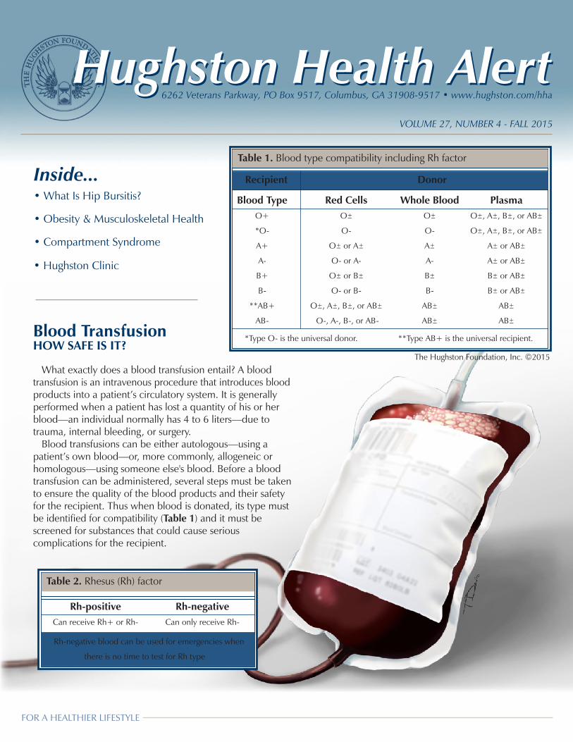
Table 1. Blood type compatibility including Rh factor
*Type O- is the universal donor. **Type AB+ is the universal recipient.
Blood TypeO+
*O-
A+
A-
B+
B-
**AB+
AB-
Red CellsO±
O-
O± or A±
O- or A-
O± or B±
O- or B-
O±, A±, B±, or AB±
O-, A-, B-, or AB-
Whole BloodO±
O-
A±
A-
B±
B-
AB±
AB±
PlasmaO±, A±, B±, or AB±
O±, A±, B±, or AB±
A± or AB±
A± or AB±
B± or AB±
B± or AB±
AB±
AB±
Table 2. Rhesus (Rh) factor
Rh-positiveCan receive Rh+ or Rh-
Rh-negativeCan only receive Rh-
Recipient Donor
Rh-negative blood can be used for emergencies when
there is no time to test for Rh type
Blood Transfusion
HOW SAFE IS IT?
What exactly does a blood transfusion entail? A blood transfusion is an intravenous procedure that introduces blood products into a patient’s circulatory system. It is generally performed when a patient has lost a quantity of his or her blood—an individual normally has 4 to 6 liters—due to trauma, internal bleeding, or surgery.
Blood transfusions can be either autologous—using a patient’s own blood—or, more commonly, allogeneic or homologous—using someone else's blood. Before a blood transfusion can be administered, several steps must be taken to ensure the quality of the blood products and their safety for the recipient. Thus when blood is donated, its type must be identified for compatibility (Table 1) and it must be screened for substances that could cause serious complications for the recipient.
The Hughston Foundation, Inc. ©2015
Inside...• What Is Hip Bursitis?
• Obesity & Musculoskeletal Health
• Compartment Syndrome
FOR A HEALTHIER LIFESTYLE
VOLUME 27, NUMBER 4 - FALL 2015
• Hughston Clinic

What is blood typing?Early blood transfusions were usually whole blood
transfusions. They were performed throughout the 19th century, but were considered risky business as, in the absence of knowledge of blood typing and compatibility, they often resulted in death. The mystery of blood compatibility was unraveled when Austrian physician Karl Landsteiner (1868-1943) identified and categorized specific blood proteins called antigens, thus laying the groundwork for the modern system of blood type classification and safe transfusions. According to this system, a person’s blood type depends on the presence or absence of different kinds of antigens or protein molecules—more specifically, glycoproteins or sugars—on his or her erythrocytes (red blood cells). Common medical practice today relies on this blood classification system and generally uses only those components of the blood—such as red blood cells, white blood cells, plasma, clotting factors, and platelets—necessary for the patient’s condition.
Currently, there are 35 different human blood group systems. When it comes to transfusions, the most important are the ABO and Rh systems. These systems are based on the different types of red blood cell glycoproteins or antigens—short for “antibody generators”—that cause the immune system to produce antibodies against them. The ABO typing scheme comprises just 2 antigens, designated A and B. Every individual has 1, both, or neither one of these types of red blood cell antigens. If, for example, a person’s blood is type A, he or she has antigen A present on his or her erythrocytes; likewise, the presence of antigen B makes a person’s blood type B. If an individual has both antigens, he or she belongs to the AB blood group. If neither antigen is present, that person’s blood group is O (Table 1).
What are antibodies?Antibodies are another type of blood protein. In contrast to
antigens, which are found on the surface of red blood cells, antibodies are found in the plasma or clear, yellowish liquid portion of the blood. An antibody, also known as an immunoglobulin (Ig), is a large Y-shaped protein produced by plasma cells. Antibodies identify and neutralize pathogens, such as bacteria and viruses, by reacting to their antigens. Thus each blood group with its respective antigen type (A, B, AB, or O) also produces the corresponding antibodies— anti-B, anti-A, both, or none—in the blood plasma. As individuals have immunological tolerance only toward what can be found in their own bodies, their antigens produce antibodies that attack what is recognized as foreign.
What is Rh factor?Another blood grouping system important for the practice
of blood transfusions is the Rh system. Around 1940, Landsteiner, in collaboration with other scientists, discovered the Rhesus blood group system while studying animal transfusions in which Rhesus monkeys (small monkeys
indigenous to India) were involved. This system includes 50 other blood antigens, the most important of which for compatibility is an antigen designated D and commonly known as Rhesus (Rh) factor. Many people have this antigen on the surface of their red blood cells, making them Rh+ while those without it are Rh-. This difference is the cause of most adverse transfusion reactions (Table 2). For example, a person who is Rh- does not normally have antibodies in his or her blood plasma, but if he or she receives blood from a person who is Rh+, the newly introduced Rh antigens can trigger the production of Rh antibodies (an Rh+ individual, however, can receive blood from a person who is Rh- without any adverse reaction). To indicate the Rh status in addition to the ABO classification, a person’s blood type is often designated with a plus or minus sign, for example, A+ or A- and B+ or B-.
What is meant by blood compatibility?Blood antibodies are also hemagglutinins or substances that
cause particles to coagulate to form a thickened mass. Thus if group-B blood is given to a recipient who belongs to blood group A, anti-B antibodies will agglutinate or clump together with the A antigens in the donor blood at the antigen-binding sites, causing them to change from a fluid state into a thickened mass. After a period of time, these agglutinated red blood cells will break down and their contents leak out into the bloodstream and become toxic. Thus if the donor blood type is not a good match, a serious transfusion reaction called acute hemolytic reaction can occur when the body attacks the transfused red blood cells. To prevent these complications, exact blood type matching is essential. Finally, there are a number of other risks associated with blood transfusions. These include blood-borne infections, acute lung injury, hemochromatosis (iron overload), and allergic reactions such as hives, fever, and chills.
How are safe outcomes ensured?Blood transfusions are to date one of the most frequently
performed medical procedures: approximately 85 million units of red blood cells are transfused globally each year and the rate of success is outstanding. In order to minimize risks, 70% of all countries, as noted by the World Health Organization (WHO) in 2012, have put into effect a national blood policy, and 62% have specific legislation covering issues related to the safety and quality of blood transfusions. These measures, along with increasingly rigorous blood screening methods, help to ensure safer and more satisfactory outcomes for patients receiving lifesaving blood transfusions.
Eric Stanford, DOAthens, Alabama
2 FOR A HEALTHIER LIFESTYLE

What Is Hip Bursitis?Hip bursitis is a condition that affects just one of the several
bursae or bursal sacs around the hip joint. These types of sacs, which can be found in many other places throughout the body, are filled with synovial fluid—a clear, slippery substance that allows structures such as tendons, muscles, and bone to glide over one another by reducing the amount of friction between them. The hip bursa is located between the iliotibial tract (a thick connective tissue band that runs along the lateral side of the hip and thigh and inserts just below the outside of the knee) and the outer surface of the upper portion of the femur (thighbone) at the level of the greater trochanter (the bony prominence at the top of the femur) (Fig. 1). It is thus also referred to as the greater trochanteric bursa. Hip bursitis is the most common cause of hip pain in the general population.
Symptoms: where does it hurt? The pain from hip bursitis usually
presents on the outside of your hip where the top of the front pocket of your pants is located. Localizing the pain will help you to distinguish this condition from other problems of the hip joint more commonly felt in the groin. However, while hip bursitis pain may start out sharp and remain localized over your outer hip, as the condition progresses, it may become dull, achy, or burning, and travel down the outside of your leg to your knee or lower back. The unpredictable nature of the pain may make it seem as if it is coming from your knee or back, making a correct diagnosis difficult. The pain may become worse if you sleep on the affected side; it may also worsen after you arise in the morning, but ease off after a period of time. Additionally, hip bursitis can sometimes cause you to experience a sharp pain when getting up from a sitting position.
What causes hip bursitis?Common causes of hip bursitis include obesity, leg length
discrepancy, rheumatoid arthritis, overuse, injury, or calcification of the bursa. Moreover, prior surgery on the hip, including a hip replacement, may alter the mechanics around your joint in such a way that the bursa becomes irritated, leading to hip bursitis.
How is hip bursitis diagnosed?To diagnose hip bursitis, your physician will start by taking
a thorough history. If, like many patients, you have already seen a number of physicians with little or no relief from your
pain, you may arrive at your appointment feeling frustrated. The physical examination will most likely elicit pain over your lateral (outer) hip. Your physician should also perform specific examination maneuvers to rule out problems with the spine, pelvic bone, or hip joint. X-rays of the hip are usually normal for hip bursitis sufferers, but can sometimes show calcification within the bursa.
How is hip bursitis treated?Treatment for hip bursitis begins
with conservative management. For example, your physician may prescribe physical therapy to help correct muscle imbalances while electrotherapy or ultrasound therapy can work to reduce pain and inflammation. To compensate for faulty foot biomechanics, you may want to have orthotic insoles for your shoes made. Rest and nonsteroidal anti-inflammatory medications (NSAIDs), such as ibuprofen or naproxen, are also recommended. If your pain becomes chronic or does not respond to physical therapy and oral medication, a combination of steroids and numbing medication injected into the affected bursa will often bring relief. Also, if necessary, your doctor can aspirate fluid from the bursa with a needle. Most cases of hip bursitis can be resolved with
these nonsurgical interventions; however, sometimes symptoms persist. If so, the offending bursal tissue can be removed through an incision on the outside of the hip in a procedure called open bursectomy. By contrast, endoscopic hip bursectomy—inserting a camera through a small incision into the hip area and removing the bursa through another small incision—has an outcome comparable to that of open bursectomy, but with the advantages that it is less invasive and can be performed as an outpatient procedure (Fig. 2).
Can hip bursitis be prevented?Using proper footwear when walking and playing sports
can go a long way toward preventing hip bursitis. Additionally, you should make an effort to stretch consistently—both before and after activity—to help head off the condition. Doing yoga may also benefit you. If you are experiencing hip bursitis, minimize any activity that might aggravate symptoms, such as going up and down stairs. If you are an athlete, avoid uphill running and overuse. In most cases, with proper care and planning, the pain of hip bursitis can be managed effectively or even eliminated without surgery.
George Sutherland, MDOkatie, South Carolina
Fig. 1. Hip anatomy
Fig. 2. Camera inserted for hip bursectomy
Pelvic bone
Bursal sac
Iliotibial tract
Femur (thighbone)
Camera
Greater trochanter
The Hughston Foundation, Inc. ©2015
FOR A HEALTHIER LIFESTYLE 3

Obesity and Musculoskeletal HealthThe percentage of the population that qualifies as obese has
been rising steadily, making obesity a national health concern. Obesity occurs when a person has accumulated too much body fat. The condition has been linked to increased health risks as well as reduced life expectancy. More specifically, obesity can lead to diabetes, heart disease, peripheral vascular disease, obstructive sleep apnea (shallow or infrequent breathing during sleep), and some forms of cancer. Obesity can also have an overall negative impact on a person’s bones and joints and carries with it increased risks for complications and less than desirable outcomes from surgery (Fig. 1).
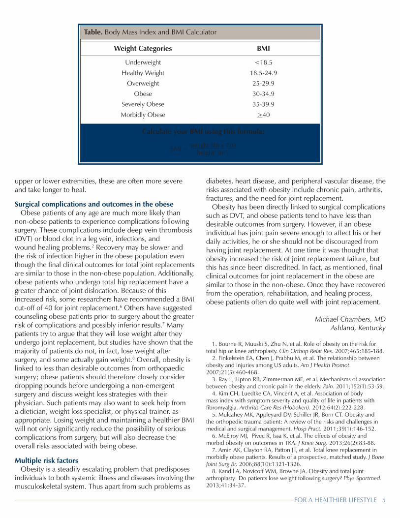
Obesity is often determined by using the body mass index (BMI) or measure of lean body mass, which is calculated by dividing a person’s body weight by his or her height squared (Table). In the general population, obesity is indicated by a BMI of 30 or more. Morbid obesity is defined as having a BMI greater than 40 while a BMI greater than 50 qualifies a person as super-obese. BMI, however, is not always the best tool for determining obesity. For example, for growing children, BMI represents a relative rather than absolute value that must be mapped into a percentile for age and gender before it can be interpreted. For athletes, who tend to have a high quantity of muscle—which weighs more than the same volume of fat—body fat assessments, such as waist circumference measurements or waist-to-hip ratio, are more accurate. Other measures used to determine obesity include waist-to-height ratio, body composition, percentage of body fat, and bioelectrical impedance analysis.
A link to osteoarthritis and arthroplastyObesity can cause abnormal wear and tear in weight-bearing
joints, which directly links it to both osteoarthritis (chronic wear and tear of the joint cartilage resulting in pain) and the need for total knee and hip arthroplasties (replacements) (Fig. 2). As the incidence of obesity has increased in the general population, so has the incidence of osteoarthritis. There are several theories that attempt to explain why obesity is linked to osteoarthritis,
including more stress on the joints (due to decreased muscle mass), increased joint load (due to excess pounds), and the presence of a low-grade systemic inflammatory reaction. Moreover, while osteoarthritis is the most common reason for joint arthroplasties in both obese and non-obese populations, osteoarthritis patients with a higher BMI are more likely to have total knee replacements.1
Musculoskeletal problems in the obeseObese people have a greater likelihood than non-obese
individuals of suffering from musculoskeletal problems. Statistics show that musculoskeletal injuries occur at a 15% higher rate for those with a BMI between 25 and 30, and as BMI increases, so does the risk of injury.2 Moreover, obese people are more likely to sustain certain types of orthopaedic injuries. For example, ankle fractures tend to be both more common and more severe in the obese population. Furthermore, low velocity knee dislocations—for instance, those that occur as a result of falling from a standing height—are typically seen in the obese. Additionally, obesity is associated with musculoskeletal pain syndromes and can worsen fibromyalgia (chronic pain in the muscles and soft tissues). Lastly, in the elderly population, the overall risk of developing chronic musculoskeletal pain for obese patients is almost double that for non-obese patients.3, 4
Traumatic injury in the obeseAfter traumatic injury, obese patients show a higher
rate of organ failure, respiratory issues, infections, increased length of hospital stays, and even death compared to those who are not obese. This is thought to be a function of possible organ damage already present at the time of the traumatic event.5 Moreover, obese patients have a greater risk of artery and nerve damage following a knee dislocation. Furthermore, when obese patients sustain fractures in either the
Fig. 1. Normal knee joint (straight)Fig. 2. Knee joint affected by osteoarthritis
Femur (thighbone)
Bent knee/front view Straight knee/side view
Cartilage
Worn cartilage
FibulaTibia (shinbone)
The Hughston Foundation, Inc. ©2015The Hughston Foundation, Inc. ©2015
4 FOR A HEALTHIER LIFESTYLE
upper or lower extremities, these are often more severe and take longer to heal.
Surgical complications and outcomes in the obeseObese patients of any age are much more likely than
non-obese patients to experience complications following surgery. These complications include deep vein thrombosis (DVT) or blood clot in a leg vein, infections, and wound healing problems.2 Recovery may be slower and the risk of infection higher in the obese population even though the final clinical outcomes for total joint replacements are similar to those in the non-obese population. Additionally, obese patients who undergo total hip replacement have a greater chance of joint dislocation. Because of this increased risk, some researchers have recommended a BMI cut-off of 40 for joint replacement.6 Others have suggested counseling obese patients prior to surgery about the greater risk of complications and possibly inferior results.7 Many patients try to argue that they will lose weight after they undergo joint replacement, but studies have shown that the majority of patients do not, in fact, lose weight after surgery, and some actually gain weight.8 Overall, obesity is linked to less than desirable outcomes from orthopaedic surgery; obese patients should therefore closely consider dropping pounds before undergoing a non-emergent surgery and discuss weight loss strategies with their physician. Such patients may also want to seek help from a dietician, weight loss specialist, or physical trainer, as appropriate. Losing weight and maintaining a healthier BMI will not only significantly reduce the possibility of serious complications from surgery, but will also decrease the overall risks associated with being obese.
Multiple risk factorsObesity is a steadily escalating problem that predisposes
individuals to both systemic illness and diseases involving the musculoskeletal system. Thus apart from such problems as
diabetes, heart disease, and peripheral vascular disease, the risks associated with obesity include chronic pain, arthritis, fractures, and the need for joint replacement.
Obesity has been directly linked to surgical complications such as DVT, and obese patients tend to have less than desirable outcomes from surgery. However, if an obese individual has joint pain severe enough to affect his or her daily activities, he or she should not be discouraged from having joint replacement. At one time it was thought that obesity increased the risk of joint replacement failure, but this has since been discredited. In fact, as mentioned, final clinical outcomes for joint replacement in the obese are similar to those in the non-obese. Once they have recovered from the operation, rehabilitation, and healing process, obese patients often do quite well with joint replacement.
Michael Chambers, MDAshland, Kentucky
1. Bourne R, Muuski S, Zhu N, et al. Role of obesity on the risk for
total hip or knee arthroplasty. Clin Orthop Relat Res. 2007;465:185-188.2. Finkelstein EA, Chen J, Prabhu M, et al. The relationship between
obesity and injuries among US adults. Am J Health Promot. 2007;21(5):460-468.
3. Ray L, Lipton RB, Zimmerman ME, et al. Mechanisms of association between obesity and chronic pain in the elderly. Pain. 2011;152(1):53-59.
4. Kim CH, Luedtke CA, Vincent A, et al. Association of body mass index with symptom severity and quality of life in patients with fibromyalgia. Arthritis Care Res (Hoboken). 2012;64(2):222-228.
5. Mulcahey MK, Appleyard DV, Schiller JR, Born CT. Obesity and the orthopedic trauma patient: A review of the risks and challenges in medical and surgical management. Hosp Pract. 2011;39(1):146-152.
6. McElroy MJ, Pivec R, Issa K, et al. The effects of obesity and morbid obesity on outcomes in TKA. J Knee Surg. 2013;26(2):83-88.
7. Amin AK, Clayton RA, Patton JT, et al. Total knee replacement in morbidly obese patients. Results of a prospective, matched study. J Bone Joint Surg Br. 2006;88(10):1321-1326.
8. Kandil A, Novicoff WM, Browne JA. Obesity and total joint arthroplasty: Do patients lose weight following surgery? Phys Sportmed. 2013;41:34-37.
Table. Body Mass Index and BMI Calculator
Weight Categories
Underweight
Healthy Weight
Overweight
Obese
Severely Obese
Morbidly Obese
BMI
<18.5
18.5-24.9
25-29.9
30-34.9
35-39.9
>40
Calculate your BMI using this formula:
BMI = weight (lb) x 703 height2 (in2)
FOR A HEALTHIER LIFESTYLE 5

Compartment SyndromeWHAT IS COMPARTMENT SYNDROME?
Increased pressure within a compartment of the body— defined as a group of muscles or organs, including the blood vessels and nerves, that is surrounded by fascia or connective tissue—is known as compartment syndrome, a condition that can be either acute or chronic (Fig. 1). Acute compartment syndrome is overall the most common form of compartment syndrome. When the condition is chronic, the most common type is called chronic exertional compartment syndrome (CECS) as it is often exercise-induced.
What causes compartment syndrome?Acute compartment syndrome can be caused by a fracture
to the arm or leg. Following an injury to a limb, fluid, such as blood, may accumulate in the compartment, causing the intracompartmental pressure to increase beyond the tissue pressure. This is because the connective tissue surrounding the compartment is inelastic and cannot easily expand. Consequently, the blood flow into the compartment is restricted and oxygen does not reach the tissues, resulting in severe tissue damage and even tissue necrosis (tissue death). Acute compartment syndrome can also develop from treating a fracture. For example, if a cast or splint is too tight, it can restrict the flow of blood and oxygen to a compartment. After removal, reperfusion (the restoration of blood flow to the area) and swelling can greatly increase the intra-compartmental pressure. Acute compartment syndrome can also occur as a consequence of other kinds of injuries that do not involve fractures—for instance, crush injuries, burns, extreme vigorous exercise, or the prolonged compression and consequent swelling upon reperfusion of a limb.
By contrast, CECS occurs primarily in athletes under the age of 30 who engage in intense, repetitive, high-impact activities such as running. Greater blood flow during exercise, the intensity of training, or the use of either anabolic steroids or creatine (a nitrogenous organic acid that helps supply energy to cells, particularly muscle cells) may cause the muscles of a compartment to increase in size. They may then exert increased pressure on the tough surrounding fascial tissue which is unable to expand sufficiently to accommodate the growth (Fig. 2). Over time, the blood
supply to the muscles can be cut off. CECS tends to occur in the legs, particularly the lower leg, of runners and cyclists, and in the arms of swimmers. Of the 4 compartments of the lower leg, it most commonly affects the anterior or front compartment, which generally causes problems with dorsiflexion or the upward movement of the ankles and toes needed to walk and run. On the whole, CECS is a frequently overlooked cause of muscle pain in athletes.
What are the symptoms?Patients with acute compartment syndrome usually
experience pain, the severity of which seems out of proportion to the injury. Passive stretching of the involved muscle or compartment worsens the pain. Pain can be difficult to assess if a patient is suffering from multiple traumas and almost impossible to assess if he or she has been sedated or intubated (when a flexible, plastic tube is surgically
inserted into the trachea or windpipe to assist breathing). Both decreased 2-point discrimination (the ability to discern that 2 nearby objects touching the skin are truly 2 distinct points, not 1) and diminished vibration sense have been reported in patients with acute compartment syndrome. Other late symptoms include numbness, paralysis, and absent peripheral pulse.
In cases of CECS, the primary symptom is extreme tightness in the
affected muscle group followed by pain and a burning sensation when exercising. Other signs and symptoms include numbness or a tingling sensation in the affected area and pain that can be described as aching, cramping, sharp, or stabbing. The symptoms tend to recur at a very specific threshold of exertion which is different, but remains consistent, for each individual. When the person stops exercising, the pressure in the compartment decreases and the symptoms subside. A typical scenario would be the development of pain in the anterior compartment of the calf 15 minutes into a run that resolves within 30 minutes of stopping. Overall, the pain of CECS may go on intermittently for months or even years.
How is compartment syndrome diagnosed?For patients with acute compartment syndrome, a prompt
and thorough evaluation will minimize the risk of permanent damage or death. Acute compartment syndrome is a clinical diagnosis based on the type and mechanism of injury, clinical signs and symptoms, a physical examination,
Fig. 1. Lower leg compartments
Tibia (shinbone)
Superficial posterior compartmentDeep posterior
compartment
Anterior compartment
Lateral compartment
Fibula
Fascia
The Hughston Foundation, Inc. ©2015
6 FOR A HEALTHIER LIFESTYLE

and thorough history. It is therefore best if the patient’s mental state is intact. In cases involving patients who have been intubated or are nonresponsive, a physician can insert a needle device that includes a pressure scale into the injured area and measure the pressure inside the compartment directly. If the difference between the patient’s diastolic (resting) blood pressure and the compartment pressure is less than or equal to 30 mm Hg, a diagnosis of acute compartment syndrome is made.
CECS is diagnosed initially through a process of elimination, as in the early stages it can mimic other conditions such as tendonitis, fascial defect, and medial tibial stress syndrome (shin splints). X-rays should also be taken to rule out stress fractures (hairline fractures caused by repetitive stress) or other bone problems. When these have been excluded, the presence of the condition is confirmed by using a needle device to measure the pressure within the affected compartment prior to beginning activity and after exercising to the point of pain. Usually, the compartmental pressure will remain high even after exercise. As a rule, if the resting pressure is equal to or greater than 15 mm Hg and the 5-minute post-exercise pressure is equal to or greater than 20 mm Hg, the diagnosis is CECS.
Who is at risk?Patients taking anticoagulants are at higher risk
for developing acute compartment syndrome because when they bleed, as a result of trauma or injury, they cannot clot normally, causing blood to accumulate and intracompartmental pressure to rise. Athletes who train intensely or take steroids or other performance-enhancing substances are also at increased risk for developing CECS as greater muscle mass can put pressure on the fascia.
How is compartment syndrome treated?When acute compartment syndrome is diagnosed, surgery
must be done immediately to decrease the compartmental pressure. A fasciotomy or incision through both the skin and the fascia covering the compartment is performed (Fig. 3). Supportive measures, such as elevating the affected extremity, increasing oxygen saturation, intravenous fluids, and pain medication, can also be taken, but there is no truly effective nonsurgical option for the condition.
CECS is initially treated with anti-inflammatory and pain medications, stretching and strengthening, and therapeutic
massage. Recommendations for the athlete suffering from CECS also include taking time out from training, undergoing physical therapy, changing biomechanics (how one moves), using orthotics, switching training surfaces, and cross-training with low-impact activities. If symptoms do persist, the most effective treatment is to operate on the fascia. Either a fasciotomy (releasing the fascia) or, in some cases, a fasciectomy (excising strips of fascia) will decompress the compartment and relieve the pain. A fasciotomy can be open—where a deep incision gives the surgeon a full view of the compartment—or subcutaneous (beneath the skin). The latter is a less invasive, but more complicated procedure. It often requires the use of an endoscope and, statistics show, carries with it a greater likelihood that the patient will suffer a recurrence of the problem.
What are the complications of treatment?Despite the well-documented efficacy of fascial
surgery, complications can nevertheless arise. These include possible infection, hemorrhage, lymphocele (a collection of lymphatic fluid in the body), vascular injury, skin breakdown, permanent nerve damage, numbness, and scarring. Tissue necrosis, severe infection, or plummeting blood pressure can necessitate the amputation of a
limb. Therefore, if a patient has chronic rather than acute compartment syndrome, it is worth trying less invasive or radical means of relieving the pain and pressure before resorting to surgery. Following fasciotomy for CECS, there is reportedly a 6 to 11% recurrence rate for athletes. This is generally due to incomplete release of the fascia, poor rehabilitation,
or over-active scarring. In the vast majority of such cases, performing a second fasciotomy will resolve the symptoms.
PrecautionsWhether acute or chronic, the sooner compartment
syndrome is diagnosed and treated by a qualified health professional, the better the outcome is likely to be. While acute compartment syndrome cannot usually be foreseen or prevented, precautions can be taken to avoid developing CECS. For active individuals, these include warming-up and cooling down sufficiently, stretching, cross-training, increasing exercise gradually, and staying hydrated. Ideally, it is important for athletes of all levels to respect their own limitations when it comes to training and competing.
Wasik Ashraf, DO, and Chris Maisto, PhD, CMTColumbus, Georgia
Fig. 2. Pressure building up in the tibialis anterior muscle group
Fig. 3. Fasciotomy to release pressure
Skin
Vein
ArteryNerve
Fascia
The Hughston Foundation, Inc. ©2015
FOR A HEALTHIER LIFESTYLE 7

Editor - Thomas N. Bernard, Jr., MD
Managing Editor - Dennise Brogdon
Senior Editor - Chris Maisto, PhD, CMT
Art Director - Belinda J. Klein, MA
Layout Editor - Tiffany C. Davis, MS
Editorial BoardChamp L. Baker III, MDMark A. Baker, PT, CEO William C. Etchison, MSAndy J. Grubbs, Jr., MEd, ATC Rob Hopkins, PT, SCS Cholly P. MintonSteve Young, PT
The Hughston Health Alert is a quarterly publication of The Hughston Foundation, Inc. The Founda-tion’s mission is to help people of all ages attain the highest possible standards of musculoskeletal health, fitness, and athletic prowess. Information in the Hughston Health Alert reflects the experience and training of physicians at The Hughston Clinic, P.C., of physical therapists and athletic trainers at Hughston Rehabilitation, of physicians who trained as residents and fellows under the auspices of The Hughston Foundation, Inc., and of research scientists and other professional staff at The Hughston Foundation, Inc. The information in the Hughston Health Alert is intended to supplement the advice of your personal phy-sician and should not be relied on for the treatment of an individual’s specific medical problems.
Special written permission is required to reproduce, by any manner, in whole or in part, the material herein contained.
Send inquiries to Medical Writing, The Hughston Foundation, Inc., P.O. Box 9517, 6262 Veterans Parkway, Columbus GA 31908-9517 USA.
Copyright 2015, The Hughston Foundation, Inc. ISSN# 1070-7778www.hughston.com
6262 Veterans ParkwayP.O. Box 9517
Columbus GA 31908-9517Appointments:706-324-6661
1-800-331-2910
Hughston Health AlertThe Hughston Foundation, Inc.6262 Veterans Parkway P.O. Box 9517 Columbus, Georgia 31908-9517
4401 River Chase DrivePhenix City, AL 36867Phone: 334-732-3000
Fax: 334-732-3020
Locations:
SCAN ME for more Hughston Health Alert articles.
2002-2015
NONPROFIT ORGUS POSTAGE
PAIDCOLUMBUS GAPERMIT NO 99
You may receive the Hughston Health Alert via e-mail by registering at: www.hughston.com/health-alert-sign-up.aspx
Locations:
Champ L. Baker Jr., MD - Arthroscopy & Sports Medicine
Champ L. Baker III, MD - Arthroscopy & Sports Medicine
Thomas N. Bernard Jr., MD - Orthopaedic Spine Surgery
Jared A. Brummel, DO - Sports Medicine & General Orthopaedics
J. Kenneth Burkus, MD - Orthopaedic Spine Surgery
Kevin J. Collins, MD - General Orthopaedics & Sports Medicine
Norman L. Donati Jr., MD - General Orthopaedics, Foot & Ankle
John D. Dorchak, MD - Orthopaedic Spine Surgery
Patrick J. Fernicola, MD - Shoulder, Knee, Total Joint Replacement
Fred Flandry, MD, FACS - Trauma, Arthroscopy & Sports Medicine
John C. P. Floyd, MD, FACS - Orthopaedic Traumatologist
Ryan M. Geringer, DO - General Orthopaedics & Sports Medicine
Garland K. Gudger, MD - General Orthopaedics & Sports Medicine
Robert M. Harris, MD - Director of Orthopaedic Trauma
• Albany • Auburn, AL • Columbus • Dothan, AL • Gwinnett • LaGrange • Moultrie • Nashville, TN • Thomaston • Thomasville • Phenix City • Valdosta
Locations:
J. Matthew Heaton, MD - General Orthopaedics & Sports Medicine
Kurt E. Jacobson, MD, FACS - Knee, Sports Medicine & General Orthopaedics
Philip J. Kregor, MD - Orthopaedic Trauma
David H, MacDonald, DO - Hand & Upper Extremities, Arthroscopic Surgery
James E. McGrory, MD - Orthopaedic Spine Surgery & Total Joint Replacement
William Min, MD, MS, MBA - Orthopaedic Traumatologist
Jesse L. Pace, DO - Arthroscopy, General Orthopaedics & Sports medicine
Douglas W. Pahl, MD - Orthopaedic Spine Surgery
David C. Rehak, MD - Hand, Wrist & Upper Extremities
Randall J. Ruark, MD - Hip & Knee Total Joint Replacement
Carlton G. Savory, MD, FACS - Hip, Knee, Total Joint Replacement
Michael M. Tucker Jr., MD - Knee, Shoulder, Foot, Ankle & Sports Medicine
John I. Waldrop, MD - General Orthopaedics, Total Joint Replacement
Bruce H. Ziran, MD - Director of Orthopaedic Trauma
YESTERDAY. TODAY. TOMORROW.
HUGHSTON DIFFERENCE
THE
It’s our privilege to serve you