hospital acquired pressure injury education module
TRANSCRIPT
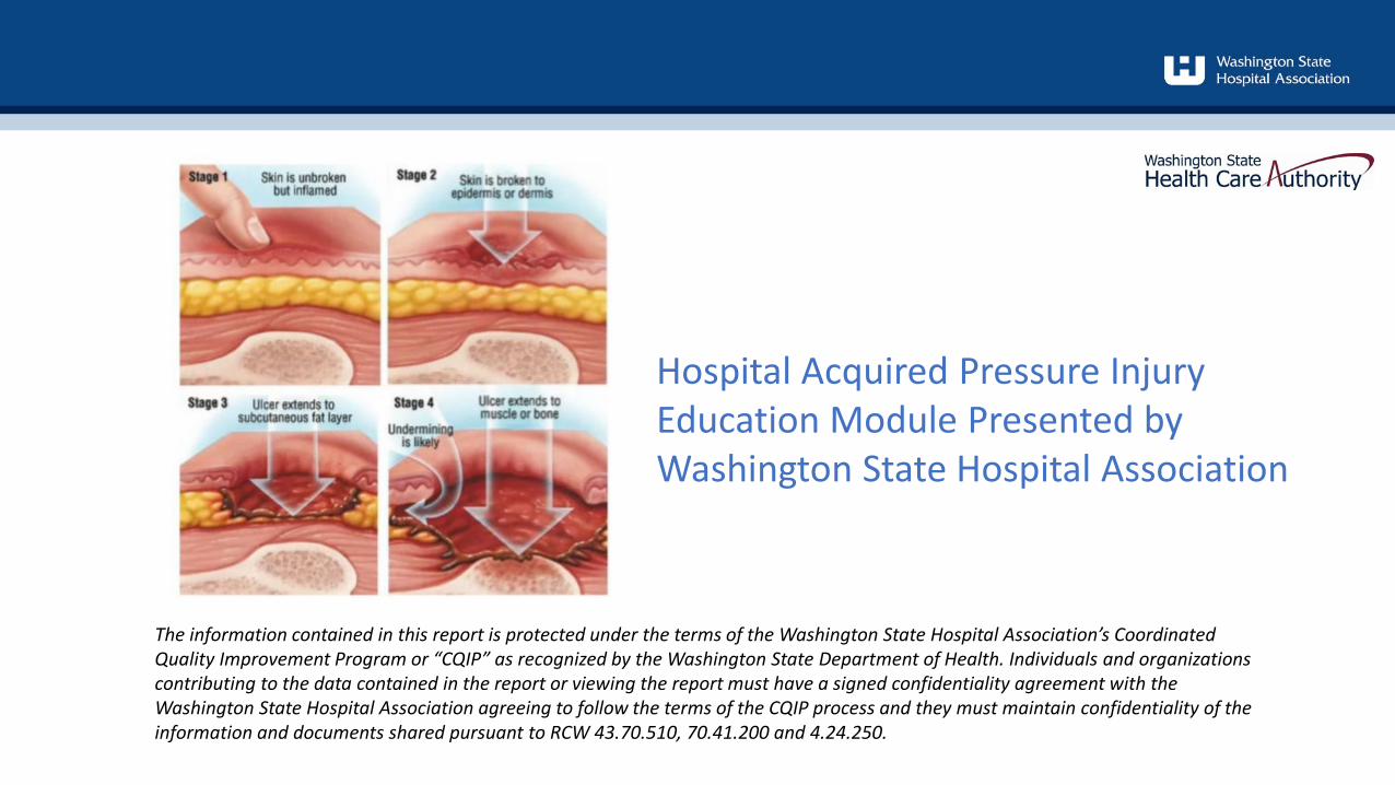
The information contained in this report is protected under the terms of the Washington State Hospital Association’s CoordinatedQuality Improvement Program or “CQIP” as recognized by the Washington State Department of Health. Individuals and organizations contributing to the data contained in the report or viewing the report must have a signed confidentiality agreement with the Washington State Hospital Association agreeing to follow the terms of the CQIP process and they must maintain confidentiality of the information and documents shared pursuant to RCW 43.70.510, 70.41.200 and 4.24.250.
Hospital Acquired Pressure Injury Education Module Presented by Washington State Hospital Association
Amy AndersonDirector Safety and Quality
MN, BSN, [email protected]
Tina Seery Senior Director Safety and Quality
MHA RN CPHQ CPPS [email protected]
Understand recent data trends
Identify vulnerable populations as it relates to pressure injuries
Describe the importance of early recognition with a complete skin assessment
Recognize devices that may increase patient risk for pressure injuries
Objectives:
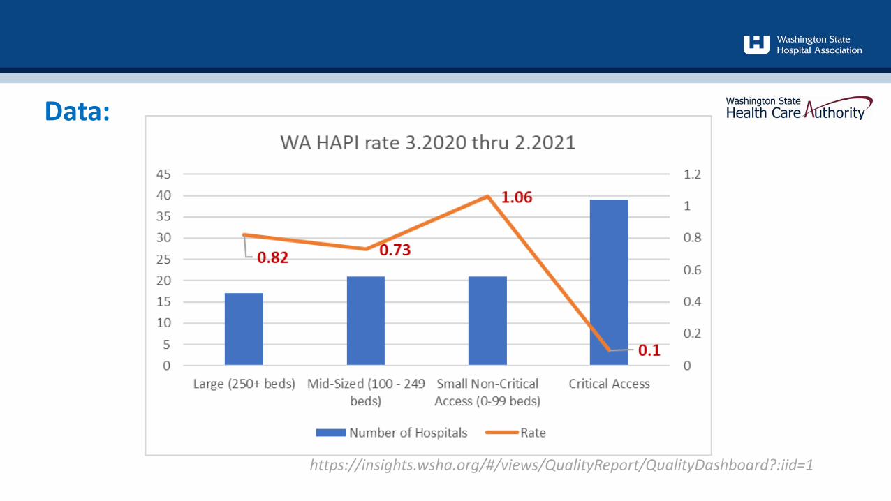
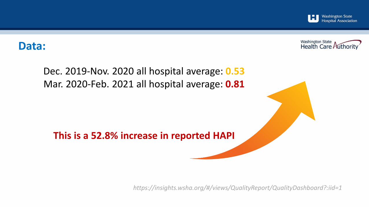
Data:
https://insights.wsha.org/#/views/QualityReport/QualityDashboard?:iid=1
Data:
https://insights.wsha.org/#/views/QualityReport/QualityDashboard?:iid=1
Dec. 2019-Nov. 2020 all hospital average: 0.53Mar. 2020-Feb. 2021 all hospital average: 0.81
This is a 52.8% increase in reported HAPI
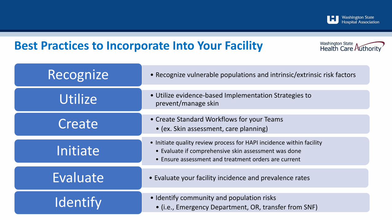
• Recognize vulnerable populations and intrinsic/extrinsic risk factors Recognize• Utilize evidence-based Implementation Strategies to
prevent/manage skinUtilize• Create Standard Workflows for your Teams
• (ex. Skin assessment, care planning)Create• Initiate quality review process for HAPI incidence within facility
• Evaluate if comprehensive skin assessment was done • Ensure assessment and treatment orders are current
Initiate
• Evaluate your facility incidence and prevalence ratesEvaluate• Identify community and population risks
• (i.e., Emergency Department, OR, transfer from SNF)Identify
Best Practices to Incorporate Into Your Facility
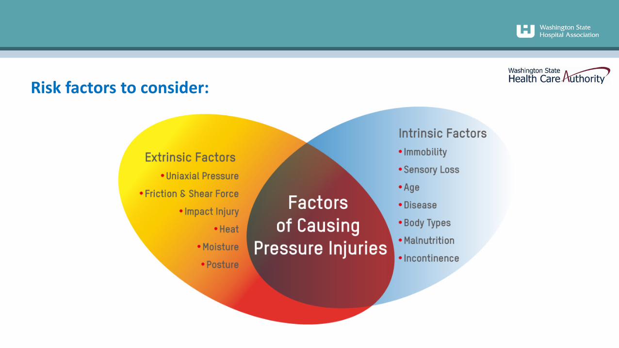
Risk factors to consider:
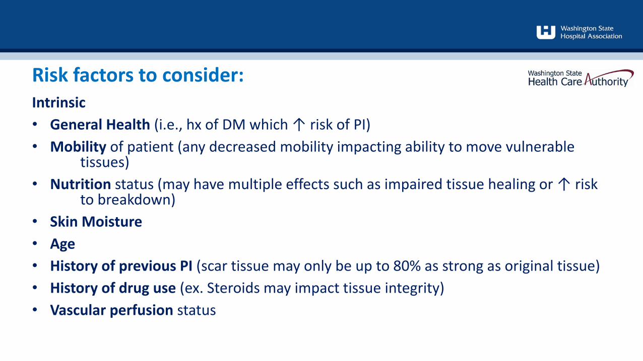
Intrinsic• General Health (i.e., hx of DM which ↑ risk of PI)• Mobility of patient (any decreased mobility impacting ability to move vulnerable
tissues)• Nutrition status (may have multiple effects such as impaired tissue healing or ↑ risk
to breakdown)• Skin Moisture• Age• History of previous PI (scar tissue may only be up to 80% as strong as original tissue)• History of drug use (ex. Steroids may impact tissue integrity)• Vascular perfusion status
Risk factors to consider:
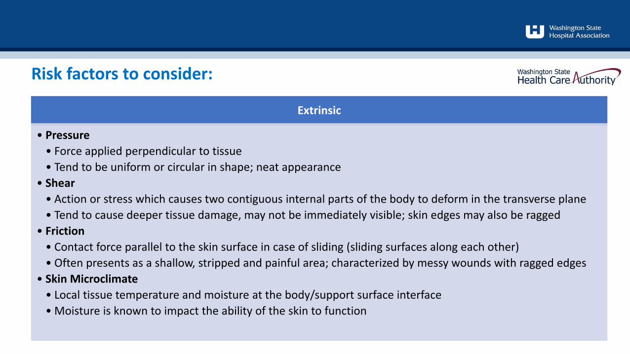
Extrinsic
• Pressure • Force applied perpendicular to tissue• Tend to be uniform or circular in shape; neat appearance
• Shear • Action or stress which causes two contiguous internal parts of the body to deform in the transverse plane• Tend to cause deeper tissue damage, may not be immediately visible; skin edges may also be ragged
• Friction • Contact force parallel to the skin surface in case of sliding (sliding surfaces along each other)• Often presents as a shallow, stripped and painful area; characterized by messy wounds with ragged edges
• Skin Microclimate • Local tissue temperature and moisture at the body/support surface interface• Moisture is known to impact the ability of the skin to function
Risk factors to consider:
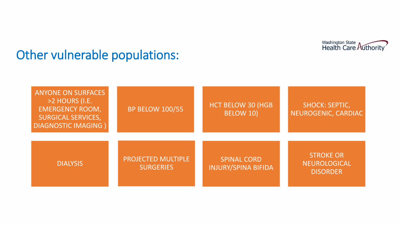
Other vulnerable populations:
ANYONE ON SURFACES >2 HOURS (I.E.
EMERGENCY ROOM, SURGICAL SERVICES,
DIAGNOSTIC IMAGING )
BP BELOW 100/55 HCT BELOW 30 (HGB BELOW 10)
SHOCK: SEPTIC, NEUROGENIC, CARDIAC
DIALYSIS PROJECTED MULTIPLE SURGERIES
SPINAL CORD INJURY/SPINA BIFIDA
STROKE OR NEUROLOGICAL
DISORDER
Medical devices account for more than 30 percent of all hospital-acquired pressure injuries (Health & Education Trust, 2017)• Clinicians should assess skin under/around medical devices to identify for early signs of
pressure injury• Medical devices should be re-positioned frequently in order to redistribute the force
and pressure• Ensure device is proper size, in proper location and secured properly; additionally,
some medical devices may require padding to reduce friction (follow manufacturers guidelines)
• Documentation and communication: use standardized forms, tools and technologies to assist clinicians in documentation and communication amongst each other
Medical Devices Increasing Risk of Pressure Injuries:
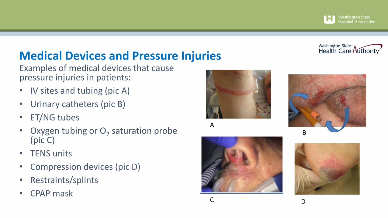
Examples of medical devices that cause pressure injuries in patients:• IV sites and tubing (pic A)• Urinary catheters (pic B)• ET/NG tubes• Oxygen tubing or O2 saturation probe
(pic C) • TENS units• Compression devices (pic D)• Restraints/splints• CPAP mask
Medical Devices and Pressure Injuries
AB
C D
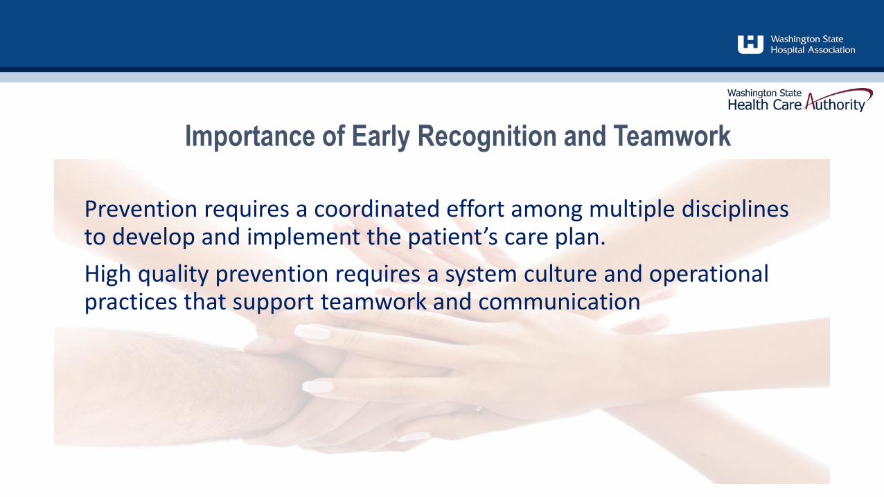
Prevention requires a coordinated effort among multiple disciplines to develop and implement the patient’s care plan. High quality prevention requires a system culture and operational practices that support teamwork and communication
Importance of Early Recognition and Teamwork
Importance of early recognition and teamwork
Utilize comprehensive skin assessment and risk assessment from the Braden to build an individualized plan of care for patient
RN scope of practice is to assess and document findings of comprehensive skin assessment (“4 EYES IN 4 HOURS” – this method uses 2 RNs to complete the initial skin assessment)
• Note: Other care providers may be within scope of practice to perform and document a comprehensive skin assessment
CNA should examine skin each time patient is repositioned or cleaned and share results with RN
Don’t be afraid to ask for extra help from a peer!
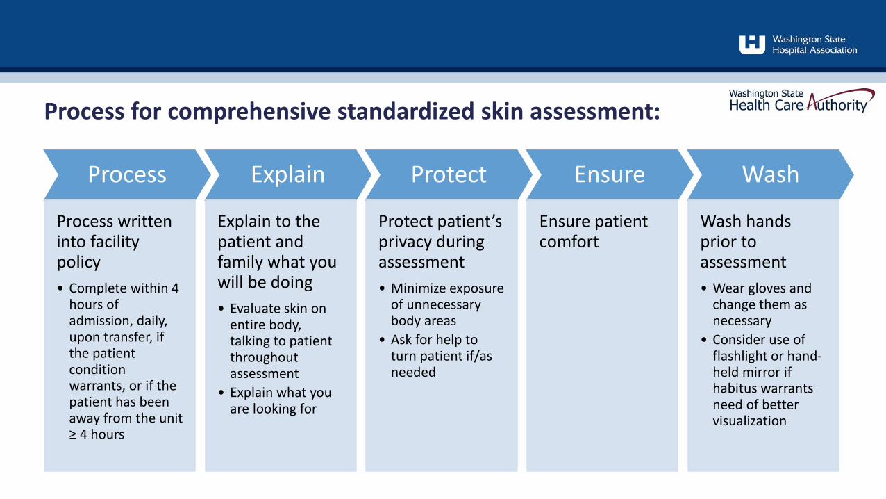
Process for comprehensive standardized skin assessment:
Process
Process written into facility policy• Complete within 4
hours of admission, daily, upon transfer, if the patient condition warrants, or if the patient has been away from the unit ≥ 4 hours
Explain
Explain to the patient and family what you will be doing• Evaluate skin on
entire body, talking to patient throughout assessment
• Explain what you are looking for
Protect
Protect patient’s privacy during assessment• Minimize exposure
of unnecessary body areas
• Ask for help to turn patient if/as needed
Ensure
Ensure patient comfort
Wash
Wash hands prior to assessment• Wear gloves and
change them as necessary
• Consider use of flashlight or hand-held mirror if habitus warrants need of better visualization
• Areas to focus on:• Skin under/around medical or compression devices• Bony prominences (heels, sacrum, occiput)• Areas of decreased sensation or previous skin breakdown (hx of
Diabetes Mellitus, Neuropathy, etc.)• Patient is receiving medication that decreases sensation (i.e.,
epidural/spinal)• Areas where skin to skin contact is occurring (i.e., penis, back of knees,
buttocks, and inner thighs)• Areas where skin has not been repositioned in ≥ 2 hours (i.e.,
Emergency Department, EMS transport, Surgical services)
Comprehensive standardized skin assessment:
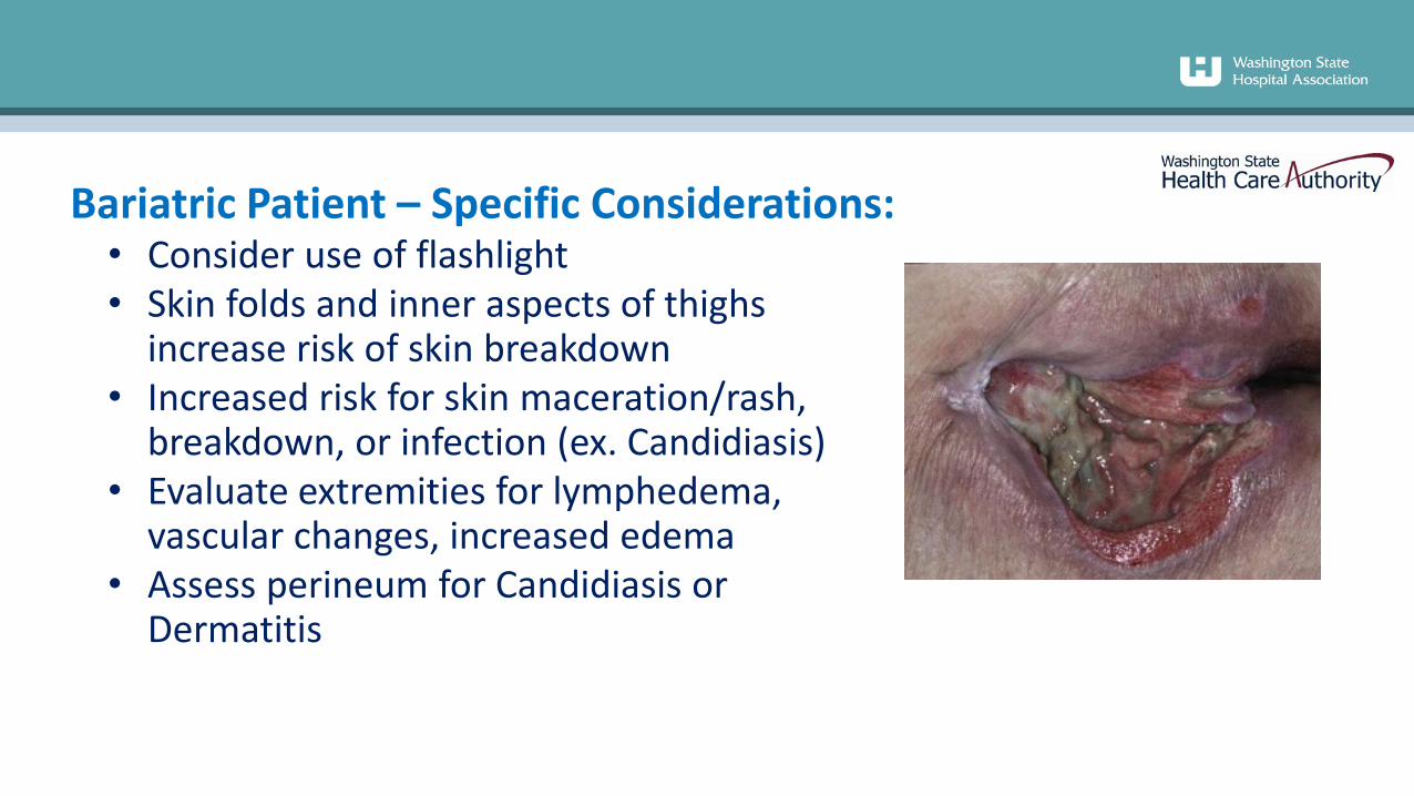
• Consider use of flashlight• Skin folds and inner aspects of thighs
increase risk of skin breakdown• Increased risk for skin maceration/rash,
breakdown, or infection (ex. Candidiasis)• Evaluate extremities for lymphedema,
vascular changes, increased edema• Assess perineum for Candidiasis or
Dermatitis
Bariatric Patient – Specific Considerations:
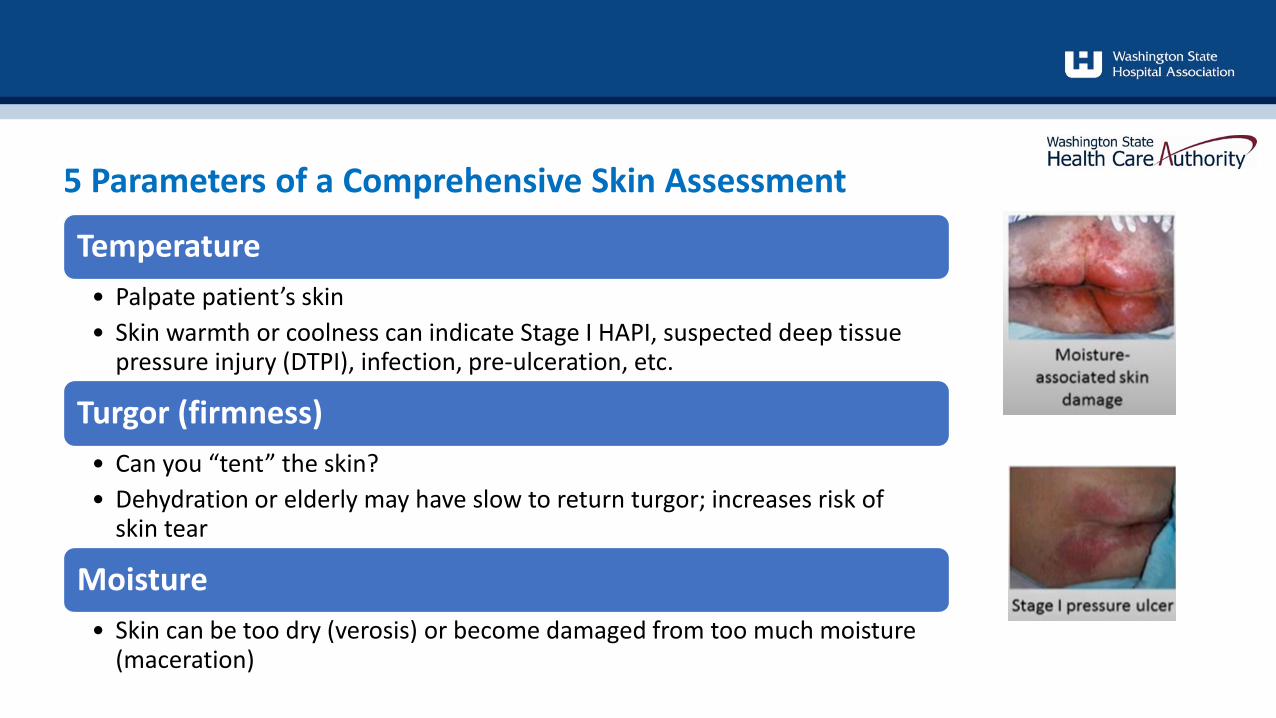
Temperature• Palpate patient’s skin• Skin warmth or coolness can indicate Stage I HAPI, suspected deep tissue
pressure injury (DTPI), infection, pre-ulceration, etc.
Turgor (firmness)• Can you “tent” the skin?• Dehydration or elderly may have slow to return turgor; increases risk of
skin tear
Moisture• Skin can be too dry (verosis) or become damaged from too much moisture
(maceration)
5 Parameters of a Comprehensive Skin Assessment
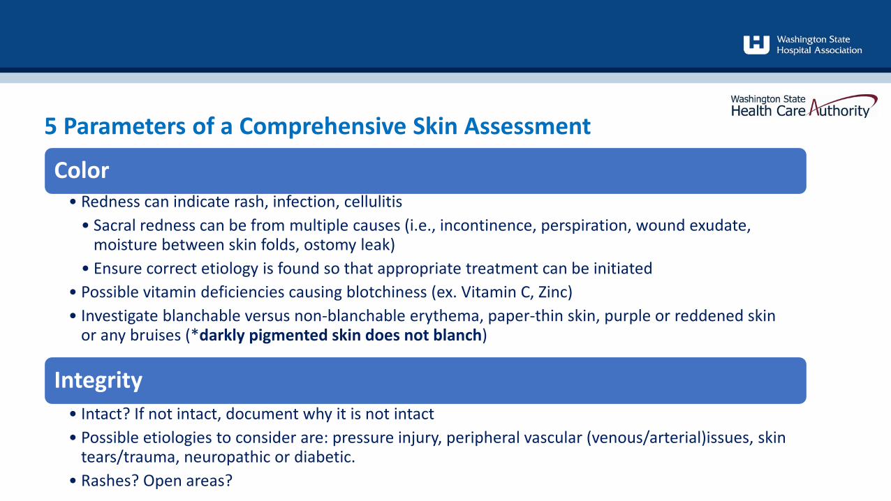
Color• Redness can indicate rash, infection, cellulitis
• Sacral redness can be from multiple causes (i.e., incontinence, perspiration, wound exudate, moisture between skin folds, ostomy leak)
• Ensure correct etiology is found so that appropriate treatment can be initiated• Possible vitamin deficiencies causing blotchiness (ex. Vitamin C, Zinc)• Investigate blanchable versus non-blanchable erythema, paper-thin skin, purple or reddened skin
or any bruises (*darkly pigmented skin does not blanch)
Integrity• Intact? If not intact, document why it is not intact• Possible etiologies to consider are: pressure injury, peripheral vascular (venous/arterial)issues, skin
tears/trauma, neuropathic or diabetic. • Rashes? Open areas?
5 Parameters of a Comprehensive Skin Assessment
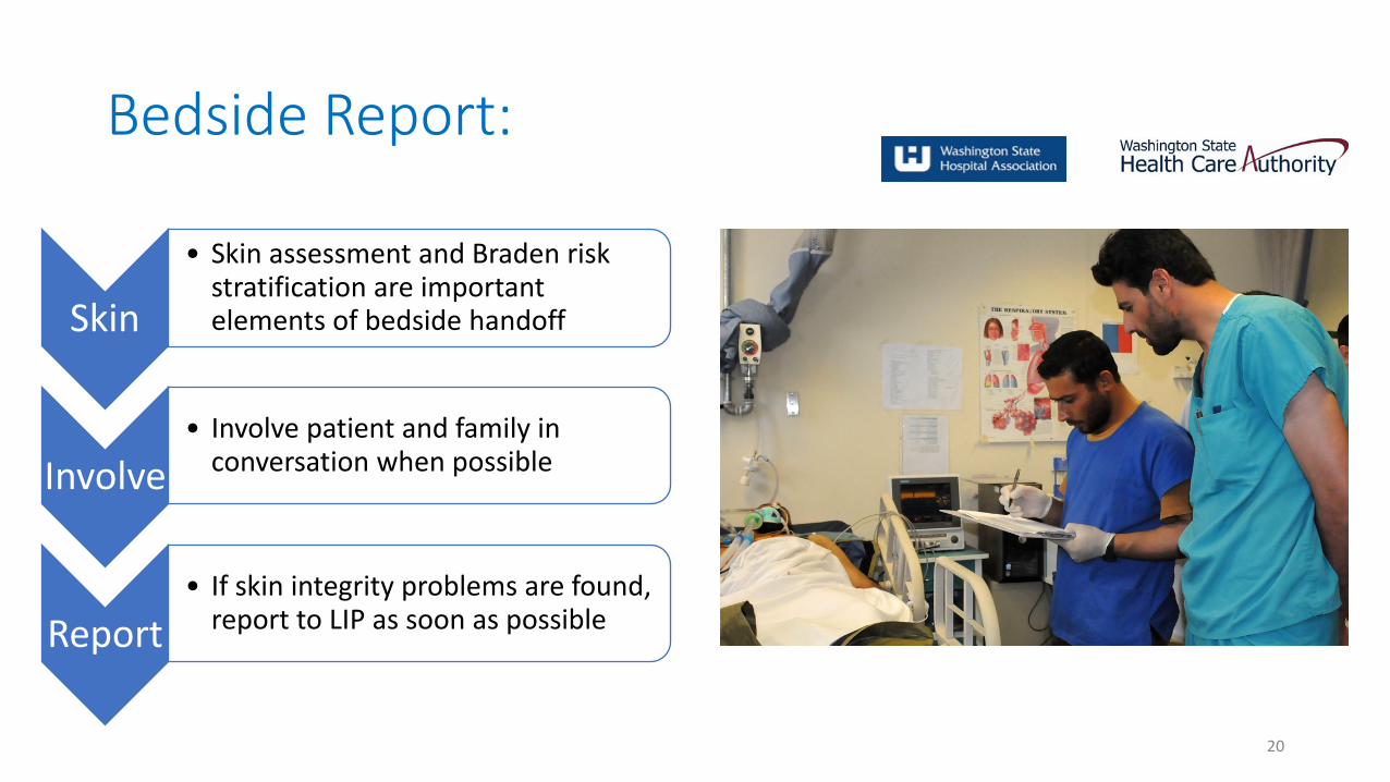
Bedside Report:
20
Skin
• Skin assessment and Braden risk stratification are important elements of bedside handoff
Involve• Involve patient and family in
conversation when possible
Report• If skin integrity problems are found,
report to LIP as soon as possible
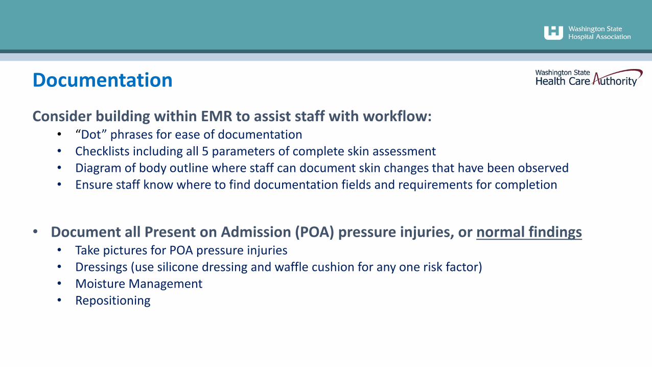
Consider building within EMR to assist staff with workflow:• “Dot” phrases for ease of documentation• Checklists including all 5 parameters of complete skin assessment• Diagram of body outline where staff can document skin changes that have been observed• Ensure staff know where to find documentation fields and requirements for completion
• Document all Present on Admission (POA) pressure injuries, or normal findings• Take pictures for POA pressure injuries• Dressings (use silicone dressing and waffle cushion for any one risk factor)• Moisture Management• Repositioning
Documentation
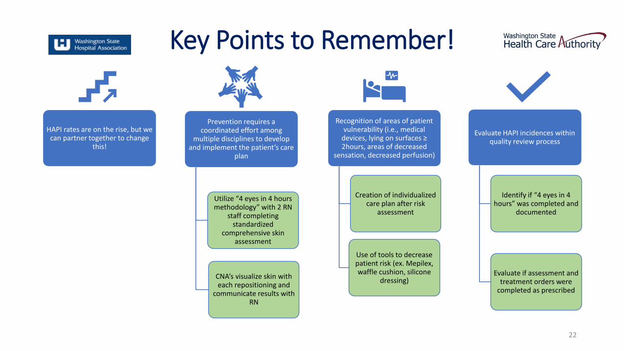
Key Points to Remember!
HAPI rates are on the rise, but we can partner together to change
this!
Prevention requires a coordinated effort among
multiple disciplines to develop and implement the patient’s care
plan
Utilize “4 eyes in 4 hours methodology” with 2 RN
staff completing standardized
comprehensive skin assessment
CNA’s visualize skin with each repositioning and
communicate results with RN
Recognition of areas of patient vulnerability (i.e., medical
devices, lying on surfaces ≥ 2hours, areas of decreased
sensation, decreased perfusion)
Creation of individualized care plan after risk
assessment
Use of tools to decrease patient risk (ex. Mepilex, waffle cushion, silicone
dressing)
Evaluate HAPI incidences within quality review process
Identify if “4 eyes in 4 hours” was completed and
documented
Evaluate if assessment and treatment orders were
completed as prescribed
22
References:
1. Health Research & Educational Trust. Hospital Acquired Pressure Ulcers/Injuries (HAPU/I): April 2017. Chicago, IL: Health Research & Educational Trust.
2. https://www.ahrq.gov/patient-safety/settings/hospital/resource/pressureinjury/workshop/guide3.html
3. https://npiap.com/4. https://npiap.com/page/FreeMaterials5. https://npiap.com/page/2019Guideline6. Kirkland-Kyhn, Teleten et al. OWM, 2017;63(2):42-47, 2017.7. Kirkland-Kyhn, Teleten et al. WMP, 2019;65(2):14-19.8. Pan Pacific Pressure Injury Alliance. (2019). Prevention and treatment of
pressure ulcers/injuries: clinical practice guideline: the quick reference guideline 2019.
9. Pressure ulcer prevention: pressure, shear, friction and microclimate in context. A consensus
10. Preventing Pressure Ulcers in Hospitals: A Toolkit for Improving Quality of Care. (ahrq.gov)
11. The Joint Commission. Managing medical device-related pressure injuries. Quick Safety. Issue 43, July 2018. Oakbrook Terrace, IL: The Joint Commission
12. World Union of Wound Healing Societies (WUWHS). Consensus Document: Role of dressings in pressure ulcer prevention. London, UK: Wounds Int; 2016 document. London: Wounds International (2010).
23