history taking for pharmacist -...
TRANSCRIPT

History Taking for Pharmacist
Chris Fujitnirun M.D., M.Sc.Division of Infectious Diseases, Department of Medicine, Bhumipol Adulyadej Hospital,
Directorate of Medical Services, Royal Thai Air Force
November, 25 2018

Outline
• URI symptoms
• Headache
• Abdominal pain
• Diarrhea
• Fever

History Taking – Overview
• History
• Physical examination
• Lab and radiological investigation
• Diagnosis
• Management

• Introduction (WIIPP)– Wash your hands
– Introduce yourself
– Identity: confirm you’re speaking to the correct patient
–
– Permission: confirm the reason for seeing the patient
– Positioning: patient sitting in chair approximately a metre away from you
History Taking – Overview
http://www.oxfordmedicaleducation.com

• Presenting Complaint“expressed in the patient’s own words”
“Try to elicit the patient’s ideas, concerns and expectations (ICE)”
• History of Presenting Complaint• Site
• Onset
• Character
• Radiation
• Alleviating factors
• Timing
• Exacerbating factors
• Severity (1-10)
History Taking – Overview
http://www.oxfordmedicaleducation.com
Anatomical localization
Etiology

• Past Medical History
• All previous medical problems (they may forget some)
• Top ensure none are missed ask about these important conditions specifically (mnemonic: “MJTHREADS Ca”)
History Taking – Overview
Myocardiac infarctionJaundiceTuberculosisHypertension and HIVRheumatic fever
EpilepsyAsthmaDiabetes and DyslipidemiaStrokeCancer (and treatment if so)
http://www.oxfordmedicaleducation.com

• Risk factors
• Clarification of past medical history– COPD; diagnosis, severity, treatment
– Myocardial infarction; previous heart attacks, any previous angiograms , previous stenting
– Diabetes: treatment, insulin and usual control of diabetes
– HIV: medication, CD4
History Taking – Overview
http://www.oxfordmedicaleducation.com

• Drug History– All medications that they take for each medication ask them to specify:
– Dose, frequency, route and compliance (i.e whether they regularly take these medication).
– Recreational drugs
– Intravenous drug use (current or previous)
– Over the counter (OTC) medications
History Taking – Overview
http://www.oxfordmedicaleducation.com

• Allergies– Does the patient have any allergies?
– If allergic to medications, clarify the type of medication and the exact reaction to that medication.
– Specifically ask about whether there’s been a history of anaphylaxise.g. “throat swelling, trouble breathing or puffy face”
History Taking – Overview
Allergy vs. Side Effects
http://www.oxfordmedicaleducation.com

• Social History– Alcohol intake
– Tobacco use
– Employment history
– Home situation
– Travel history
History Taking – Overview
http://www.oxfordmedicaleducation.com

Upper Respiratory Tract Symptoms
• Common cold
• Pharyngitis
• Sinusitis
• Bronchitis
FeverCough
Runny noseSore throat

Common Cold
• Clinical manifestations– Onset: 1 to 3 days after viral infection
– First symptom noted is frequently a sore or “scratchy” throat
– Nasal obstruction and rhinorrhea
– Sore throat resolves quickly and nasal symptoms predominate
– Cough is associated with approximately 30% of colds and usually begins after the onset of nasal symptoms
– Systemic symptoms are uncommon
– The usual cold persists about 1 week, although 25% last 2 weeks
– A change in the color or consistency of the secretions is common during the course of the illness and is not indicative of sinusitis or bacterial superinfection
Ronald B. Turner. Mandell, Douglas, and Bennett’s Principles and Practice of Infectious Diseases, 8th Edition, 2015

Common Cold: Etiology
Ronald B. Turner. Mandell, Douglas, and Bennett’s Principles and Practice of Infectious Diseases, 8th Edition, 2015

Differential Diagnosis
• Allergic rhinitis; nasal or conjunctival itching
• Foreign body
• Streptococcosis
• Catarrhal phase of pertussis
• Complication of common cold; sinusitis– Bacterial sinusitis is more likely to be present if symptoms persist for
more than 10 days, if severe illness is present, or if symptoms worsen after improvement >> occur 8%
Ronald B. Turner. Mandell, Douglas, and Bennett’s Principles and Practice of Infectious Diseases, 8th Edition, 2015
Treatment
• Symptomatic Therapies
• Given the absence of demonstrated benefit and the potential for toxicity, symptomatic common cold therapies are not recommended for children younger than 4 years.
Ronald B. Turner. Mandell, Douglas, and Bennett’s Principles and Practice of Infectious Diseases, 8th Edition, 2015

Treatment
• Nasal congestion– Topical and oral adrenergic agents
– Prolonged use of the topical adrenergic agents should be avoided to prevent the development of rhinitis medicamentosa (use longer than 2 weeks)
• Topical– Imidazoline derivatives
– Beta phenylethylamine derivatives: ephedrine, phenylephrine
• Oral– Pseudoephedrine, phenylephrine, phenylpropanolamine
Side effects: central nervous system stimulation, hypertension, and palpitations
Ronald B. Turner. Mandell, Douglas, and Bennett’s Principles and Practice of Infectious Diseases, 8th Edition, 2015

• Rhinorrhea– Blockade of cholinergic stimulation of glandular secretion
• Ipratropium
• First-generation antihistamines
• Second-generation or “nonsedating” antihistamines have had no effect on common cold symptoms in a limited number of studies
Treatment
Side effects: sedation and drying of the eyes, mouth, and nose
Ronald B. Turner. Mandell, Douglas, and Bennett’s Principles and Practice of Infectious Diseases, 8th Edition, 2015
• Sneezing >> antihistamine
• Sore Throat >> acetaminophen, NSAIDS
• Cough (caused by nasal secretion or virus-induced reactive airway) >> anti histamines and bronchodilator
– Codeine or dextromethorphan hydrobromide >> no benefit
Treatment
Ronald B. Turner. Mandell, Douglas, and Bennett’s Principles and Practice of Infectious Diseases, 8th Edition, 2015

Treatment
Ronald B. Turner. Mandell, Douglas, and Bennett’s Principles and Practice of Infectious Diseases, 8th Edition, 2015

Pharyngitis
• Triad of sore throat, fever, and pharyngeal inflammation characterized by erythema and edema, although exudates, vesicles, or ulcerations may also be present
Anthony R. Flores. Mandell, Douglas, and Bennett’s Principles and Practice of Infectious Diseases, 8th Edition, 2015
• Viruses are the single most common cause of pharyngitis
• What virus?
• Some clinical clues
Pharyngitis: Etiology
Anthony R. Flores. Mandell, Douglas, and Bennett’s Principles and Practice of Infectious Diseases, 8th Edition, 2015

Anthony R. Flores. Mandell, Douglas, and Bennett’s Principles and Practice of Infectious Diseases, 8th Edition, 2015

• Bacteria
• Streptococcus pyogenes, group A Streptococcus (GAS) – GAS and acute rheumatic fever (ARF)
– GAS is responsible for approximately 10% to 15% of cases of pharyngitis in adults >> no association in non-group A streptococci
• Fusobacterium necrophorum, 10% of cases of pharyngitis– 23% of cases of peritonsillar abscess
• Arcanobacterium haemolyticum <1%
• Corynebacterium diphtheriae
• Mycoplasma and Chlamydia
• Syphilis, Gonococci
Pharyngitis: Etiology
Anthony R. Flores. Mandell, Douglas, and Bennett’s Principles and Practice of Infectious Diseases, 8th Edition, 2015

Anthony R. Flores. Mandell, Douglas, and Bennett’s Principles and Practice of Infectious Diseases, 8th Edition, 2015

Throat Examination


Clinical Manifestations
• Group A Streptococcus• Sudden in onset, fever, headache, and gastrointestinal symptoms (nausea,
vomiting, abdominal pain)
• Pharyngeal erythema, tonsillar enlargement, and a gray-white exudatecovering the posterior pharynx and tonsillar pillars
• Petechiae are sometimes observed on the soft palate, with erythema and edema of the uvula
• Anterior cervical lymphadenopathy
• Scarlatiniform rash
Anthony R. Flores. Mandell, Douglas, and Bennett’s Principles and Practice of Infectious Diseases, 8th Edition, 2015
Signs and symptoms most indicative of GAS pharyngitis are tonsillar or pharyngeal exudates, tender anterior cervical nodes,
fever or history of fever, and absence of cough

• Fusobacterium necrophorum, 10% of cases of pharyngitis– 23% of cases of peritonsillar abscess >> severe complication
• Arcanobacterium haemolyticum <1%– Rash is more common than GAS
• Corynebacterium diphtheriae– Membrane on the tonsil or pharyngeal
– White early in the course of the illness, becomes dark gray, and leather-like, with attempts to dislodge the membrane potentially causing bleeding
– Swelling of the neck
• Mycoplasma and Chlamydia– Lower respiratory tract infection
• Syphilis >> chancre
• Gonococci >> mild symptom
Clinical Manifestations
Anthony R. Flores. Mandell, Douglas, and Bennett’s Principles and Practice of Infectious Diseases, 8th Edition, 2015

Bull neck in diphtheria patient

• Epstein-Barr Virus >> Infectious Mononucleosis– Fever, pharyngitis, and adenopathy
– Other symptoms included cough, myalgia, arthralgia, and nausea
– Rash was uncommon and is typically described as a diffuse maculopapular eruption in patients given ampicillin or related compounds
– Painful anterior and posterior cervical lymphadenopathy
– Hepatosplenomegaly
– Mild-to-moderate enlargement of the tonsils as well as exudates and palatal petechiae
Clinical Manifestations
Anthony R. Flores. Mandell, Douglas, and Bennett’s Principles and Practice of Infectious Diseases, 8th Edition, 2015

• Human Immunodeficiency Virus >> acute HIV infection– Occur in 40% to 90% of primary infection
– 5 to 29 days after infection
– Fever, rash, pharyngitis, fatigue, weight loss, myalgia, arthralgia, headache, night sweats, cervical adenopathy, nausea, vomiting, or diarrhea
– Pharyngitis is recognized in 50% to 70% of patients, whereas cervical adenopathy is noted in 25% to 50%
– Low incidence of exudate
– Ulceration
Clinical Manifestations
Anthony R. Flores. Mandell, Douglas, and Bennett’s Principles and Practice of Infectious Diseases, 8th Edition, 2015

• Enterovirus– Herpangina
– Hand foot mouth syndrome
Clinical Manifestations
Anthony R. Flores. Mandell, Douglas, and Bennett’s Principles and Practice of Infectious Diseases, 8th Edition, 2015

• Adenovirus– Pharyngoconjunctival fever is a specific syndrome caused by
adenovirus infections, often occurring in outbreaks and associated with swimming or bathing
• Herpes Simplex Virus– Fever, pharyngeal erythema, exudates, and enlarged tender cervical
adenopathy
– Gingivostomatitis
Clinical Manifestations
Anthony R. Flores. Mandell, Douglas, and Bennett’s Principles and Practice of Infectious Diseases, 8th Edition, 2015

• Group A Streptococcus is the most improtant pathogens
• 10 to 15% in adults
Anthony R. Flores. Mandell, Douglas, and Bennett’s Principles and Practice of Infectious Diseases, 8th Edition, 2015
Signs and symptoms most indicative of GAS pharyngitis are tonsillar or pharyngeal exudates, tender anterior cervical nodes,
fever or history of fever, and absence of cough
Pharyngitis

One Slide ID

Anthony R. Flores. Mandell, Douglas, and Bennett’s Principles and Practice of Infectious Diseases, 8th Edition, 2015

Sinusitis
• Most cases of acute bacterial sinusitis are secondary to viral upper respiratory infection (URI) or allergic inflammation
• The first pattern is that of persistent symptoms characterized by nasal discharge and/or cough that last more than 10 days without improvement
– Lack of improvement that is a sign of an acute bacterial process. Accompanying symptoms may include periorbital edema, malodorous breath, or low-grade fever.
Gregory P. DeMuri. Mandell, Douglas, and Bennett’s Principles and Practice of Infectious Diseases, 8th Edition, 2015

Sinusitis
• The second presentation is characterized by the onset of severe symptoms. Fever will accompany purulent nasal discharge that is present over a 3- to 4-day period
• The third presentation; “double sickening”
• Pain, tenderness, swelling and pressure around your eyes, cheeks, nose or forehead that worsens when bending over
Gregory P. DeMuri. Mandell, Douglas, and Bennett’s Principles and Practice of Infectious Diseases, 8th Edition, 2015www.mayoclinic.org

Clinical Diagnosis
• (1) onset with persistent symptoms or signs, lasting at least 10 days without evidence of clinical improvement
• (2) onset with severe symptoms or signs of high fever (≥39° C) and purulent nasal discharge lasting for 3 to 4 consecutive days
• (3) onset with worsening symptoms or signs characterized by the new development of fever, headache, or increased nasal discharge after a typical viral URI that lasted 5 to 6 days with initial improvement (“double sickening”)
Gregory P. DeMuri. Mandell, Douglas, and Bennett’s Principles and Practice of Infectious Diseases, 8th Edition, 2015

Sinusitis: Pathogens
Gregory P. DeMuri. Mandell, Douglas, and Bennett’s Principles and Practice of Infectious Diseases, 8th Edition, 2015

Invasive fungal sinusitis
• Serious underlying diseases, such as diabetes mellitus, malignancy and associated neutropenia, or those using high-dose corticosteroids
Gregory P. DeMuri. Mandell, Douglas, and Bennett’s Principles and Practice of Infectious Diseases, 8th Edition, 2015

Invasive Fungal Sinusitis

Sinusitis: Treatment
• High rate of spontaneous improvement within 2 weeks of presentation
• Overall, antimicrobial agents reduce the rate of clinical failure 25% to 30% within 7 to 14 days of initiating therapy
• Some guidelines give an option to observe signs and symptoms before starting treatment
Gregory P. DeMuri. Mandell, Douglas, and Bennett’s Principles and Practice of Infectious Diseases, 8th Edition, 2015

Sinusitis: Treatment
Gregory P. DeMuri. Mandell, Douglas, and Bennett’s Principles and Practice of Infectious Diseases, 8th Edition, 2015

Acute Bronchitis
• Dry or productive cough of less than 3 weeks’ duration
• Acute bronchitis begins with signs and symptoms typical of the common cold syndrome
• Followed shortly by the onset of cough, which becomes the dominant sign in acute bronchitis
Edward E. Walsh. Mandell, Douglas, and Bennett’s Principles and Practice of Infectious Diseases, 8th Edition, 2015

Acute Bronchitis: Microbiology

Acute Bronchitis: Treatment
• Narcotic cough suppressants, expectorants, antihistamines, decongestants, and β2-agonists
• Not clear benefits
• Infectious Diseases Society of America, do not recommend the routine use of antibiotics for uncomplicated acute bronchitis in otherwise normal persons.

URI
• Common cold: nasal discharge
• Pharyngitis: sore throat
• Sinusitis: complicated nasal discharge
• Bronchitis: cough
• Clinical spectrum
• Few cases need antibiotic

Headache
• Primary headache– Patients with a history of headache who do not have red flag signs
and symptoms are at low risk of serious headache
– Migraine, Tension-type, Cluster, Other (e.g., cold stimulus headache)
• Secondary headache– Headache attributed to any of the following: head or neck trauma,
cranial or cervical vascular disorder, nonvascular intracranial disorder, substance use or withdrawal, infection, disturbance of homeostasis, psychiatric disorder
– Headache or facial pain attributed to disorder of the cranium, neck, eyes, ears, nose, sinuses, teeth, mouth, or other facial or cranial structures

TENSION-TYPE HEADACHE
• Bilateral mild to moderate pressure without other associated symptoms
www.aafp.org

Migraine Headache
• Nausea, photophobia (sensitivity to light), and phonophobia(sensitivity to sound)
• Physical activity often exacerbates migraine headache
• Pulsatile quality, duration of four to 72 hours, unilateral location, nausea or vomiting, and disabling intensity
• Aura may be present in some cases of migraine
www.aafp.org

Migraine without Aura
www.aafp.org

Migraine with Typical Aura
www.aafp.org
Case
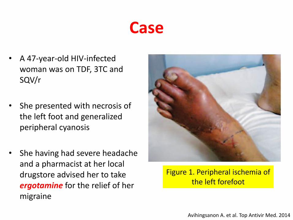
• A 47-year-old HIV-infected woman was on TDF, 3TC and SQV/r
• She presented with necrosis of the left foot and generalized peripheral cyanosis
• She having had severe headache and a pharmacist at her local drugstore advised her to take ergotamine for the relief of her migraine
Figure 1. Peripheral ischemia of the left forefoot
Avihingsanon A. et al. Top Antivir Med. 2014

Ergotism and Antiretrovirals
• Ergotamine, typically used to treat migraine, has less than 5% bioavailability due to extensive first-pass metabolism by cytochrome P450 3A4 (CYP3A4)
• Concurrent intake of ergotamine and strong CYP3A4inhibitors, such as the HIV protease inhibitors (PIs), can lead to clinical ergotism
• Peripheral vasoconstriction
• Pain, cyanosis, gangrene
Avihingsanon A. et al. Top Antivir Med. 2014
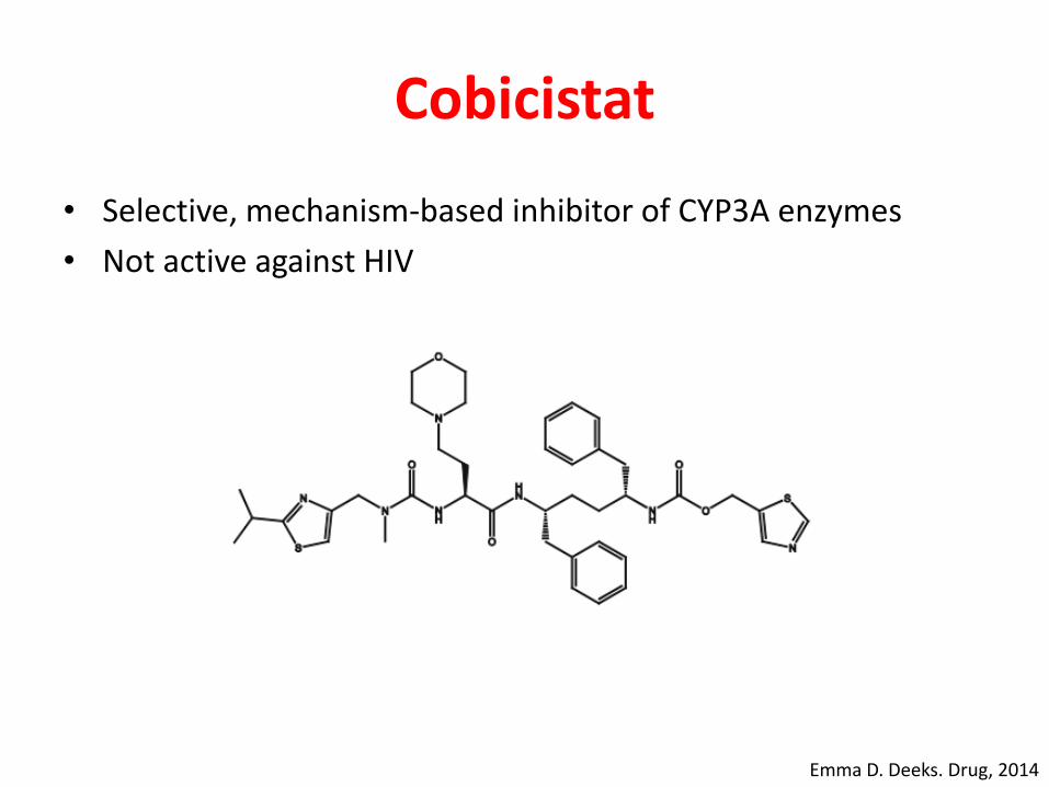
Cobicistat
• Selective, mechanism-based inhibitor of CYP3A enzymes
• Not active against HIV
Emma D. Deeks. Drug, 2014

• Use of cobicistat, a CYP3A inhibitor prescribed as an alternative boosting agent to ritonavir, is contraindicated in patients taking ergot alkaloids
Ergotism and Antiretrovirals
Avihingsanon A. et al. Top Antivir Med. 2014

CLUSTER HEADACHES
• Brief (15 to 180 minutes) episodes of severe head pain with associated autonomic symptoms
• Most commonly describe the pain as sharp, but some report that it can also be pulsating and pressure-like
• Can occur on both sides of the head, most patients report unilateral pain
• Retro-orbital area, followed by the temporal region, upper teeth, jaw, cheek, lower teeth, and neck
• Ipsilateral autonomic symptoms such as eyelid edema, nasal congestion, lacrimation, or forehead sweating
www.aafp.org

CLUSTER HEADACHES
www.aafp.org

Dangerous Headaches
• Associated red flag symptoms
www.aafp.org

Headache
• Primary or Secondary
• Beware of Red Flag Signs
• Consult doctor
www.aafp.org

Abdominal pain
• Visceral pain: internal organ, dull aching, difficult to localized• Epigastrium >> foregut: stomach, duodenum, liver, pancreas,
biliary
• Periumbilicus >> midgut: jejunum, appendix, cecum, ascending colon
• Hypogastrium >> hindgut: transverse colon to rectum
• Somatic pain: abdominal wall or peritoneum, sudden, severe, localized


Abdominal pain
• Duration, onset and severity– Acute and severe >> acute abdomen:
– Acute appendicitis, cholecystitis, gut obstruction, pancreatitis, ureteric stone, peptic perforation, diverticulitis, complication of pregnancy, Gynae condition
• Consult doctor
www.aafp.org

Dyspepsia
• Epigastric discomfort
• Lasting at least 1 month
• Can be associated with any other upper gastro intestinal symptom such as epigastric fullness, nausea, vomiting, or heartburn, provided epigastric pain is the patient’s primary concern.
The American Journal of GASTROENTEROLOGY, 2017

Dyspepsia: DDx
• Functional dyspepsia• Presence of at least one of
the following:
• Bothersome postprandial
fullness
• Early satiation
• Epigastric pain
• Epigastric burning and
• No evidence of structural disease (including at upper endoscopy) that is likely to explain the symptoms
www.aafp.org

Dyspepsia: DDx
The American Journal of GASTROENTEROLOGY, 2017

Dyspepsia: DDx
www.aafp.org

Diarrhea
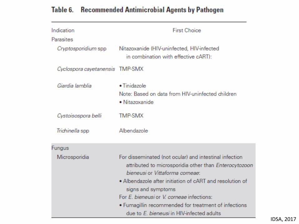
IDSA, 2017

Acute Mucus Bloody Stool
• CMV• Shigella• Salmonella• Vibrio parahaemolyticus• Plesiomonas• Yersinia enterocolitica• EHEC• EIEC• C. difficile• Campylobacter• Entamoeba histolytica• Balantidium coli
Need W/U

Clinical Clues

Hx of Exposure
IDSA, 2017

Hx of Exposure
IDSA, 2017

Patient Evaluation
• People of all ages with acute diarrhea should be evaluated for dehydration, which increases the risk of life-threatening illness and death, especially among the young and older adults
• Hx of voluminous diarrhea
• Tachycardia
• Hypotension
• Sunken eye ball
• Poor skin turgor
• Adequate Volume Replacement
IDSA, 2017

Infectious Diarrhea Tx
• In immunocompetent children and adults, empiric antimicrobial therapy for bloody diarrhea while waiting for results of investigations is not recommended (strong, low), except for the following– a. Infants <3 months of age with suspicion of a bacterial etiology.
– b. Ill immunocompetent people with fever documented in a medical setting, abdominal pain, bloody diarrhea, and bacillary dysentery (frequent scant bloody stools, fever, abdominal cramps, tenesmus) presumptively due to Shigella.
– c. People who have recently travelled internationally with body temperatures ≥38.5°C and/or signs of sepsis (weak, low)
IDSA, 2017

• Fluoroquinolone such as ciprofloxacin, or azithromycin, depending on the local susceptibility patterns and travel history
• Children: third-generation cephalosporin for infants <3 months of age and others with neurologic involvement, or azithromycin, depending on local susceptibility patterns and travel history
• Antimicrobial therapy for people with infections attributed to STEC O157 and other STEC that produce Shiga toxin 2 (or if the toxin genotype is unknown) should be avoided
Infectious Diarrhea Tx
IDSA, 2017

IDSA, 2017
IDSA, 2017

Watery Diarrhea
• In most people with acute watery diarrhea and without recent international travel, empiric antimicrobial therapy is not recommended– Except; infant, immunocompromised, ill-appearing
IDSA, 2017

Supportive Tx
• Antimotility, antinausea, or antiemetic agents can be considered once the patient is adequately hydrated
• Antimotility drugs (eg, loperamide) should not be given to children
IDSA, 2017

Acute Diarrhea
• Mucus Bloody or Watery
• Clinical Clue
• Patient evaluation
• Few cases need ABx

• “A state of elevated core temperature, which is often, but not necessarily, part of the defensive responses of multicellular organisms (host) to the invasion of live (microorganisms) or inanimate matter recognized as pathogenic or alien by the host.”
• Find the causes of fever
Fever
Sajadi et al. Mandell, Douglas, and Bennett's Principles and Practice of Infectious Diseases, 8th edition, 2015

Sajadi et al. Mandell, Douglas, and Bennett's Principles and Practice of Infectious Diseases, 8th edition, 2015
increase in the hypothalamic set point

Sajadi et al. Mandell, Douglas, and Bennett's Principles and Practice of Infectious Diseases, 8th edition, 2015
Pyrogens
• Pyrogen (Greek pyro, “fire”) is used to describe any substance that causes fever.
• Exogenous pyrogens are derived from outside the patient; most are microbial products, microbial toxins, or whole microorganisms (including viruses)
• Examples: • lipopolysaccharide (endotoxin) produced by
all gram-negative bacteria• Pyrogenic products of gram-positive
organisms include the enterotoxins of Staphylococcus aureus and the groups A and B streptococcal toxins, also called superantigens
• One staphylococcal toxin of clinical importance is that associated with isolates of S. aureus from patients with toxic shock syndrome

Very High Fever
• A fever of >41.5°C (>106.7°F) is called hyperpyrexia– This extraordinarily high fever can develop in patients with severe
infections but most commonly occurs in patients with central nervous system (CNS) hemorrhages
• In rare cases, the hypothalamic set point is elevated as a result of local trauma, hemorrhage, tumor, or intrinsic hypothalamic malfunction >> hypothalamic fever
Dinarello at al. Harrison's Principles of Internal Medicine, 19th edition, 2015

• An uncontrolled increase in body temperature that exceeds the body’s ability to lose heat
• The setting of the hypothalamic thermoregulatory center is unchanged
Hyperthermia
Dinarello at al. Harrison's Principles of Internal Medicine, 19th edition, 2015

OneSlideID

Etiology of Fever
• Infection– Bacteria;
• Pyogenic bacteria; Staphylococcus aureus, Streptococcus pneumoniae, E. coli • Higher order bacteria; mycobacteria, Nocardia, Rhodococcus• Others: rickettsioses, Chlamydia, Mycoplasma, Leptospirosis
– Fungus;• Mold; Aspergillosis, Mucormycosis• Yeast; Cryptococcosis, Candidiasis• Dimorphic fungi: Histoplasmosis, Talaromycosis
– Virus– Parasite;
• Helminths; Cestodes, Nematodes, Trematodes• Protozoa; Coccidia, Ciliate, Flagellate, Amoeba
• Inflammation; autoimmune disease, tissue inflammation• Malignacies; hematologic, solid organ• Miscellaneous; drugs, factitious

Clinical Approach
• Fever with systemic symptoms or organ-specific symptoms
• For organ-specific >> anatomical localization
• Use all clues to predict the most likely cause of fever– History; clinical course, clinical manifestation (some clues for some
pathogens), risk factor (patient information, habitat, occupation, recreational activity, sexual behavior, traveling, pets etc.), incubation peroid, previous medical condition and drugs
– Physical examination– Laboratory and imaging– Epidemiology; common pathogens for the disease community acquired or
hospital acquired infection
• Choose the proper management– Antibiotics, surgery, immunosuppressive agents etc.

• A 34-year-old male presented with fever for 4 days
• Please take a history
Clinical Approach

• 4 วนัก่อนมา รพ. ผู้ ป่วยมีไข้สงู หนาวสัน่ วนัละครัง้ ร่วมกบัอ่อนเพลีย และปวดหวัมาก ปวดหวัขมบัสองข้างและร้าวไปรอบศีรษะ อาการปวดหวัเบาลงเมื่อไมมี่ไข้ สามารถไปท างานได้แตอ่่อนเพลีย มีปวดตามแขนขา เป็นมากเวลาไข้ ปัสสาวะ อจุจาระเหลว 1 ครัง้
• Physical examination normal
• Problem: acute fever with fatigue and headache for 4 days >> acute undifferentiated fever
Clinical Approach

Differential Diagnosis
• Bacteremia; Staphylococcus aureus, E. coli, Melioidosis etc., Salmonella
• Rickettsiosis
• Leptospirosis
• Influenza
• Dengue (plus other virus)
• Malaria
• ผู้ ป่วยเป็นต ารวจตระเวณชายแดน อยูช่ายแดนไทยกมัพชูา จงัหวดัสริุนทร์ นอนในป่า มานาน 2 เดือน เพิ่งออกจากป่ามา 4 วนั
Rick M. Fairhurst. Mandell Douglas and Benett’s Principles and Practice of Infectious Diseases, 2015

• DDx Malaria
Rickettsial infection
Meliodosis
• Blood smear: ring form trophozoites of Plasmodium flaciparum
Clinical Approach
Point of learning: Occupation and Travelling Hx are important

• A 38-year-old female presented with fever for 10 days
• Please take a history
Clinical Approach

• 10 วนัก่อนมา รพ. ผู้ ป่วยมีไข้สงู ไมห่นาวสัน่ วนัละครัง้ ร่วมกบัออ่นเพลีย และปวดหวัมาก ปวดหวัขมบัสองข้างและร้าวไปรอบศีรษะ อาการปวดหวัเบาลงเมื่อไมมี่ไข้ สามารถไปท างานได้แตอ่่อนเพลีย มีปวดตามแขนขา เป็นมากเวลาไข้ น า้หนกัลด 3 กก.
• Problem: acute fever with fatigue and headache for 10 days >> acute undifferentiated fever
• Lab: SGOT/SGPT 260/300
Clinical Approach

Clinical Approach
• Physical examination showed small painless erythematous plaque with central necrosis >> eschar
• ผู้ ป่วยขอบเดินทาง Trekking ลา่สดุเม่ือ 2 สปัดาห์ก่อน

OneSlideID

Pathogens/diseases Incubation period (range)Incubation period
(usual)
Gonorrhoea 1 - 10 days (3) 2 - 5 days (3)
Non gonococcal urethritis 2 – 35 days (17) 7 - 14 days (16)
Herpes simplex 2 – 12 days (1) 4 days (1)
Pathogens/diseasesIncubation period
(range)Incubation period
(usual)
Syphilis 10 – 90 days (3) 3 weeks (3)
Chancroid 1 – 14 days (13) 4 – 7 days (12)
Acute HIV 2–6 weeks (15) 2 – 4 weeks (14)
Pathogens/diseasesIncubation period
(range)Incubation period
(usual)
EHEC 1 - 10 days (1) 3 - 4 days (1)
Shigella 1 – 7 days (10) 0.5 – 4 days (1)
Vibrio cholerae 2 h – 5 days (1) 2 – 3 days (1)
Campylobacter 2 – 5 days (1) 1 – 10 days (11)
Norovirus 12 -48 h (1) 33 h (1)
Rotavirus 1 – 3 days (2) less than 48 h (1)
Pathogens/diseases Incubation period (range)Incubation period
(usual)
HAV 15 - 48 days (3) 30 (3)
HBV 30 - 180 days (3) 60 - 90 days (3)
HCV 15 - 160 days (3) 50 days (3)
HEV 14 - 60 days (3) 40 days (3)
Pathogens/diseasesIncubation period
in rangeUsual Incubation
period
Influenza 1 - 4 days (1) 2 days (1)
RSV 3 - 7 days (9) 5 days (9)
MERSCoV 2 – 14 days (1) 5 days (1)
SARS 2 - 10 days (9) 5 days (9)
Rhinovirus 2 – 4 days (9) 2 days (9)
Adenovirus 4 -8 days (9) 6 days (9)
Parainfluenza virus 2 – 6 days (9) 4 days (9)
Metapneumovirus 5–6 days (9)
Diphtheria 1 – 10 days (1) 2 - 5 days (1)
Pertussis 4 – 21 days (1) 7 – 10 days (1)
Pathogens/diseases Incubation period (range)Incubation period
(usual)
Dengue 3 - 10 days (1) 5 - 7 days (1)
Zika virus 3 - 14 days (1) 6 days (5)
Ebola 2 - 21 days (1) 8 - 12 days (2)
Yellow fever 3 - 6 days (4) 3 - 6 days (2)
Chikungunya 1 - 12 days (1) 3 - 7 days (1)
Scrub typhus 5 - 20 days (2) 10 - 12 days (2)
Leptospirosis 2 - 30 days (2) 5 - 14 days (1)
Pathogens/diseaseIncubation period
In rangeUsual Incubation
period
Scarlet fever (Gr. A Strep) 1 – 7 days (7) 2 - 5 days (1)
Measles 7 - 21 days (1) 14 days (1)
Rubella 12 - 23 days (1) 14 days (1)
Chicken pox 10 - 21 days (1) 14 - 16 days (1)
Roseola infantum(HHV – 6 and 7)
5 – 15 days (8) 12 days (8)
Erythema infectiosum(Parvovirus B19)
Upto 20 days (6) 4 – 14 days (6)
Mumps 12 - 25 days (1) 16 - 18 days (1)
OneSlideID

• Weil-Felix test positive for OX-K 1:160 with 4 fold rising
• Dx: Scrub typhus
• Her fever dramatically responded to doxycycline
Clinical Approach
Point of learning: Some Signs are Pathognomonic

Clinical Approach
• A 39-year-old female, housewife presented with fever and headache for 5 days
• Please take a history.

• 5 วนัก่อนมารพ. ผู้ ป่วยมีอาการไข้ ปวดศีรษะ น า้มกูไหล อาการไข้ไม่มีหนาวสัน่ ปวดศีรษะเป็นพร้อมๆ ไข้ดีขึน้เมื่อไข้ลง มีอาการปวดเม่ือยตามตวั มาห้องฉกุเฉิน ได้รับการตรวจ nasal swab for influenza virus ได้ผล positive for type A Influenza ได้รับการรักษาด้วย oseltamivirแล้วกลบับ้าน
• 3 วนัก่อนมารพ. อาการปวดศีรษะไมด่ีขึน้ เป็นตลอดทัง้วนั มคีลื่นไส้อาเจียน ไอจามเบง่ปวดมากขึน้ กลอกตาเจ็บ สู้แสงสวา่งไมไ่ด้ ปวดเม่ือย
• 1 วนัก่อนมารพ. เร่ิมนอนมากกวา่ปกต ิตอบค าถามช้าญาตจิงึพามารพ.
Clinical Approach

• Physical examination showed high grade of fever
• Slowly responded to one step command
• Nuchal rigidity
• Problem: fever with meningism for 5 days
• Anatomical localization: meningitis with complication
• DDx: Influenza meningitis, acute bacterial meningitis
Clinical Approach
Point of learning: Don’t Ignore the Red Flags

• WBC more than 20000 cell/µL
• Lumbar puncture
• OP 30, CP 16
• WBC 600, mostly PMN
• Glucose very low
• Protein 250 mg/dL
• Bacterial meningitis
Clinical Approach

Common Pathogens for Bacterial Meningitis
• Streptococcus pneumoniae– Typical bacterial meningitis– Invasive infection appear as influenza complication
• Group B Streptococcus; – may be associated with other foci of infection such as skin and soft tissue infection, IE,
osteomyelitis
• Neisseria meningitidis; – very short clinical course, very severe, purpura fulminans may be found
• Haemophilus influenzae– Uncommon in Thai adult– Typical bacterial meningitis– Invasive infection appear as influenza complication
• Listeria monocytogenes– Meningitis with cerebritis or brain abscess– Rhombencepahilis– Extreme age, neonate, pregnant, immunocompromised pt.
CDC, 2017

Clinical Approach
• Empirically treated with ceftriaxone 2 g IV q 12 h
• CSF Gram stain showed Gram positive diplococci
• Her hemoculture grew Streptococcus pneumoniae for 2 specimens

A 47-year-old male
Progressive dyspnea on exertion for 2 weeks then sudden dyspnea for 2 hours
Low grade fever
Scant sputum
SpO2 80%
Pneumocystis pneumonia (PCP)

PCP: Pathogen
• Pneumocystis is closely related unicellular fungi but lacks ergosterol
• Pneumocystis isolated from humans was described as P. jirovecii
– P. carinii and P. wakefieldiae are for rats
– P. murina for mice
– P. oryctolagi for rabbits
• Lack of a reliable Pneumocystis in vitro cultivation
• Lifestyles of obligate parasites
• P. jirovecii infection is acquired early in life, so by age 2 or 3 years of age most (approximately 80%) children have been exposed
Walzer et al, Mandell, Douglas, and Bennett’s Principles and Practice of Infectious Diseases, 2015
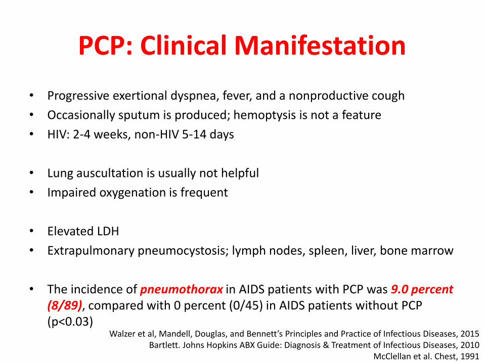
• Progressive exertional dyspnea, fever, and a nonproductive cough
• Occasionally sputum is produced; hemoptysis is not a feature
• HIV: 2-4 weeks, non-HIV 5-14 days
• Lung auscultation is usually not helpful
• Impaired oxygenation is frequent
• Elevated LDH
• Extrapulmonary pneumocystosis; lymph nodes, spleen, liver, bone marrow
• The incidence of pneumothorax in AIDS patients with PCP was 9.0 percent (8/89), compared with 0 percent (0/45) in AIDS patients without PCP (p<0.03)
PCP: Clinical Manifestation
Walzer et al, Mandell, Douglas, and Bennett’s Principles and Practice of Infectious Diseases, 2015Bartlett. Johns Hopkins ABX Guide: Diagnosis & Treatment of Infectious Diseases, 2010
McClellan et al. Chest, 1991

Summary