history & physical exam and pfts pulmonary …roushmedicine.com/pulmonary.pdf · pulmonary...
TRANSCRIPT

Roushmedicine.com
HISTORY & PHYSICAL EXAM AND PFTsPulmonary diseases associated with smoking:include Asthma, COPD, lung cancer, but alsoDesquamative interstitial pneumonitis, Respiratory bronchiolitis–interstitial lung disease, Pulmonary Langerhans cell histiocytosis, Goodpasture's disease, and Pulmonary alveolar proteinosis
Acute pleuritic chest pain differential diagnosis includes asthma, pneumonia, pulmonaryembolus, pneumothorax, and cholecystitis.
A fruity odor on the breath is usually found in diabetic ketoacidosis, which is associated withKussmaul's respirations (deep, gasping respirations), or tachypnea with large tidal volumes.
Increased fremitus, bronchial breath sounds, egophony (E to A), whispered pectoriloquy(whisper 1,2,3) >> consolidated pneumonia.
Decreased tactile fremitus, dullness to percussion, decreased breath sounds, no voicetransmission point to pleural effusion or atelectasis.
Absent fremitus, hyper-resonance, and absent breath sounds >> pneumothorax.
Tracheal deviation to left >> right pleural effusion OR left loss of volume (atelectasis due toobstructed bronchus).
Cyanosis that involves the skin and extremities: DVT, Raynaud’s, cold exposure, andperipheral vascular disease.Cyanosis that involves the skin and mucous membranes: methemoglobinemia and COpoisoning, right to left shunting, pulmonary AV malformations, and severe lung disease.
Digital clubbing can be caused by >Many intrathoracic tumors: lung cancer and mesothelioma.>Chronic intrathoracic non-malignant processes: bronchiectasis, lung abscess, pulmonary
fibrosis>Cardiac: SBE and congenital heart defects>GI: cirrhosis and inflammatory bowel disease.
Severe kyphoscoliosis is associated with restrictive lung disease, alveolar hypoventilation,pulmonary hypertension, and v/q abnormalities with hypoxemia.
Cheyne-Stokes respirations, in which tidal volume and frequency wax and wane alternately,indicates decompensated heart failure. The mechanism is not known, but is possibly due to

Roushmedicine.com
increased circulatory time, alterations in acid-base status, and affects on the brainstemchemoreceptors that govern the respiratory pattern. PFTs:Reductions in DLCO results from obliteration of capillary bed volume: pulmonary fibrosis (e.g.,due to scleroderma or bleomycin), pulmonary hypertension, chronic PTE,
Normal DLCO occurs in asthma and chronic bronchitis.
Increased DLCO is seen a) when pulmonary capillary blood volume is increased: Obesity, early CHF, L.R shunts,Exercise, altitude, supine position.b) With increased RBC’s, e.g., With increased hematocrit or with alveolar hemorrhage.
In emphysema, one does not use spirometry to measure total lung capacity because it cannotaccount for residual volume. The most frequently used and accurate measures of lung volumesare steady-state helium dilution lung volumes and body plethysmography. Helium lung volumesare easier to perform for patients and staff and give reliable results in most circumstances.In cardiopulmonary exercise testing:
Cardiopulmonary testing:In patients with cardiovascular dysfunction, findings include a heart rate more than 85% of themaximum heart rate, a low anaerobic threshold, reduced maximum oxygen consumption,arrhythmia or ischemia on electrocardiography, and a drop in blood pressure without desaturationor achievement of the maximal predicted ventilation.
Conversely, pulmonary dysfunction is suggested by achieving or exceeding maximal ventilation,desaturation, a drop in FEV1 with exercise, and stability or an increase in the ratio of dead spaceto tidal volume without reaching 85% of the maximal heart rate or ischemic changes onelectrocardiography.
In COPD, administration of oxygen does not suppress the respiratory drive. Rather the increasein the PCO2 is due to a) V/Q mismatch and b) the Haldane effect (Oxygen drives the release ofCO2 bound to hemoglobin).

Roushmedicine.com
COUGH:ACUTE: URI, pertussis, allergic rhinitis, COPD exacerbation, asthma, acute sinusitis.
CHRONIC:Post nasal dripVasomotor rhinitisChronic sinusitisCerumen impaction and ear canal hairs (irritating the tympanic membrane)Chronic bronchitisPost-infectious
Gastroesophageal reflux can be associated with a bitter taste or sore throat on awakening. 24-hour pH monitor accurately identifies this problem and can be used to correlate episodes ofcough with reflux. However, A 24-hour pH monitor is invasive and often not necessary. Protonpump inhibitor therapy to block gastric acid production generally is required to attenuate coughassociated with reflux. An empiric trial of an H-2 blocker; if unsuccessful, a proton pumpinhibitor such as omeprazole may be used before 24 hour pH monitoring. The usual anti-refluxmeasures, such as avoiding fatty foods, alcohol, and food before bedtime, should be instituted aswell. Prescribers must be aware that sometimes a complete resolution of cough takes months.
Occult asthma is also a common cause of undiagnosed chronic cough. Cough-variantasthma can be associated with normal pulmonary function tests but increased sensitivity of theairways to inhalation of methacholine or histamine. Treatment of cough-variant asthma can bedifficult because the use of inhalers may precipitate coughing episodes. Therefore, oral agentssuch as oral long-acting $-agonists or leukotriene inhibitors may be more useful. In addition,short, tapering courses of corticosteroids may be very useful in decreasing cough resulting fromasthma.
Bronchiectasis is also a cause of chronic cough, asthma is not a cause of this problem.Chronic cough resulting from bronchiectasis responds to rotating antibiotics.
Medications: ACE inhibitors, especially the nonselective agents, cause cough inapproximately 10% of treated patients. The associated cough is generally nonproductive and maynot clear for several days after discontinuation of the drug. Most asthma medications includingalbuterol and monteleukast. Amiodorone.
Malignancy.Left ventricular dysfunction.

Roushmedicine.com
HEMOPTYSISCAUSES OF HEMOPTYSIS:Vascular: PE, vasculitides (Goodpastures, Wegener’s), arteriovenous malformation Cardiac: CHF, mitral stenosisNeoplastic: Bronchogenic carcinoma, metastatic disease Connective tissue: Lupus, rheumatoid arthritis Infectious: TB, bronchitis, pneumonia, abscess, aspergilloma (has opacities within cavitieschanging with position)Drugs: tobacco, Anticoagulants, cocaine, solvents Congenital: Hereditary hemorrhagic telangiectasia [ 1) personal epistaxis; 2) telangiectasias ofmouth, nose or fingers; 3) AV malformations of the lung; 4) Family history.Miscellaneous: Bronchiectasis, Trauma, foreign body, epistaxis, hematemesis, Antiphospholipidantibody syndrome.In massive hemoptysis of bronchiectasis, the most likely cause is bronchial artery erosion, whichis treated with embolization.
PNEUMONIACAP manifestations: tachypnea, purulent sputum, fever, crackles, and egophany. Rhinitis is anegative predictor.
RISK FACTORS FOR SPECIFIC BACTERIA in Community Acquired PneumoniaDrug Resistant Strep pneumoniae:age > 65use of beta lactam Ab’cs in past 3 monthsimmuno-suppressionmultiple medical co-morbiditiesregular exposure to a child in day care
Gram negative organisms:All of those for S. pneumoniae above PLUSResidence in a nursing homeregular alcohol consumption.
Anaerobic bacteria:Witnessed or suspected aspiration
Pseudomonas aeruginosa:Cystic fibrosis and Bronchiectasis and other structural lung diseasesmechanical ventilationdaily steroidsrecent broad spectrum antibiotic treatmentsevere malnutrition.
Staphyloccocus aureusPost Influenza

Roushmedicine.com
Cystic Fibrosis

Roushmedicine.com
SETTINGS FOR SPECIFIC PNEUMONIASBronchiectasis is associated with MoraxellaPseudomonasH FluStaphStrep pneumoniae(Rx with flouroquinoloines and aminoglycosides.)
Cystic FibrosisDitto
COPDDitto plusAtypical MycobacteriaInfluenza virus and parainfluenza virus
Atypical pneumonia, (e.g. from L. pneumophila, M. pneumoniae, or C. pneumoniae) is morecommonly associated with systemic symptoms such as myalgias, anorexia, and pharyngitis.
Younger patients tend to get mycoplasma; older patients Chlamydia.
Approximately 50 to 70% of patients with mycoplasma pneumonia will develop coldagglutinins as a result of the development of IgM antibodies to the I-antigen on red blood cells.Hemolytic anemia may occur with a significant fall in the hematocrit (increase retics, MCV,indirect bilirubin, decrease haptoglobin). The Findingof myringitis bullosa on the tympanicmembrane supports the diagnosis of mycoplasma pneumonia.
Post influenza: Although the incidence of pneumonia resulting from S. aureus is increased afterinfluenza infection, the most common bacterial pathogen is S. pneumoniae. P. aeruginosa andH.. influenzae also occur. In fact, H. influenzae received its name because it was initiallyisolated from a patient with clinical influenza.
Ways in which organisms gain access to lower respiratory tract and cause pneumonia: 1) microaspiration of colonized oropharyngeal contents. 2) gross aspiration (stroke or seizures) 3) aerosol inhalation (histo, cocci, TB), 4) blood born (bacterial endocarditis), 5) extension froma contiguously infected site.
Prevention of CAP generally involves immunization with pneumococcal (every 6 years) orinfluenza (yearly) vaccine.
In CAP, patients treated with fluoroquinolones within the past 3 months, should not be retreated

Roushmedicine.com
with these.
In CAP, patients treated within 8 hours of arrival to the ER have a lower mortality than those inwhom treatment is delayed.
CURB65 Stands for Confusion, uremia, respiratory rate greater than 30, blood pressure less than90/60, and age 65+. A score of 2 indicates need for hospitalization; a score of 3+ is an indicationfor the ICU.
CAP recommendations for antibiotics. IDSA/ATS Guidelines 2007
OPD
Healthy, no AB’cs in prior 3 months: Macrolide (or Doxy)
Comorbidities or use of AB’cs in prior 3 months:
Levofloxacin or moxifloxacin OR
Beta lactam + macrolide
Hi rate of infection with macrolide resistant Strep, consider use of Levofloxacin
INPATIENTS
Non-ICU
Levofloxacin or moxifloxacin OR
Beta lactam + macrolide
ICU: beta lactam (cefotaxime, cetriaxone, or amp-sulbactam + either azithromycin ORrespiratory fluoraquinolone.
Pseudomonas is a consideration (structural lung disease)
Piperacillin-tazobactam, cefepime, imipenem or meropenem +
Either cipro or levofloxacin (750mg)
OR
Beta lactam + aminoglycoside and azithromycin
OR
Beta lactam + aminoglycoside + cipro or levofloxacin
CAP-MRSA is a consideration: add vancomycin or linezolid.
Trade names:
3rd Gen

Roushmedicine.com
Ceftriaxone= Rocephin 1 to 2 gm
Cefotaxime: = Clarofan 1gm Q12
Cefixime = Suprax 400mg PO
4th Gen
Cefepime (Maxipime) 1 to 2gm Q12Hr
Piperacillin-Tazobactim= Zosyn (4th generation penicillin) 3.375 gm iv Q6Hr.
Ampicillin-sulbactam =Unasyn (3rd generation penicillin) 2gm iv Q6Hr.
Levofloxacin=Levoquin (3rd generation fluoroquinolone) 750 mg
Moxifloxacin=Avelox (4th generation fluoroquinolone) 400 mg
Linezolid=Zyvox 600mg Q12
Imipenem-cilastatin=Primaxin 500 Q6Hr
Meropenem=Merrem 1gm Q8Hr
Oseltamivir (Tamiflu) 75 PO BID
Eosinophilic pneumonia can be caused by nitrofurantoin, sulfa drugs, NSAIDS,penicillins,INH, thiazides, tricyclic antidepressants, hydralazine, and chlorpropramide.
BOOP (SEE BELOW)
ASTHMA and COPD:
Pathophysiology
> Histamine, a preformed mast cell mediator, has direct vasoactive and smooth musclespasmogenic activity.
>Platelet activating factor is a mast cell-derived mediator that induces bronchospasm. >Leukotriene B4 is a precursor to the sulfidopeptide leukotrienes C4, D4, and E4. All ofthese leukotrienes have some effect on airflow by modulating vascular permeability,smooth muscle constriction, and mucus secretion.
>Interleukin 5 supports the differentiation of cells into eosinophils, is a specific

Roushmedicine.com
chemotaxin for eosinophils, is important in immediate hypersensitivity and is a target of drugtherapy investigation.
>Prostacyclin, a prostaglandin metabolite of arachidonic acid, has bronchodilating effects(hence the potential for bronchospasm with ASA and NSAIDS).
For exercise-induced asthma use inhaled beta agonists before exercise. Diagnose it by PFTspre and post exercise.
Exercise induced asthma: Exercise-induced asthma is very common, occurring in 50 to 90% ofasthmatics. Asthmatics manifest bronchodilation at early stages of exercise. Airway water lossduring exercise may cause bronchospasm. and dry air worsens exercise-induced bronchospasm inaffected asthmatics. In contrast, humid air improves symptoms.
Occupational Asthma: Allergy to agricultural grasses, etc. Acute wheezing and obstruction. NOFEVER.
>Isocyanates, chemicals used in spray paints and other industrial products, are anexample of low-molecular-weight substances that cause asthma.
>Flour dust contains high-molecular-weight components, specifically proteins that induceantibody formation in some individuals and asthma in a subgroup of those individuals.
The initial symptom of byssinosis is chest tightness.
RADS: Reactive Airway disease syndrome) is caused by a ingle toxic irritant exposure (e.g.,chlorine gas). The subsequent asthma is confirmed by methacoline challenge test. A similarsyndrome can follow a severe pulmonary infection.
Allergic bronchopulmonary aspergillosis is characterized by asthma, central bronchiectasis,peripheral eosinophilia, an immediate type of reaction to skin test reactivity, increased IgE inserum, and aspergilla precipitins in serum. It is a hypersensitivity pneumonitis that involves anallergic reaction to antigens from Aspergillus spp., most commonly A. fumigatus.
Diagnostic criteria:
1) Asthma,
2) Antigenic skin testing is positive in immediate (type I, wheal-and-flare)reaction, and reaction after 4 to 6 h (type III, erythema and induration).
3) Serum precipitins to aspergilli.
Supportive criteria:
IgE > 1 mgm/ml
Peripheral eosinophilia > 500/ml

Roushmedicine.com
Lung infiltrates
Bronchiectasis
Aspergilli in the sputum.
Lymphocytic alveolitis on BAL (sensitive but not specific).
COPD MAINTENANCE: Emerging data suggest that ipratropium bromide treatment improvesprognosis in patients with COPD. Patients with this disorder commonly respond to ipratropiummore favorably than asthmatics.
Anticholinergics are as, or more effective, than beta 2 agonists in COPD !! (Differs fromasthma).
DIFFERENTIAL DIAGNOSIS OF ACUTE ASTHMA:
*Mechanical obstruction (a peanut)
*Vocal cord dysfunction
*Bronchiolitis
*Pneumonia
*CHF
*PE
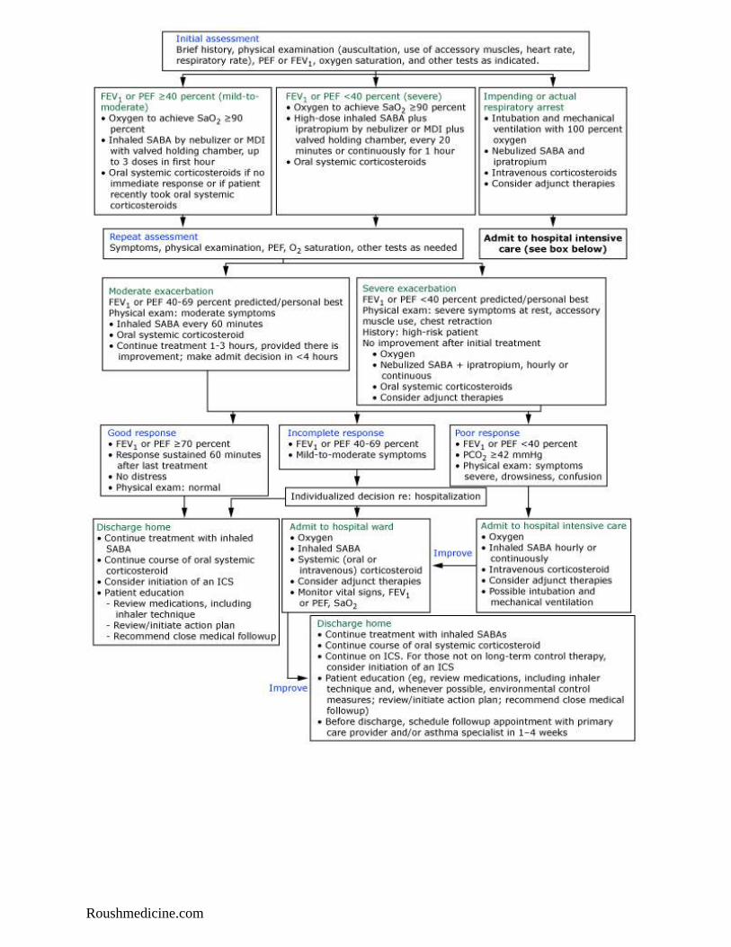
ACUTE EXACERBATION OF ASTHMA & COPD
ASTHMA exacerbation:
Causes: Dust, chemicals, allergens, Beta antagonists, NSAIDs, GERD, Exercise
Assessing severity:
Clinical: Pulsus paradoxus (> 12 mmHg decrease in arterial pressure with inspiration), accessorymuscles, diaphoresis, orthopnea.
Peak Flow:
<200 (“ml/min” is the unit) means severe. If < 100, this is life threatening.
PEFR nl is 80%-100% of patient’s best.
PEFR < 70% means to treat

Roushmedicine.com
Treatment:
Albuterol (Beta agonist) – MDI or via nebulization.
Methylprednisolone IV.
Ipratroprium, if asthma is very severe or if there is no improvement with albuterol
Mg Sulfate IV (smooth muscle relaxant)
NO Chest physiotherapy
NO Antibiotics unless there is pneumonia
NO inhaled steroids for acute attack.
Re-Evaluate after 4 to 6 hours:
If PEFR > 70%, discharge on prednisone with taper.
If PEFR < 40%, admit.
If PEFR 40%-70% individualize. Any of the following factors might be sufficient foradmission:
Poor Self care
Poor Home support
New Onset Asthma (1st ever attack)
Multiple prior hospitalizations or ER visits
Steroids at presentation.
Theophylline:
Conditions & Drugs reducing theopylline levels via liver metabolism:
>cigarette smoking, rifampin, phenytoin, and barbiturates
Conditions & drugs increasing theophylline levels:
>cimetidine, allopurinol, erythromycin, ciprofloxacin, oral contraceptives,propranolol, and clarithromycin, and cirrhosis, congestive heart failure, and febrile viral illness.
Ciprofloxacin inhibits the CYP 1A2 in the cyt p 450 system. Theophylline clearance is reduced.
Beneficial effects of theophylline include augmentation of cardiac output, enhancement ofdiaphragmatic strength, and bronchodilation.
COPD exacerbation (from Sthi S, Murfphy T. NEJM 2008; 359:2355.)

Roushmedicine.com
Cardinal symptoms: increased dyspnea, sputum volume, and purulence.
Risk factors: age 65+, FEV1 < 50% predicted, 3+ exacerbations annually, heart disease.
Mild: 1 cardinal symptom: No antibiotics. Increase bronchodilator.
Moderate, uncomplicated: 2+ cardinal symptoms, no risk factors: Ab’c: azithromycin,cefuroxime, doxy, or TMP/SMX.
Moderate-Severe, complicated: 2+ cardinal symptoms, 1+ risk factors: Moxi, Amox-clav, orcipro (if pseudomonas expected). (Use alternative class if antibiotics were given < 3 monthsago.)
Albuterol doses are much lower than in asthma
Alpha 1 anti-trypsin deficiency now has a synonym: alpha 1 anti-protease deficiency. Prevalence is 0.06% in Sweden.
CM’s: COPD in those less than 40. No cigarette smoking, positive family history. PE:Tympanitic chest, and decreased breast sounds, Chest x-ray: lower lobe emphysema (panacinar–entire alveolus), cirrhosis, and panniculitis (with subcutaneous nodules and infiltration offat lobules with acute inflammatory cells). Associated liver disease.
Obtain alpha 1 antitrypsin level.
Smoking Lung apices central acinar, central part of alveolus
alpha 1 anti-trypsin Lung bases pan-acinar, entire alveolus
Therapy is with infusion and is costly.
Cystic fibrosis has persistent asthma, airflow obstruction, and sputum cultures growing P. aeruginosa and S. aureus
coupled with bilateral upper lobe infiltrates.
Bronchitis: Cotton dust and grain dust exposures. Endotoxins within these dusts are probably the inciting
components for this syndrome. Both of these dusts can also cause fever in the absence of pulmonary symptoms, also
probably resulting from endotoxin inhalation. Bronchitis secondary to cotton dust exposure tends to improve with
continued exposure during the work week but worsen once again after returning to work from a period off the job.
Bronchiectasis:
>Symptoms: cough, copious mucopurulent sputum, and fetid breath.
>Causes of bronchiectasis by type:
Focal:
>Infections: Viral, staph, klebsiella, mycobacterium (tuberculous or avium)
>enlarged lymph nodes, endobronchial carcinoid or endobronchial lung cancer.
Central: Allergic bronchopumonary aspergillosis.
Diffuse: Impaired host defense mechanisms such as cystic fibrosis, immunodeficiency such as with
panhypogammaglobulinemia and HIV, and ciliary dysfunction as in Kartagener's syndrome (immotile sperm,
infertility).
>Treatment includes reversal (if possible) of the underlying cause and reduction in recurrent infections

Roushmedicine.com
using prophylactic rotating antibiotic regimens.
IPF, idiopathic pulmonary fibrosis, is characterized by oxygen desaturation with exercise and velcro like
inspiratory crackles.
Pulmonary fibrosis is associated with
>Connective tissue disease: rheumatoid arthritis, scleroderma, and SLE.
>Pulmonary hypertension, pleural effusions, aspiration resulting from esophageal dysfunction, and
obstructive lung disease
>Ankylosing spondylitis
>Neurofibromatosis (neurofibromas in the skin and other sites, cafe au lait spots, and axillary freckles).
Interstitial lung disease:
>PM N’s occur in: asbestosis and idiopathic pulmonary fibrosis.
>Lymphocytes occur in sarcoid and hypersensitivity pneumonitis.
>Langerhans cells, a form of monocyte, points to eosinophilc granuloma (histiocytosis X)
>Pro liferating smooth muscle cells: lympangioleiomyomatosis.
Methotrexate pulmonary toxicity can include hypersensitivity pneumonitis, pulmonary fibrosis, pleural effusion, or
non-cardiogenic pulmonary edema.
Asbestosis refers to interstitial lung disease, generally with fibrosis, seen in the lower lung fields of a chest radiogram
or chest CT and an associated restrictive ventilatory defect. This patient, although he has pleural plaques suggesting
asbestosis exposure, does not have interstitial changes on chest radiography and has no restriction on pulmonary
function tests; therefore, he does not have asbestosis.
There are three types of lung disease in farmers
Asthma Allergy to agricultural grasses, etc. Acute wheezing and obstruction. NO FEVER.
Silo Filer’s Disease: Oxides of nitrogen released at top of silo lead to initial chest tightness. After an interval of non-
exposure of days to weeks, with relatively few symptoms, there can develop an irreversible obstruction due to
bronhiolitis obliterans with a marked decrease in FEV1
Farmer’s lung: A hypersensitivity pneumonities, a delayed hypersensitivity reaction, due to inhalation of spores of
thermoactinomyces from moldy hay, which induces interstitial inflammation and loosely formed granbulomas. Onset
is within hours of exposure, with fever, cough, dyspnea, bibasilar crackles. Diagnosis is by History of exposure and
removal of exposure. I
In hypersensitivity pneumonitis (extrinsic allergic alveolitis), the pathogenic immune response is not an allergic
reaction, but rather a delayed hypersensitivity reaction.
Most hypersensitivity pneumonitis syndromes (e.g. bagassosis (sugar cane) bird fancier’s, humidifier lung, etc)
involve exposure to organic compounds, usually molds. Also mycobacterium avium (hot tub lung). And
methotrexate.

Roushmedicine.com
Hypersensitivity pneumonitis can develop to rat urine.
Sarcoidosis
>Sarcoid inheritance is supported by monozygotic twin studies.
> Kveim antigen is a suspension of sarcoid tissue that causes characteristic skin granulomas when injected
intradermally in up to 80% of patients with early sarcoidosis.
>Absolute indications for corticosteroid therapy of sarcoidosis include (1) hypercalcemia, (2) cardiac
involvement, (3) ocular involvement (uveitis), (4) disfiguring skin disease, and (5) central nervous system
involvement. The utility of corticosteroids in the treatment of other sequelae of sarcoidosis, including lung
involvement, is controversial.
>Slit lamp examination is imperative for all patients with sarcoidosis to prevent resulting blindness(uveitis
in 20%).
>For biopsy, choose skin and lung which are rarely be involved with noncaseating granulomatous
inflammation from other disorders. (Don’t biopsy liver or lymph nodes where granulomas occur from other disease.)
Sarcoid can have cranial nerve palsies and EN.
Berylliosis, like sarcoid, has inflammatory granulomas and is treated with steroids. Exposures occur in aluminum
alloy processes and fluorescent light industry.
Pulmonary cavities:
Less likely to cause cavities: H. influenzae, M. pneumoniae, and other type serotypes of S. pneumoniae other than
III.
Pulmonary cavities due to tissue necrosis: S. aureus, S. penumoniae serotype III, aerobic gram-negative bacilli, oral
anaerobes, M. tuberculosis, and fungi.
Upper lobe cavities: M TB.
Lower lobe cavity with air fluid level and sulfur granules in subcutaneous tissues: The anaerobe, Actinomyces.
OSA may present with personality changes !
Triple test for an pleural effusion exudates: Any of :
Cholesterol >45
Protein >3
LDH >1.6 x upper limit of normal serum level.
Adenosine deaminase concentrations are elevated in tuberculous pleural effusions: the sensitivity of an adenosine
deaminase pleural fluid level greater than 47 IU/L was 100% and specificity was 91% .
An indication for IVC filter is severe COPD, wherein minor compromise due to PE would cause death.
In pneumocystis, LDH is elevated. Levels greater than 600 IU/ml indicate a poor prognosis.

Roushmedicine.com
In pleural fluid exudates, low glucose can be caused by rheumatoid arthritis (classic), esophogeal rupture, TB, and
malignancy.
PLEURAL FLUID ANALYSIS: The Rule of “15" or 29-49-60 for 2.9 protein, 49 cholesterol, and 60 LDH.
For an exudate, any of the following must be true:
Protein > 2.9 gm/dL
Cholesterol > 45 mg/dL
LDH > 60% of upper limit of normal of serum LDH (usually 60U/L).
Otherwise, it is a transudate.
DRU G IND UCED PU LMONARY DISEASE (from Robbins) July 14, 2004
Pneumonitis & Fibrosis Bleomycin
Amiodarone
Hypersensitivity pneumonitis Methotrexate
Nitrofurantoin
Bronchospasm Aspirin
beta-blockers

Roushmedicine.com
PATHOLOGY OF SELECTED LUNG CONDITIONS July 14, 2004
Name pathology Radiologic CMs RX
Idiopathic*
pulmonary
fibrosis (=
Usual
interstitial
pneumonitis)
Patchy, heterogenous disease
Sub-pleural honeycomb fibrosis
Proliferating fibroblast foci
Minimal inflammation.
Non-Uniform.
Lower lobe, pleura
Honey combing
Cough,
Dyspnea
Desquamative
Interstitial
pneumonitis*
Pigmented Macrophages in distal air spaces.
Peribronchiolar inflammation with little fibrosis.
Desquamation of epithelial cells into air spaces.
Uniform
Lower lobe
Ground glass
Smokers Steroids
Bronchiolitis
obliterans
organizing
pneumonia
(BOOP)=
Cryptogenic
Organizing
Pneumonia
(COP) *, (2)
Bronchiolocentric inflammation.
Airspaces plugged with granulation tissue
Non-uniform
ground glass
peripheral
H/o Rx for
penumonia. No
fever.
Bilateral
crackles.
PFTS: Mixed
obstructive &
restrictive.
Steroids
Hypersensi-
tivity
pneumonitis
(Delayed
hypersensitivit
y)
(2)
Interstitial pneumonitis with lymphs, plasma cells,
& macrophages.
Interstitial fibrosis.
Obliterative bronchiolitis.
Granuloma formation
Ground glass
Nodular
Patchy
Acute(4-6hrs)
^T, cough,
dyspnea.
Resolves in 2
ds. ^ PM Ns.
Ab’s to specific
Ag (2)
PFT ’s:
Restrictive.
Goodpasture’s
syndrome
Necrotizing, hemorrhaging IS pneumonitis: Intra-
alveolar hemorrage, fibrous thickening of septa,
hemosiderin laden macrophages. Linear deposits of
Immunoglobulins along BM of septal walls (as in
glomerular BM).
Idiopathic
pulmonary
hemosiderosis
shedding & hyperplasia of alveolar epithelial cells.
Blood in alveoli.
Pulmonary
alveolar
proteinosis
Intra-alveolar dense granular material: lipid and
PAS positive.
Cough with
gelatinous
sputum.
*An “Idiopathic Insterstial Pneumonia”.
(1) BOOP has many causes ore associations other than “idopathic”, including post-infectious, drug related
(amiodarine, bleo , MTX , cocaine), rheumatologic (RA, SLE), Immunologic (common variable immuno deficiency,
essential mixed cryoglobulinemia), organ transplant, and other associations.
(2) Etiologies are myriad organic compounds: e.g., Farmer’s lung (moldy hay, grain, silage), Bird Fancier’s lung (
Avian droppings), woodworker’s lung, hot tub lung (mold on ceiling). Methotrexate. HVAC lung. Tissue Bx is not
necessary for Dx. M ethod of diagnosis is by history of exposure and removal from agent.
Roushmedicine.com
NOTES:
Solitary pulmonary nodule
If all of these are true
—
Age < 40
Non-smoker
no prior CA
Nodule < 1 CM
Benign calcification pattern (diffuse, central, or laminar (“popcorn”)
Middle or lower lobe location (NOT upper lobe)
Not a spiculated edge and with smooth borders
—
Then you can follow patient with CXR.
If there is any doubt, do a PET scan. (Sens & Spec > 95%).
False negatives are due to bronchoalveolar ca or hyperglycemia.
Presentations of lung tumors:
Adenocarcinoma: peripheral location and non-calcified. M ost likely to have a single brain met. Associated with
IPF.
Broncho-alveolar ca: non-resolving pulmonary infiltrate with air bronchogram. Associated with IPF.
Squamous cell ca: central lung mass, hypercalcemia.
Small cell ca: Central lung mass, hepatomegaly, lymphadenopathy, proximal muscle weakness, paresthesias (Eaton-
Lambert) or SIADH.
Bronchial carcinoid: Atelectasis, hypervascular pedunculated mass, liver nodules, wheezing, diarrhea, and
flushing.

Roushmedicine.com
TYPES OF DYSPNEA
SETTING ONSET AIRWAY & LUNGPATHOLOGY
Persulfates in hair dressers Minutes to hours irritant asthma
Sugar Can, Bird Droppings,or moldy hay
5 hours Hypersensitivity pneumonitis
Latex exposure (healthworkers)
weeks to years immunologic asthma
Silo-fillers disease or oxidesof nitrogen
immediate
(but see to right )
Chest tightness, rinorrhea,laryngeal symptoms (later,there can be irreversiblebronchiolitis obliterans)
Phosgene (fire fighters) 6 hours to days Bronchitis, alveolitis
ABNORMAL HEMOGLOBIN OR 02 CARRYING CAPACITY
CO poisoning Methemoglobinemia*
O2% by Pulse oximetry NL Decreased but still overestimate & plateaus at 85%
O2% by usual ABGs NL NL
PaO2 NL NL
O2% by co-oximetry Decreased (accurate) Decreased (accurate)
*Acquired methemoglobinemia can result from dapsone (e.g., for dermetitis herpetiformis or forprophylaxis against pneumocystis in HIV patients) which oxidizes ferrous (Fe2+) to ferric (Fe3+)state. This results in a functional anemia and hypoxemia and may even be fatal (Pallais NEJM2011;364:957).
IF THERE IS HYPOXEMIA ON ROOM AIR, and the FiO2 is increased to 100%,
the PaO2 may increase dramatically, e.g., to from 50 to 613, indicates
V/Q mismatch, as in pulmonary embolus, COPD, or interstitial lung disease.
If the PaO2 increases modestly from 50 to 75, this indicates a
Shunt, due to atelectasis, pneumonia, ARDS, cardiogenic pulmonary edema, pulmonary infarct orintra-cardiac right to left shunt. (See Cecil, Figure 99-2.)
Even when the PaO2 is correctable, there is still lung disease, manifest bythe increased A-a Gradient. Partial pressure of air = 760 mmHg - 47 mmHg = 713. (Where 47= partial pressure of water vapor).
A-a gradient = 713*FiO2 - 5/4 * PCO2 - PaO2 For example,
at FiO2 = 21%, PCO2=52, and PaO2 = 50
=713*.21 - 5/4 * 52 - 50
= 150 - 65 - 50 = 35.

Roushmedicine.com
Or, at the FiO2, where the PaO2 is 613, the result is 713-65-613=35.
Normally the A-a gradient is 10 to 30, from 30 to 100 years of age.
At age 65 it is normally around 20.

Roushmedicine.com
Acid-Base disturbances April 21, 2004 — based on MKSAP XIII
Summary ch inHC03
HCO3 ch inpCO2
pCO2 phchng
ph
Normal 0 0
24 40 7.40
Met Acidosis ch in pCO2 is 110% of ch in HCO3 -10 -11
14 29
Met Alkalosis ch in pCO2 is 70% of ch in HCO3 +10 +7
34 47
Resp Acidosis-Acute ch in HCO3 is 10% of ch in pCO2 +1 +10 -.08
(DUE to buffering) 25 50 7.32
Resp Acidosis-Chron ch in HCO3 is 40% of ch in pCO2 +4 +10 -.04
(Due to kidneys) 28 50 7.36
Resp Alkalosis-Acute ch in HCO3 is 20% of ch in pCO2 -2 -10 +.08
(Due to buffering) 22 30 7.48
Resp Alkalosis-Chron ch in HCO3 is 40% of ch in pCO2 -4 -10 +.04
(Due to kidneys) 20 30 7.44
For metabolic acidosis, you can use the winter formula: PC02 = 1.5 * Bicarb + 8.
For anion gap metabolic acidosis, you can calculate
Change in Anion Gap / Change in Bicarb. = 1 to 2
If the entire process is attributable to the abnormal anions.
If Ratio > 2, there is also a metabolic alkalosis.
If the ratio is < 1, there is an additional non-anion gap metabolic acidosis.
Passive smoking is associated with increased cardiac mortality, respiratory illness, and lungcancer.
In TB, the pathology is the caseating or necrotizing granuloma. Pleural biopsy is positive for non-caseating granulomas and TB, but cultures of pleural fluid are usually negative.
Patients from high risk areas of the world where tuberculosis is highly prevalent (as evidenced byuse of BCG vaccine) and who have been vaccinated with BCG, have their BCG vaccine ignoredin interpreting the PPD and are treated for latent TB on the basis of an induration of greater than10 MM (because they are from a location with high prevalence and therefore have intermediatepre-test probability of TB).

Roushmedicine.com
Treat hepatic hydrothorax (transudative, right pleural effusion associated with hepatitis) witha)diuretics and salt restriction, b) possibly TIPS, c) and, finally, if those don’t work, pleurodesis.
In lung transplant patients, the greatest risk of infection at 1 week is bacterial, at 1 - 6 months it isCMV, and then after 6 months it is bacterial again.
In HIV, a person with latent tuberculosis who acquires HIV infection is estimated to have a 3 to15% annual risk of developing active tuberculosis, which is substantially higher than the risk inHIV-uninfected individuals. Active tuberculosis may develop at any point in the course of HIVinfection. The clinical presentation varies with the degree of immune suppression. Typical upperlobe cavitary disease is common early in the course of HIV infection, when immune suppressionis least. As the degree of immune suppression becomes more advanced, extrapulmonary oratypical presentations such as mediastinal lymph node, disseminated, and meningeal diseasebecome more common. HIV-infected patients have a decreased frequency of sputumsmear–positive disease that in conjunction with the atypical presentations makes diagnosis oftenmore difficult than is the case in HIV-uninfected individuals.
In post transplant patients, human herpesvirus type 8 (HHV-8) is causally associated with primaryeffusion lymphoma as well as Kaposi's sarcoma and multicentric Castleman's disease.
In these patients, Epstein-Barr virus can cause posttransplant B cell lymphoproliferative disease.
It is stated that in a patient with chronic hypoxemia and Obesity Hypoventilation Syndrome(Pickwickian), the over correction of hypoxemia led to decrease respiratory drive and respiratoryarrest. In OHS, remember to screen for hypothyroidism.
Patients who develop postpolio syndrome tend to have affliction of the same muscle groups thatwere affected in the original presentation. This patient presents with symptoms of diaphragmaticmuscle weakness 40 years after the initial presentation with poliomyelitis.
The most common cause of ambulatory care visits in the US is rhinovirus infection.
Reverse pulmonary edema and peripheral blood eosinophilia: hypereosinophilic syndrome.
Factor V Leiden deficiency – with protein C resistance --is the most frequent cause of PE in whitepopulations.
Loffler’s syndrome: pneumonitis+eosinophilia 2nd to Ascaris Lumbricoides.
For PE, the classic EKG is an SI, QIII

Roushmedicine.com
This is a left lower lobe lung abscess in a 60 y.o. man with a 2week history of cough and fever. Sputum is foul smelling. This is an abscess, likely due to anaerobes. It is best treated byclindamycin or metronidazole. (It used to be treated withpenicillin, but not anymore.)
MEDSTUDY Pulmonary questions
ciprofloxacin increases theophylline levels.
Chronic eosinophilic pneumonia: peripheral infiltrates (i.e.,photo negative of pulmonary edema ), eosinophilia.
In INH therapy, baseline and repeat LFTs are not indicated unless there is a co-existing liverdisease. Clinical monitoring is done monthly for signs of INH-hepatitis : Anorexia, N,V, darkurine, rash, icterus, persistant parathesias of hands and feet, fatigue, weakness, fever, abodominalpain, arthralgias, and easy bruising.
Eosinophilic granuloma: In a smoker, on cxr honey comb changes with small cystic spaces inupper lung fields, and may have abnormalities related to pituitary insufficiency due to granulomasthere (e.g., diabetes insipidus).
Length of treatment for DVT/PE:
Warfarin Life long: 2+ unprovoked DVT, 2 DVTs with any type of thrombophilia, or one DVT inan unusual site, or PE with APLAB.
Heparin Life long: DVT or PE secondary to cancer.
Warfarin for 12 months: 1 unprovoked PE (or one of which would be with an unprovokedproximal DVT) OR one unprovoked PE with an irremovable risk factor (e.g., ACLA or Factor VLeiden).
Warfarin for 6 months: 1st unprovoked DVT.
Warfarin 3 - 6 months: one DVT with removable risk factor.
CPAP must be used cautiously because it can increase the work of breathing. It is particularlycontraindicated in ALS because it can cause tiring and death.
BI-PAP means that the pressure can be set differently for inspiration and expiration. E.G.: forexpiration the pressure can be set to drop and make breathing more easily. It is particularlyimportant to use BI-PAP for neuromuscular (ALS) or chest wall disease (scoliosis).
Alpha 1 ATT deficiency: CM’s: Emphysema < 45y.o. or in absence of smoking oroccupation or in the lower lungs. Bronchiectasis with etiology. Asthma with persistent airflowobstruction post treatment. Unexplained liver disease. Necrotizing panniculitis.
Serum ATT is used for presumptive diagnosis: 100 - 300 mg/dL. < 80 indicates significant risk.
Phenotyping is required to confirm, if the serum ATT is borderline or if considering Rx.
PiZZ phenotype in serum is responsible for nearly all cases. Genotyping can be done on mouth

Roushmedicine.com
swabs. In ATT deficiency pulmonary disease, the pathogenesis is proteolysis.
In ATT liver disease, the pathogenesis is accumulation of ATT protein in hepatocytes.
Rx for pulmonary disease is by ATT mist. Rx for liver disease is renal transplant.
Cystic fibrosis (JAMA 2007;298:1787): This is a pearl: Think of cystic fibrosis in the followingsitutions:
Bronchiectasis plus any of the following a) male infertility (congenital bilateral absence of the vasdeferens), b) recurrent idiopathic pancreatitis (10 - 20% of individuals with chronic idiopathicpancreatitis care cystic fibrosis mutations), or c) recurrent sinusitis with nasal polyposis.
Recent treatments include:
Better manual compression techniques.
Mucolytic dornase alfa (nebulized) (breaks up DNA from PMNs)
Nebulized antibiotics.
Oral azithromycin
Nutritional support (because of malaborption these patients are undernourished).
Replacement of ADEK.
EOSINOPHILIC LUNG DISEASE: Increased eosinophils in the lung with or without peripheralblood eosinophilia.
These can be divided into Airway disorders and parenchymal disorders
Airway Disorders
Asthma
Non asthmatic eosinophilic bronchitis
Allergic bronchopulmonary aspergillosis
Parenchymal disorders
Hypereosinophilic syndromes
Eosinophilic pneumonias
Pulmonary vasculitis
Malignancies
Infections
Drugs
Interstitial lung disease
Non asthmatic eosinophilic bronchitis: Hallmark=Chronic cough. (No bronchial hyper-responsiveness.
Allergic bronchopulmonary aspergillosis: Hallmark=Asthma, infiltrates on CXR, High IgE. Precipitins for aspergilla. Stages are: 1. Acute asthma with infiltrates. 2. Remission. 3.Exacerbation with 2x higher IgE. 4. Steroid dependency where the taper worsens the asthma. 5.A fibrotic end stage with cor pulmonale. Rx: oral steroids until CXR clears. Ketoconazole orItraconazole.
Acute eosinophilic pneumonia: Hallmark=pneumonia with BAL eosinophils > 25%. Steroidresponsive without relaps.
Roushmedicine.com
Chronic eosinophilic pneumonia. Peripheral infiltrates (“Photonegative infiltrates”) BAL eos >40%. Steroid responsive but relapses occur.
Idiopathic hyper-eosinophilic syndrome: Hallmark = multi-organ disease. Arterial and venousthrombi.
Churg Strauss syndrome: pulmonary-renal syndrome. Ashma, rhinitis, and peripheral bloodeosinophilia. Vasculitis. P-ANCA positive.
Parasitic infections causing pulmonary disease and peripheral blood eosinophilia.
Parenchymal invasion with hemoptysis, CXR infiltrates and pleural effusions: Paragonimus,echinococcus, and cystercicosis.
Hematogenous spread: Hookworm, Ascaris, and Strongyloides. Cough & wheezing.
Various other disease can give eosinophilic pulmonary disease: sacrcoid, lung cancer, Hodgkin’sdisease, medications such as ASA, amiodarone, bleomycin, captopril, dilantin.
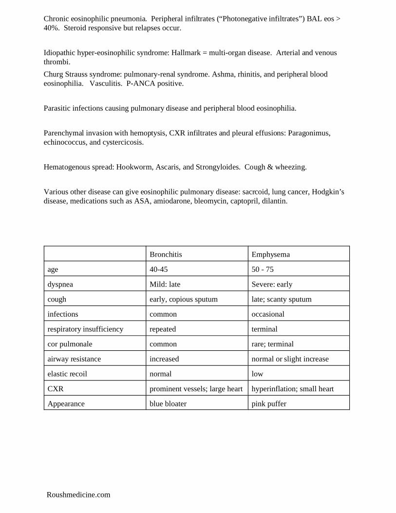
Bronchitis Emphysema
age 40-45 50 - 75
dyspnea Mild: late Severe: early
cough early, copious sputum late; scanty sputum
infections common occasional
respiratory insufficiency repeated terminal
cor pulmonale common rare; terminal
airway resistance increased normal or slight increase
elastic recoil normal low
CXR prominent vessels; large heart hyperinflation; small heart
Appearance blue bloater pink puffer
Roushmedicine.com
Roushmedicine.com
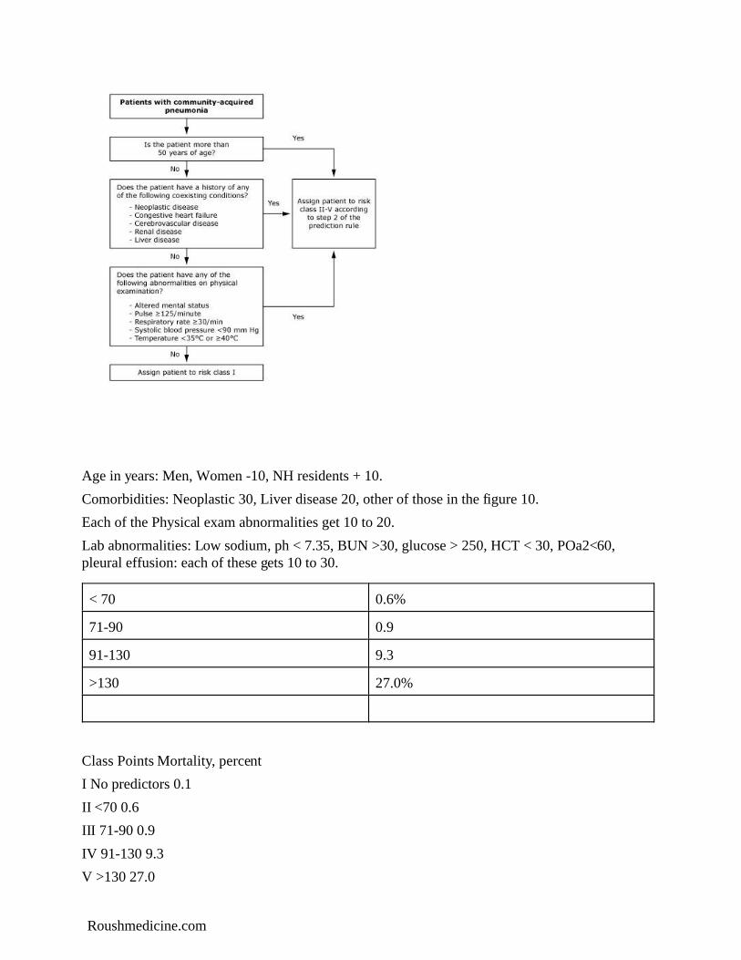
Age in years: Men, Women -10, NH residents + 10.
Comorbidities: Neoplastic 30, Liver disease 20, other of those in the figure 10.
Each of the Physical exam abnormalities get 10 to 20.
Lab abnormalities: Low sodium, ph < 7.35, BUN >30, glucose > 250, HCT < 30, POa2<60,pleural effusion: each of these gets 10 to 30.
< 70 0.6%
71-90 0.9
91-130 9.3
>130 27.0%
Class Points Mortality, percent
I No predictors 0.1
II <70 0.6
III 71-90 0.9
IV 91-130 9.3
V >130 27.0

Roushmedicine.com
CURB 65
Confusion
Urea Nitrogen > 20
Respiratory Rate >30
Blood pressure < 0-
Age > 65
Number of factors & 30 day mortality rate:
0: 0.6%,
1: 2.1%,
2: 9.2%,
3: 14.5%,
4: 40%
SHUNTING AND DISEASE CORELATIONS
Shunting absent:
IPF (idiopathic pulmonary fibrosis)
COPD
Asthma
A1ATD
Shunting present:
Hereditary hemorrhagic telangiectasia (Osler-Rendu-Weber)
Right to left shunts in heart disease; VSD
Pulmonary edema
HHT: Classically: epistaxis, GI bleed, iron deficiency, and telangiectasias of the mouth andfingers. The criteria are: 1) epistaxis, 2) telangiectasia of the mouth and fingers, 3) AVM of thelung, cerebrum and liver, 4) autosomal dominant pattern. The condition is associated withpulmonary hypertension.
Roushmedicine.com
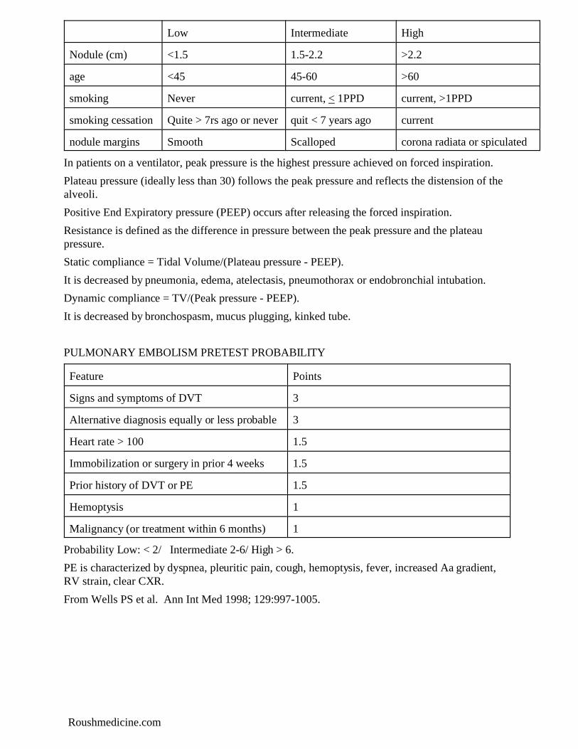
Low Intermediate High
Nodule (cm) <1.5 1.5-2.2 >2.2
age <45 45-60 >60
smoking Never current, < 1PPD current, >1PPD
smoking cessation Quite > 7rs ago or never quit < 7 years ago current
nodule margins Smooth Scalloped corona radiata or spiculated
In patients on a ventilator, peak pressure is the highest pressure achieved on forced inspiration.
Plateau pressure (ideally less than 30) follows the peak pressure and reflects the distension of thealveoli.
Positive End Expiratory pressure (PEEP) occurs after releasing the forced inspiration.
Resistance is defined as the difference in pressure between the peak pressure and the plateaupressure.
Static compliance = Tidal Volume/(Plateau pressure - PEEP).
It is decreased by pneumonia, edema, atelectasis, pneumothorax or endobronchial intubation.
Dynamic compliance = TV/(Peak pressure - PEEP).
It is decreased by bronchospasm, mucus plugging, kinked tube.
PULMONARY EMBOLISM PRETEST PROBABILITY
Feature Points
Signs and symptoms of DVT 3
Alternative diagnosis equally or less probable 3
Heart rate > 100 1.5
Immobilization or surgery in prior 4 weeks 1.5
Prior history of DVT or PE 1.5
Hemoptysis 1
Malignancy (or treatment within 6 months) 1
Probability Low: < 2/ Intermediate 2-6/ High > 6.
PE is characterized by dyspnea, pleuritic pain, cough, hemoptysis, fever, increased Aa gradient,RV strain, clear CXR.
From Wells PS et al. Ann Int Med 1998; 129:997-1005.

Roushmedicine.com
DEEP VEIN THROMBOSIS PRETEST PROBABLITY:
History of Cancer 1
Immobilization 1
Bedd ridden 1
Thigh or calf tenderness 1
Calf swelling 1
Pitting edema 1
Collateral vein distension 1
Varicosities 1
Alternative diagnosis: -2
Pulmonary hypertension can be caused by nocturnal hypoxemia: Q 48, MKSAP 14, Pulmonary.