histopathological characterization and whole exome...
TRANSCRIPT

Research ArticleHistopathological Characterization and Whole ExomeSequencing of Ectopic Thyroid: Fetal Architecture in a FunctionalEctopic Gland from Adult Patient
Rosalinda Yasato Camargo,1 Cristina Takami Kanamura,2 Celso Ubirajara Friguglietti,3
Célia Regina Nogueira,4 Sonia Iorcansky,5 Alfio José Tincani,6 Ana Karina Bezerra,7
Ester Brust,8,9 Fernanda Christtanini Koyama,10 Anamaria Aranha Camargo,10
Fernanda Orpinelli R. Rego,10 Pedro Alexandre Favoretto Galante,10
Geraldo Medeiros-Neto,1 and Ileana Gabriela Sanchez Rubio 8,9
1Thyroid Unit, Cellular and Molecular Endocrine Laboratory, LIM-25, Faculdade de Medicina da Universidade de São Paulo(FMUSP), Avenida Doutor Arnaldo 455, Cerqueira César, 01246-904 São Paulo, SP, Brazil2Adolfo Lutz Institute, São Paulo Public Health Service, Av. Dr. Arnaldo 355, Cerqueira César, 01246-000 São Paulo, SP, Brazil3Head and Neck Surgery of Santa Catarina Hospital, Av. Paulista 200, Bela Vista, 01310-000 São Paulo, SP, Brazil4Department of Internal Medicine, Botucatu School of Medicine, UNESP, Av. Prof. Montenegro, s/n Distrito de Rubião Junior,18618-687 Botucatu, SP, Brazil5Servicio de Endocrinología, Hospital de Pediatría Dr. Juan Garrahan, Combate de los Pozos 1881,C1245AAM Buenos Aires, Argentina6Departamento de Cirurgia na Disciplina de Cirurgia de Cabeça e Pescoço da Faculdade de Ciências Médicas da UNICAMP,R. Tessália Vieira de Camargo 126, 13083-887 Campinas, SP, Brazil7Medicine School, Universidade de Fortaleza (Unifor), Av. Washington Soares 1321, Edson Queiroz, 60811-905 Fortaleza, CE, Brazil8Postgraduate Program in Biotechnology, Universidade Federal de São Paulo (UNIFESP), Pedro de Toledo 669, 040399-032 SãoPaulo, SP, Brazil9Thyroid Molecular Sciences Laboratory, Universidade Federal de São Paulo, Departamento de Ciências Biológicas, PostgraduationPrograms in Biotechnology and Structural and Functional Biology, UNIFESP, Pedro de Toledo 669, 040399-032 São Paulo,SP, Brazil10Molecular Oncology Center, Hospital Sírio-Libanés, Rua Prof. Daher Cutait 69, 01308-060 São Paulo, SP, Brazil
Correspondence should be addressed to Ileana Gabriela Sanchez Rubio; [email protected]
Received 30 August 2017; Accepted 16 November 2017; Published 8 February 2018
Academic Editor: Maria L. Dufau
Copyright © 2018 Rosalinda Yasato Camargo et al. This is an open access article distributed under the Creative CommonsAttribution License, which permits unrestricted use, distribution, and reproduction in any medium, provided the original workis properly cited.
Ectopic thyroid results from a migration defect of the developing gland during embryogenesis causing congenital hypothyroidism.But it has also been detected in asymptomatic individuals. This study aimed to investigate the histopathological, functional, andgenetic features of human ectopic thyroids. Six samples were histologically examined, and the expression of the specific thyroidproteins was assessed by immunohistochemistry. Two samples were submitted to whole exome sequencing. An oropharynxsample showed immature fetal architecture tissue with clusters or cords of oval thyrocytes and small follicles; one sampleexhibited a normal thyroid pattern while four showed colloid goiter. All ectopic thyroids expressed the specific thyroid genesand T4 at similar locations to those observed in normal thyroid. No somatic mutations associated with ectopic thyroid werefound. This is the first immature thyroid fetal tissue observed in an ectopic thyroid due to the arrest of structural differentiationearly in the colloid stage of development that proved able to synthesize thyroid hormone but not to respond to TSH. Despite theability of all ectopic thyroids to synthetize specific thyroid proteins and T4, at some point in life, it may be insufficient tosupport body growth leading to hypothyroidism, as observed in some of the patients.
HindawiInternational Journal of EndocrinologyVolume 2018, Article ID 4682876, 10 pageshttps://doi.org/10.1155/2018/4682876
1. Introduction
The thyroid gland is a bilobar gland composed of two endo-crine cells, namely, the thyroid follicular cells (TFCs) thatproduce the thyroid hormones T3 and T4 and the parafolli-cular cells that secrete calcitonin. TFCs are derived fromthe thyroid primordium, or thyroid anlage, that originatesfrom a thickening of the endodermal epithelium in the fore-gut at the base of the prospective tongue. This structuredevelops the thyroid diverticulum that proliferates, invadesthe surrounding mesenchyme, and migrates through theanterior midline of the neck. During migration, the primitivethyroid acquires a bilobed structure while the thyroglossalduct, the initial connection to the primitive pharyngeal floor,loses its lumen and turns into disconnected fragments.When the developing thyroid reaches its final position atthe base of the neck in front of the trachea, the fusion ofthe ultimobranchial bodies takes place to give rise to C cells.Finally, folliculogenesis occurs concomitantly with differen-tiation of thyroid progenitor cells into functional thyroid fol-licular cells, expressing specific thyroid genes such as TSHreceptor (TSHR), Na/I symporter (NIS), and thyroglobulin(TG) [1–3].
In mouse models, normal embryogenesis of the thyroidwas shown to be controlled by a well-integrated regulatorynetwork of transcription factors [4, 5]. During the formationof the thyroid anlage (specification phase), the expression ofthyroid transcription factors TITF1, PAX8, FOXE1, andHEX1 can be observed. FOXE1 is believed to be requiredfor migration of the mouse thyroid primordium after detach-ment of the bud [6], and survival of the thyroid primordiumdepends on the expression of FOXE1 and PAX8 [4].
Ectopic thyroid is a rare malformation that resultsfrom a migration defect of the developing gland duringembryogenesis [1]. The glands can be found at any pointalong the path of migration, from the foramen cecum tothe mediastinum, and also in distal subdiaphragmatic areas.However, the most frequent site is at the base of the tongueor lingual thyroid [7]. Ectopia is the most common formof thyroid dysgenesis (TD) and congenital hypothyroidism[8, 9] but has also been detected in asymptomatic individualsor as a cause of hypothyroidism, dysphonia, dysphagia,cough, snoring, or sleep apnea and coexisting with a normallylocated thyroid [10–12] [13, 14].
The pathogenesis of ectopic thyroid, and the fact thatsome ectopic patients remain euthyroid throughout life,has not yet been elucidated. Few studies have character-ized the expression of important thyroid proteins [10, 14].In addition, mutations in genes FOXE1, PAX8, TSHR,NKX2.1, or NKX2.5 associated with TD were identified inonly 2-3% of TD cases [15]. Recently, with whole exomesequencing (WES), a powerful tool for investigating geneticcauses of human diseases, novel genes were identified inTD cases [16, 17].
Thus, the aim of the present study was to investigate thehistopathological and functional characteristics of humanectopic thyroid tissues through the expression of T4, specificthyroid proteins TG, NIS, TSHR, and thyroid transcriptionfactors NKX2.1 and PAX8 by immunohistochemistry and
to identify potential pathogenic mutations with expandedwhole exome sequencing.
2. Material and Methods
2.1. Samples. Paraffin blocks of four human ectopic thyroidsamples were retrospectively collected from patients whounderwent thyroid resection, and two flash-frozen tissuesamples removed due to symptoms (sample 4) or suspiciouscytology for papillary thyroid lesion (patient 2) were alsoincluded in the study. The control sample was taken fromarchived paraffin blocks of the normal thyroid. The studyprotocol was approved by the local Research Ethics Commit-tees (HC 893/01 and CEP 1078/11).
2.2. Patients. All patients were born before the establishmentof the mandatory Neonatal Screening Program in Brazil,except for patient 5. Clinicopathologic information on theectopic patients was obtained from medical records. Patient1 is a 34-year-old female with an oropharyngeal ectopicthyroid mass detected at 7 years of age. The patient was nevertreated with levothyroxine (LT4) and had two cesarean deliv-eries. Preoperative thyroid hormonal status was TSH:11.19mIU/L (ref. 0.5–4.2mIU/L), and total T4: 2.48μg/dL(ref. 5.3–12.6μg/dL) (to convert to nmol/L, multiply by12.87). Patient 2 is a 46-year-old female with ectopic thyroidlocated at the hyoid bone. The patient had been treated withLT4 (100μg/day) for hypothyroidism for the last 30 years.Goiter was diagnosed 4 years before surgery. Patient 3 is a7-year-old euthyroid girl. Lingual thyroid was detected at 5years of age when investigating swallowing and sleeping dif-ficulties. The patient was never treated with LT4. Patient 4 isa 38-year-old male with lingual thyroid causing excessivesnoring. The patient was never treated with LT4, and preop-erative thyroid hormonal status was FT4: 0.6 ng/dL (ref. 0.6–1.54 ng/dL) (to convert to pmol/L, multiply by 12.87), TSH:46.7mIU/L (ref. 0.5–4.2mIU/L), and negative anti-TPOand anti-Tg. Patient 5 is a one-month-old girl with midlinedefect and lingual thyroid, diagnosed after surgery for sus-pected lingual tumor. Neonatal test after surgery indicatedTSH> 200mIU/L. Patient 6 is a 13-year-old girl with a necknodule noted at birth that grew during childhood develop-ment. At 11 years of age, lingual thyroid was diagnosed andhypothyroidism confirmed with serum TSH: 11.42mIU/L,FT4: 0.73 ng/dL (ref. 0.897–1.794 ng/dL), and negative anti-TPO. Levothyroxine was prescribed, but the patient showedpoor compliance.
2.3. Immunohistochemistry. Three μm thick sections offormalin-fixed and paraffin-embedded tissue samples fromthe six patients (samples 1–6) were first stained withhematoxylin/eosin for histological observation and thensubmitted to immunohistochemical procedures. Antigenretrieval was performed in 10mM citrate buffer/pH6.0 ina pressure cooker for three minutes, and endogenous per-oxidase was inactivated in 6% hydrogen peroxide solutionin an incubation step. The antibodies used in this studywere monoclonal anti-thyroid peroxidase (TPO) (MoAb47,1 : 500, DakoCytomation, Glostrup, Denmark), monoclonal
2 International Journal of Endocrinology
anti-sodium-iodide symporter (NIS) (FP5A, 1 : 200, MayoClinic, USA), monoclonal anti-TTF1 (NKX2.1) (8G7G3/1,1 : 500, Cell Marque, Rocklin, USA), monoclonal anti-thyroid-stimulating hormone receptor (TSHR) (4C1/E1/G8,1 : 200, NeoMarkers, Fremont, USA), polyclonal anti-PAX8(rabbit, 1 : 500, Cell Marque), polyclonal anti-thyroglobulin(rabbit, 1 : 400.000; DakoCytomation), and polyclonalanti-thyroxine (T4) (rabbit, 1 : 1.000; Cloud-Clone Corp.,Houston, USA). Tissue sections were incubated overnightwith the primary antibodies, and amplifications wereobtained by peroxidase-conjugated polymer (UltravisionTL015-HDS, Thermo Fisher Scientific, Fremont, USA) andthen revealed by diaminobenzidine/hydrogen peroxidesubstrate chromogen. Images were acquired using a LeicaTCS SP8 microscope. The samples were examined by twoof the authors.
2.4. Whole Exome Sequencing and Analysis. Total DNA fromectopic tissue from patients 2 and 4 was used to prepare theDNA library with Agilent SureSelectXT reagent kit (AgilentTechnologies®, Santa Clara, USA), and the whole exomeand UTR regions were sequenced in Illumina NextSeq 500platform (Illumina® Inc., San Diego, USA) with a calculatedcoverage average of 140x per sample. Sequenced readswere aligned to a reference genome (GRCh37/hg19) usingBurrows-Wheeler Alignment (BWA), and calling was per-formed with Genome Analysis Toolkit (GATK). Singlenucleic variants and small insertions-deletions were anno-tated with ANNOVAR [18]. Data were filtered with 12sample controls of Brazilian familial thyroid cancer tissues,variants with low-quality score, and common variants(MAF> 1%), and neutral variants were eliminated. Pre-dictions of deleterious effect were evaluated using onlinebioinformatics tools: SIFT, PolyPhen-2, mutationassessor,fathmm, Condel, MutationTaster, and PROVEAN [19–21].
3. Results
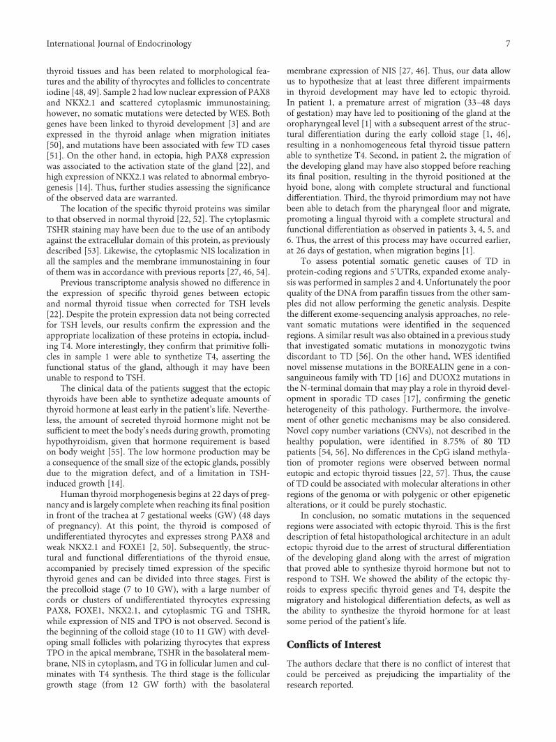
3.1. Histopathology of Ectopic Thyroids. Fetal thyroid archi-tecture was observed in sample 1 which exhibited isolatedepithelial oval cells that were unpolarized or arranged in clus-ters and cords and primitive small follicles lined by cuboidalcells with a small lumen, some of them with colloid embed-ded in stroma (Figures 1 and 2). Small areas of the sectionwith normal-sized follicles were also observed.
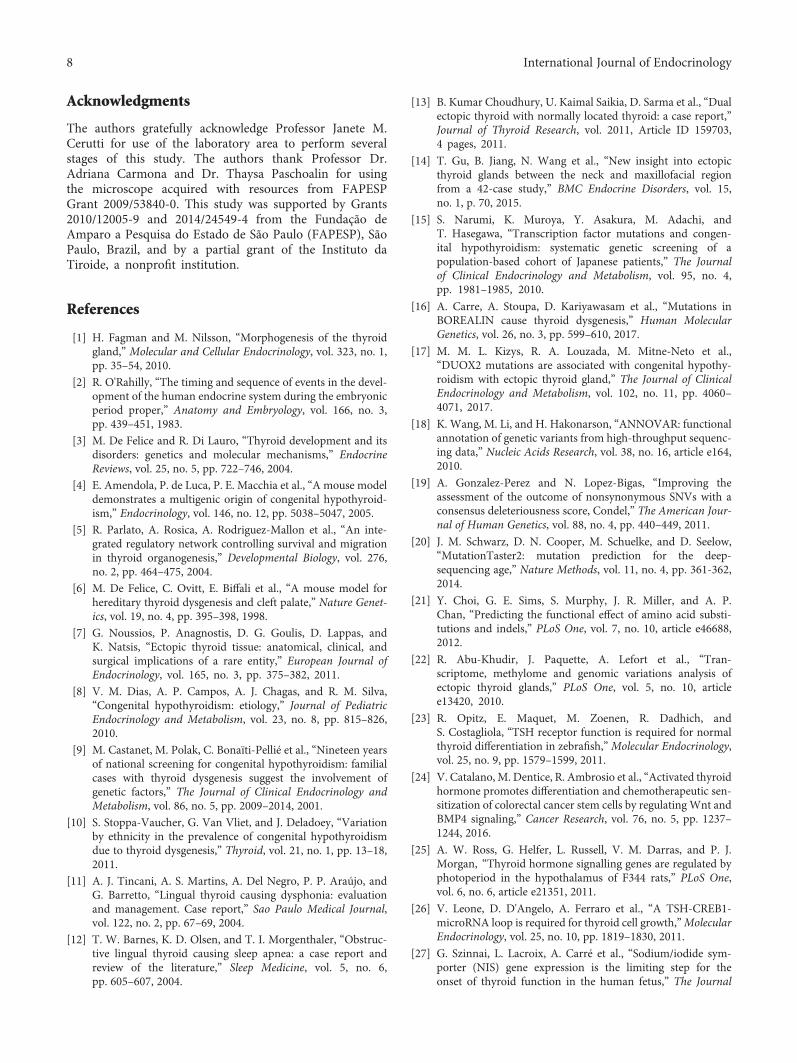
Histopathological examination of samples 2, 3, 4, and 6revealed colloid goiter with highly enlarged follicles lined byflattened epithelial cells (Figure 2(A)). Focal areas (10–25%)composed of normal-sized follicles with cuboidal cells werealso detected in these samples. Sample 5 showed normalthyroid tissue (Figure 2(B)).
3.2. Immunohistochemistry Expression. TG expression wasintense and diffuse in the lumen of all follicles of the samplesunder investigation, even in microfollicles from tissue withfetal architecture (sample 1). Cytoplasmic TG staining wasweak or negative in the normal-sized follicles and also inthe macrofollicles of the ectopic samples, similarly to nor-mal thyroid sample. However, strong cytoplasmic staining
was observed in isolated thyrocytes and microfolliclesfrom the fetal thyroid tissue (sample 1) (Figures 1(a), 1(b),and 2, a–c).
TPO staining was strong and diffuse within the cyto-plasm of thyrocytes and was also observed in the apical mem-brane of all follicles of the ectopic samples examined, similarto normal thyroid tissue. Only cytoplasmic TPO wasobserved in isolated thyrocytes from fetal thyroid tissue(sample 1) (Figures 1(c) and 2, d–f).
TSHR expression was intense and scattered within thecytoplasm of thyrocytes of the ectopic samples, akin to a nor-mal sample (Figures 1(d) and 2, g–i).
NKX2.1 diffuse immunostaining was strong in themajority of the nuclei of the ectopic thyroid samples, exceptfor sample 2 which had approximately 10% low positivenuclei and scattered cytoplasmic expression. Low expressionwas observed in most of the nuclei of normal thyroid(Figures 1(e) and 2, j–l).
PAX8 moderate to intense diffuse expression wasobserved for all ectopic samples in approximately 70–80%of follicular cell nuclei, except for sample 2 which had a lowexpression in around 20% of nuclei and scattered cytoplas-mic expression. Expression in normal thyroid tissue waslow in most of the nuclei (Figures 1(f) and 2, m–o).
NIS was positive in the basolateral membrane in up to10% of the cells of ectopic samples 3, 4, and 5. Cytoplas-mic staining was observed in all samples. Normal thyroidsample had a similar pattern of 20% positivity in the baso-lateral membrane and cytoplasmic staining (Figures 1(g)and 2, p–r).
T4 expression was diffuse and intense in colloid of micro-and normal-sized follicles from the fetal thyroid sample(Figure 1(h)). In the other ectopic samples, the colloid T4expression was moderate or intense, although some folliclesshowed low or negative immunostaining, similarly to normalthyroid (Figure 2, s–u).
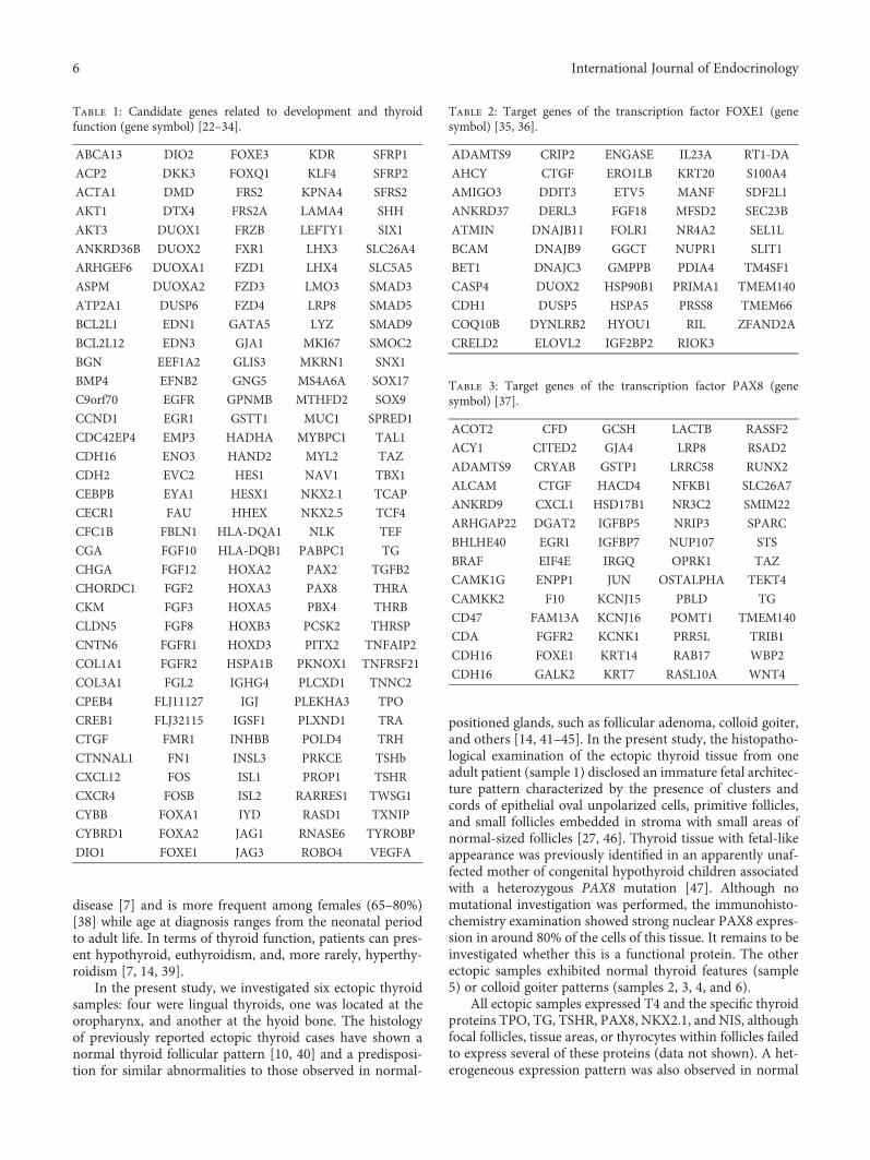
3.3. Whole Exome Sequencing. After filtering the sequencingdata, 3501 and 3287 variants (indel or SNVs) were identifiedin samples 2 and 4, respectively. The approaches to selectdeleterious mutations were (a) identification of commonvariants, (b) investigation of homozygous alterations (161and 165, resp.) due to a possible recessive pattern of inheri-tance, (c) the candidate gene approach along with a list of190 genes related to development and thyroid function(Table 1) [22–34] because no family members were available,and (d) identification of variants in the 5’UTRs of genes withbinding motifs for FOXE1 (Table 2) [35, 36] and PAX8(Table 3) [37] because these transcription factors areexpressed from the early stages of thyroid embryogenesis.None of the selected variants were predicted deleterious bythe specific programs. Thus, this analysis did not identifysomatic mutations in the sequenced regions that could corre-late with the disease.
4. Discussion
Ectopic thyroid has a prevalence of 1 : 100,000–300,000 indi-viduals and 1 : 4000–8000 among patients with thyroid
3International Journal of Endocrinology
(a) (b)
(c) (d)
(e) (f)
(g) (h)
Figure 1: Hematoxylin/eosin staining (a) (10x) and immunohistochemistry expression of specific thyroid genes in ectopic thyroid tissuesample 1 showing fetal architecture with clusters, cords and isolated epithelial oval cells, small follicles lined by cuboidal cells, and smallareas of normal-sized follicles (b–h) (40x). TG: intense diffuse expression in the lumen of follicles and in cytoplasm of microfollicles andisolated thyrocytes (b). TPO: intense immunostaining in the apical membrane of micro- and normal-sized follicles; cytoplasmic TPO wasobserved in follicular cells and isolated thyrocytes (c). TSHR: scattered staining in the cytoplasm of thyrocytes (d). NKX2.1: strong diffusenuclear immunostaining observed in most nuclei (e). PAX8: moderate or intense expression observed in around 80% of nuclei (f). NIS:diffuse cytoplasmic staining observed (g). Thyroxine (T4): diffuse and intense expression observed in the colloid of micro- and normal-sized follicles (h).
4 International Journal of Endocrinology

TG
(a) (b) (c)
(d) (e) (f)
(g) (h) (i)
(j) (k) (l)
(m) (n) (o)
(p) (q) (r)
(s) (t) (u)
(A) Ectopic thyroids withcolloid goiter
(B) Ectopic thyroid withnormal thyroid pattern
(C) Normal thyroid
TPO
TSHR
NKX2.1
PAX8
NIS
T4
Figure 2: Immunohistochemistry expression of specific thyroid genes in ectopic and normal thyroid tissue. (A) Representative images ofsamples 2, 3, 4, and 6 showing goiter; (B) sample 5 exhibiting normal thyroid pattern; and (C) normal thyroid sample. TG: intense diffuseTG expression in the lumen of all follicles of the ectopic and normal samples; weak or negative cytoplasmic staining in cell follicles (a–c).TPO: intense immunostaining in the cytoplasm of thyrocytes and in the apical membrane of the follicles of ectopic samples examined,similar to the normal thyroid sample (d–f). TSHR: scattered staining in the cytoplasm of thyrocytes in ectopic and normal samples (g–i);magnified representative image showing basolateral membrane staining in some cells. NKX2.1: strong diffuse nuclear immunostaining wasobserved in most of the nuclei of the ectopic and normal thyroid tissues (j–l), except for sample 2, showing 10% positive nuclei. PAX8:moderate expression observed in around 80% of nuclei of normal and ectopic thyroid tissues (m–o) with focal areas of low nuclearexpression in the ectopic samples. In sample 2, only 20% of positive nuclei were positive and scattered cytoplasmic expression was alsoobserved. NIS: cytoplasmic staining was observed in all samples (p–r), and basolateral membrane positivity was observed in up to 10% ofcells of samples 3, 4, and 5 (p, q). Normal samples had a similar pattern of 20% positivity in the basolateral membrane and cytoplasmicstaining (r). Thyroxine (T4): moderate or intense diffuse expression in follicular colloid was observed, although some follicles showed lowimmunostaining, similar to the normal thyroid (s–u). Magnification at 40x except for the images of a, c, s, and u at 20x.
5International Journal of Endocrinology
disease [7] and is more frequent among females (65–80%)[38] while age at diagnosis ranges from the neonatal periodto adult life. In terms of thyroid function, patients can pres-ent hypothyroid, euthyroidism, and, more rarely, hyperthy-roidism [7, 14, 39].
In the present study, we investigated six ectopic thyroidsamples: four were lingual thyroids, one was located at theoropharynx, and another at the hyoid bone. The histologyof previously reported ectopic thyroid cases have shown anormal thyroid follicular pattern [10, 40] and a predisposi-tion for similar abnormalities to those observed in normal-
positioned glands, such as follicular adenoma, colloid goiter,and others [14, 41–45]. In the present study, the histopatho-logical examination of the ectopic thyroid tissue from oneadult patient (sample 1) disclosed an immature fetal architec-ture pattern characterized by the presence of clusters andcords of epithelial oval unpolarized cells, primitive follicles,and small follicles embedded in stroma with small areas ofnormal-sized follicles [27, 46]. Thyroid tissue with fetal-likeappearance was previously identified in an apparently unaf-fected mother of congenital hypothyroid children associatedwith a heterozygous PAX8 mutation [47]. Although nomutational investigation was performed, the immunohisto-chemistry examination showed strong nuclear PAX8 expres-sion in around 80% of the cells of this tissue. It remains to beinvestigated whether this is a functional protein. The otherectopic samples exhibited normal thyroid features (sample5) or colloid goiter patterns (samples 2, 3, 4, and 6).
All ectopic samples expressed T4 and the specific thyroidproteins TPO, TG, TSHR, PAX8, NKX2.1, and NIS, althoughfocal follicles, tissue areas, or thyrocytes within follicles failedto express several of these proteins (data not shown). A het-erogeneous expression pattern was also observed in normal
Table 1: Candidate genes related to development and thyroidfunction (gene symbol) [22–34].
ABCA13 DIO2 FOXE3 KDR SFRP1
ACP2 DKK3 FOXQ1 KLF4 SFRP2
ACTA1 DMD FRS2 KPNA4 SFRS2
AKT1 DTX4 FRS2A LAMA4 SHH
AKT3 DUOX1 FRZB LEFTY1 SIX1
ANKRD36B DUOX2 FXR1 LHX3 SLC26A4
ARHGEF6 DUOXA1 FZD1 LHX4 SLC5A5
ASPM DUOXA2 FZD3 LMO3 SMAD3
ATP2A1 DUSP6 FZD4 LRP8 SMAD5
BCL2L1 EDN1 GATA5 LYZ SMAD9
BCL2L12 EDN3 GJA1 MKI67 SMOC2
BGN EEF1A2 GLIS3 MKRN1 SNX1
BMP4 EFNB2 GNG5 MS4A6A SOX17
C9orf70 EGFR GPNMB MTHFD2 SOX9
CCND1 EGR1 GSTT1 MUC1 SPRED1
CDC42EP4 EMP3 HADHA MYBPC1 TAL1
CDH16 ENO3 HAND2 MYL2 TAZ
CDH2 EVC2 HES1 NAV1 TBX1
CEBPB EYA1 HESX1 NKX2.1 TCAP
CECR1 FAU HHEX NKX2.5 TCF4
CFC1B FBLN1 HLA-DQA1 NLK TEF
CGA FGF10 HLA-DQB1 PABPC1 TG
CHGA FGF12 HOXA2 PAX2 TGFB2
CHORDC1 FGF2 HOXA3 PAX8 THRA
CKM FGF3 HOXA5 PBX4 THRB
CLDN5 FGF8 HOXB3 PCSK2 THRSP
CNTN6 FGFR1 HOXD3 PITX2 TNFAIP2
COL1A1 FGFR2 HSPA1B PKNOX1 TNFRSF21
COL3A1 FGL2 IGHG4 PLCXD1 TNNC2
CPEB4 FLJ11127 IGJ PLEKHA3 TPO
CREB1 FLJ32115 IGSF1 PLXND1 TRA
CTGF FMR1 INHBB POLD4 TRH
CTNNAL1 FN1 INSL3 PRKCE TSHb
CXCL12 FOS ISL1 PROP1 TSHR
CXCR4 FOSB ISL2 RARRES1 TWSG1
CYBB FOXA1 IYD RASD1 TXNIP
CYBRD1 FOXA2 JAG1 RNASE6 TYROBP
DIO1 FOXE1 JAG3 ROBO4 VEGFA
Table 2: Target genes of the transcription factor FOXE1 (genesymbol) [35, 36].
ADAMTS9 CRIP2 ENGASE IL23A RT1-DA
AHCY CTGF ERO1LB KRT20 S100A4
AMIGO3 DDIT3 ETV5 MANF SDF2L1
ANKRD37 DERL3 FGF18 MFSD2 SEC23B
ATMIN DNAJB11 FOLR1 NR4A2 SEL1L
BCAM DNAJB9 GGCT NUPR1 SLIT1
BET1 DNAJC3 GMPPB PDIA4 TM4SF1
CASP4 DUOX2 HSP90B1 PRIMA1 TMEM140
CDH1 DUSP5 HSPA5 PRSS8 TMEM66
COQ10B DYNLRB2 HYOU1 RIL ZFAND2A
CRELD2 ELOVL2 IGF2BP2 RIOK3
Table 3: Target genes of the transcription factor PAX8 (genesymbol) [37].
ACOT2 CFD GCSH LACTB RASSF2
ACY1 CITED2 GJA4 LRP8 RSAD2
ADAMTS9 CRYAB GSTP1 LRRC58 RUNX2
ALCAM CTGF HACD4 NFKB1 SLC26A7
ANKRD9 CXCL1 HSD17B1 NR3C2 SMIM22
ARHGAP22 DGAT2 IGFBP5 NRIP3 SPARC
BHLHE40 EGR1 IGFBP7 NUP107 STS
BRAF EIF4E IRGQ OPRK1 TAZ
CAMK1G ENPP1 JUN OSTALPHA TEKT4
CAMKK2 F10 KCNJ15 PBLD TG
CD47 FAM13A KCNJ16 POMT1 TMEM140
CDA FGFR2 KCNK1 PRR5L TRIB1
CDH16 FOXE1 KRT14 RAB17 WBP2
CDH16 GALK2 KRT7 RASL10A WNT4
6 International Journal of Endocrinology
thyroid tissues and has been related to morphological fea-tures and the ability of thyrocytes and follicles to concentrateiodine [48, 49]. Sample 2 had low nuclear expression of PAX8and NKX2.1 and scattered cytoplasmic immunostaining;however, no somatic mutations were detected by WES. Bothgenes have been linked to thyroid development [3] and areexpressed in the thyroid anlage when migration initiates[50], and mutations have been associated with few TD cases[51]. On the other hand, in ectopia, high PAX8 expressionwas associated to the activation state of the gland [22], andhigh expression of NKX2.1 was related to abnormal embryo-genesis [14]. Thus, further studies assessing the significanceof the observed data are warranted.
The location of the specific thyroid proteins was similarto that observed in normal thyroid [22, 52]. The cytoplasmicTSHR staining may have been due to the use of an antibodyagainst the extracellular domain of this protein, as previouslydescribed [53]. Likewise, the cytoplasmic NIS localization inall the samples and the membrane immunostaining in fourof them was in accordance with previous reports [27, 46, 54].
Previous transcriptome analysis showed no difference inthe expression of specific thyroid genes between ectopicand normal thyroid tissue when corrected for TSH levels[22]. Despite the protein expression data not being correctedfor TSH levels, our results confirm the expression and theappropriate localization of these proteins in ectopia, includ-ing T4. More interestingly, they confirm that primitive folli-cles in sample 1 were able to synthetize T4, asserting thefunctional status of the gland, although it may have beenunable to respond to TSH.
The clinical data of the patients suggest that the ectopicthyroids have been able to synthetize adequate amounts ofthyroid hormone at least early in the patient’s life. Neverthe-less, the amount of secreted thyroid hormone might not besufficient to meet the body’s needs during growth, promotinghypothyroidism, given that hormone requirement is basedon body weight [55]. The low hormone production may bea consequence of the small size of the ectopic glands, possiblydue to the migration defect, and of a limitation in TSH-induced growth [14].
Human thyroid morphogenesis begins at 22 days of preg-nancy and is largely complete when reaching its final positionin front of the trachea at 7 gestational weeks (GW) (48 daysof pregnancy). At this point, the thyroid is composed ofundifferentiated thyrocytes and expresses strong PAX8 andweak NKX2.1 and FOXE1 [2, 50]. Subsequently, the struc-tural and functional differentiations of the thyroid ensue,accompanied by precisely timed expression of the specificthyroid genes and can be divided into three stages. First isthe precolloid stage (7 to 10 GW), with a large number ofcords or clusters of undifferentiated thyrocytes expressingPAX8, FOXE1, NKX2.1, and cytoplasmic TG and TSHR,while expression of NIS and TPO is not observed. Second isthe beginning of the colloid stage (10 to 11 GW) with devel-oping small follicles with polarizing thyrocytes that expressTPO in the apical membrane, TSHR in the basolateral mem-brane, NIS in cytoplasm, and TG in follicular lumen and cul-minates with T4 synthesis. The third stage is the folliculargrowth stage (from 12 GW forth) with the basolateral
membrane expression of NIS [27, 46]. Thus, our data allowus to hypothesize that at least three different impairmentsin thyroid development may have led to ectopic thyroid.In patient 1, a premature arrest of migration (33–48 daysof gestation) may have led to positioning of the gland at theoropharyngeal level [1] with a subsequent arrest of the struc-tural differentiation during the early colloid stage [1, 46],resulting in a nonhomogeneous fetal thyroid tissue patternable to synthetize T4. Second, in patient 2, the migration ofthe developing gland may have also stopped before reachingits final position, resulting in the thyroid positioned at thehyoid bone, along with complete structural and functionaldifferentiation. Third, the thyroid primordium may not havebeen able to detach from the pharyngeal floor and migrate,promoting a lingual thyroid with a complete structural andfunctional differentiation as observed in patients 3, 4, 5, and6. Thus, the arrest of this process may have occurred earlier,at 26 days of gestation, when migration begins [1].
To assess potential somatic genetic causes of TD inprotein-coding regions and 5’UTRs, expanded exome analy-sis was performed in samples 2 and 4. Unfortunately the poorquality of the DNA from paraffin tissues from the other sam-ples did not allow performing the genetic analysis. Despitethe different exome-sequencing analysis approaches, no rele-vant somatic mutations were identified in the sequencedregions. A similar result was also obtained in a previous studythat investigated somatic mutations in monozygotic twinsdiscordant to TD [56]. On the other hand, WES identifiednovel missense mutations in the BOREALIN gene in a con-sanguineous family with TD [16] and DUOX2 mutations inthe N-terminal domain that may play a role in thyroid devel-opment in sporadic TD cases [17], confirming the geneticheterogeneity of this pathology. Furthermore, the involve-ment of other genetic mechanisms may be also considered.Novel copy number variations (CNVs), not described in thehealthy population, were identified in 8.75% of 80 TDpatients [54, 56]. No differences in the CpG island methyla-tion of promoter regions were observed between normaleutopic and ectopic thyroid tissues [22, 57]. Thus, the causeof TD could be associated with molecular alterations in otherregions of the genoma or with polygenic or other epigeneticalterations, or it could be purely stochastic.
In conclusion, no somatic mutations in the sequencedregions were associated with ectopic thyroid. This is the firstdescription of fetal histopathological architecture in an adultectopic thyroid due to the arrest of structural differentiationof the developing gland along with the arrest of migrationthat proved able to synthesize thyroid hormone but not torespond to TSH. We showed the ability of the ectopic thy-roids to express specific thyroid genes and T4, despite themigratory and histological differentiation defects, as well asthe ability to synthesize the thyroid hormone for at leastsome period of the patient’s life.
Conflicts of Interest
The authors declare that there is no conflict of interest thatcould be perceived as prejudicing the impartiality of theresearch reported.
7International Journal of Endocrinology
Acknowledgments
The authors gratefully acknowledge Professor Janete M.Cerutti for use of the laboratory area to perform severalstages of this study. The authors thank Professor Dr.Adriana Carmona and Dr. Thaysa Paschoalin for usingthe microscope acquired with resources from FAPESPGrant 2009/53840-0. This study was supported by Grants2010/12005-9 and 2014/24549-4 from the Fundação deAmparo a Pesquisa do Estado de São Paulo (FAPESP), SãoPaulo, Brazil, and by a partial grant of the Instituto daTiroide, a nonprofit institution.
References
[1] H. Fagman and M. Nilsson, “Morphogenesis of the thyroidgland,” Molecular and Cellular Endocrinology, vol. 323, no. 1,pp. 35–54, 2010.
[2] R. O'Rahilly, “The timing and sequence of events in the devel-opment of the human endocrine system during the embryonicperiod proper,” Anatomy and Embryology, vol. 166, no. 3,pp. 439–451, 1983.
[3] M. De Felice and R. Di Lauro, “Thyroid development and itsdisorders: genetics and molecular mechanisms,” EndocrineReviews, vol. 25, no. 5, pp. 722–746, 2004.
[4] E. Amendola, P. de Luca, P. E. Macchia et al., “A mouse modeldemonstrates a multigenic origin of congenital hypothyroid-ism,” Endocrinology, vol. 146, no. 12, pp. 5038–5047, 2005.
[5] R. Parlato, A. Rosica, A. Rodriguez-Mallon et al., “An inte-grated regulatory network controlling survival and migrationin thyroid organogenesis,” Developmental Biology, vol. 276,no. 2, pp. 464–475, 2004.
[6] M. De Felice, C. Ovitt, E. Biffali et al., “A mouse model forhereditary thyroid dysgenesis and cleft palate,” Nature Genet-ics, vol. 19, no. 4, pp. 395–398, 1998.
[7] G. Noussios, P. Anagnostis, D. G. Goulis, D. Lappas, andK. Natsis, “Ectopic thyroid tissue: anatomical, clinical, andsurgical implications of a rare entity,” European Journal ofEndocrinology, vol. 165, no. 3, pp. 375–382, 2011.
[8] V. M. Dias, A. P. Campos, A. J. Chagas, and R. M. Silva,“Congenital hypothyroidism: etiology,” Journal of PediatricEndocrinology and Metabolism, vol. 23, no. 8, pp. 815–826,2010.
[9] M. Castanet, M. Polak, C. Bonaïti-Pellié et al., “Nineteen yearsof national screening for congenital hypothyroidism: familialcases with thyroid dysgenesis suggest the involvement ofgenetic factors,” The Journal of Clinical Endocrinology andMetabolism, vol. 86, no. 5, pp. 2009–2014, 2001.
[10] S. Stoppa-Vaucher, G. Van Vliet, and J. Deladoey, “Variationby ethnicity in the prevalence of congenital hypothyroidismdue to thyroid dysgenesis,” Thyroid, vol. 21, no. 1, pp. 13–18,2011.
[11] A. J. Tincani, A. S. Martins, A. Del Negro, P. P. Araújo, andG. Barretto, “Lingual thyroid causing dysphonia: evaluationand management. Case report,” Sao Paulo Medical Journal,vol. 122, no. 2, pp. 67–69, 2004.
[12] T. W. Barnes, K. D. Olsen, and T. I. Morgenthaler, “Obstruc-tive lingual thyroid causing sleep apnea: a case report andreview of the literature,” Sleep Medicine, vol. 5, no. 6,pp. 605–607, 2004.
[13] B. Kumar Choudhury, U. Kaimal Saikia, D. Sarma et al., “Dualectopic thyroid with normally located thyroid: a case report,”Journal of Thyroid Research, vol. 2011, Article ID 159703,4 pages, 2011.
[14] T. Gu, B. Jiang, N. Wang et al., “New insight into ectopicthyroid glands between the neck and maxillofacial regionfrom a 42-case study,” BMC Endocrine Disorders, vol. 15,no. 1, p. 70, 2015.
[15] S. Narumi, K. Muroya, Y. Asakura, M. Adachi, andT. Hasegawa, “Transcription factor mutations and congen-ital hypothyroidism: systematic genetic screening of apopulation-based cohort of Japanese patients,” The Journalof Clinical Endocrinology and Metabolism, vol. 95, no. 4,pp. 1981–1985, 2010.
[16] A. Carre, A. Stoupa, D. Kariyawasam et al., “Mutations inBOREALIN cause thyroid dysgenesis,” Human MolecularGenetics, vol. 26, no. 3, pp. 599–610, 2017.
[17] M. M. L. Kizys, R. A. Louzada, M. Mitne-Neto et al.,“DUOX2 mutations are associated with congenital hypothy-roidism with ectopic thyroid gland,” The Journal of ClinicalEndocrinology and Metabolism, vol. 102, no. 11, pp. 4060–4071, 2017.
[18] K. Wang, M. Li, and H. Hakonarson, “ANNOVAR: functionalannotation of genetic variants from high-throughput sequenc-ing data,” Nucleic Acids Research, vol. 38, no. 16, article e164,2010.
[19] A. Gonzalez-Perez and N. Lopez-Bigas, “Improving theassessment of the outcome of nonsynonymous SNVs with aconsensus deleteriousness score, Condel,” The American Jour-nal of Human Genetics, vol. 88, no. 4, pp. 440–449, 2011.
[20] J. M. Schwarz, D. N. Cooper, M. Schuelke, and D. Seelow,“MutationTaster2: mutation prediction for the deep-sequencing age,” Nature Methods, vol. 11, no. 4, pp. 361-362,2014.
[21] Y. Choi, G. E. Sims, S. Murphy, J. R. Miller, and A. P.Chan, “Predicting the functional effect of amino acid substi-tutions and indels,” PLoS One, vol. 7, no. 10, article e46688,2012.
[22] R. Abu-Khudir, J. Paquette, A. Lefort et al., “Tran-scriptome, methylome and genomic variations analysis ofectopic thyroid glands,” PLoS One, vol. 5, no. 10, articlee13420, 2010.
[23] R. Opitz, E. Maquet, M. Zoenen, R. Dadhich, andS. Costagliola, “TSH receptor function is required for normalthyroid differentiation in zebrafish,”Molecular Endocrinology,vol. 25, no. 9, pp. 1579–1599, 2011.
[24] V. Catalano, M. Dentice, R. Ambrosio et al., “Activated thyroidhormone promotes differentiation and chemotherapeutic sen-sitization of colorectal cancer stem cells by regulating Wnt andBMP4 signaling,” Cancer Research, vol. 76, no. 5, pp. 1237–1244, 2016.
[25] A. W. Ross, G. Helfer, L. Russell, V. M. Darras, and P. J.Morgan, “Thyroid hormone signalling genes are regulated byphotoperiod in the hypothalamus of F344 rats,” PLoS One,vol. 6, no. 6, article e21351, 2011.
[26] V. Leone, D. D'Angelo, A. Ferraro et al., “A TSH-CREB1-microRNA loop is required for thyroid cell growth,”MolecularEndocrinology, vol. 25, no. 10, pp. 1819–1830, 2011.
[27] G. Szinnai, L. Lacroix, A. Carré et al., “Sodium/iodide sym-porter (NIS) gene expression is the limiting step for theonset of thyroid function in the human fetus,” The Journal
8 International Journal of Endocrinology
of Clinical Endocrinology and Metabolism, vol. 92, no. 1,pp. 70–76, 2007.
[28] Y. Kurihara, H. Kurihara, K. Maemura, T. Kuwaki,M. Kumada, and Y. Yazaki, “Impaired development of the thy-roid and thymus in endothelin-1 knockout mice,” Journal ofCardiovascular Pharmacology, vol. 26, no. Suppl 3, pp. S13–S16, 1995.
[29] O. A. Elsalini, J. Gartzen, M. Cramer, and K. B. Rohr,“Zebrafish hhex, nk2.1a, and pax2.1 regulate thyroid growthand differentiation downstream of Nodal-dependent tran-scription factors,” Developmental Biology, vol. 263, no. 1,pp. 67–80, 2003.
[30] M. A. Cooley, C. B. Kern, V. M. Fresco et al., “Fibulin-1 isrequired for morphogenesis of neural crest-derived struc-tures,” Developmental Biology, vol. 319, no. 2, pp. 336–345,2008.
[31] F. B. Davis, S. A. Mousa, L. O'Connor et al., “Proangiogenicaction of thyroid hormone is fibroblast growth factor-dependent and is initiated at the cell surface,” CirculationResearch, vol. 94, no. 11, pp. 1500–1506, 2004.
[32] S. Hannenhalli and K. H. Kaestner, “The evolution of Foxgenes and their role in development and disease,” NatureReviews Genetics, vol. 10, no. 4, pp. 233–240, 2009.
[33] Y. Kameda, M. Ito, T. Nishimaki, and N. Gotoh, “FRS2a isrequired for the separation, migration, and survival ofpharyngeal-endoderm derived organs including thyroid, ulti-mobranchial body, parathyroid, and thymus,” in Yearbook ofPediatric Endocrinology, J.-C. Carel and Z. E. Rochberg, Eds.,pp. 32-33, Karger, Basel, Switzeland, 2009.
[34] V. Panicker, “Genetics of thyroid function and disease,”Clinical Biochemist Reviews, vol. 32, no. 4, pp. 165–175,2011.
[35] T. Di Palma, A. Conti, T. de Cristofaro, S. Scala, L. Nitsch,and M. Zannini, “Identification of novel Pax8 targets inFRTL-5 thyroid cells by gene silencing and expressionmicroarray analysis,” PLoS One, vol. 6, no. 9, article e25162,2011.
[36] S. Ruiz-Llorente, E. C. de Pau, A. Sastre-Perona et al.,“Genome-wide analysis of Pax8 binding provides new insightsinto thyroid functions,” BMC Genomics, vol. 13, no. 1, p. 147,2012.
[37] L. P. Fernandez, A. López-Márquez, A. M. Martínez,G. Gómez-López, and P. Santisteban, “New insights intoFoxE1 functions: identification of direct FoxE1 targets inthyroid cells,” PLoS One, vol. 8, no. 5, article e62849,2013.
[38] J. Deladoey, J. Ruel, Y. Giguère, and G. Van Vliet, “Is theincidence of congenital hypothyroidism really increasing?A 20-year retrospective population-based study in Quebec,”The Journal of Clinical Endocrinology & Metabolism, vol. 96,no. 8, pp. 2422–2429, 2011.
[39] J. S. Yoon, K. C. Won, I. H. Cho, J. T. Lee, and H. W. Lee,“Clinical characteristics of ectopic thyroid in Korea,” Thyroid,vol. 17, no. 11, pp. 1117–1121, 2007.
[40] I. Vandernoot, H. Sartelet, R. Abu-Khudir, J. P. Chanoine, andJ. Deladoëy, “Evidence for calcitonin-producing cells inhuman lingual thyroids,” The Journal of Clinical Endocrinologyand Metabolism, vol. 97, no. 3, pp. 951–956, 2012.
[41] K. Kamijo, “Lingual thyroid associated with Graves’ diseaseand Graves’ ophthalmopathy,” Thyroid, vol. 15, no. 12,pp. 1407-1408, 2005.
[42] Y. J. Wang, P. Y. Chu, and S. K. Tai, “Ectopic thyroid papil-lary carcinoma presenting as bilateral neck masses,” Journalof the Chinese Medical Association, vol. 73, no. 4, pp. 219–221, 2010.
[43] B. C. Shah, C. S. Ravichand, S. Juluri, A. Agarwal, C. S.Pramesh, and R. C. Mistry, “Ectopic thyroid cancer,” Annalsof Thoracic and Cardiovascular Surgery, vol. 13, no. 2,pp. 122–124, 2007.
[44] G. Tucci and F. Rulli, “Follicular carcinoma in ectopic thyroidgland. A case report,” Il Giornale di Chirurgia, vol. 20, no. 3,pp. 97–99, 1999.
[45] Y. Y. Mishriki, B. P. Lane, M. S. Lozowski, and H. Epstein,“Hurthle-cell tumor arising in the mediastinal ectopic thyroidand diagnosed by fine needle aspiration. Light microscopicand ultrastructural features,” Acta Cytologica, vol. 27, no. 2,pp. 188–192, 1983.
[46] T. H. Shepard, H. Andersen, and H. J. Andersen, “Histochem-ical studies of the human fetal thyroid during the first half offetal life,” The Anatomical Record, vol. 149, no. 3, pp. 363–379, 1964.
[47] S. Narumi, A. Yoshida, K. Muroya et al., “PAX8 mutationdisturbing thyroid follicular growth: a case report,” The Jour-nal of Clinical Endocrinology and Metabolism, vol. 96, no. 12,pp. E2039–E2044, 2011.
[48] H. Studer, R. Forster, A. Conti, H. Kohler, A. Haeberli, andH. Engler, “Transformation of normal follicles intothyrotropin-refractory “cold” follicles in the aging mouse thy-roid gland,” Endocrinology, vol. 102, no. 5, pp. 1576–1586,1978.
[49] A. Faggiano, B. Caillou, L. Lacroix et al., “Functional character-ization of human thyroid tissue with immunohistochemistry,”Thyroid, vol. 17, no. 3, pp. 203–211, 2007.
[50] S. S. Trueba, J. Augé, G. Mattei et al., “PAX8, TITF1, andFOXE1 gene expression patterns during human development:new insights into human thyroid development and thyroiddysgenesis-associated malformations,” The Journal of ClinicalEndocrinology and Metabolism, vol. 90, no. 1, pp. 455–462,2005.
[51] L. Montanelli andM. Tonacchera, “Genetics and phenomics ofhypothyroidism and thyroid dys- and agenesis due to PAX8and TTF1 mutations,” Molecular and Cellular Endocrinology,vol. 322, no. 1-2, pp. 64–71, 2010.
[52] A. R. Laury, R. Perets, H. Piao et al., “A comprehensiveanalysis of PAX8 expression in human epithelial tumors,”The American Journal of Surgical Pathology, vol. 35, no. 6,pp. 816–826, 2011.
[53] M. Sequeira, B. Jasani, D. Fuhrer, M.Wheeler, andM. Ludgate,“Demonstration of reduced in vivo surface expression of acti-vating mutant thyrotrophin receptors in thyroid sections,”European Journal of Endocrinology, vol. 146, no. 2, pp. 163–171, 2002.
[54] A. Thorwarth, I. Mueller, H. Biebermann et al., “Screeningchromosomal aberrations by array comparative genomichybridization in 80 patients with congenital hypothyroid-ism and thyroid dysgenesis,” The Journal of Clinical Endo-crinology and Metabolism, vol. 95, no. 7, pp. 3446–3452,2010.
[55] M. Devdhar, R. Drooger, M. Pehlivanova, G. Singh, andJ. Jonklaas, “Levothyroxine replacement doses are affectedby gender and weight, but not age,” Thyroid, vol. 21, no. 8,pp. 821–827, 2011.
9International Journal of Endocrinology
[56] F. Magne, R. Serpa, G. van Vliet, M. E. Samuels, andJ. Deladoëy, “Somatic mutations are not observed by exomesequencing of lymphocyte DNA from monozygotic twinsdiscordant for congenital hypothyroidism due to thyroiddysgenesis,” Hormone Research in Pædiatrics, vol. 83, no. 2,pp. 79–85, 2015.
[57] R. Abu-Khudir, F. Magne, J. P. Chanoine, C. Deal, G. van Vliet,and J. Deladoëy, “Role for tissue-dependent methylation dif-ferences in the expression of FOXE1 in nontumoral thyroidglands,” The Journal of Clinical Endocrinology and Metabo-lism, vol. 99, no. 6, pp. E1120–E1129, 2014.
10 International Journal of Endocrinology

Stem Cells International
Hindawiwww.hindawi.com Volume 2018
Hindawiwww.hindawi.com Volume 2018
MEDIATORSINFLAMMATION
of
EndocrinologyInternational Journal of
Hindawiwww.hindawi.com Volume 2018
Hindawiwww.hindawi.com Volume 2018
Disease Markers
Hindawiwww.hindawi.com Volume 2018
BioMed Research International
OncologyJournal of
Hindawiwww.hindawi.com Volume 2013
Hindawiwww.hindawi.com Volume 2018
Oxidative Medicine and Cellular Longevity
Hindawiwww.hindawi.com Volume 2018
PPAR Research
Hindawi Publishing Corporation http://www.hindawi.com Volume 2013Hindawiwww.hindawi.com
The Scientific World Journal
Volume 2018
Immunology ResearchHindawiwww.hindawi.com Volume 2018
Journal of
ObesityJournal of
Hindawiwww.hindawi.com Volume 2018
Hindawiwww.hindawi.com Volume 2018
Computational and Mathematical Methods in Medicine
Hindawiwww.hindawi.com Volume 2018
Behavioural Neurology
OphthalmologyJournal of
Hindawiwww.hindawi.com Volume 2018
Diabetes ResearchJournal of
Hindawiwww.hindawi.com Volume 2018
Hindawiwww.hindawi.com Volume 2018
Research and TreatmentAIDS
Hindawiwww.hindawi.com Volume 2018
Gastroenterology Research and Practice
Hindawiwww.hindawi.com Volume 2018
Parkinson’s Disease
Evidence-Based Complementary andAlternative Medicine
Volume 2018Hindawiwww.hindawi.com
Submit your manuscripts atwww.hindawi.com