histone variants and epigenetics - cshl pcshperspectives.cshlp.org/content/7/1/a019364.full.pdf ·...
TRANSCRIPT
Histone Variants and Epigenetics
Steven Henikoff1 and M. Mitchell Smith2
1Howard Hughes Medical Institute, Fred Hutchinson Cancer Research Center, Seattle, Washington 98109-1024;2Department of Microbiology, University of Virginia, Charlottesville, Virginia 22908
Correspondence: [email protected]
SUMMARY
Histones package and compact DNA by assembling into nucleosome core particles. Most histones aresynthesized at S phase for rapid deposition behind replication forks. In addition, the replacement ofhistones deposited during S phase by variants that can be deposited independently of replication providethe most fundamental level of chromatin differentiation. Alternative mechanisms for depositing differentvariants can potentially establish and maintain epigenetic states. Variants have also evolved crucial rolesin chromosome segregation, transcriptional regulation, DNA repair, and other processes. Investigationsinto the evolution, structure, and metabolism of histone variants provide a foundation for understandingthe participation of chromatin in important cellular processes and in epigenetic memory.
Outline
1 DNA is packaged by architectural proteins inall organisms
2 Eukaryotic core histones evolvedfrom archaeal histones
3 Bulk histones are deposited afterDNA replication
4 Variant histones are deposited throughoutthe cell cycle
5 Centromeres are identified bya special H3 variant
6 The replacement histone variant H3.3is found at active chromatin
7 H3.3 functions in the germline
8 Phosphorylation of H2A.X functions in DNAdouble-strand break repair
9 H2A.Z plays diverse roles in chromatinregulation
10 H3.3 and H2A.Z occupy discrete chromatinlocations
11 H2A.Z nucleosome occupancy is dynamicand changes the properties of chromatin
12 H2A.Z functions in epigenetic inheritance
13 Other H2A variants differentiate chromatin,but their functions are as yet unknown
14 Many histones have evolved to more tightlypackage DNA
15 Histone variants and human disease
16 Conclusions and future research
References
Editors: C. David Allis, Marie-Laure Caparros, Thomas Jenuwein, and Danny Reinberg
Additional Perspectives on Epigenetics available at www.cshperspectives.org
Copyright # 2015 Cold Spring Harbor Laboratory Press; all rights reserved; doi: 10.1101/cshperspect.a019364
Cite this article as Cold Spring Harb Perspect Biol 2015;7:a019364
1
on May 30, 2020 - Published by Cold Spring Harbor Laboratory Press http://cshperspectives.cshlp.org/Downloaded from

OVERVIEW
Histones package DNA by assembling into nucleosome coreparticles, whereas the double helix wraps around. Over evo-lutionary time, histone-fold domain proteins have diversifiedfrom archaeal ancestors into the four distinct subunits thatcomprise the familiar octamer of the eukaryotic nucleosome.Further diversification of histones into variants results in dif-ferentiation of chromatin that can have epigenetic conse-quences. Investigations into the evolution, structure, andmetabolism of histone variants provides a foundation for un-derstanding the participation of chromatin in important cel-lular processes and in epigenetic memory.
Most histones are synthesized at S phase for rapid de-position behind replication forks to fill in gaps resultingfrom the distribution of preexisting histones. In addition, thereplacement of canonical S-phase histones by variants, inde-pendent of replication, can potentially differentiate chroma-tin. The replacement of a canonical histone bya noncanonicalvariant is a dynamic process that changes the composition ofchromatin.
The differentiation of chromatin by a histone variant isespecially conspicuous at centromeres, in which the H3 var-iant, CENP-A, is assembled into specialized nucleosomes thatform the foundation for kinetochore assembly. A centromericH3 (cenH3) counterpart of CENP-A is found in all eukaryotes.In plants and animals, the faithful assembly of cenH3-con-taining nucleosomes at centromeres does not appear to re-quire centromeric DNA sequences, a spectacular example ofepigenetic inheritance. Some cenH3s have evolved adaptive-ly in regions that contact DNA, which suggests that centro-meres compete with each other, and cenH3s and othercentromere-specific DNA-binding proteins have adapted inresponse. This process could account for the large size andcomplexity of centromeres in plants and animals.
Chromatin can also be differentiated outside of centro-meres by incorporation of a constitutively expressed form of
H3, called H3.3, which is the substrate for replication-in-dependent nucleosome assembly. Replacement with H3.3occurs at active genes, a dynamic process with potential epi-genetic consequences. Differences between H3 and H3.3 intheir complement of covalent modifications might underliechanges in the properties of chromatin at actively transcribedloci.
Several H2A variants can also differentiate or regulatechromatin. H2A.X is defined as a variant by a four-amino-acid carboxy-terminal motif whose serine residue is the sitefor phosphorylation at sites of DNA double-stranded breaks.Phosphorylation of H2A.X is an early event in double-strandbreak repair, in which it is thought to concentrate componentsof the repair machinery. H2A.X phosphorylation also marksthe inactive XY bivalent during mammalian spermatogenesisand is required for condensation, pairing, and fertility.
H2A.Z is a structurally diverged variant that has long pre-sented an enigma. Studies in yeast have implicated H2A.Z inestablishing transcriptional competence and in counteractingheterochromatic silencing. The biochemical complex that re-places H2Awith H2A.Z in nucleosomes is an ATP-dependentnucleosome remodeler, providing the first example of a spe-cific function for a member of this diverse class of chromatin-associated machines.
Two vertebrate-specific variants, macroH2A and H2A.B(also called H2A.Bbd), display contrasting features whenpackaged into nucleosomes in vitro, with macroH2A imped-ing and H2A.B facilitating transcription. These features areconsistent with their localization patterns on the epigeneti-cally inactivated mammalian X chromosome: macroH2Ashowing enrichment and H2A.B showing depletion.
The emerging view from these studies is that histone var-iants and the processes that deposit them into nucleosomesprovide a primary differentiation of chromatin that mightserve as the basis for epigenetic processes.
S. Henikoff and M.M. Smith
2 Cite this article as Cold Spring Harb Perspect Biol 2015;7:a019364
on May 30, 2020 - Published by Cold Spring Harbor Laboratory Press http://cshperspectives.cshlp.org/Downloaded from

1 DNA IS PACKAGED BY ARCHITECTURALPROTEINS IN ALL ORGANISMS
The enormous length of the DNA double helix relative tothe size of the chromosome that contains it requires tightpackaging, and architectural proteins have evolved for thispurpose. The first level of packaging shortens the doublehelix and protects it from damage while still allowing DNApolymerase to gain full access to each base pair every cellcycle. In addition, these architectural proteins facilitatehigher-order folding to further reduce the length of a chro-mosome. Perhaps because of stringent requirements forpackaging DNA, only two structural classes of architecturalproteins are found in nearly all cellular life-forms (Talbertand Henikoff 2010): HU proteins that package bacterialDNA, and histones that package eukaryotic DNA. ArchaealDNA is packaged by either HU proteins or histones.
Histones package DNA into nucleosome particles, andthis architectural role can account for the fact that histonescomprise half of the mass of a eukaryotic chromosome.However, histones have also been found to play diverse
roles in gene expression, chromosome segregation, DNArepair, and other basic chromosomal processes in eukary-otes. Specific requirements of these chromosomal process-es have led to the evolution of distinct histone variants.The incorporation of a variant histone into a nucleosomerepresents a potentially profound alteration of chromatin.Indeed, some histone variants are deposited by distinctnucleosome assembly complexes, which suggests that chro-matin is diversified, at least in part, by the incorporationand replacement of histone variants.
The four core histones, H2A, H2B, H3, and H4, differwith respect to their propensity to diversify into variants.For example, humans have only one H4 isotype but severalH2A paralogs with different properties and functions. Ev-idently, the different positions of the core histones withinthe nucleosome particle have subjected them to differentevolutionary forces, leading to important diversificationsof H2A and H3 but not to H2B and H4 (Fig. 1). Theavailability of genomic sequences from a wide variety ofeukaryotes allows us to conclude that these diversificationshave occurred at various times during eukaryotic evolu-tion. However, the evident diversification of an ancestralhistone-fold protein into the familiar four core histonesmust have occurred early in the evolution of the eukary-otic nucleus or perhaps before. By considering these an-cient events, we gain insight into the forces that haveresulted in subsequent diversification into present-dayvariants.
2 EUKARYOTIC CORE HISTONES EVOLVEDFROM ARCHAEAL HISTONES
The eukaryotic nucleosome is a complex structure, consist-ing of an octamer of four core histones wrapped nearlytwice by DNA, with histone tails and linker histones me-diating a variety of packaging interactions outside the coreparticle (Arents et al. 1991; Wolffe 1992; Luger et al. 1997).Archaeal nucleosomes are much simpler, and it is evidentthat they resemble the ancestral particle from which eu-karyotic nucleosomes evolved (Malik and Henikoff 2003).An archaeal nucleosome consists of histone-fold domainproteins that lack tails and form a tetrameric particle that iswrapped only once by DNA. The genomes of all ancientarchaeal lineages encode histones (Fig. 2), which impliesthat the eukaryotic nucleosome evolved from an as-yetunidentified archaeal ancestor. The kinship between ar-chaeal and eukaryotic nucleosomes can be seen by com-paring their structures: The backbone of the archealtetramer nearly superimposes over that of the (H3-H4)2
tetramer. When archaeal nucleosomes are reconstituted toform chromatin, the resulting fiber behaves similarly to“tetrasomes” of (H3-H4)2, and when mapped in vivo,
Histone H3
Histone H4
Histone H2A
Histone H2B
Histone H1
H3
H3.3
cenH3
H4
H2A
H2A.Z
H2A.X
macroH2A
H2B
HFD
HFD
HFD
HFD
HFD
HFD
HFD
HFD
WHD
MACRO
135
135
133
102
129
127
115
355
125
179–223
α1 α3
α1 α2 α3
αN α1 α2 α3 αC
α1 α2 α3 αC
α1 α2 α3 β β
α2αN
HFD
H2A.B HFD
142
H1
Figure 1. Histone variants. Protein domain structure for the corehistones (H3, H4, H2A, and H2B), linker histone H1, and vari-ants of histones H3 and H2A. The histone-fold domain (HFD)is where histone dimerization occurs. Regions of sequence varia-tion in histone variants are indicated in red. WHD, winged-helixdomain.
Histone Variants and Epigenetics
Cite this article as Cold Spring Harb Perspect Biol 2015;7:a019364 3
on May 30, 2020 - Published by Cold Spring Harbor Laboratory Press http://cshperspectives.cshlp.org/Downloaded from

they show phasing downstream from transcriptional startsites analogous to what is seen for eukaryotic nucleosomes(Ammar et al. 2012). Therefore, it is thought that eukary-otic nucleosomes evolved from an archaeal ancestor bydoubling the number of subunits to allow for a secondDNAwrap, and by acquisition of histone tails. In addition,DNAwraps into a right-handed superhelix around archaealcores, but into a left-handed superhelix around eukaryoticcores.
Further insight into the origin of the eukaryotic nucle-osomes comes from examination of the subunit structuresof archaeal nucleosomes. Whereas most archaeal histonesare undifferentiated monomers or are differentiated intostructurally interchangeable variants that come togetherto form a tetramer, some are head-to-tail dimeric fusionsthat come together to form a dimer of fused dimers. Whentwo of these fused dimers assemble into a nucleosome par-ticle, each member of the fused pair is in a structurallydistinguishable position. By occupying distinct positionsin the particle, each member of the archaeal fused dimerwill evolve independently, allowing it to adapt to a singleposition in the nucleosome particle. In contrast, monomersthat occupy interchangeable positions are not free to adapt
to particular positions. Indeed, the two members of archae-al dimers have diverged from one another in both indepen-dent lineages in which they are found. This process providesa possible scenario for the differentiation of an ancestralhistone-fold domain protein into four distinct subunitsthat occupy distinct positions in the eukaryotic nucleo-some. Like their presumed archaeal ancestors, eukaryotichistones form dimers, where H2A dimerizes with H2B, andH3 with H4 (which also stably tetramerizes in solution).The structural backbone of an archaeal histone dimer su-perimposes with those of H2A-H2B and H3-H4 at 2-Aresolution, with the first member of the dimeric repeatsuperimposing on H2A or H3 and the second membersuperimposing on H2B or H4. So, although all four eu-karyotic histones lack significant sequence similarity toone another and to archaeal histones, the striking structuralsuperposition of dimeric units suggests that eukaryotichistones evolved and differentiated from simpler archaealancestors.
The asymmetry of H2A-H2B and H3-H4 dimers,which appears to have originated from archaeal tandemdimers, could have led the way to subsequent diversifica-tion of eukaryotic histone variants. Both H2A and H3 cor-respond to the first member of archaeal tandem histonedimers and both have subsequently diversified multipletimes in eukaryotic evolution. In contrast, H2B and H4correspond to the second member and have shown little(H2B) or no (H4) functional diversification. Both H3 andH2A make homodimeric contacts in the octamer (Fig. 3),whereas H4 and H2B only contact other histones. As aresult, changes in the residues involved in homodimeriza-tion of either H2A or H3 can potentially resist formation ofmixed octamers, allowing nucleosomes containing an H2Aor H3 variant to evolve independently of parental nucleo-somes. In general, structural features that facilitated inde-pendent evolution of subunits may have been prerequisitesfor diversification of nucleosome particles.
Although we can rationalize the descent of the eukary-otic core histones from archaeal tandem dimers, other basicquestions remain. Where did histone tails come from? Did(H3-H4)2 tetrasomes evolve before acquiring flankingH2A-H2B dimers, or perhaps the four core histones evolvedfirst as (H2A-H2B-H4-H3) “hemisomes” before doublingto form the octamer, or was there some other evolutionaryprogression from tetramer to octamer? Did these eventsoccur before, during, or after the evolution of the eukaryot-ic nucleus? Did the emergence of octameric nucleosomeswith two DNAwraps allow for the tight packaging of mitot-ic chromosomes, a eukaryotic-specific invention? Perhapsthe sequences of more archaea or of primitive eukary-otes will reveal intermediate forms that can answer thesequestions.
Halobacteriales
Methanomicrobia
Archaeoglobales
Thermoplasmatales
Methanopyrales
Methanococcales
Methanobacteriales
Thermococcales
Nanoarchaeota
Korarchaeum
Desulfurococcales
Sulfolobales
Thermoproteales
Cenarchaeum
Nitrosphera
Caldiarchaeum
Histones present
Histones absent
Archaeal lineages
Figure 2. Archaeal cladogram indicating the presence of histones inall ancestral clades. Losses are attributable to horizontal transfer ofHU proteins from bacteria. (Modified, with permission, from Broch-ier-Armanet et al. 2011, # Elsevier.)
S. Henikoff and M.M. Smith
4 Cite this article as Cold Spring Harb Perspect Biol 2015;7:a019364
on May 30, 2020 - Published by Cold Spring Harbor Laboratory Press http://cshperspectives.cshlp.org/Downloaded from
3 BULK HISTONES ARE DEPOSITED AFTERDNA REPLICATION
The packaging of essentiallyall DNA in a eukaryotic cell intonucleosomes requires that chromatin is duplicated whenDNA replicates. Thus, canonical histones are produced dur-ing the DNA synthesis (S) phase of the cell cycle. S-phasecoupling of histone synthesis to DNA synthesis is undertight cell-cycle control (Marzluff and Duronio 2002). Thisis especially evident in animals, in which special processingof histone transcripts by the U7 small nuclear ribonuclearprotein complex, and messenger RNA (mRNA) stabiliza-
tion by the stem-loop-binding protein (SLBP) contributesto the tight coordination of histone synthesis with DNAreplication. The need for rapid and massive production ofhistones during S phase is very likely responsible for the factthat replication-coupled (RC) histones in animals are en-coded in clusters that comprise many histone genes. Forexample, there are 14 H4 genes in the human genome,most of which are found in two major clusters, where theseH4 genes are interspersed with other RC histone genes(Marzluff et al. 2002). In animals, RC histones are recog-nizable by the presence of a 26-bp 3′ sequence that forms astem-loop for recognition by SLBP when transcribed intohistone mRNA. Canonical plant histones are also encodedby multiple genes and are deposited during S phase, al-though plant histone transcripts are polyadenylated andthere does not appear to be a counterpart to SLBP.
To the extent that epigenetic inheritance results frominheritance of a chromatin “state,” the process of RC nucle-osome assembly has been of intense interest. The biochem-istry of the process was elucidated with the development ofin vitro systems that could assemble nucleosomes onto rep-licating DNA. These studies revealed that a three-subunitcomplex, CAF-1 (chromatin assembly factor 1), acts as ahistone chaperone that facilitates the incorporation of H3-H4 as a first step in nucleosome assembly (reviewed in Loy-ola and Almouzni 2004). CAF-1 was shown to interact withthe replication processivity clamp, PCNA, which impliesthat DNA replication and RC assembly occur in close prox-imity (Fig. 4). Work in budding yeast revealed that none ofthe subunits of complexes involved in RC assembly in vitrois essential for growth, suggesting that in vivo, there areredundant mechanisms for RC assembly. The fact thatmuch of yeast chromatin is assembled in a replication-in-dependent (RI) manner (Altheim and Schultz 1999) pro-vides a rationale for this evident redundancy. As we shall see,histone variants are typically deposited by RI nucleosomeassembly.
RC assembly is not completely redundant in buddingyeast. An intriguing finding is that absence of the largeCAF-1 subunit leads to loss of epigenetic silencing at telo-meres (Loyola and Almouzni 2004). Moreover, in humancells, depletion of CAF-1 results in the deposition of H3.3 atsites of DNA replication (Ray-Gallet et al. 2011). The con-nection between RC assembly and epigenetic silencing hasbeen extended to Arabidopsis, in which loss of CAF-1 sub-units results in a variety of defects attributable to loss ofepigenetic memory (Kaya et al. 2001). Although the mech-anistic basis for these observations is unknown, it seemsclear that the proper deposition of new nucleosomes be-hind the replication fork is important for maintaining anepigenetically silenced state. An example of the importanceof replication-coupled assembly for maintaining a devel-
Loop 1CenH3
Loop 1H2AZ
DockingH2AZ
N
C C
N
N
C
N
C
C
C
N
N
N
N
C
C
Carboxy-terminal motifH2AX
Macro H2A
Carboxy-terminal motifH2AXMacro H2A
Loop 1H2AZ
DockingH2AZ
H2A
H3
Loop 1CenH3
Loop 1CenH3
Loop 1CenH3
Carboxy-terminal motifH2AXMacro H2A
Carboxy-terminal motifH2AX
Macro H2A
Amino-terminaltailH3.3
CenH3
Amino-terminal tailH3.3CenH3
Amino-terminal tailH3.3
CenH3
Amino-terminal tailH3.3CenH3
α -helix 2H3.3
α-helix 2H3.3
α-helix 2H3.3
α-helix 2H3.3
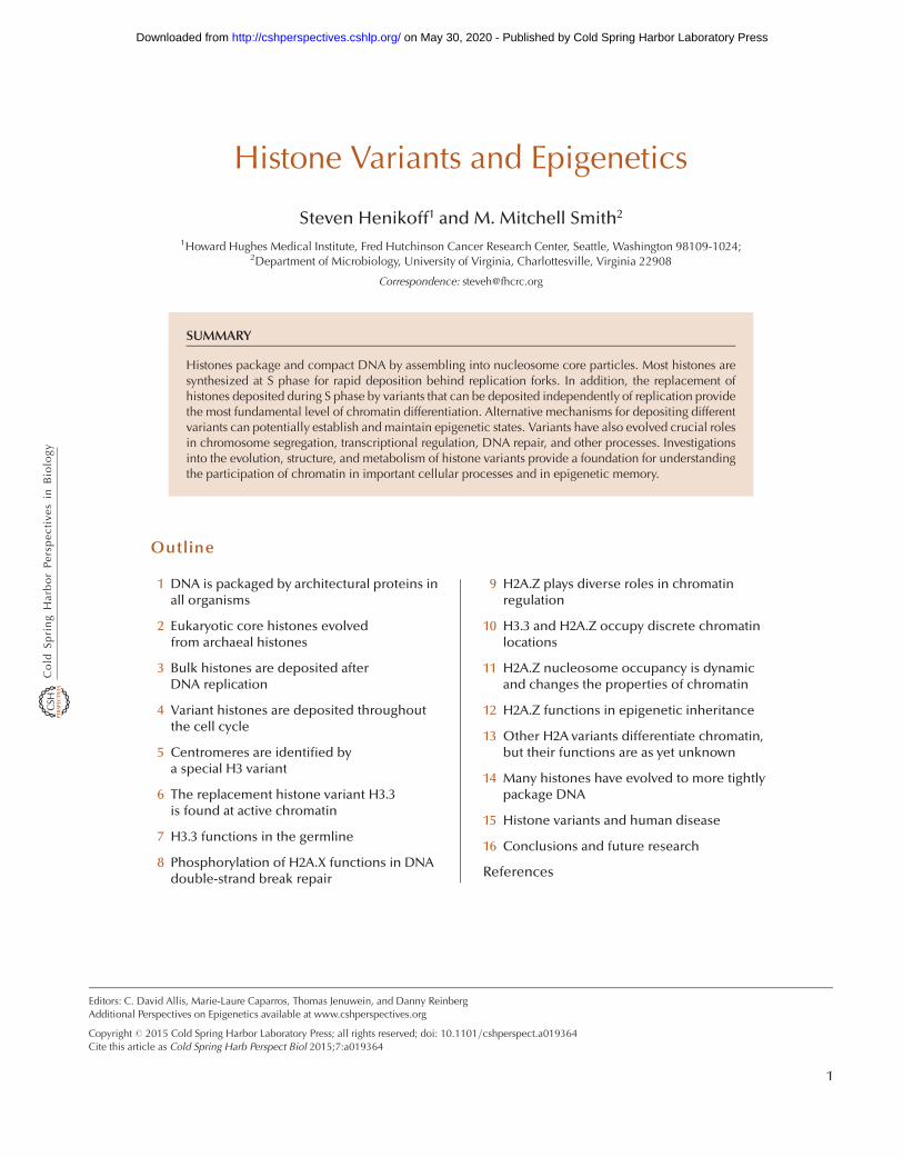
Figure 3. Location of histones H3 (blue) and H2A (brown) in thenucleosome core particle. Differences between variants are highlight-ed in yellow. (Reprinted, with permission, from Henikoff and Ahmad2005.)
Histone Variants and Epigenetics
Cite this article as Cold Spring Harb Perspect Biol 2015;7:a019364 5
on May 30, 2020 - Published by Cold Spring Harbor Laboratory Press http://cshperspectives.cshlp.org/Downloaded from

opmental state is the observation that the identity of one oftwo sister neurons in Caenorhabditis elegans is disrupted bymutating either the H3-H3 dimerization interface or CAF-1 (Nakano et al. 2011).
A prerequisite for epigenetic inheritance of a nucleo-some state is that preexisting nucleosomes must be distrib-uted to daughter chromatids following replication. Indeedthis is the case; classical studies have shown that old nucle-osomes are inherited intact and evidently at random todaughter chromatids (Annunziato 2005). However, morerecent studies of particular cell types and particular locihave challenged this dogma. Germline stem cells in Dro-sophila male early embryos show asymmetric inheritanceof RC H3, but not RI H3.3, whereby old nucleosomes re-main in the stem cell and new nucleosomes segregate to thedifferentiating daughter cell (Tran et al. 2012). Moreover,although RC H3 was not found to split at replication (Xuet al. 2010), the small amount of RI H3.3 that splits wasshown to be enriched at active human genes and cell-type-specific enhancers (Huang et al. 2013). Taken together,these two findings suggest that RC nucleosome assemblyis profoundly involved in initiating developmental deci-sions and RI assembly in epigenetic inheritance, resusci-tating ideas proposed long before these pathways wereelucidated (Weintraub et al. 1976). More work is needed
to test these exciting possibilities and to explore the impor-tance of nucleosome deposition pathways in establishingand maintaining epigenetic states (Jenuwein 2001; Henik-off and Ahmad 2005).
4 VARIANT HISTONES ARE DEPOSITEDTHROUGHOUT THE CELL CYCLE
As we have seen, core histones can be classified based ontheir ancestral sequence and position in the nucleosome.Linker histones are characterized by a winged-helix do-main, rather than a histone-fold domain, and bind to thelinker DNA that separates nucleosomes (Wolffe 1992). Al-though minor variants of these canonical histones exist,they appear to be interchangeable with the major form.For example, mammalian H3.1 and H3.2 differ by a singleamino acid that is not known to impart different functionalproperties to the two isoforms. The existence of multiplegenes that produce large amounts of canonical histones forS-phase deposition is typical of eukaryotic genomes. Thenear ubiquity and overwhelming abundance of canonicalS-phase histones has resulted in relatively little attentionbeing paid to histone variants until recently.
The renaissance of interest in histone variants came inpart from the realization that they differ from canonicalS-phase histones in ways that can lead to profound differ-entiation of chromatin. One way that they differ is in theirmode of incorporation into chromatin. RC assembly in-corporates new nucleosomes into gaps between old nucle-osomes genome-wide, whereas RI assembly involves localreplacement of an existing nucleosome or subunit (Mar-zluff et al. 2002). RI assembly therefore has the potential ofswitching a chromatin state by replacing a canonical his-tone with a variant. Replacing one histone with anotheralso could erase or alter the pattern of posttranslationalmodifications. Therefore, RI assembly can potentially resetepigenetic states that are thought to be mediated by his-tones and their modifications. Recent progress in studyinghistone variants and the processes by which they are depos-ited has led to new insights into the basis for epigeneticinheritance and remodeling. Below we discuss features ofparticular histone variants that contribute to chromatindifferentiation and might be involved in propagating epi-genetic information.
5 CENTROMERES ARE IDENTIFIED BYA SPECIAL H3 VARIANT
A defining feature of the eukaryotic chromosome is thecentromere, which is the site of attachment of spindle mi-crotubules at mitosis. The first centromeres to be describedin molecular detail were those of budding yeast (Saccharo-
CAF1
CAF1
11111
CCAFAFAFFC 1111
CCCAFCAFAFAFF11111
DNA Pol
DNA Pol
PCNA
H3-H4
Figure 4. Distribution of old and new nucleosomes at a replicationfork. Old nucleosomes (gray disks) are randomly distributed behindthe replication fork and new nucleosomes (cyan disks) are depositedin the gaps. CAF-1-mediated nucleosome assembly is depicted onthe leading and lagging strand in magnification. DNA polymerase(green); replication processivity clamp, PCNA (gray ring); histoneH3-H4 tetramers (cyan); newly synthesized DNA (red lines).
S. Henikoff and M.M. Smith
6 Cite this article as Cold Spring Harb Perspect Biol 2015;7:a019364
on May 30, 2020 - Published by Cold Spring Harbor Laboratory Press http://cshperspectives.cshlp.org/Downloaded from

myces cerevisiae), in which a 125-bp sequence is necessaryand sufficient for centromere formation (Amor et al.2004b). However, centromeres of plants and animals arevery different, typically consisting of megabase arrays ofshort tandem repeats. Unlike the situation for buddingyeast, the role of DNA sequence at these complex centro-meres is uncertain because fully functional human neo-centromeres are known to form spontaneously at ectopicsites that entirely lack sequences resembling centromericrepeats (Fig. 5A). These and other observations argueagainst a direct role of DNA sequence in determining thelocation of centromeres (see Allshire and Ekwall 2014).
A key insight into the basis for centromere identity andinheritance came from the identification of a histone H3variant, CENP-Awhich was found to localize specifically tocentromeres and be incorporated into nucleosomal parti-cles in place of H3 itself (Palmer et al. 1991). Remarkably,CENP-A remains associated with centromeres during thetransition from histones to protamines during spermato-genesis, when essentially all other histones are lost (Palmeret al. 1990). This early observation in the study of CENP-Asuggested that CENP-A contributes to centromere identityof the male genome. The generality of this insight was notfully appreciated until it was realized that CENP-A is a muchbetter marker for centromeres than DNA sequence (Fig. 5)(Amor et al. 2004b), and that counterparts of CENP-A(cenH3s) can be found in the genomes of all eukaryotes(Talbert and Henikoff 2010). So, although budding yeastcentromeres are determined by a 125-bp consensus se-quence, this is also the site of a centromeric nucleosomethat contains the cenH3 variant. In fission yeast (Schizosac-charomyces pombe), an array of cenH3-containing nucleo-somes occupies the central core region of the centromereflanked by H3-containing nucleosomes that display hetero-chromatic features (see Fig. 3 of Allshire and Ekwall 2014;Amor et al. 2004b). In flies and vertebrates, cenH3s arepresent in arrays that alternate with H3-containing arraysthat display a unique pattern of histone modifications (Sul-livan and Karpen 2004). Alternation can account for the factthat centromeres occupy only the outside edge of the cen-tromeric constriction of metaphase chromosomes. This isconsistent with the observation that in worm “holokinetic”chromosomes, microtubules attach throughout the lengthof each anaphase chromosome, and cenH3 occupies theleading edge all along its length (Fig. 5C) (Malik and Henik-off 2003). Indeed a unique cenH3 variant is found to pre-cisely mark the centromere in nearly all eukaryotes (Fig.6A). This apparent ubiquity, and the presence of centro-meres to perform mitosis in all eukaryotes, raises the pos-sibility that the first canonical H3 evolved from a cenH3.
Genetic experiments in a variety of eukaryotes haveconfirmed the essentiality of cenH3 for formation of thekinetochore and for chromosome segregation (Amor et al.2004b). Because they remain in place throughout the cellcycle, cenH3-containing nucleosomes form the foundationfor assembly of other kinetochore proteins during mitosisand meiosis (see Allshire and Ekwall 2014). An outstandingquestion in chromosome research is just how these proteinsinteract to provide a linkage between the centromere andspindle microtubules that can hold up to the strong pullingforces exerted on kinetochores at anaphase. Several dozenkinetochore-specific proteins have been identified in yeast(for more detail, see Allshire and Ekwall 2014), althoughexactly how they interact with cenH3-containing nucleo-
A
B C
Figure 5. cenH3s at centromeres of eukaryotes. (A) Human neocen-tromeres (indicated by an arrow) lack centromeric a-satellite DNA,but have CENP-A and heterochromatin. Anti-CENP-A stainingin green and Anti-CENP-B staining in red (which marks a-satelliteDNA) identifies a Chromosome 4 neocentromere that lacks a-satel-lite (main panel). This Chromosome 4 is otherwise normal, havingbeen transmitted for at least three meiotic generations in normalindividuals. Inset shows anti-HP1 staining, which indicates that de-spite the lack of satellite DNA, heterochromatin forms around activeneocentromeres (indicated by arrow). (Reprinted, with permission,from Amor et al. 2004a, # National Academy of Sciences.) (B)Drosophila melanogaster anti-cenH3 antibody (red) stains centro-meres in metaphase chromosomes and throughout interphase. (Im-age courtesy of Suso Platero.) (C) C. elegans anti-cenH3 antibody(green) stains the end-to-end holocentromeres of prophase chromo-somes (red). (Image courtesy of Landon Moore.)
Histone Variants and Epigenetics
Cite this article as Cold Spring Harb Perspect Biol 2015;7:a019364 7
on May 30, 2020 - Published by Cold Spring Harbor Laboratory Press http://cshperspectives.cshlp.org/Downloaded from

Homo H1.0Mus H1.0
Xenopus H1.0
Taenopygia H5
Gallus H5
Danio H1.0
Salmo H1.0
Xenopus H1t
Homo H1.1
Mus H
1.1H
omo H
1.3M
us H1.4
Hom
o H1.4
Hom
o H1.2
Bos H
1.1
Hom
o H
1.5
Hom
o H
1tC
anis
H1t
Mus
H1t
Sal
mo
H1t
Hom
o H
1xG
allu
s H1x
Xenop
us H
1x
Osmerus H
1x
Salmo H1x
Monopleutes H1.1
Clonorchis H1.1
Monopleutes H1.2
Mus H1T2Homo H1T2
Clonorchis H1.2Clonorchis H1.4
Clonorchis H1.5
Aspergillus H1.3
Mus H1oo
Xenopus B4
Rana B4
Danio H
1M1
Saccoglossus H
1oo
Arabidopsis H
1.1
Arabidopsis H
1.2
Arabidopsis H
1.3
Tha
lass
iosi
ra H
1
Aed
es H
1γ
Str
ongy
loce
ntro
tus
H1β
Oik
ople
ura
H1.
2
Cae
norh
abdi
tis H
1.1
Tryp
anos
oma
H1.
1
Caeno
rhab
ditis
H1.
4
Caeno
rhab
ditis
H1.5
Oikopleura H
1.3
Harpegnathos H1.1
Drosophila H1ooDrosophila H1.3
Strongylocentrotus early H1Clonorchis H1.3
Fungi
Plants
Alveoloates
Stramenopiles
Animals
Excavates 0.1
Tetrahymena CenH3Arabidopsis CenH3
Drosophila CenH3Mus CenH3
Caenorhabditis CenH3Saccharomyces CenH3
Tetrahymena H3Tetrahymena H3.3
Dictyostelium H3Saccharomyces H3.3
Human H3.3Human H3
Arabidopsis H3Arabidopsis H3.3
Trypanosoma H2AHuman H2A.B
Chimp H2A.BCow H2A.B
Mouse H2A.BTrypanosoma H2A.Z
Arabidopsis H2A.ZHuman H2A.ZDrosophila H2A.Z.X
Tetrahymena H2A.ZSaccharomyces H2A.Z
Human macroH2ADanio macroH2A
Gallus macroH2AArabidopsis H2A
Arabidopsis H2A.XTetrahymena H2A.XTetrahymena H2A
Saccharomyces H2A.XDrosophila H2A
Human H2AMouse H2A.X
Human H2A.XCow H2A.X
B
A
C
H3 and H3.3
H2A.B
H2A.Z
macroH2A
H2A and H2A.X
cenH3
Mus Hils1Homo Hils1
Figure 6. Histone variant phylogenies. Histone sequences from selected species were multiply aligned and neighbor-joining trees were produced using the EBI server (http://www.ebi.ac.uk/Tools/phylogeny). (A) Histone H3s. (B)Histone H2As. Note that there are no clear phylogenetic distinctions between RC H3 and RI H3.3, and between RCH2A and RI H2A.X. (C) H1 variants from diverse eukaryotes show a “star” phylogeny, which suggests that they arefunctionally interchangeable. (C, Modified from Talbert et al. 2012.)
S. Henikoff and M.M. Smith
8 Cite this article as Cold Spring Harb Perspect Biol 2015;7:a019364
on May 30, 2020 - Published by Cold Spring Harbor Laboratory Press http://cshperspectives.cshlp.org/Downloaded from

somes and other foundation proteins, such as CENP-C,remains unclear.
The evolution of cenH3s is unlike that of any otherhistone class. Whereas histone H3 is almost invariant insequence, which reflects extraordinarily strong purifyingselection on every residue, cenH3s are evolving rapidly,especially in plant and animal lineages (Talbert and Henik-off 2010). This is most evident from the amino-terminaltails, which differ in length and sequence to such an extentthat they cannot be aligned between the cenH3s of differenttaxonomic groups. Even the histone-fold domain of cenH3is evolving orders of magnitude faster than that of H3.What is the reason for this striking evolutionary differencebetween an H3 that functions at centromeres and an H3that functions everywhere else?
Rapidly evolving regions of Drosophila and ArabidopsiscenH3 genes display an excess of replacement nucleotidesubstitutions over what would be expected from the rate ofsynonymous substitutions (Malik and Henikoff 2009).This excess is a hallmark of adaptive evolution. Adaptiveevolution in plants and animals is also seen for anothermajor centromere foundation protein, CENP-C (Malikand Henikoff 2009). Although adaptive evolution is welldocumented for genes involved in genetic conflicts suchas arms races between hosts and parasites, these are theonly known essential single-copy genes that are adap-tively evolving in any organism. In the case of cenH3 andCENP-C, the regions of adaptive evolution correspond toregions of DNA binding and targeting. This suggests thatthe major centromere-binding proteins are adapting tothe evolving centromeric DNA, thus allowing centromericchromatin to interact with the conserved kinetochore ma-chinery that connects the centromere to spindle micro-tubules. It has been proposed that centromeres competeduring female meiosis to be included in the egg nucleusrather than being lost as polar bodies (Malik and Henikoff2009). An arms race would develop leading to expansion ofcentromeres, probably by unequal crossing-over betweensister chromatids. Host suppression of this meiotic driveprocess by cenH3 and CENP-C would lead to an excess ofreplacement changes in regions that interact with DNA.Organisms in which there is no opportunity for centro-meres to compete, such as budding yeast, would not un-dergo centromere drive, and this might account for the factthat they have small centromeres, and their cenH3 andCENP-C proteins are under strong purifying selection.
Thus, we see that a special region of the genome, thecentromere, is distinguished by a single histone variantclass, whose sequences reveal remnants of an arms racethat may have led to the extraordinary complexity of cen-tromeres. The RI assembly process that targets new cenH3-containing nucleosomes to centromeres every cell cycle has
been elucidated by the discovery and detailed characteriza-tion of the related Scm3 (yeast) and HJURP (mammalian)cenH3-specific chaperones (Stoler et al. 2007; Dunleavyet al. 2009; Foltz et al. 2009). Detailed biochemical andstructural characterization of Scm3/HJURP complexes(Shuaib et al. 2010; Cho and Harrison 2011; Hu et al. 2011)indicates a role in CenH3 nucleosome assembly that paral-lels that of other nucleosome assemblycomplexes, discussedfurther in Almouzni and Cedar (2014). Centromeric nu-cleosomes show a remarkable lack of sequence specificityin that they not only can faithfully localize to neocentro-meres that are completely unlike native centromeres (Fig.5A), but also the yeast homolog Cse4 can functionallyreplace human CENP-A (Wieland et al. 2004). It is ex-traordinary that our centromeres have remained in thesame positions for tens of millions of years without anyevident sequence determinants involved in the process thatmaintains them. To the extent that epigenetics refers toinheritance that does not depend on DNA sequence, theinheritance of centromeres on a geological timescale is themost extreme form imaginable. Yet, we are still seeking amechanism to explain how they have maintained them-selves for even a single cell cycle.
There is now general agreement in the centromere fieldthat the cenH3 nucleosome is the key to understandingthe epigenetic inheritance of centromeres (Black andCleveland 2011; Henikoff and Furuyama 2012). It is notonly necessary for recruitment of the other structural com-ponents of the centromere, in some experimental systemsit is also sufficient (Guse et al. 2011; Mendiburo et al.2011). However, its molecular structure has been thesubject of controversy for several years. In vivo evidencefrom flies, humans, and yeast is most consistent with aright-handed hemisome (Henikoff and Furuyama 2012),whereas several groups have shown that reconstitutionof cenH3-containing particles generally results in the for-mation of a partially unwrapped left-handed octamericnucleosome (Black and Cleveland 2011). Indeed, since2007, the authors of this article coauthored the studiesthat provided the first evidence for nonoctameric cenH3particles (Dalal et al. 2007; Mizuguchi et al. 2007), butthe composition and structure of these proposed particleswere completely different! In light of the continuing con-troversy, we leave a final resolution of this important issueto the future.
6 THE REPLACEMENT HISTONE VARIANT H3.3IS FOUND AT ACTIVE CHROMATIN
Like centromeres, transcriptionally active chromatin isthought to be maintained epigenetically and is enrichedin an H3 variant, H3.3, which is the substrate for RI dep-
Histone Variants and Epigenetics
Cite this article as Cold Spring Harb Perspect Biol 2015;7:a019364 9
on May 30, 2020 - Published by Cold Spring Harbor Laboratory Press http://cshperspectives.cshlp.org/Downloaded from

osition (Filipescu et al. 2013). H3.3 is very similar in se-quence to the canonical forms of H3, differing by only fouramino acids. With so few differences, it might have beenassumed that these two forms are interchangeable. How-ever, H3.3 is deposited exclusively by RI nucleosome as-sembly, whereas H3 is deposited only at replication fociin an RC manner. This difference between the two variantsis encoded in the protein itself, with three of the four dif-ferences between H3 and H3.3 evidently involved in pre-venting H3 from being deposited by an RI pathway(illustrated in the a-helix 2 of Fig. 3). Purification of solu-ble nucleosome assembly complexes confirmed that thesetwo forms participate in distinct assembly processes: H3.1copurifies with CAF-1 for RC assembly, and H3.3 copuri-fies with other components, including the HirA and Daxxhistone chaperones, and participates in RI assembly.
Although the four-amino-acid difference might seempractically insignificant, when one considers that humans,flies, and clams have precisely the same H3.3 sequence,these differences from H3 stand out. Phylogenetic analysisreveals that the H3/H3.3 pair evolved at least four separatetimes during eukaryotic evolution, in plants, animals/fun-gi, ciliates, and apicomplexans (Fig. 6A) (Talbert and He-nikoff 2010). Despite having a separate origin from animalsand fungi, the animal H3/H3.3 pair and the pair fromplants (called H3.1 [RC] and H3.2 [RI]—to avoid confu-sion, we will refer to all RC isoforms as H3 and all RIisoforms as H3.3) are strikingly similar. The same clusterof amino acids (positions 87–90) that prevents RI deposi-tion of H3 in Drosophila are found to differ in plants, andthe remaining difference in animals (position 31 is Ala forH3 and either Ser or Thr for H3.3) is also found in plants.Fungi are especially interesting. Ancestrally, they have bothH3 and H3.3; however, ascomycetes, which include yeastsand molds, have lost the H3 form. Thus, the obligate RCform of histone 3 that has received the most attention inanimals is not even present in yeast.
Studies of H3.3 in bulk chromatin showed that it isenriched in transcriptionally active chromatin fractions(Filipescu et al. 2013). However, various factors contributedto the obscurityof this potential “mark” of active chromatinduring a time of great excitement in the chromatin fieldwhen it was realized that histone modifications can distin-guish active from silent chromatin. For one thing, no anti-bodies were available that could effectively distinguish H3from H3.3 in chromatin (positions 87–90 are blocked bythe DNA gyres in the nucleosome), whereas excellent anti-bodies against many different posttranslational modifica-tions were readily available. Also, the seemingly slightsequence differences between H3 and H3.3 did not suggestany fundamental distinctions in chromatin, whereas his-tone modifications were mostly on tail lysines that were
known to affect chromatin interactions or to bind chroma-tin-associated proteins. This perception that the two his-tone 3 forms should be interchangeable was confirmed bythe finding in Tetrahymena and Drosophila that the S-phaseform can in general substitute for its replacement counter-part. Finally, the influential “histone code” hypothesis en-visioned nucleosomes as fixed targets of modificationenzymes during chromatin differentiation (Jenuwein andAllis 2001). However, it has become increasingly evidentthat chromatin is highly dynamic, and even heterochroma-tin-associated proteins bind with residence times of a mi-nute or less (Phair et al. 2004). It appears that the chromatinof actively transcribed genes is in constant flux, character-ized by continual histone replacement (Dion et al. 2007).The three core amino acid differences that distinguish H3and H3.3 make H3.3-H4 dimers the substrate for RI assem-bly, and RI assembly itself profoundly changes chromatin.As a result of this process, actively transcribed regions be-come marked by H3.3 (Fig. 7), and evidence for this processcomes from the observation of RI replacement of H3 meth-ylated on lysine-9 (H3K9me) with tagged H3.3 at RNApolymerase I and II (RNA Pol I and II) transcribed loci(Schwartz and Ahmad 2005).
The dynamic nature of chromatin at active loci resultsin the erasure of preexisting histone modifications, andyet histone modification states persist through multiplerounds of cell division. Therefore, the enzymes responsiblefor modifying histones must be targeted to their sites ofaction. For histone modifications that are typically as-sociated with transcriptionally active chromatin, this isachieved by association with the carboxy-terminal do-main (CTD) of RNA Pol II, a tandem array of YSPTSPS
DAPI H3.3 DAPI + H3.3
Figure 7. H3.3 preferentially localizes to actively transcribed regionsof Drosophila polytene chromosomes. DAPI staining (red) shows theDNA banding pattern (left), and H3.3-GFP (green) localizes to in-terbands (middle), which are sites of RNA Pol II localization. Themerge (Schwartz and Ahmad 2005) is shown on the right. In eachimage, the shorter arrow points to a decondensed interband that isenriched in H3.3, and the longer arrow points to a condensed bandthat lacks H3.3.
S. Henikoff and M.M. Smith
10 Cite this article as Cold Spring Harb Perspect Biol 2015;7:a019364
on May 30, 2020 - Published by Cold Spring Harbor Laboratory Press http://cshperspectives.cshlp.org/Downloaded from

heptamers. For example, the Set1 H3K4 methyltransferaseassociates with the CTD when it is heavily phosphorylatedon Serine-5 during transcriptional initiation, and so en-counters its substrate primarily near initiation sites. Simi-larly, the Set2 H3K36 methyltransferase associates with theCTD when it becomes heavily phosphorylated on Serine-2during transcriptional elongation and encounters its sub-strate within gene bodies. When a nucleosome is evictedand replaced with unmodified histones during transcrip-tional elongation, the newly deposited H3.3 is thereforemodified appropriately.
It appears that an analogous process maintains histonemodifications that are typically associated with silent chro-matin. Nucleosomes that are lost at sites of short-periodtandem repeats, such as occur at mammalian telomeres andpericentric regions, are replaced by the Daxx H3.3-specifichistone chaperone complex and the ATRX ATP-dependentnucleosome remodeling protein (Fig. 8) (Drane et al. 2010;Goldberg et al. 2010). ATRX has a bifunctional histone tailrecognition domain with high affinity for an H3 tail that isboth unmethylated at K4 and trimethylated at K9 (Euster-mann et al. 2011), and so likely is recruited to telomericsites that are enriched for H3K9me and lack H3K4me.Telomeres are also enriched for heterochromatin-associat-ed protein 1 (HP1), which recruits the Su(var)3-9 H3K9methyltransferase, and binds its H3K9 methylated product(Hines et al. 2009), and so an enzyme that methylates thetails of replacement H3.3 at telomeres is present at a highlocal concentration in which new H3.3 is incorporated.This implies that all of the components necessary for main-taining H3K9 methylation are present at telomeres: the
enzyme that performs the modification, the modifica-tion-specific-binding module on the machine that usesATP to provide energy for the replacement process, andthe fresh unmodified H3.3 substrate that becomes incor-porated into the new nucleosome (Fig. 8). It seems likelythat a similar process occurs at other sites of short-periodtandem repeats in DNAwhere nucleosomes frequently turnover, insofar as ATRX is also abundant in mammals at C +G-rich sites (Law et al. 2010), which are found at mostpromoters, and the Drosophila ortholog of ATRX, XNP, isabundantly present at a single site of a (GATA)n repeatwhere H3.3 is actively incorporated (Schneiderman et al.2009). The incorporation of H3.3 nucleosomes at sites oftelomeric heterochromatin belies the common notion thatH3.3 is a “mark” of active chromatin. As the general sub-strate for replacement of nucleosomes wherever they arelost, the finding that H3.3 is mostly incorporated at activegenes rather implies that these are sites where nucleosometurnover is most intense. This generic replacement func-tion of H3.3 has important relevance to human disease asdescribed in Section 15.
7 H3.3 FUNCTIONS IN THE GERMLINE
When cells exit the cell cycle and differentiate, they nolonger produce or incorporate S-phase histones, andH3.3 accumulates as a result. For example, H3.3 accumu-lates in rat brains to a level of 87% of the histone 3 by thetime that rats are 400 days old (Pina and Suau 1987). Thisclassical observation suggested that replacement by H3.3has no functional significance except to prevent holes in the
ATRXXX
DA
XX
Suv39h
HP1Me
HP1
SSuSuv39huv39hv39hv39hv39hv39hv39h39393939SS
HP1MMMMMMMMMMMMeeeeeeeeeeeeeeeeeeeeeeeeeeeeeeeeeeeeeeeeeeeee
HP1 Me
ATRX
Suv39h
HP1Me
HP1
SSuv39hSuvuv39hv39hv39hv39h39h39h339h9SS
HP1HMMMMMMMMMMMMeeeeeeeeeeeeeeeeeeeeeeeeeeeeeeeeeeeeeeeeeeeeeeeeeee
HP1H Me
Me
ATRRXX
Me
X
Me
A B
ATP
ADP
ADDdomain
Figure 8. Model for maintenance of histone modifications by the concerted action of multiple chromatin regulatorsvia RI replacement with H3.3. We address the question of how a histone modification can be inherited whena nucleosome is lost and replaced. (A) The Suv39h H3K9 methyltransferase (an ortholog of fly Su(var)3-9) isrecruited by HP-1 protein, which binds specifically to methylated H3K9. To perpetuate this mark when thenucleosome turns over, we speculate that the ATRX ATPase is recruited to the site via its ATRX-DNMT3-DNMT3L(ADD) domain, which binds with high specificity to methylated H3K9 on tails that entirely lack H3K4 methylation(because there are no H3K4 methyltransferases in this region of the genome). (B) ATRX provides the energy of ATPand works together with the H3.3-specific DAXX histone chaperone complex to incorporate the new nucleosome(Goldberg et al. 2010), or half-nucleosome in the case of partial eviction (Xu et al. 2010). The high local concen-tration of Suv39h results in a new nucleosome with the same H3K9 methylation as the nucleosome that was lost.
Histone Variants and Epigenetics
Cite this article as Cold Spring Harb Perspect Biol 2015;7:a019364 11
on May 30, 2020 - Published by Cold Spring Harbor Laboratory Press http://cshperspectives.cshlp.org/Downloaded from

nucleosomal landscape. Consistent with this view, H3.3 hasbeen found to be nonessential for Drosophila development,as flies lacking both H3.3 genes develop normally to thepupal stage, with occasional adult escapers that die shortlyafter eclosion but show no specific morphological defects(Hodl and Basler 2009; Sakai et al. 2009). Moreover, H3.3can functionally substitute for H3 and allow at least somedevelopmental decisions to be made in Drosophila embry-os, further suggesting that RC and RI substrates are largelyinterchangeable (Hodl and Basler 2012). It appears thathistone replacement in H3.3 null proliferating cells canbe accomplished by incorporating H3 nucleosomes usingthe RI pathway. However, there is no germline developmentin flies lacking key RI pathway components, such as HirA(e.g., Fig. 9). Females that lack the ChD1 ATP-dependentnucleosome remodeler protein are also sterile, evidentlybecause ChD1 is required in the zygote for sperm nucleidecondensation and the replacement of protamines by ma-ternally encoded RI histones (Orsi et al. 2009). This essen-tial germline function of the RI pathway is conserved in
mammals, in which H3.3 is required for remodeling ofboth maternal and paternal gametes (Santenard et al.2010; Akiyama et al. 2011). Similar RI processes havebeen documented in both C. elegans and Arabidopsis, inwhich maternal H3.3 is incorporated into the paternal ge-nome of the zygote (Ooi et al. 2006; Ingouff et al. 2007).Therefore, germline remodeling via RI assembly of H3.3nucleosomes is a universal process that has evolved in bothanimals and plants, most likely to “reset” the chromatinlandscape to a totipotent state.
Remodeling events in the zygote, where RI assembly ofH3.3 nucleosomes plays a key role, is analogous to nuclearreprogramming, which can be artificially induced in Xe-nopus eggs, mouse embryonic stem cells, and inducedpleuripotent cells. In Xenopus, transfer of an embryonicnucleus into an enucleated egg can result in the productionof mostly normal embryos that nevertheless sometimes(mis)express genes that were active in the differentiateddonor nucleus (Ng and Gurdon 2008). The lack of observ-able gene expression during the intervening 12 embryonic
H2A-H2B
H3 -H4
FACT
H3-H4
H3.3-H4
H2A -H2B H2AZ-H2B
SWR1Pol
H2A-H2B
REPLACE H3-H4 REPLACE H2A-H2B
H3.3-H4
HIRA
Figure 9. Models for RI replacement or exchange. A large molecular machine (either the SWR1 complex or RNApolymerase) partially or completely unravels a nucleosome during transit. The result is either retention of hetero-dimeric subunits, such as the FACT-facilitated transfer of H2A-H2B from in front of RNA polymerase to behind(Formosa et al. 2002; Belotserkovskaya et al. 2003) or loss of a heterodimer. In the latter case, chromatin repairreplaces the lost heterodimer with either H3.3-H4 (left) or H2A.Z-H2B (right).
S. Henikoff and M.M. Smith
12 Cite this article as Cold Spring Harb Perspect Biol 2015;7:a019364
on May 30, 2020 - Published by Cold Spring Harbor Laboratory Press http://cshperspectives.cshlp.org/Downloaded from

divisions implies that the persistence of an epigenetic markmaintains the memory of prior gene activity. Overexpres-sion of H3.3 in developing embryos improved epigeneticmemory and mutation of H3.3K4 to glutamine erasedmemory of the active state, whereas no effect was seen usinga general DNA methyltransferase inhibitor. Further evi-dence for the importance of H3.3 in nuclear reprogram-ming comes from the finding that the shift from somatic tooocyte transcription does not require replication, but doesrequire transcription and the H3.3-specific chaperone,HirA (Jullien et al. 2012). It is attractive to think of H3.3and its histone partners as general mediators of totipotencyboth in the zygote and during nuclear reprogramming,insofar as histones are likely to be most accessible to theenzymes that posttranslationally modify their tails duringnucleosome assembly (e.g., Fig. 8).
8 PHOSPHORYLATION OF H2A.X FUNCTIONSIN DNA DOUBLE-STRAND BREAK REPAIR
The H2A histones also comprise a family of distinct vari-ants found throughout eukaryotes (Fig. 6B). The H2A.Xvariant is defined by the presence of a carboxy-terminalamino acid sequence motif, SQ(E or D)Ø, in which Øindicates a hydrophobic amino acid. The serine in thissequence motif is the site of phosphorylation, producinga modified protein designated “gH2A.X.” The dynamicnature of chromatin, and H2A.X phosphorylation, is espe-cially evident when double-strand breaks (DSBs) occurin DNA (Morrison and Shen 2005). The lethality of evena single double-stranded (ds) break requires immediateaction to repair the lesion and restore the continuity ofthe double helix. The detection of a ds break normallyoccurs within a minute or so of its formation and this,in turn, triggers the rapid phosphorylation of H2A.X inthe immediate vicinity of a break site. This phosphory-lation is performed by members of the phosphoinositol3-kinase-like kinase family. Following this initial event,H2A.X phosphorylation then spreads quickly along thechromosome marking a relatively large chromatin domainsurrounding the break. Finally, the ds break is eventuallyrepaired by either homologous recombination or nonho-mologous end-joining and the phosphorylation mark isremoved.
Phosphorylation of H2A.X is not essential for detectionor repair of DSBs because deletion of the gene or mutationof the target serine residue does not abolish repair. How-ever, H2A.X is not just a marker of damage because suchmutants have reduced efficiency of repair and are hyper-sensitive to radiation damage and genotoxic agents. Cur-rently, H2A.X is thought to function in ds break repair in atleast two ways. First, it may help recruit or retain proteins
required for repair at the site of the break (Morrison andShen 2005). Second, it may stabilize the chromosome sur-rounding the broken ends through the recruitment of co-hesin, the protein complex responsible for keeping sisterchromatids together (Lowndes and Toh 2005).
The evolution of H2A.X is unlike that of other histonevariants. Although a gene for H2A.X is found in nearly alleukaryotes, it has had multiple relatively recent divergencesfrom H2A (Fig. 6B) (Malik and Henikoff 2003; Talbert andHenikoff 2010). For example, the version of H2A.X foundin Drosophila is different from that found in another dip-teran insect, Anopheles. Some organisms, such as yeasts,have an H2A.X but lack an H2A, consistent with the pos-sibility that most present-day canonical H2As have evolvedfrom ancestral H2A.Xs (Talbert and Henikoff 2010). Pre-sumably, the ability to evolve either a canonical H2A froman H2A.X or vice versa is a consequence of the simplicityof the SQE motif. Depending on selective constraints,the loss or gain of such a simple motif at the carboxylterminus of a protein might occur repeatedly over evolu-tionary time. Occasional loss of an existing H2A.X with anewly minted version might be fueled by the need forH2A.X to be very uniformly distributed because DSBscan occur anywhere in the genome. If mutations occur inan existing H2A.X gene that reduce its similarity to thecanonical H2A in such a way that its assembly becomesless efficient or uniform, then there will be strong selectionto replace it with a version that is more similar to canonicalH2A. This rationale could help account for the exceptionalcase of Drosophila H2A.X, which unlike other eukaryotes,is not derived from its canonical H2A, but rather from thedistant H2A.Z variant lineage (Baldi and Becker 2013).If all that is necessary to be an H2A.X is to be in the H2Aposition in a nucleosome and to have the carboxy-terminalmotif for phosphorylation, then an H2A.Z can evolve thiscapability.
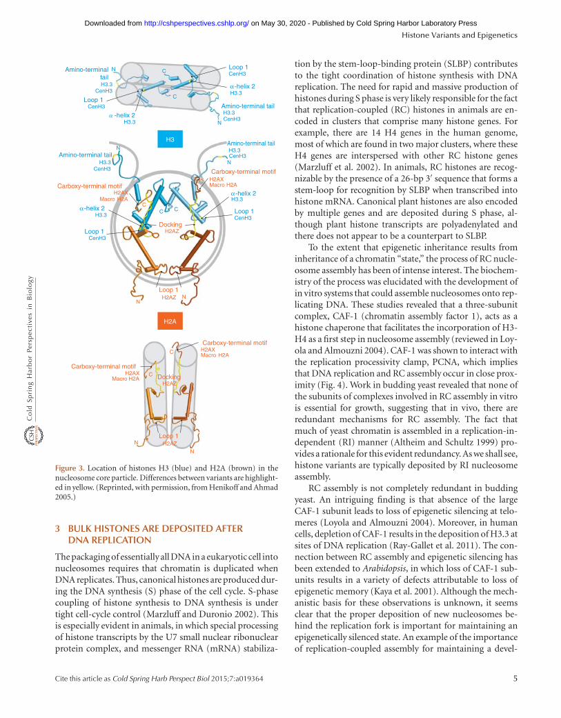
DSB repair is clearly the universal function of H2A.Xphosphorylation, and there would seem to be no stableepigenetic aspect to this process. However, H2A.X nullmice are sterile, and cytological examination of mammali-an spermatogenesis has revealed a striking epigenetic fea-ture in which H2A.X is specifically phosphorylated on theXY bivalent (Fig. 10) (Fernandez-Capetillo et al. 2003).This chromosome pair occupies a distinct “sex body” dur-ing meiotic prophase, which has been implicated in silenc-ing of sex-linked genes during male meiosis. H2A.Xphosphorylation is essential for normal sex-body forma-tion, and H2A.X-deficient spermatocytes fail to pair orcondense and fail to inactivate X and Y genes during mei-osis. H2A.X phosphorylation of the XY bivalent is distinctfrom the process that occurs at DSBs. XY phosphorylationin the sex body does not require breaks, but rather occurs
Histone Variants and Epigenetics
Cite this article as Cold Spring Harb Perspect Biol 2015;7:a019364 13
on May 30, 2020 - Published by Cold Spring Harbor Laboratory Press http://cshperspectives.cshlp.org/Downloaded from
most conspicuously at unpaired regions of the chromo-somes. The mechanisms whereby H2A.X phosphorylationis targeted to unpaired chromosomes and how this eventleads to condensation, pairing, and silencing are currentlyunknown. However, it is interesting to speculate that thisrole may be related to its ability to interact with and recruitcohesin.
9 H2A.Z PLAYS DIVERSE ROLES IN CHROMATINREGULATION
Histone variant H2A.Z is found throughout most eukary-otic lineages and it has been intensely studied for its struc-ture and function in chromatin biology (Zlatanova andThakar 2008; Draker and Cheung 2009; Marques et al.2010; Talbert et al. 2012). H2A.Z diverged from an ancestralH2A early in evolution and shares only �60% similaritywith its major histone H2A counterpart. Consistent withthis separate lineage, genetic experiments in yeast, plants,flies, and mammals have shown that histones H2A andH2A.Z have evolved to play separate nonoverlapping func-tions. H2A.Z is an essential histone in most organisms, fromciliated protozoans to mammals. However, in budding andfission yeasts, cells that carry a deletion of the single-copyH2A.Z gene are viable, although the null mutants show a
varietyof conditional lethal phenotypes. Plants, as exempli-fied by Arabidopsis thaliana, have three closely relatedH2A.Z genes, HTA8, HTA9, and HTA11, with roughly90% identity and a more distantly related HTA4 gene (Tal-bert and Henikoff 2010). There appears to be functionalredundancy among these genes as organisms with singledeletions of either HTA8, HTA9, or HTA11 are normal,whereas the hta9 hta11 double mutant shows developmen-tal defects. Interestingly, vertebrates have evolved two closelyrelated H2A.Z variants, H2A.Z.1 and H2A.Z.2, which differat only three amino-acid residues (Dryhurst et al. 2009;Matsuda et al. 2010; Mehta et al. 2010). These two variantsshowdifferent patterns of chromatindistribution (Dryhurstet al. 2009) and are apparently nonredundant as the sin-gle deletion of H2A.Z.1 in the mouse is lethal (Faast et al.2001).
Evidence for functional diversification of H2A.Z comesfrom the discovery of an alternatively spliced form of hu-man H2A.Z.2 mRNA that results in a protein with a re-duced CTD. This shorter H2A.Z isoform destabilizesnucleosomes and is most enriched in brain (Bonisch et al.2012), in which H2A.Z, like H3.3, is known to be especiallyabundant (Pina and Suau 1987). Although this particularisoform appears to be limited to primates, evidence for alonger alternatively spliced isoform with potential for nu-cleosome destabilization was reported in carp brain (Simo-net et al. 2013). With the increasing popularity of RNA-seqfor identification of alternatively processed variants, weexpect that other examples of potential H2A.Z functionaldiversification will be discovered.
The high-resolution structure of an H2A.Z-containingnucleosome reveals several unique properties of the variant(Suto et al. 2000). Compared with H2A nucleosomes,H2A.Z presents an extended acidic patch domain on thesurface of the nucleosome and mutational studies haveshown this to have functional significance. The acidic patchis then part of a larger “docking domain,” an essential partof the protein necessary for interaction with H3 in thenucleosome. Like other histones, H2A.Z is subject to avariety of posttranslational modifications, including acet-ylation, ubiquitylation, and sumoylation. There is stronggenetic and biochemical evidence that these modificationsaffect the localization, dynamics, and function of H2A.Znucleosomes (Talbert and Henikoff 2010).
H2A.Z has been linked to a wide variety of differentand sometimes contradictory nuclear functions, includ-ing transcriptional activation, transcriptional repression,RNA Pol II elongation, heterochromatin, antisilencing,cell-cycle control, DNA replication, DNA damage repair,chromosome segregation, and genome integrity (Zlatano-va and Thakar 2008; Altaf et al. 2009; Marques et al. 2010;Talbert and Henikoff 2010; Xu et al. 2012; Adkins et al.
X
Y
Y
X
H2AX+/+
H2AX–/–
SCP3 SCP3 + XMR
Figure 10. Pachytene stage of spermatogenesis showing the depen-dence of sex-body formation on H2A.X. In normal mammalianspermatocytes, a nuclear structure, the sex body (arrow, green, inright panels), is seen to encompass the unpaired XY bivalent (labeledin left panels). The synaptonemal complex, which aligns paired chro-mosomes, is stained red. H2A.X is normally enriched in the sex body(H2A.X+/+). In H2A.X2/2 spermatocytes, the sex body does notform and a sex-body epitope becomes dispersed (lower right). Scalebar, 10 mm. (Images courtesy of Shantha Mahadevaiah and PaulBurgoyne; Fernandez-Capetillo et al. 2003.)
S. Henikoff and M.M. Smith
14 Cite this article as Cold Spring Harb Perspect Biol 2015;7:a019364
on May 30, 2020 - Published by Cold Spring Harbor Laboratory Press http://cshperspectives.cshlp.org/Downloaded from
2013). It likely has direct mechanistic roles in transcriptioninitiation, elongation, antisilencing, and DNA damage re-pair. In some other cases, evidence suggests that H2A.Zfunction is manifest indirectly through its involvementin transcription. For example, delay in the G1-S transitionobserved in budding yeast deleted for H2A.Z is likely dueto the misregulation of cyclin gene expression and notdefects in DNA replication initiation (Dhillon et al.2006). In S. pombe, and perhaps other organisms, H2A.Zcooperates with heterochromatin-silencing factors (Clr4/SUV39H) to enforce RNA-processing fidelity and pre-vent deleterious antisense transcription (Zofall et al. 2009).Loss of this enforcement may account for the genome in-stability and chromosome segregation defects observedin H2A.Z deletion mutants. Indeed, at least part of thechromosome segregation defects observed in H2A.Z mu-tants of S. pombe might be caused by decreased trans-criptional expression of the centromere protein, CENP-C(Hou et al. 2010). Nevertheless, such examples of indirectfunction are relatively rare. Indeed, ruling out indirect ac-tivities for any chromatin regulator is a challenging prob-lem, particularly for H2A.Z in organisms for which it isessential.
10 H3.3 AND H2A.Z OCCUPY DISCRETECHROMATIN LOCATIONS
Much of our understanding of histone variant functionis inferred from its patterns of genomic chromatin oc-cupancy. H3.3 makes up �15%–25% of total H3 proteinand H2A.Z makes up �5%–10% of the total H2A pro-tein in most organisms examined to date. Abundances in-crease when cells exit from the cell cycle and no longerreplicate their DNA, such as during development (Pinaand Suau 1987). These variants are widely, but not uni-formly, distributed throughout the genome. High-res-olution chromatin immunoprecipitation experiments ina number of model organisms have revealed that bothH3.3 and H2A.Z preferentially occupy nucleosomes thatflank gene promoters and both are particularly enrichedat the +1 nucleosome bordering transcriptional start sites(TSSs). They are often enriched at the 21 or 22 nucleo-some as well and thus flank a nucleosome-depleted re-gion at the TSS (Talbert and Henikoff 2010). In animals,the H3.3 over gene bodies correlates with transcriptionallevels, suggesting that it replaces nucleosomes that are oc-casionally lost during transcription (illustrated in the leftpanel of Fig. 9). Direct evidence for this interpretationcomes from measuring nucleosome turnover by metaboliclabeling, which showed nucleosome turnover patternsclosely matching H3.3 patterns genome-wide (Deal et al.2010).
H2A.Z has been mapped genome-wide in a variety ofeukaryotes. In budding yeast, nematodes, and plants,H2A.Z occupancy around the promoter is correlatedwith nontranscribing genes “poised” for activation (Zhanget al. 2005; Mavrich et al. 2008; Whittle et al. 2008; Kumarand Wigge 2010). However, in flies and mammals, pro-moter H2A.Z occupancy appears to correlate more withactively transcribing genes (Barski et al. 2007; Mavrichet al. 2008; Hardy et al. 2009; Hardy and Robert 2010; Kellyet al. 2010), similar to the situation for H3.3. Althoughpreferentially found at promoters and regulatory sites,H2A.Z nucleosomes can also be found at lower frequencyin gene bodies and elsewhere (Hardy et al. 2009; Weber etal. 2010; Santisteban et al. 2011). Enrichment of H2A.Zover gene bodies closely corresponds to that of chromatinthat is extracted with low salt, suggesting that H2A.Zchanges the physical properties of nucleosomes (Weberet al. 2010).
H2A.Z is also specifically deposited near or within het-erochromatin. In budding yeast, H2A.Z is enriched neartelomeres where it serves as an antisilencing factor. Dele-tion of the H2A.Z gene results in extended spreading ofsilent chromatin inward from the telomeres and this defectcan be suppressed by the additional deletion of genes en-coding the silencing factors themselves (see Grunstein andGasser 2013 for more detail). Indeed, this function mayact globally, in parallel with the Set1 histone H3 methyl-transferase, to prevent large-scale aberrant distributionof silencing factors (Venkatasubrahmanyam et al. 2007).In metazoans, H2A.Z is also localized in facultative andconstitutive heterochromatin, the inactive X chromosome,transposable elements, and pericentric heterochromatin(Greaves et al. 2007; Draker and Cheung 2009; Boyarchuket al. 2011; Zhang and Pugh 2011).
In contrast to the chromosomal features that are cor-related with H2A.Z, there is a remarkable anticorrela-tion between histone H2A.Z nucleosome occupancy andDNA methylation (Zilberman et al. 2008; Kobor and Lor-incz 2009; March-Diaz and Reyes 2009; Conerly et al.2010; Edwards et al. 2010; Zemach et al. 2010; see Li andZhang 2014 for a discussion of DNA methylation). Thereis strong evidence that this mutual antagonism is causaland not simply correlative. Mutants in A. thaliana withdecreased DNA methylation show an increase in H2A.Zoccupancy at loci where it is normally not found, indepen-dent of transcriptional activity. Conversely, mutants defec-tive in H2A.Z deposition show increased DNA methylationover gene bodies normally occupied by H2A.Z nucleo-somes. While the precise molecular pathways that accountfor this mutual exclusion remain to be elucidated, thisfunctional relationship has important implications for de-velopment and carcinogenesis. For example, the stochastic
Histone Variants and Epigenetics
Cite this article as Cold Spring Harb Perspect Biol 2015;7:a019364 15
on May 30, 2020 - Published by Cold Spring Harbor Laboratory Press http://cshperspectives.cshlp.org/Downloaded from
or environmentally effected loss of H2A.Z from the pro-moter of a tumor-suppressor gene could well contribute tolocally increased DNA methylation and heritable epigenet-ic repression.
11 H2A.Z NUCLEOSOME OCCUPANCY ISDYNAMIC AND CHANGES THE PROPERTIESOF CHROMATIN
The dynamic exchange of H2A.Z nucleosomes in chroma-tin appears to be an important part of its function. (SeeBecker and Workman 2013 for an in-depth discussion ofhistone exchange.) Unlike the major core histones, H2A.Zexpression is not restricted to S phase and it can be incor-porated into chromatin independent of DNA replication.The deposition of H2A.Z into nucleosomes is performedby multisubunit protein complexes, which have been con-served throughout the eukaryotic kingdom (Lu et al. 2009;March-Diaz and Reyes 2009; Morrison and Shen 2009).First identified in budding yeast, the SWR1 complexes con-tain, as their catalytic subunits, homologs of the proteinSwr1, a member of the SWI/SNF family of ATP-dependentchromatin remodelers. The substrate for SWR1 is anH2A.Z-H2B dimer, which is used to replace one of theexisting H2A-H2B dimers in the nucleosome in an ATP-dependent exchange reaction (Fig. 9). This reaction is step-wise and unidirectional, in vitro, resulting in the completereplacement of H2A-H2B dimers with H2A.Z-H2B dimers(Luk et al. 2010). In vivo, unidirectional replacement ofH2A with H2A.Z by SWR1 is enforced by acetylation ofH3K56, which allows the reverse reaction to occur, result-ing in local reduction in H2A.Z incorporation, thus mod-ulating transcription (Watanabe et al. 2013). SWR1 is likelydedicated to the task of replacing H2Awith H2A.Z becausethe effects of eliminating SWR1 function are similar to theeffects of deleting the gene encoding H2A.Z itself. Indeed,the activity of the SWR1 complex is actually deleterious tothe cell in the absence of its H2A.Z-H2B substrate (Halleyet al. 2010; Morillo-Huesca et al. 2010).
The removal of H2A.Z from chromatin proceeds by atleast two pathways. Nucleosome exchange and eviction oc-curs in many contexts such as the remodeling and evictionof complete nucleosomes that can happen at promoters.Any H2A.Z that is a part of those nucleosomes will beremoved as well. However, there is also evidence thatH2A.Z-H2B dimers may be specifically removed from nu-cleosomes by the INO80 complex, a close relative of SWR1(Morrison and Shen 2009; Papamichos-Chronakis et al.2011). In budding yeast, the elimination of INO80 resultsin the global mislocalization of H2A.Z and a decrease in itsapparent exchange rate. Additional mutational results areconsistent with this interpretation. In vitro, purified INO80
is reported to catalyze the replacement of a nucleosomalH2A.Z-H2B dimer with a canonical H2A-H2B dimer, thatis, the reverse of the SWR1 reaction (Luk et al. 2010; Papa-michos-Chronakis et al. 2011), and this reaction may alsobe regulated byacetylation of H3K56 (Watanabe et al. 2013).
The factors that determine where H2A.Z is depositedare incompletely understood. At present, there is little ev-idence that H2A.Z templates its own deposition epigenet-ically (Viens et al. 2006). In budding yeast, a DNA sequencerelated to the binding site of transcription factor Reb1 isable to target the enrichment of H2A.Z at ectopic sites,independent of Reb1 (Raisner et al. 2005). In other cases,transcription factors themselves are implicated in targetingH2A.Z deposition (Updike and Mango 2006; Zachariou-dakis et al. 2007; Gevry et al. 2009). The unifying themeunderlying the function of these various factors appears tobe the creation of a nucleosome-depleted region necessaryfor H2A.Z deposition, although how this recruits H2A.Z isnot currently known (Hartley and Madhani 2009). Inter-estingly, SWR1 complexes often contain homologs of yeastBrd1, a protein containing dual bromodomain motifs ca-pable of binding acetylated lysines. Thus, SWR1 may berecruited to, or stabilized at, chromatin neighborhoodsrich in acetylated histones. H2A.Z deposition may also beblocked at specific loci. The S. pombe SWR1 complex con-tains a regulatory subunit, Msc1, which is dispensable forthe loading of H2A.Z at promoter nucleosomes, but whichis required to prevent H2A.Z deposition within the chro-matin of the inner centromere and subtelomeric regions(Buchanan et al. 2009; Zofall et al. 2009). It has also beensuggested that an additional pathway directing localizedoccupancy by H2A.Z may involve its random depositionand then specific eviction, perhaps as a consequence oftranscription (Hardy and Robert 2010).
In any case, the consequence of H2A.Z deposition andreplacement can be complex. Because of the exchange re-actions, nucleosomes in cellular chromatin may be “ZZ,”“ZA,” or “AA,” containing two, one, or zero H2A.Z-H2Bdimers, respectively (Luk et al. 2010; Weber et al. 2010).In Drosophila, the pattern of ZZ and ZA nucleosome oc-cupancy is different, and homotypic H2A.Z nucleosomesare enriched over the bodies of active genes, perhaps as aconsequence of transcription elongation. In mouse tro-phoblast cells, there are distinct changes in the promotercontent of ZZ and ZA nucleosomes during G1, S, and Mphase, independent of actual transcription activity, sug-gesting a major remodeling pathway dependent on thecell-division cycle (Nekrasov et al. 2012). Differential post-translational modifications further enrich the situation asH2A.Z acetylation has been linked with both SWR1 andINO80 functions (Millar et al. 2006; Papamichos-Chron-akis et al. 2011).
S. Henikoff and M.M. Smith
16 Cite this article as Cold Spring Harb Perspect Biol 2015;7:a019364
on May 30, 2020 - Published by Cold Spring Harbor Laboratory Press http://cshperspectives.cshlp.org/Downloaded from
A quantitative evaluation of the +1 nucleosome inyeast revealed that at steady state the relative abundanceof ZZ, ZA, and AA nucleosomes is roughly 32%, 24%,and 44% respectively (Luk et al. 2010). How can H2A.Zdrive chromatin function with this level of heterogeneity?One explanation is that H2A.Z nucleosomes are in dynam-ic exchange at these sites. In budding yeast, nucleosomeswith rapid turnover were identified by the kinetic incorpo-ration of newly synthesized histone H3. These “hot” nucle-osomes preferentially map to promoter regions, includingthe nucleosomes at the TSS, which are enriched in H2A.Zhistones (Dion et al. 2007). Remarkably, in budding yeastH2A.Z appears to increase the global turnover of nucleo-somes and not simply those into which it is most abun-dantly incorporated (Dion et al. 2007; Santisteban et al.2011). The mechanistic basis for this influence is currentlyunknown.
The stability of H2A.Z-containing nucleosomes in vitrohas been examined in many studies with contrasting results(Zlatanova and Thakar 2008; Talbert and Henikoff 2010).In vivo it is clear that not all H2A.Z nucleosomes are createdequal and that the lability of H2A.Z nucleosomes is affectedby posttranslational modifications and the presence of oth-er variant histones. Nucleosomes containing both H2A.Zand H3.3, especially H2A.Z/H2A nucleosomes with H3.3,are particularly unstable and sites occupied by these dou-ble-variant nucleosomes may be erroneously scored asentirely free of nucleosomes, depending on how the chro-matin is isolated (Jin et al. 2009; Nekrasov et al. 2012).H2A.Z acetylation generally destabilizes nucleosomes invitro and in vivo is associated with gene activation (Tanabeet al. 2008; Wan et al. 2009; Halley et al. 2010).
A particularly striking example of the differential prop-erties of H2A.Z nucleosomes is the transcriptional re-sponse of cells to temperature. Plants possess a signalingpathway that senses ambient temperature and regulatesgene expression (see Baulcombe and Dean 2014 for a dis-cussion of plant responses to environmental factors). Thedevelopmental program for flowering, for example, is ac-celerated at higher temperature. To identify factors thatregulate this response, A. thaliana was screened for mutantswith a constitutive high temperature expression patternand the mutations turned out to be in ARP6, which en-codes one of the conserved subunits of the SWR1 complex(Kumar and Wigge 2010). Indeed, H2A.Z occupancy at thepromoters of heat responsive genes was found to decreasewith increasing temperature and this loss was independentof transcriptional activity per se. These findings have po-tentially broad implications for agriculture in a globallywarming environment, insofar as depletion of H2A.Z in amodel cereal phenocopies the ambient temperature re-sponse and impacts grain yield (Boden et al. 2013).
12 H2A.Z FUNCTIONS IN EPIGENETICINHERITANCE
There is increasing evidence that H2A.Z participates in theheritable specification of chromatin function. Chromo-somes undergo extensive remodeling each mitotic genera-tion and the specification of silent and active chromatindomains across division cycles is essential for normal de-velopment. In budding yeast, H2A.Z is transiently lost froma subset of genes during telophase, as measured by chro-matin immunoprecipitation and this displacement is re-quired for the establishment of heterochromatin in aninducible model (Martins-Taylor et al. 2011). In mamma-lian cells most active transcription is repressed during mi-tosis and must be reactivated in the next cell cycle. The +1nucleosome containing H2A.Z is a prime candidate formediating this control and its translational positioning atTSSs has been examined in G0/G1-phase, S-phase, and M-phase cells (Kelly et al. 2010; Nekrasov et al. 2012). Indeed,the positions of these nucleosomes were found to be shiftedduring mitosis, altering the size of the nucleosome-deplet-ed region and remodeling chromatin at the TSS. Togetherthese features may help establish epigenetic memory dur-ing mitosis and rapid gene reactivation following cell divi-sion. The pathways regulating these cell-cycle behaviors ofH2A.Z are currently unknown.
The location of a gene within the nucleus can be impor-tant for its expression. Positioning at the nuclear peripheryis often associated with gene inactivation with the exceptionof localization at nuclear pores, which is a positive factor inexpression (reviewed in Akhtar and Gasser 2007). Severalgenes in budding yeast, such as GAL1 and INO1, have beenfound to move to the nuclear periphery on activation andremain there for multiple mitotic cell cycles even after re-pression. It has been argued that this localization contrib-utes to “transcriptional memory,” which is the observationthat recently expressed genes are more rapidly reactivatedthan genes that have experienced long-term repression(Brickner 2009). The molecular mechanisms responsiblefor transcriptional memory are currently controversial (Za-charioudakis et al. 2007; Halley et al. 2010; Kundu andPeterson 2010). Nevertheless, H2A.Z appears to have arole in maintaining the localization of recently repressedgenes at the nuclear periphery for multiple mitotic genera-tions. In the case of INO1, this pathway requires a cis-act-ing DNA element in the INO1 promoter and interactionwith the nuclear pore protein NUP100 to direct the loadingof H2A.Z (Light et al. 2010). If the H2A.Z gene is deleted,recently repressed INO1 fails to remain at the nuclear pe-riphery and becomes nucleoplasmic (Brickner 2009).
H2A.Z is also linked to the epigenetic specification ofcell fates in mammalian stem cells (Creyghton et al. 2008;
Histone Variants and Epigenetics
Cite this article as Cold Spring Harb Perspect Biol 2015;7:a019364 17
on May 30, 2020 - Published by Cold Spring Harbor Laboratory Press http://cshperspectives.cshlp.org/Downloaded from
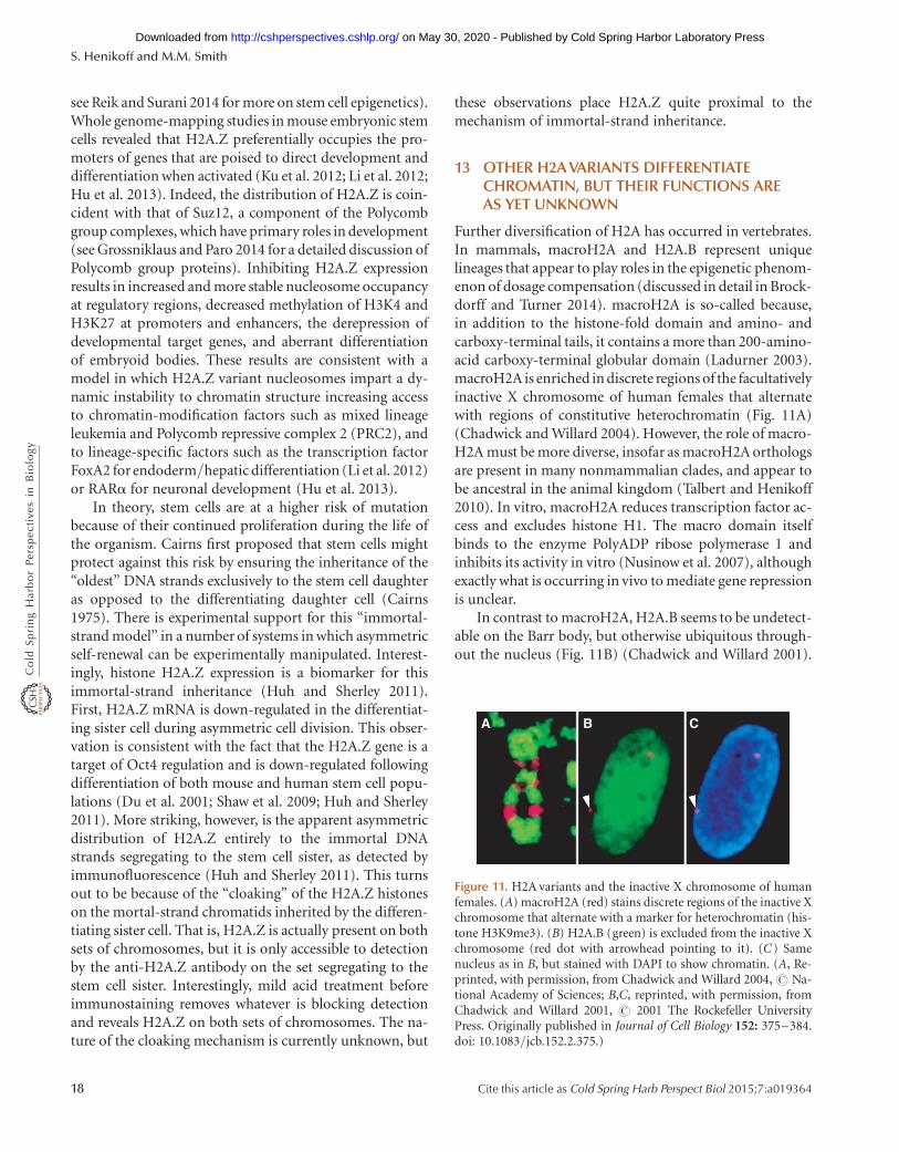
see Reik and Surani 2014 for more on stem cell epigenetics).Whole genome-mapping studies in mouse embryonic stemcells revealed that H2A.Z preferentially occupies the pro-moters of genes that are poised to direct development anddifferentiation when activated (Ku et al. 2012; Li et al. 2012;Hu et al. 2013). Indeed, the distribution of H2A.Z is coin-cident with that of Suz12, a component of the Polycombgroup complexes, which have primary roles in development(see Grossniklaus and Paro 2014 for a detailed discussion ofPolycomb group proteins). Inhibiting H2A.Z expressionresults in increased and more stable nucleosome occupancyat regulatory regions, decreased methylation of H3K4 andH3K27 at promoters and enhancers, the derepression ofdevelopmental target genes, and aberrant differentiationof embryoid bodies. These results are consistent with amodel in which H2A.Z variant nucleosomes impart a dy-namic instability to chromatin structure increasing accessto chromatin-modification factors such as mixed lineageleukemia and Polycomb repressive complex 2 (PRC2), andto lineage-specific factors such as the transcription factorFoxA2 for endoderm/hepatic differentiation (Li et al. 2012)or RARa for neuronal development (Hu et al. 2013).
In theory, stem cells are at a higher risk of mutationbecause of their continued proliferation during the life ofthe organism. Cairns first proposed that stem cells mightprotect against this risk by ensuring the inheritance of the“oldest” DNA strands exclusively to the stem cell daughteras opposed to the differentiating daughter cell (Cairns1975). There is experimental support for this “immortal-strand model” in a number of systems in which asymmetricself-renewal can be experimentally manipulated. Interest-ingly, histone H2A.Z expression is a biomarker for thisimmortal-strand inheritance (Huh and Sherley 2011).First, H2A.Z mRNA is down-regulated in the differentiat-ing sister cell during asymmetric cell division. This obser-vation is consistent with the fact that the H2A.Z gene is atarget of Oct4 regulation and is down-regulated followingdifferentiation of both mouse and human stem cell popu-lations (Du et al. 2001; Shaw et al. 2009; Huh and Sherley2011). More striking, however, is the apparent asymmetricdistribution of H2A.Z entirely to the immortal DNAstrands segregating to the stem cell sister, as detected byimmunofluorescence (Huh and Sherley 2011). This turnsout to be because of the “cloaking” of the H2A.Z histoneson the mortal-strand chromatids inherited by the differen-tiating sister cell. That is, H2A.Z is actually present on bothsets of chromosomes, but it is only accessible to detectionby the anti-H2A.Z antibody on the set segregating to thestem cell sister. Interestingly, mild acid treatment beforeimmunostaining removes whatever is blocking detectionand reveals H2A.Z on both sets of chromosomes. The na-ture of the cloaking mechanism is currently unknown, but
these observations place H2A.Z quite proximal to themechanism of immortal-strand inheritance.
13 OTHER H2A VARIANTS DIFFERENTIATECHROMATIN, BUT THEIR FUNCTIONS AREAS YET UNKNOWN
Further diversification of H2A has occurred in vertebrates.In mammals, macroH2A and H2A.B represent uniquelineages that appear to play roles in the epigenetic phenom-enon of dosage compensation (discussed in detail in Brock-dorff and Turner 2014). macroH2A is so-called because,in addition to the histone-fold domain and amino- andcarboxy-terminal tails, it contains a more than 200-amino-acid carboxy-terminal globular domain (Ladurner 2003).macroH2A is enriched in discrete regions of the facultativelyinactive X chromosome of human females that alternatewith regions of constitutive heterochromatin (Fig. 11A)(Chadwick and Willard 2004). However, the role of macro-H2A must be more diverse, insofar as macroH2A orthologsare present in many nonmammalian clades, and appear tobe ancestral in the animal kingdom (Talbert and Henikoff2010). In vitro, macroH2A reduces transcription factor ac-cess and excludes histone H1. The macro domain itselfbinds to the enzyme PolyADP ribose polymerase 1 andinhibits its activity in vitro (Nusinow et al. 2007), althoughexactly what is occurring in vivo to mediate gene repressionis unclear.
In contrast to macroH2A, H2A.B seems to be undetect-able on the Barr body, but otherwise ubiquitous through-out the nucleus (Fig. 11B) (Chadwick and Willard 2001).
A B C
Figure 11. H2A variants and the inactive X chromosome of humanfemales. (A) macroH2A (red) stains discrete regions of the inactive Xchromosome that alternate with a marker for heterochromatin (his-tone H3K9me3). (B) H2A.B (green) is excluded from the inactive Xchromosome (red dot with arrowhead pointing to it). (C) Samenucleus as in B, but stained with DAPI to show chromatin. (A, Re-printed, with permission, from Chadwick and Willard 2004, # Na-tional Academy of Sciences; B,C, reprinted, with permission, fromChadwick and Willard 2001, # 2001 The Rockefeller UniversityPress. Originally published in Journal of Cell Biology 152: 375–384.doi: 10.1083/jcb.152.2.375.)
S. Henikoff and M.M. Smith
18 Cite this article as Cold Spring Harb Perspect Biol 2015;7:a019364
on May 30, 2020 - Published by Cold Spring Harbor Laboratory Press http://cshperspectives.cshlp.org/Downloaded from

The short wrap of DNA around H2A.B nucleosomes (Baoet al. 2004) and its enrichment at TSSs (Soboleva et al.2012) is consistent with it playing a role in facilitating tran-scription. H2A.B and its closest relative, H2A.L (Govinet al. 2007), are rapidly evolving relative to other H2A var-iant classes, perhaps related to the fact that they are bothtestes-specific variants (Talbert and Henikoff 2010). Theroles that H2A.B and H2A.L play in mammalian malegerm cell development remain to be elucidated.
14 MANY HISTONES HAVE EVOLVED TO MORETIGHTLY PACKAGE DNA
When it is no longer necessary to gain access to DNA forreplication and transcription, chromatin typically becomesfurther condensed, and this often involves replacement ofcanonical histones. This is obviously the case for sperm,and in some lineages, histone paralogs have evolved spe-cialized packaging roles. For example, sea urchin spermcontains H1 and H2B variants with repeated tail motifsthat bind to the minor grooves of DNA (Talbert and Henik-off 2010), presumably an adaptation to tightly packagechromosomes for inclusion into sperm heads. A similaradaptation is found in pollen-specific H2Avariants in flow-ering plants. In vertebrates, a sperm-specific H2B spe-cialized histone variant is found in mammalian testes,including an H2B paralog (subH2Bv) that localizes to theacrosome and a testes-specific H3 variant (Witt et al. 1996).
The replacement of histones during sperm maturationby protamines and other proteins provides a potentialmeans of erasing epigenetic information in the male germ-line. However, evidence for transgenerational inheritance(Rakyan and Whitelaw 2003; discussed in more detail inBlewitt and Whitelaw 2013), especially in animals that lackDNA methylation, raises the possibility that a subset ofnucleosomal histones survive this transition and transmitepigenetic information. As already pointed out, this is justwhat occurs for CENP-A at centromeres (Palmer et al.1990), and it is possible that a small fraction of other var-iants, such as H3.3, remain with sperm for epigenetic in-heritance of gene-expression information. Although ourunderstanding of the process that replaces histones duringsperm development is rudimentary, we expect that muchmore can be learned by understanding how CENP-A sur-vives this transition.
Increased compaction also occurs in somatic cells thathave finished dividing and undergo differentiation. In somecases, compaction involves quantitative and qualitativechanges in linker histones. The stoichiometry of histoneH1 relative to nucleosomes determines the average spacingwithin nucleosome arrays in vivo (Fan et al. 2003). In addi-tion, the presence of H1 in chromatin promotes higher-
order chromatin structure that generally inhibits transcrip-tion (Wolffe 1992). Linker histones are much more mobilethan core histones in vivo. Residence times for H2A andH2B are hours in length, and cannot even be measuredfor H3 and H4, whereas the residence time of H1 is a fewminutes (Phair et al. 2004). As a result, the incorporation ofvariant linker histones is unlikely to differentiate chromatinin a heritable manner. Rather, the role of H1 variants isthought to change the bulk properties of chromatin thatcan affect overall compaction (Wolffe 1992).
H1 variants share with core histones a distinction be-tween RC and RI forms (Marzluff et al. 2002). RC variantforms of H1 appear to be interchangeable with one anoth-er, based on the fact that knockout mice lacking one or twoof the five RC H1 variants are phenotypically normal (Fanet al. 2003). The functional interchangeability of histonevariants is implicit in the “star” phylogeny of the H1 family,in which there is little if any evidence for evolutionaryconservation of H1 branches in the tree (Fig. 6C). Rather,the greater divergence of H1 relative to core histone variantsis likely to reflect weaker structural constraints, and theexistence of multiple H1 genes in many lineages mightrepresent an adaptation for regulating levels of linker his-tone. For example, in birds, the H1.0 linker histone variant(previously referred to as H5) is deposited during erythro-cyte maturation, which accompanies extreme compactionof the nucleus. The mammalian H1.0 ortholog (previouslyreferred to as H1˚) is deposited at high levels in nondivid-ing cells. Overexpression of H1.0 renders chromatin lessaccessible to nucleases than similar overexpression of a ca-nonical form. The natural accumulation of H1.0 in nondi-viding cells might be a general mechanism for chromatincompaction as cells become quiescent.
15 HISTONE VARIANTS AND HUMAN DISEASE
The diverse roles of histone variants in basic epigeneticprocesses leads to the expectation that their loss or misex-pression can result in disease, and indeed some examples ofthis have recently come to light. For example, ATRX, whichis the DNA translocase component of the ATRX-Daxx-H3.3 pathway (Fig. 8), was originally identified as causinga-thalassemia mental retardation on the X (ATRX) syn-drome. In this syndrome, loss of ATRX results in defectsat the CpG island of the a-globin gene promoter, and cannow be understood as resulting from frequent nucleosomeloss at CpG islands (Law et al. 2010). Importantly, muta-tions causing the ATRX syndrome are most frequentlyfound within the ADD histone tail-binding domain (Eu-stermann et al. 2011), strongly implicating H3/H3.3 tailinteractions as being key causal factors in the syndrome.The study of ATRX syndrome has also led to the realization
Histone Variants and Epigenetics
Cite this article as Cold Spring Harb Perspect Biol 2015;7:a019364 19
on May 30, 2020 - Published by Cold Spring Harbor Laboratory Press http://cshperspectives.cshlp.org/Downloaded from

that another histone variant, macroH2A, is also depletedfrom specific sites, including thea-globin CpG island (Rat-nakumar et al. 2012).
A remarkable relationship has recently emerged be-tween the ATRX-Daxx-H3.3 nucleosome assembly processand cancer that provides important insights into both theprocess itself and oncogenesis. Genomic sequencing ofhuman pancreatic neuroendocrine tumors (PanNETs) re-vealed that �40% harbored likely loss-of-function muta-tions in either Daxx or ATRX (Jiao et al. 2011). Notably, allsuch tumors displayed a distinct phenotype called “ALT”(alternative lengthening of telomeres) in which telomeresshow massive lengthening without induction of telomerase(see Fig. 1 of Liu et al. 2014; Heaphy et al. 2011a). Basedon examination of more than 6000 human tumors, 3.73%showed ALT occurring in cancers of all different types(Heaphy et al. 2011b). ATRX or Daxx are mutated in thevast majority of cases of ALTexamined by sequencing. Al-though the precise molecular basis for telomere lengthen-ing in ALT is not well understood, lengthening is likelyinitiated by recombinational strand invasion between telo-meres, which suggests that failure to replace a nucleosomeat a simple sequence array can result in DNA breakage andstrand invasion. In the case of PanNETs, ALT predicts amore favorable outcome than telomerase induction, as ifthe ALT pathway is less efficient than the telomerase path-way in preventing the senescence that occurs when telo-meres erode (Jiao et al. 2011).
Sequencing of tumors has also revealed the very sur-prising presence of specific mutations in H3.3 itself in alarge fraction of pediatric glioblastomas (the topic of Liuet al. 2014; also Schwartzentruber et al. 2012; Wu et al.2012). One of the two H3.3 genes in these tumors is foundto encode either K27M or G34R/V, and is most likely to bea gain-of-function mutation that drives accelerated tumor-igenesis (see Fig. 1 of Liu et al. 2014). G34R/V is associatedwith ATRX loss and probably enhances ALT, whereas K27Mcan occur on an H3 as well, so would appear to be inde-pendent of the ATRX-Daxx-H3.3 pathway. Rather, K27 is akey substrate for methylation and acetylation involved inPolycomb silencing, and the presence of a nonmodifiableresidue at position 27 of the H3/H3.3 tail causes gain-of-function inhibition of methylation by the EZH2 subunitof PRC2 (Lewis et al. 2013). It would then appear that theresulting global reduction in H3K27 trimethylation con-tributes to this aggressive pediatric tumor.
Misregulation of histone variant expression has alsobeen implicated in cancer. In malignant melanoma cells,macroH2A levels are sharply reduced and restoration ofmacroH2A levels suppresses metastasis (Kapoor et al.2010). Reduced levels of macroH2A result in up-regulationof many genes, including the CDK8 oncogene. macroH2A
is also reduced in more rapidly proliferating lung cancers,and is elevated in cells undergoing senescence, which sug-gests that reduced senescence of tumor cells is a mechanismwhereby loss of macroH2A contributes to oncogenesis(Sporn et al. 2009). It is possible that other defects in theexpression of histone variants and in pathways that depositthem can contribute to cancer and other human diseases,but there is as yet insufficient evidence of causality as op-posed to mere correlation. For example, excessively highlevels of H2A.Z are found to correlate with poor out-comes in estrogen-positive breast cancer (Hua et al. 2008).In addition, CENP-A is found to be overexpressed and ec-topically localized in a variety of cancers, suggesting thattransient or permanent neocentromere formation mightresult in some of the aneuploidies that are hallmarks ofcancer cells (Dalal 2009). We anticipate that as genome-wide technologies improve, other possible roles of histonevariants incorporation pathways in human diseases will beuncovered.
16 CONCLUSIONS AND FUTURE RESEARCH
Histone variants provide the most fundamental level ofdifferentiation of chromatin, and alternative mechanismsfor depositing different variants can potentially establishand maintain epigenetic states. Histones H2A, H2B, H3,and H4 occupy distinct positions in the core particle as aresult of an evolutionary process that began before the lastcommon ancestor of eukaryotes. Key evolutionary innova-tions remain uncertain, including the emergence of an oc-tamer from an ancestral tetramer, and we look forwardto the sequencing of more archaeal and primitive eukary-otic genomes that might provide missing links. Subsequentelaborations of the four core histones into distinct variantshave provided the basis for epigenetic processes, includingdevelopment and chromosome segregation. For a full un-derstanding of epigenetic inheritance, we need a betterunderstanding of the processes that incorporate variantsby replacing canonical histones. An important recent de-velopment is the initial characterization of RI assemblypathways dedicated to particular variants.
Centromeres are the most conspicuous examples ofprofoundly different chromatin that is attributable to spe-cial properties of a histone variant. Although it is clear thatcenH3-containing nucleosomes form the foundation ofthe centromere, just how they are deposited in the sameplace every cell generation without any hint of sequencespecificity is a major challenge for future research.
It is becoming evident that histone variants are alsoinvolved in epigenetic properties of active genes. BothH3.3 and H2A.Z are enriched at transcriptionally activeloci, and understanding the assembly processes that are
S. Henikoff and M.M. Smith
20 Cite this article as Cold Spring Harb Perspect Biol 2015;7:a019364
on May 30, 2020 - Published by Cold Spring Harbor Laboratory Press http://cshperspectives.cshlp.org/Downloaded from
responsible for their enrichment is an exciting area of cur-rent research. The dynamic behavior of chromatin leads tothe realization that transcription, chromatin remodeling,and histone modification might be coupled to nucleosomeassembly and disassembly. The study of dynamic processescoupled to histone turnover is only at an early stage, and welook forward to technological advances in molecular biol-ogy, cytogenetics, biochemistry, and structural biology thatcan be harnessed to better understand the dynamic natureof chromatin.
In addition to these universal processes, histone vari-ants are also involved in particular epigenetic phenomena.In the case of the mammalian X chromosome, three dif-ferent H2A variants, phospho-H2A.X, macroH2A, andH2A.B have been recruited to participate in silencing oractivation of genes for purposes of germline inactivation ordosage compensation. Understanding the function of thesevariants in epigenetic processes remains a major challengefor the future.
The availability of the first high-resolution structure ofthe nucleosome core particle (Luger et al. 1997) was a semi-nal advance in elucidating the properties of chromatin. Byelaborating this basic structure in a way that has biologicalconsequences, histone variants provide an opportunity todeepen our understanding of how these fascinating archi-tectural proteins have evolved to play diverse roles in epi-genetic processes.
REFERENCES
∗Reference is also in this collection.
Adkins NL, Niu H, Sung P, Peterson CL. 2013. Nucleosome dynamicsregulates DNA processing. Nat Struct Mol Biol 20: 836–842.
Akhtar A, Gasser SM. 2007. The nuclear envelope and transcriptionalcontrol. Nat Rev Genet 8: 507–517.
Akiyama T, Suzuki O, Matsuda J, Aoki F. 2011. Dynamic replacement ofhistone h3 variants reprograms epigenetic marks in early mouse em-bryos. PLoS Genet 7: e1002279.
∗ Allshire RC, Ekwall K. 2014. Epigenetic regulation of chromatin statesin Schizosaccharomyces pombe. Cold Spring Harb Perspec Biol doi:10.1101/cshperspect.a018770.
∗ Almouzni G, Cedar H. 2014. Maintenance of epigenetic information.Cold Spring Harb Perspec Biol doi: 10.1101/cshperspect.a019372.
Altaf M, Auger A, Covic M, Cote J. 2009. Connection between histoneH2Avariants and chromatin remodeling complexes. Biochem Cell Biol87: 35–50.
Altheim BA, Schultz MC. 1999. Histone modification governs the cellcycle regulation of a replication-independent chromatin assemblypathway in Saccharomyces cerevisiae. Proc Natl Acad Sci 96: 1345–1350.
Ammar R, Torti D, Tsui K, Gebbia M, Durbic T, Bader GD, Giaever G,Nislow C. 2012. Chromatin is an ancient innovation conserved be-tween Archaea and Eukarya. eLife 1: e00078.
Amor DJ, Bentley K, Ryan J, Perry J, Wong L, Slater H, Choo KH. 2004a.Human centromere repositioning ‘in progress’. Proc Natl Acad Sci 101:6542–6547.
Amor DJ, Kalitsis P, Sumer H, Choo KH. 2004b. Building the centromere:From foundation proteins to 3D organization. Trends Cell Biol 14:359–368.
Annunziato AT. 2005. Split decision: What happens to nucleosomes dur-ing DNA replication? J Biol Chem 280: 12065–12068.
Arents G, Burlingame RW, Wang BC, Love WE, Moudrianakis EN. 1991.The nucleosomal core histone octamer at 3.1 A resolution: A tripartiteprotein assembly and a left-handed superhelix. Proc Natl Acad Sci 88:10148–10152.
Baldi S, Becker PB. 2013. The variant histone H2A.V of Drosophila—Three roles, two guises. Chromosoma 122: 245–258.
Bao Y, Konesky K, Park YJ, Rosu S, Dyer PN, Rangasamy D, TremethickDJ, Laybourn PJ, Luger K. 2004. Nucleosomes containing the histonevariant H2A.Bbd organize only 118 base pairs of DNA. EMBO J 23:3314–3324.
Barski A, Cuddapah S, Cui K, Roh TY, Schones DE, Wang Z, Wei G,Chepelev I, Zhao K. 2007. High-resolution profiling of histone meth-ylations in the human genome. Cell 129: 823–837.
∗ Baulcombe D, Dean C. 2014. Epigenetic regulation in plant responsesto the environment. Cold Spring Harb Perspec Biol 6: a019471.
∗ Becker PB, Workman JL. 2013. Nucleosome remodeling and epigenetics.Cold Spring Harb Perspec Biol 5: 017905.
Belotserkovskaya R, Oh S, Bondarenko VA, Orphanides G, Studitsky VM,Reinberg D. 2003. FACT facilitates transcription-dependent nucleo-some alteration. Science 301: 1090–1093.
Black BE, Cleveland DW. 2011. Epigenetic centromere propagation andthe nature of CENP-A nucleosomes. Cell 144: 471–479.
∗ Blewitt M, Whitelaw E. 2013. The use of mouse models to study epige-netics. Cold Spring Harb Perspec Biol 5: 017939.
Boden SA, Kavanova M, Finnegan J, Wigge PA. 2013. Thermal stresseffects on grain yield in Brachypodium distachyon occur via H2A.Z-nucleosomes. Genome Biol 14: R65.
Bonisch C, Schneider K, Punzeler S, Wiedemann SM, Bielmeier C, Bo-cola M, Eberl HC, Kuegel W, Neumann J, Kremmer E, et al. 2012.H2A.Z.2.2 is an alternatively spliced histone H2A.Z variant thatcauses severe nucleosome destabilization. Nucleic Acids Res 40:5951–5964.
Boyarchuk E, Montes de Oca R, Almouzni G. 2011. Cell cycle dynamicsof histone variants at the centromere, a model for chromosomal land-marks. Curr Opin Cell Biol 23: 266–276.
Brickner JH. 2009. Transcriptional memory at the nuclear periphery.Curr Opin Cell Biol 21: 127–133.
Brochier-Armanet C, Forterre P, Gribaldo S. 2011. Phylogeny and evo-lution of the Archaea: One hundred genomes later. Curr Opin Micro-biol 14: 274–281.
∗ Brockdorff N, Turner B. 2014. Dosage compensation in mammals. ColdSpring Harb Perspec Biol doi: 10.1101/cshperspect.a019406.
Buchanan L, Durand-Dubief M, Roguev A, Sakalar C, Wilhelm B, Stral-fors A, Shevchenko A, Aasland R, Shevchenko A, Ekwall K et al. 2009.The Schizosaccharomyces pombe JmjC-protein, Msc1, prevents H2A.Zlocalization in centromeric and subtelomeric chromatin domains.PLoS Genet 5: e1000726.
Cairns J. 1975. Mutation selection and the natural history of cancer.Nature 255: 197–200.
Chadwick BP, Willard HF. 2001. A novel chromatin protein, distantlyrelated to histone H2A, is largely excluded from the inactive X chro-mosome. J Cell Biol 152: 375–384.
Chadwick BP, Willard HF. 2004. Multiple spatially distinct types of fac-ultative heterochromatin on the human inactive X chromosome. ProcNatl Acad Sci 101: 17450–17455.
Cho US, Harrison SC. 2011. Recognition of the centromere-specifichistone Cse4 by the chaperone Scm3. Proc Natl Acad Sci 108: 9367–9371.
Conerly ML, Teves SS, Diolaiti D, Ulrich M, Eisenman RN, Henikoff S.2010. Changes in H2A.Z occupancy and DNA methylation during B-cell lymphomagenesis. Genome Res 20: 1383–1390.
Histone Variants and Epigenetics
Cite this article as Cold Spring Harb Perspect Biol 2015;7:a019364 21
on May 30, 2020 - Published by Cold Spring Harbor Laboratory Press http://cshperspectives.cshlp.org/Downloaded from

Creyghton MP, Markoulaki S, Levine SS, Hanna J, Lodato MA, Sha K,Young RA, Jaenisch R, Boyer LA. 2008. H2AZ is enriched at Polycombcomplex target genes in ES cells and is necessary for lineage commit-ment. Cell 135: 649–661.
Dalal Y. 2009. Epigenetic specification of centromeres. Biochem Cell Biol87: 273–282.
Dalal Y, Wang H, Lindsay S, Henikoff S. 2007. Tetrameric structure ofcentromeric nucleosomes in interphase Drosophila cells. PLoS Biol 5:e218.
Deal RB, Henikoff JG, Henikoff S. 2010. Genome-wide kinetics of nu-cleosome turnover determined by metabolic labeling of histones. Sci-ence 328: 1161–1164.
Dhillon N, Oki M, Szyjka SJ, Aparicio OM, Kamakaka RT. 2006. H2A.Zfunctions to regulate progression through the cell cycle. Mol Cell Biol26: 489–501.
Dion M, Kaplan T, Friedman N, Rando OJ. 2007. Dynamics of replica-tion-independent histone turnover in budding yeast. Science 315:1405–1408.
Draker R, Cheung P. 2009. Transcriptional and epigenetic functions ofhistone variant H2A.Z. Biochem Cell Biol 87: 19–25.
Drane P, Ouararhni K, Depaux A, Shuaib M, Hamiche A. 2010. Thedeath-associated protein DAXX is a novel histone chaperone involvedin the replication-independent deposition of H3.3. Genes Dev 24:1253–1265.
Dryhurst D, Ishibashi T, Rose KL, Eirin-Lopez JM, McDonald D, Silva-Moreno B, Veldhoen N, Helbing CC, Hendzel MJ, Shabanowitz J, et al.2009. Characterization of the histone H2A.Z-1 and H2A.Z-2 isoformsin vertebrates. BMC Biol 7: 86.
Du Z, Cong H, Yao Z. 2001. Identification of putative downstream genesof Oct-4 by suppression-subtractive hybridization. Biochem BiophysRes Commun 282: 701–706.
Dunleavy EM, Roche D, Tagami H, Lacoste N, Ray-Gallet D, Nakamura Y,Daigo Y, Nakatani Y, Almouzni-Pettinotti G. 2009. HJURP is a cell-cycle-dependent maintenance and deposition factor of CENP-A atcentromeres. Cell 137: 485–497.
Edwards JR, O’Donnell AH, Rollins RA, Peckham HE, Lee C, MilekicMH, Chanrion B, Fu Y, Su T, Hibshoosh H, et al. 2010. Chromatin andsequence features that define the fine and gross structure of genomicmethylation patterns. Genome Res 20: 972–980.
Eustermann S, Yang JC, Law MJ, Amos R, Chapman LM, Jelinska C,Garrick D, Clynes D, Gibbons RJ, Rhodes D, et al. 2011. Combinatorialreadout of histone H3 modifications specifies localization of ATRX toheterochromatin. Nat Struct Mol Biol 18: 777–782.
Faast R, Thonglairoam V, Schulz TC, Beall J, Wells JR, Taylor H, MatthaeiK, Rathjen PD, Tremethick DJ, Lyons I. 2001. Histone variant H2A.Z isrequired for early mammalian development. Curr Biol 11: 1183–1187.
Fan Y, Nikitina T, Morin-Kensicki EM, Zhao J, Magnuson TR, WoodcockCL, Skoultchi AI. 2003. H1 linker histones are essential for mousedevelopment and affect nucleosome spacing in vivo. MolCell Biol 23:4559–4572.
Fernandez-Capetillo O, Mahadevaiah SK, Celeste A, Romanienko PJ,Camerini-Otero RD, Bonner WM, Manova K, Burgoyne P, Nussenz-weig A. 2003. H2AX is required for chromatin remodeling and inac-tivation of sex chromosomes in male mouse meiosis. Dev Cell 4: 497–508.
Filipescu D, Szenker E, Almouzni G. 2013. Developmental roles of his-tone H3 variants and their chaperones. Trends Genet 29: 630–640.
Foltz DR, Jansen LE, Bailey AO, Yates JR 3rd, Bassett EA, Wood S, BlackBE, Cleveland DW. 2009. Centromere-specific assembly of CENP-Anucleosomes is mediated by HJURP. Cell 137: 472–484.
Formosa T, Ruone S, Adams MD, Olsen AE, Eriksson P, Yu Y, Roades AR,Kaufman PD, Stillman DJ. 2002. Defects in SPT16 or POB3 (yFACT)in Saccharomyces cerevisiae cause dependence on the Hir/Hpc path-way: Polymerase passage may degrade chromatin structure. Genetics162: 1557–1571.
Gevry N, Hardy S, Jacques PE, Laflamme L, Svotelis A, Robert F, Gau-dreau L. 2009. Histone H2A.Z is essential for estrogen receptor signal-ing. Genes Dev 23: 1522–1533.
Goldberg AD, Banaszynski LA, Noh KM, Lewis PW, Elsaesser SJ, StadlerS, Dewell S, Law M, Guo X, Li X, et al. 2010. Distinct factors controlhistone variant H3.3 localization at specific genomic regions. Cell 140:678–691.
Govin J, Escoffier E, Rousseaux S, Kuhn L, Ferro M, Thevenon J, CatenaR, Davidson I, Garin J, Khochbin S, et al. 2007. Pericentric hetero-chromatin reprogramming by new histone variants during mousespermiogenesis. J Cell Biol 176: 283–294.
Greaves IK, Rangasamy D, Ridgway P, Tremethick DJ. 2007. H2A.Z con-tributes to the unique 3D structure of the centromere. Proc Natl AcadSci 104: 525–530.
∗ Grossniklaus U, Paro R. 2014. Transcriptional silencing by Polycomb-group proteins. Cold Spring Harb Perspec Biol 6: a019331.
∗ Grunstein M, Gasser SM. 2013. Epigenetics in Saccharomyces cerevisiae.Cold Spring Harb Perspec Biol 5: 017491.
Guse A, Carroll CW, Moree B, Fuller CJ, Straight AF. 2011. In vitrocentromere and kinetochore assembly on defined chromatin tem-plates. Nature 477: 354–358.
Halley JE, Kaplan T, Wang AY, Kobor MS, Rine J. 2010. Roles for H2A.Zand its acetylation in GAL1 transcription and gene induction, but notGAL1-transcriptional memory. PLoS Biol 8: e1000401.
Hardy S, Robert F. 2010. Random deposition of histone variants: Acellular mistake or a novel regulatory mechanism? Epigenetics 5:368–372.
Hardy S, Jacques PE, Gevry N, Forest A, Fortin ME, Laflamme L, Gau-dreau L, Robert F. 2009. The euchromatic and heterochromatic land-scapes are shaped by antagonizing effects of transcription on H2A.Zdeposition. PLoS Genet 5: e1000687.
Hartley PD, Madhani HD. 2009. Mechanisms that specify promoternucleosome location and identity. Cell 137: 445–458.
Heaphy CM, de Wilde RF, Jiao Y, Klein AP, Edil BH, Shi C, Bette-gowda C, Rodriguez FJ, Eberhart CG, Hebbar S, et al. 2011a. Al-tered telomeres in tumors with ATRX and DAXX mutations. Science333: 425.
Heaphy CM, Subhawong AP, Hong SM, Goggins MG, Montgomery EA,Gabrielson E, Netto GJ, Epstein JI, Lotan TL, Westra WH, et al. 2011b.Prevalence of the alternative lengthening of telomeres telomere main-tenance mechanism in human cancer subtypes. Am J Pathol 179:1608–1615.
Henikoff S, Ahmad K. 2005. Assembly of variant histones into chroma-tin. Ann Rev Cell Dev Biol 21: 133–153.
Henikoff S, Furuyama T. 2012. The unconventional structure of centro-meric nucleosomes. Chromosoma 121: 341–352.
Hines KA, Cryderman DE, Flannery KM, Yang H, Vitalini MW, HazelriggT, Mizzen CA, Wallrath LL. 2009. Domains of heterochromatin pro-tein 1 required for Drosophila melanogaster heterochromatin spread-ing. Genetics 182: 967–977.
Hodl M, Basler K. 2009. Transcription in the absence of histone H3.3.Curr Biol 19: 1221–1226.
Hodl M, Basler K. 2012. Transcription in the absence of histone H3.2 andH3K4 methylation. Curr Biol 22: 2253–2257.
Hou H, Wang Y, Kallgren SP, Thompson J, Yates JR 3rd, Jia S. 2010.Histone variant H2A.Z regulates centromere silencing and chromo-some segregation in fission yeast. J Biol Chem 285: 1909–1918.
Hu H, Liu Y, Wang M, Fang J, Huang H, Yang N, Li Y, Wang J, Yao X, Shi Y,et al. 2011. Structure of a CENP-A-histone H4 heterodimer in complexwith chaperone HJURP. Genes Dev 25: 901–906.
Hu G, Cui K, Northrup D, Liu C, Wang C, Tang Q, Ge K, Levens D,Crane-Robinson C, Zhao K. 2013. H2A.Z facilitates access of activeand repressive complexes to chromatin in embryonic stem cell self-renewal and differentiation. Cell Stem Cell 12: 180–192.
Hua S, Kallen CB, Dhar R, Baquero MT, Mason CE, Russell BA, Shah PK,Liu J, Khramtsov A, Tretiakova MS, et al. 2008. Genomic analysis of
S. Henikoff and M.M. Smith
22 Cite this article as Cold Spring Harb Perspect Biol 2015;7:a019364
on May 30, 2020 - Published by Cold Spring Harbor Laboratory Press http://cshperspectives.cshlp.org/Downloaded from

estrogen cascade reveals histone variant H2A.Z associated with breastcancer progression. Mol Syst Biol 4: 188.
Huang C, Zhang Z, Xu M, Li Y, Li Z, Ma Y, Cai T, Zhu B. 2013. H3.3-h4tetramer splitting events feature cell-type specific enhancers. PLoSGenet 9: e1003558.
Huh YH, Sherley JL. 2011. Molecular cloaking of H2A.Z on mortal DNAchromosomes during nonrandom segregation. Stem Cells 29: 1620–1627.
Ingouff M, Hamamura Y, Gourgues M, Higashiyama T, Berger F. 2007.Distinct dynamics of HISTONE3 variants between the two fertiliza-tion products in plants. Curr Biol 17: 1032–1037.
Jenuwein T. 2001. Re-SET-ting heterochromatin by histone methyltrans-ferases. Trends Cell Biol 11: 266–273.
Jenuwein T, Allis CD. 2001. Translating the histone code. Science 293:1074–1080.
Jiao Y, Shi C, Edil BH, de Wilde RF, Klimstra DS, Maitra A, Schulick RD,Tang LH, Wolfgang CL, Choti MA, et al. 2011. DAXX/ATRX, MEN1,and mTOR pathway genes are frequently altered in pancreatic neuro-endocrine tumors. Science 331: 1199–1203.
Jin C, Zang C, Wei G, Cui K, Peng W, Zhao K, Felsenfeld G. 2009. H3.3/H2A.Z double variant-containing nucleosomes mark ‘nucleosome-free regions’ of active promoters and other regulatory regions. NatGenet 41: 941–945.
Jullien J, Astrand C, Szenker E, Garrett N, Almouzni G, Gurdon JB. 2012.HIRA dependent H3.3 deposition is required for transcriptional re-programming following nuclear transfer to Xenopus oocytes. Epige-netics Chromatin 5: 17.
Kapoor A, Goldberg MS, Cumberland LK, Ratnakumar K, Segura MF,Emanuel PO, Menendez S, Vardabasso C, Leroy G, Vidal CI, et al.2010. The histone variant macroH2A suppresses melanoma progres-sion through regulation of CDK8. Nature 468: 1105–1109.
Kaya H, Shibahara KI, Taoka KI, Iwabuchi M, Stillman B, Araki T. 2001.FASCIATA genes for chromatin assembly factor-1 in Arabidopsis main-tain the cellular organization of apical meristems. Cell 104: 131–142.
Kelly TK, Miranda TB, Liang G, Berman BP, Lin JC, Tanay A, Jones PA.2010. H2A.Z maintenance during mitosis reveals nucleosome shiftingon mitotically silenced genes. Mol Cell 39: 901–911.
Kobor MS, Lorincz MC. 2009. H2A.Z and DNA methylation: Irrecon-cilable differences. Trends Biochem Sci 34: 158–161.
Ku M, Jaffe JD, Koche RP, Rheinbay E, Endoh M, Koseki H, Carr SA,Bernstein BE. 2012. H2A.Z landscapes and dual modifications in plu-ripotent and multipotent stem cells underlie complex genome regu-latory functions. Genome Biol 13: R85.
Kumar SV, Wigge PA. 2010. H2A.Z-containing nucleosomes mediate thethermosensory response in Arabidopsis. Cell 140: 136–147.
Kundu S, Peterson CL. 2010. Dominant role for signal transduction inthe transcriptional memory of yeast GAL genes. Mol Cell Biol 30:2330–2340.
Ladurner AG. 2003. Inactivating chromosomes: A macro domain thatminimizes transcription. Mol Cell 12: 1–3.
Law MJ, Lower KM, Voon HP, Hughes JR, Garrick D, Viprakasit V,Mitson M, De Gobbi M, Marra M, Morris A, et al. 2010. ATR-Xsyndrome protein targets tandem repeats and influences allele-specificexpression in a size-dependent manner. Cell 143: 367–378.
Lewis PW, Muller MM, Koletsky MS, Cordero F, Lin S, Banaszynski LA,Garcia BA, Muir TW, Becher OJ, Allis CD. 2013. Inhibition of PRC2activity by a gain-of-function H3 mutation found in pediatric glio-blastoma. Science 340: 857–861.
∗ Li E, Zhang Y. 2014. DNA methylation in mammals. Cold Spring HarbPerspec Biol 6: a019133.
Li Z, Gadue P, Chen K, Jiao Y, Tuteja G, Schug J, Li W, Kaestner KH. 2012.Foxa2 and H2A.Z mediate nucleosome depletion during embryonicstem cell differentiation. Cell 151: 1608–1616.
Light WH, Brickner DG, Brand VR, Brickner JH. 2010. Interaction of aDNA zip code with the nuclear pore complex promotes H2A.Z incor-poration and INO1 transcriptional memory. Mol Cell 40: 112–125.
∗ Liu X, McEachron T, Schwartzentruber J, Wu G. 2014. Histone H3 mu-tations in pediatric brain tumors. Cold Spring Harb Perspec Biol 6:a018689.
Lowndes NF, Toh GW. 2005. DNA repair: The importance of phosphor-ylating histone H2AX. Curr Biol 15: R99–R102.
Loyola A, Almouzni G. 2004. Histone chaperones, a supporting role inthe limelight. Biochim Biophys Acta 1677: 3–11.
Lu PY, Levesque N, Kobor MS. 2009. NuA4 and SWR1-C: Two chroma-tin-modifying complexes with overlapping functions and compo-nents. Biochem Cell Biol 87: 799–815.
Luger K, Mader AW, Richmond RK, Sargent DF, Richmond TJ. 1997.Crystal structure of the nucleosome core particle at 2.8 A resolution.Nature 389: 251–260.
Luk E, Ranjan A, Fitzgerald PC, Mizuguchi G, Huang Y, Wei D, Wu C.2010. Stepwise histone replacement by SWR1 requires dual activationwith histone H2A.Z and canonical nucleosome. Cell 143: 725–736.
Malik HS, Henikoff S. 2003. Phylogenomics of the nucleosome. NatStruct Biol 10: 882–891.
Malik HS, Henikoff S. 2009. Major evolutionary transitions in centro-mere complexity. Cell 138: 1067–1082.
March-Diaz R, Reyes JC. 2009. The beauty of being a variant: H2A.Z andthe SWR1 complex in plants. Mol Plant 2: 565–577.
Marques M, Laflamme L, Gervais AL, Gaudreau L. 2010. Reconciling thepositive and negative roles of histone H2A.Z in gene transcription.Epigenetics 5: 267–272.
Martins-Taylor K, Sharma U, Rozario T, Holmes SG. 2011. H2A.Z (Htz1)controls the cell-cycle-dependent establishment of transcriptional si-lencing at Saccharomyces cerevisiae telomeres. Genetics 187: 89–104.
Marzluff WF, Duronio RJ. 2002. Histone mRNA expression: Multiplelevels of cell cycle regulation and important developmental conse-quences. Curr Opin Cell Biol 14: 692–699.
Marzluff WF, Gongidi P, Woods KR, Jin J, Maltais LJ. 2002. The humanand mouse replication-dependent histone genes. Genomics 80: 487–498.
Matsuda R, Hori T, Kitamura H, Takeuchi K, Fukagawa T, Harata M.2010. Identification and characterization of the two isoforms of thevertebrate H2A.Z histone variant. Nucleic Acids Res 38: 4263–4273.
Mavrich TN, Jiang C, Ioshikhes IP, Li X, Venters BJ, Zanton SJ, TomshoLP, Qi J, Glaser RL, Schuster SC, et al. 2008. Nucleosome organizationin the Drosophila genome. Nature 453: 358–362.
Mehta M, Braberg H, Wang S, Lozsa A, Shales M, Solache A, Krogan NJ,Keogh MC. 2010. Individual lysine acetylations on the N terminus ofSaccharomyces cerevisiae H2A.Z are highly but not differentially reg-ulated. J Biol Chem 285: 39855–39865.
Mendiburo MJ, Padeken J, Fulop S, Schepers A, Heun P. 2011. DrosophilaCENH3 is sufficient for centromere formation. Science 334: 686–690.
Millar CB, Xu F, Zhang K, Grunstein M. 2006. Acetylation of H2AZ Lys14 is associated with genome-wide gene activity in yeast. Genes Dev 20:711–722.
Mizuguchi G, Xiao H, Wisniewski J, Smith MM, Wu C. 2007. Nonhis-tone Scm3 and histones CenH3-H4 assemble the core of centromere-specific nucleosomes. Cell 129: 1153–1164.
Morillo-Huesca M, Clemente-Ruiz M, Andujar E, Prado F. 2010. TheSWR1 histone replacement complex causes genetic instability andgenome-wide transcription misregulation in the absence of H2A.Z.PLoS One 5: e12143.
Morrison AJ, Shen X. 2005. DNA repair in the context of chromatin. CellCycle 4: 568–571.
Morrison AJ, Shen X. 2009. Chromatin remodelling beyond transcrip-tion: The INO80 and SWR1 complexes. Nat Rev Mol Cell Biol 10: 373–384.
Nakano S, Stillman B, Horvitz HR. 2011. Replication-coupled chromatinassembly generates a neuronal bilateral asymmetry in C. elegans. Cell147: 1525–1536.
Nekrasov M, Amrichova J, Parker BJ, Soboleva TA, Jack C, Williams R,Huttley GA, Tremethick DJ. 2012. Histone H2A.Z inheritance during
Histone Variants and Epigenetics
Cite this article as Cold Spring Harb Perspect Biol 2015;7:a019364 23
on May 30, 2020 - Published by Cold Spring Harbor Laboratory Press http://cshperspectives.cshlp.org/Downloaded from

the cell cycle and its impact on promoter organization and dynamics.Nat Struct Mol Biol 19: 1076–1083.
Ng RK, Gurdon JB. 2008. Epigenetic memory of an active gene statedepends on histone H3.3 incorporation into chromatin in the absenceof transcription. Nat Cell Biol 10: 102–109.
Nusinow DA, Hernandez-Munoz I, Fazzio TG, Shah GM, Kraus WL,Panning B. 2007. Poly(ADP-ribose) polymerase 1 is inhibited by ahistone H2A variant, MacroH2A, and contributes to silencing of theinactive X chromosome. J Biol Chem 282: 12851–12859.
Ooi S, Priess J, Henikoff S. 2006. Histone H3.3 variant dynamics in thegermline of Caenorhabditis elegans. PLoS Genet 2: e97.
Orsi GA, Couble P, Loppin B. 2009. Epigenetic and replacement roles ofhistone variant H3.3 in reproduction and development. Int J Dev Biol53: 231–243.
Palmer DK, O’Day K, Margolis RL. 1990. The centromere specific histoneCENP-A is selectively retained in discrete foci in mammalian spermnuclei. Chromosoma 100: 32–36.
Palmer DK, O’Day K, Trong HL, Charbonneau H, Margolis RL. 1991.Purification of the centromere-specific protein CENP-A and demon-stration that it is a distinctive histone. Proc Natl Acad Sci 88: 3734–3738.
Papamichos-Chronakis M, Watanabe S, Rando OJ, Peterson CL. 2011.Global regulation of H2A.Z localization by the INO80 chromatin-remodeling enzyme is essential for genome integrity. Cell 144: 200–213.
Phair RD, Scaffidi P, Elbi C, Vecerova J, Dey A, Ozato K, Brown DT, HagerG, Bustin M, Misteli T. 2004. Global nature of dynamic protein-chro-matin interactions in vivo: Three-dimensional genome scanning anddynamic interaction networks of chromatin proteins. Mol Cell Biol 24:6393–6402.
Pina B, Suau P. 1987. Changes in histones H2A and H3 variant compo-sition in differentiating and mature rat brain cortical neurons. Dev Biol123: 51–58.
Raisner RM, Hartley PD, Meneghini MD, Bao MZ, Liu CL, Schreiber SL,Rando OJ, Madhani HD. 2005. Histone variant H2A.Z marks the 5′
ends of both active and inactive genes in euchromatin. Cell 123: 233–248.
Rakyan V, Whitelaw E. 2003. Transgenerational epigenetic inheritance.Curr Biol 13: R6.
Ratnakumar K, Duarte LF, LeRoy G, Hasson D, Smeets D, Vardabasso C,Bonisch C, Zeng T, Xiang B, Zhang DY, et al. 2012. ATRX-mediatedchromatin association of histone variant macroH2A1 regulates a-glo-bin expression. Genes Dev 26: 433–438.
Ray-Gallet D, Woolfe A, Vassias I, Pellentz C, Lacoste N, Puri A, SchultzDC, Pchelintsev NA, Adams PD, Jansen LE, et al. 2011. Dynamicsof histone H3 deposition in vivo reveal a nucleosome gap-fillingmechanism for H3.3 to maintain chromatin integrity. Mol Cell 44:928–941.
∗ Reik W, Surani A. 2014. Germline and pluripotent stem cells. Cold SpringHarb Perspec Biol doi: 10.1101/cshperspect.a019422.
Sakai A, Schwartz BE, Goldstein S, Ahmad K. 2009. Transcriptional anddevelopmental functions of the H3.3 histone variant in Drosophila.Curr Biol 19: 1816–1820.
Santenard A, Ziegler-Birling C, Koch M, Tora L, Bannister AJ, Torres-Padilla ME. 2010. Heterochromatin formation in the mouse embryorequires critical residues of the histone variant H3.3. Nat Cell Biol 12:853–862.
Santisteban MS, Hang M, Smith MM. 2011. Histone variant H2A.Z andRNA polymerase II transcription elongation. Mol Cell Biol 31: 1848–1860.
Schneiderman JI, Sakai A, Goldstein S, Ahmad K. 2009. The XNP re-modeler targets dynamic chromatin in Drosophila. Proc Natl Acad Sci106: 14472–14477.
Schwartz BE, Ahmad K. 2005. Transcriptional activation triggers depo-sition and removal of the histone variant H3.3. Genes Dev 19: 804–814.
Schwartzentruber J, Korshunov A, Liu XY, Jones DT, Pfaff E, Jacob K,Sturm D, Fontebasso AM, Quang DA, Tonjes M, et al. 2012. Drivermutations in histone H3.3 and chromatin remodelling genes in pae-diatric glioblastoma. Nature 482: 226–231.
Shaw ML, Williams EJ, Hawes S, Saffery R. 2009. Characterisationof histone variant distribution in human embryonic stem cells bytransfection of in vitro transcribed mRNA. Mol Reprod Dev 76:1128–1142.
Shuaib M, Ouararhni K, Dimitrov S, Hamiche A. 2010. HJURPbinds CENP-A via a highly conserved N-terminal domain andmediates its deposition at centromeres. Proc Natl Acad Sci 107:1349–1354.
Simonet NG, Reyes M, Nardocci G, Molina A, Alvarez M. 2013. Epige-netic regulation of the ribosomal cistron seasonally modulates enrich-ment of H2A.Z and H2A.Zub in response to different environmentalinputs in carp (Cyprinus carpio). Epigenetics Chromatin 6: 22.
Soboleva TA, Nekrasov M, Pahwa A, Williams R, Huttley GA, TremethickDJ. 2012. A unique H2A histone variant occupies the transcriptionalstart site of active genes. Nat Struct Mol Biol 19: 25–30.
Sporn JC, Kustatscher G, Hothorn T, Collado M, Serrano M, Muley T,Schnabel P, Ladurner AG. 2009. Histone macroH2A isoforms predictthe risk of lung cancer recurrence. Oncogene 28: 3423–3428.
Stoler S, Rogers K, Weitze S, Morey L, Fitzgerald-Hayes M, Baker RE.2007. Scm3, an essential Saccharomyces cerevisiae centromere proteinrequired for G2/M progression and Cse4 localization. Proc Natl AcadSci 104: 10571–10576.
Sullivan BA, Karpen GH. 2004. Centromeric chromatin exhibits a his-tone modification pattern that is distinct from both euchromatin andheterochromatin. Nat Struct Mol Biol 11: 1076–1083.
Suto RK, Clarkson MJ, Tremethick DJ, Luger K. 2000. Crystal structure ofa nucleosome core particle containing the variant histone H2A.Z. NatStruct Biol 7: 1121–1124.
Talbert PB, Henikoff S. 2010. Histone variants—Ancient wrap artists ofthe epigenome. Nat Rev Mol Cell Biol 11: 264–275.
Talbert PB, Ahmad K, Almouzni G, Ausio J, Berger F, Bhalla PL, BonnerWM, Cande WZ, Chadwick BP, Chan SW, et al. 2012. A unified phy-logeny-based nomenclature for histone variants. Epigenetics Chroma-tin 5: 7.
Tanabe M, Kouzmenko AP, Ito S, Sawatsubashi S, Suzuki E, Fujiyama S,Yamagata K, Zhao Y, Kimura S, Ueda T, et al. 2008. Activation offacultatively silenced Drosophila loci associates with increased acety-lation of histone H2AvD. Genes Cells 13: 1279–1288.
Tran V, Lim C, Xie J, Chen X. 2012. Asymmetric division of Drosophilamale germline stem cell shows asymmetric histone distribution. Sci-ence 338: 679–682.
Updike DL, Mango SE. 2006. Temporal regulation of foregut develop-ment by HTZ-1/H2A.Z and PHA-4/FoxA. PLoS Genet 2: e161.
Venkatasubrahmanyam S, Hwang WW, Meneghini MD, Tong AH, Mad-hani HD. 2007. Genome-wide, as opposed to local, antisilencing ismediated redundantly by the euchromatic factors Set1 and H2A.Z.Proc Natl Acad Sci 104: 16609–16614.
Viens A, Mechold U, Brouillard F, Gilbert C, Leclerc P, Ogryzko V. 2006.Analysis of human histone H2AZ deposition in vivo argues against itsdirect role in epigenetic templating mechanisms. Mol Cell Biol 26:5325–5335.
Wan Y, Saleem RA, Ratushny AV, Roda O, Smith JJ, Lin CH, Chiang JH,Aitchison JD. 2009. Role of the histone variant H2A.Z/Htz1p in TBPrecruitment, chromatin dynamics, and regulated expression of oleate-responsive genes. Mol Cell Biol 29: 2346–2358.
Watanabe S, Radman-Livaja M, Rando OJ, Peterson CL. 2013. A histoneacetylation switch regulates H2A.Z deposition by the SWR-C remod-eling enzyme. Science 340: 195–199.
Weber CM, Henikoff JG, Henikoff S. 2010. H2A.Z nucleosomes en-riched over active genes are homotypic. Nat Struct Mol Biol 17: 1500–1507.
S. Henikoff and M.M. Smith
24 Cite this article as Cold Spring Harb Perspect Biol 2015;7:a019364
on May 30, 2020 - Published by Cold Spring Harbor Laboratory Press http://cshperspectives.cshlp.org/Downloaded from

Weintraub H, Worcel A, Alberts B. 1976. A model for chromatinbased upon two symmetrically paired half-nucleosomes. Cell 9:409–417.
Whittle CM, McClinic KN, Ercan S, Zhang X, Green RD, Kelly WG, LiebJD. 2008. The genomic distribution and function of histone variantHTZ-1 during C. elegans embryogenesis. PLoS Genet 4: e1000187.
Wieland G, Orthaus S, Ohndorf S, Diekmann S, Hemmerich P. 2004.Functional complementation of human centromere protein A (CENP-A) by Cse4p from Saccharomyces cerevisiae. Mol Cell Biol 24: 6620–6630.
Witt O, Albig W, Doenecke D. 1996. Testis-specific expression of a novelhuman H3 histone gene. Exp Cell Res 229: 301–306.
Wolffe AP. 1992. Chromatin: Structure and function. Academic, San Diego.Wu G, Broniscer A, McEachron TA, Lu C, Paugh BS, Becksfort J, Qu C,
Ding L, Huether R, Parker M, et al. 2012. Somatic histone H3 alter-ations in pediatric diffuse intrinsic pontine gliomas and non-brain-stem glioblastomas. Nat Genet 44: 251–253.
Xu M, Long C, Chen X, Huang C, Chen S, Zhu B. 2010. Partitioning ofhistone H3-H4 tetramers during DNA replication-dependent chro-matin assembly. Science 328: 94–98.
Xu Y, Ayrapetov MK, Xu C, Gursoy-Yuzugullu O, Hu Y, Price BD. 2012.Histone H2A.Z controls a critical chromatin remodeling step requiredfor DNA double-strand break repair. Mol Cell 48: 723–733.
Zacharioudakis I, Gligoris T, Tzamarias D. 2007. A yeast catabolic enzymecontrols transcriptional memory. Curr Biol 17: 2041–2046.
Zemach A, McDaniel IE, Silva P, Zilberman D. 2010. Genome-wideevolutionary analysis of eukaryotic DNA methylation. Science 328:916–919.
Zhang Z, Pugh BF. 2011. Genomic organization of H2Av containingnucleosomes in Drosophila heterochromatin. PLoS One 6: e20511.
Zhang H, Roberts DN, Cairns BR. 2005. Genome-wide dynamics ofHtz1, a histone H2A variant that poises repressed/basal promotersfor activation through histone loss. Cell 123: 219–231.
Zilberman D, Coleman-Derr D, Ballinger T, Henikoff S. 2008. HistoneH2A.Z and DNA methylation are mutually antagonistic chromatinmarks. Nature 456: 125–129.
Zlatanova J, Thakar A. 2008. H2A.Z: View from the top. Structure 16:166–179.
Zofall M, Fischer T, Zhang K, Zhou M, Cui B, Veenstra TD, Grewal SI.2009. Histone H2A.Z cooperates with RNAi and heterochromatinfactors to suppress antisense RNAs. Nature 461: 419–422.
WWW RESOURCES
http://www.ebi.ac.uk/Tools/phylogeny EBI server
Histone Variants and Epigenetics
Cite this article as Cold Spring Harb Perspect Biol 2015;7:a019364 25
on May 30, 2020 - Published by Cold Spring Harbor Laboratory Press http://cshperspectives.cshlp.org/Downloaded from

2015; doi: 10.1101/cshperspect.a019364Cold Spring Harb Perspect Biol Steven Henikoff and M. Mitchell Smith Histone Variants and Epigenetics
Subject Collection Epigenetics
Metabolic Signaling to ChromatinShelley L. Berger and Paolo Sassone-Corsi
Epigenetic Determinants of CancerStephen B. Baylin and Peter A. Jones
Neuronal Development and FunctionHistone and DNA Modifications as Regulators of
Stavros Lomvardas and Tom Maniatis
Maintenance of Epigenetic InformationGeneviève Almouzni and Howard Cedar
Histone Modifications and CancerJames E. Audia and Robert M. Campbell Histone and DNA Methylation Marks
A Structural Perspective on Readout of Epigenetic
Dinshaw J. PatelEpigenetics and Human Disease
Huda Y. Zoghbi and Arthur L. Beaudet PerspectiveThe Necessity of Chromatin: A View in
Vincenzo Pirrotta
ReprogrammingInduced Pluripotency and Epigenetic
Konrad Hochedlinger and Rudolf Jaenisch
Germline and Pluripotent Stem CellsWolf Reik and M. Azim Surani
Long-Range Chromatin InteractionsJob Dekker and Tom Misteli Histone Modifications
Comprehensive Catalog of Currently Documented
Yingming Zhao and Benjamin A. GarciaRNAi and Heterochromatin Assembly
Robert Martienssen and Danesh Moazed Schizosaccharomyces pombeEpigenetic Regulation of Chromatin States in
Robin C. Allshire and Karl EkwallDrosophilaDosage Compensation in
John C. Lucchesi and Mitzi I. KurodaHistone Variants and Epigenetics
Steven Henikoff and M. Mitchell Smith
http://cshperspectives.cshlp.org/cgi/collection/ For additional articles in this collection, see
Copyright © 2015 Cold Spring Harbor Laboratory Press; all rights reserved
on May 30, 2020 - Published by Cold Spring Harbor Laboratory Press http://cshperspectives.cshlp.org/Downloaded from