structural dynamics of nucleosome core particle ... · structural dynamics of nucleosome core...
TRANSCRIPT
Structural Dynamics of Nucleosome Core Particle:Comparison with Nucleosomes Containing Histone VariantsAmutha Ramaswamy,1 Ivet Bahar,2 and Ilya Ioshikhes1*1Department of Biomedical Informatics, The Ohio State University, Columbus, Ohio2Center for Computational Biology and Bioinformatics, Department of Molecular Genetics and Biochemistry, School ofMedicine, University of Pittsburgh, Pittsburgh, Pennsylvania
ABSTRACT The present study provides in-sights on the dominant mechanisms of motions ofthe nucleosome core particle and the changes in itsfunctional dynamics in response to histone vari-ants. Comparative analysis of the global dynamicsof nucleosomes with native and variant H2A his-tones, using normal mode analysis revealed that thedynamics of the nucleosome is highly symmetric,and its interaction with the nucleosomal DNA playsa vital role in its regulation. The collective dynamicsof nucleosomes are predicted to be dominated bytwo types of large-scale motions: (1) a global stretch-ing–compression of nucleosome along the dyad axisby which the nucleosome undergoes a breathingmotion with a massive distortion of nucleosomalDNA, modulated by histone–DNA interactions; and(2) the flipping (or bending) of both the sides of thenucleosome in an out-of-plane fashion with respectto the dyad axis, originated by the highly dynamicN-termini of H3 and (H2A.Z-H2B) dimer in agree-ment with the experimentally observed perturbeddynamics of the particular N-terminus under physi-ological conditions. In general, the nucleosomeswith variant histones exhibit higher mobilities andweaker correlations between internal motions com-pared to the nucleosome containing ordinary his-tones. The differences are more pronounced at theL1 and L2 loops of the respective monomers H2Band H2A, and at the N-termini of the monomers H3and H4, all of which closely interact with the wrap-ping DNA. Proteins 2005;58:683–696.© 2004 Wiley-Liss, Inc.
Key words: nucleosome dynamics; normal modeanalysis; DNA–protein interactions;equilibrium fluctuations
INTRODUCTION
Nucleosome, the structural unit of chromatin, plays avital role in chromatin biology,1,2 and understanding thedynamics of nucleosome is of fundamental importance inimproving our knowledge of gene regulation and DNAreplication machinery. The mystery of nucleosome and theregulation of its biological function have been issues ofintense investigation over years. The structure of histones,the organization of the nucleosome, and the mechanism oftranscriptional regulation as a result of nucleosome reposi-tioning have been reviewed in several pioneering stud-
ies.2–4 Stable alterations in nucleosome structure gener-ate a transient state of chromatin as an essential step ingene regulation.4 Thus, the conformational dynamics ofthe nucleosome play a central role in determining thetranscriptional competence of any region of the chromatin.
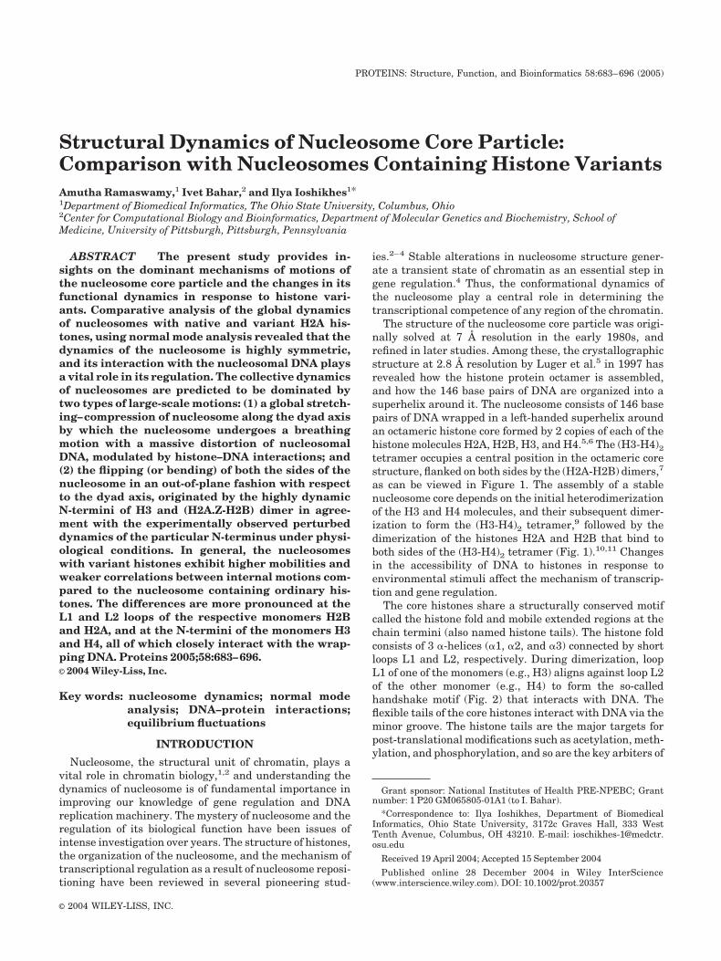
The structure of the nucleosome core particle was origi-nally solved at 7 Å resolution in the early 1980s, andrefined in later studies. Among these, the crystallographicstructure at 2.8 Å resolution by Luger et al.5 in 1997 hasrevealed how the histone protein octamer is assembled,and how the 146 base pairs of DNA are organized into asuperhelix around it. The nucleosome consists of 146 basepairs of DNA wrapped in a left-handed superhelix aroundan octameric histone core formed by 2 copies of each of thehistone molecules H2A, H2B, H3, and H4.5,6 The (H3-H4)2tetramer occupies a central position in the octameric corestructure, flanked on both sides by the (H2A-H2B) dimers,7
as can be viewed in Figure 1. The assembly of a stablenucleosome core depends on the initial heterodimerizationof the H3 and H4 molecules, and their subsequent dimer-ization to form the (H3-H4)2 tetramer,9 followed by thedimerization of the histones H2A and H2B that bind toboth sides of the (H3-H4)2 tetramer (Fig. 1).10,11 Changesin the accessibility of DNA to histones in response toenvironmental stimuli affect the mechanism of transcrip-tion and gene regulation.
The core histones share a structurally conserved motifcalled the histone fold and mobile extended regions at thechain termini (also named histone tails). The histone foldconsists of 3 �-helices (�1, �2, and �3) connected by shortloops L1 and L2, respectively. During dimerization, loopL1 of one of the monomers (e.g., H3) aligns against loop L2of the other monomer (e.g., H4) to form the so-calledhandshake motif (Fig. 2) that interacts with DNA. Theflexible tails of the core histones interact with DNA via theminor groove. The histone tails are the major targets forpost-translational modifications such as acetylation, meth-ylation, and phosphorylation, and so are the key arbiters of
Grant sponsor: National Institutes of Health PRE-NPEBC; Grantnumber: 1 P20 GM065805-01A1 (to I. Bahar).
*Correspondence to: Ilya Ioshikhes, Department of BiomedicalInformatics, Ohio State University, 3172c Graves Hall, 333 WestTenth Avenue, Columbus, OH 43210. E-mail: [email protected]
Received 19 April 2004; Accepted 15 September 2004
Published online 28 December 2004 in Wiley InterScience(www.interscience.wiley.com). DOI: 10.1002/prot.20357
PROTEINS: Structure, Function, and Bioinformatics 58:683–696 (2005)
© 2004 WILEY-LISS, INC.
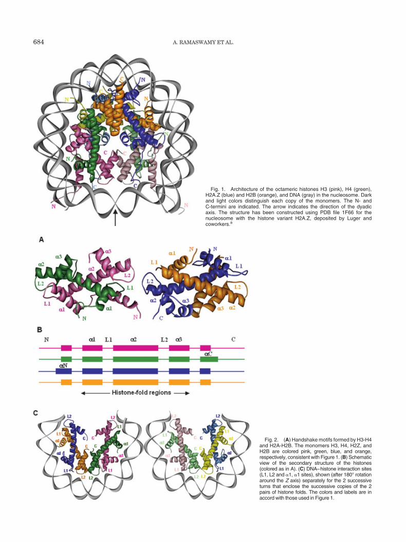
Fig. 1. Architecture of the octameric histones H3 (pink), H4 (green),H2A.Z (blue) and H2B (orange), and DNA (gray) in the nucleosome. Darkand light colors distinguish each copy of the monomers. The N- andC-termini are indicated. The arrow indicates the direction of the dyadicaxis. The structure has been constructed using PDB file 1F66 for thenucleosome with the histone variant H2A.Z, deposited by Luger andcoworkers.8
Fig. 2. (A) Handshake motifs formed by H3-H4and H2A-H2B. The monomers H3, H4, H2Z, andH2B are colored pink, green, blue, and orange,respectively, consistent with Figure 1. (B) Schematicview of the secondary structure of the histones(colored as in A). (C) DNA–histone interaction sites(L1, L2 and �1, �1 sites), shown (after 180° rotationaround the Z axis) separately for the 2 successiveturns that enclose the successive copies of the 2pairs of histone folds. The colors and labels are inaccord with those used in Figure 1.
684 A. RAMASWAMY ET AL.
chromatin function.12 The octameric histones and theDNA are highly networked by hydrogen bonds and themajor DNA–protein interaction sites of the dimers are the2 pairs of adjoining loops L1 and L2, and the �1 helices ofthe monomers [Fig. 2(C)].
Natural types of histones occur in the form of variousisoforms (H2A.1, H2A.2), variants (H2A.Z, H2A.X, H3.3,and CENP-A), and histone-like proteins (macroH2A). Dro-sophila chromatin contains, for example, 2 H2A histones,H2A.1 and H2A.2, that differ in their amino acid composi-tions and their antigenically distinct functions. H2A.Z, aminor variant of H2A, is essential for the viability of manyorganisms and has functions distinct from those of themajor H2A histone in chromatin. A number of recentstudies have focused on the chromatin structures withvariant histones given that the structure and function ofthe nucleosome are influenced by the core histone vari-ants.
Several pioneering studies have reported the impor-tance and the functional diversity of the nucleosome byH2A.Z.8,13–19 Activation of transcription within chromatinhas been correlated with the incorporation of H2A.Z intothe nucleosomes. Recently, a review article on the func-tional heterogeneity of the histone variants has beenreported by Brown.13 H2A.Z is found in a wide range oforganisms, from yeast to mammals.17 The elucidation ofthe H2A.Z nucleosome crystal structure has been instru-mental in detecting the changes in the histone–DNA andhistone–histone interactions within the nucleosome corecontaining histone variant8 compared to those in the majorhistone.
Having access to detailed sequence and structure infor-mation on the nucleosomes, it would be interesting toanalyze the factors generating the distinct behavior ofvariant histones and hence the dynamics of nucleosome.Molecular simulation techniques using conventional full-atomic force fields20,21 are prohibitively time-consumingfor exploring the dynamics of supramolecular structureslike the nucleosome (which consists of �50,000 atoms). Onthe other hand, normal modes analysis (NMA)22,23 provedto be an efficient but physically meaningful, complemen-tary tool for analyzing the equilibrium dynamics of largestructures and assemblies. Recently, simplified NMAswith uniform harmonic potentials, or methods based onelastic network formalism, have been proposed24–29 andsuccessfully applied to several molecular systems.28–31
The Gaussian Network Model (GNM)25,26 and its exten-sion, the Anisotropic Network Model (ANM),29 introducedby Bahar and coworkers to predict the sizes or directionali-ties of residue motions in different modes, have been usedadvantageously in many applications.32–35 These modelsconsider the biomolecule as an elastic network (EN) andgenerate a connectivity matrix by considering the C�
atoms as nodes. The connectivity of the network is deter-mined by defining an appropriate cutoff (rc) distance forpairs of amino acids that interact via elastic springs. Forproteins, the effective network is generated using rc � 10Å, whereas in DNA, as well as RNA, a slightly increasedcutoff distance of �14 � 2 Å has been used to include
interstrand interactions of DNA.36–38 The topology of thenetwork is represented by a connectivity (Kirchhoff) ma-trix whose eigenvalue decomposition yields the normalmodes of motion near the equilibrium structure. The GNMhas proven to be a useful technique in predicting X-raycrystallographic B factors,25 H/D exchange free energiesnear native state conditions,30 and NMR order parame-ters.31 It has also been extensively used for identifying thecooperative domain motions that underlie biomolecularfunction.36,37,39–41
In this work, the global dynamics of nucleosomes withvariant histones are analyzed with the EN models andcompared to highlight the effect of variant histones on thefunctional motions of the nucleosome. Global dynamicsrefer to the lowest frequency (and largest amplitude)modes of motions, which have been shown in severalstudies for other systems to be relevant to biologicalfunction.33–43 The analysis aims at answering a number offundamental questions: What are the dominant molecularmechanisms that control the relaxation of the nucleosome?To what degree do the variant histones influence thedynamics and intradomain interactions of the nucleo-some? What are the factors causing the divergent func-tions of histone variants?
METHODOLOGY
The structural dynamics of the nucleosome with regularhistones,44 nucleosome containing the variant histoneH2A.Z,8 and the histone with isoforms H2A.1 and H2B.245
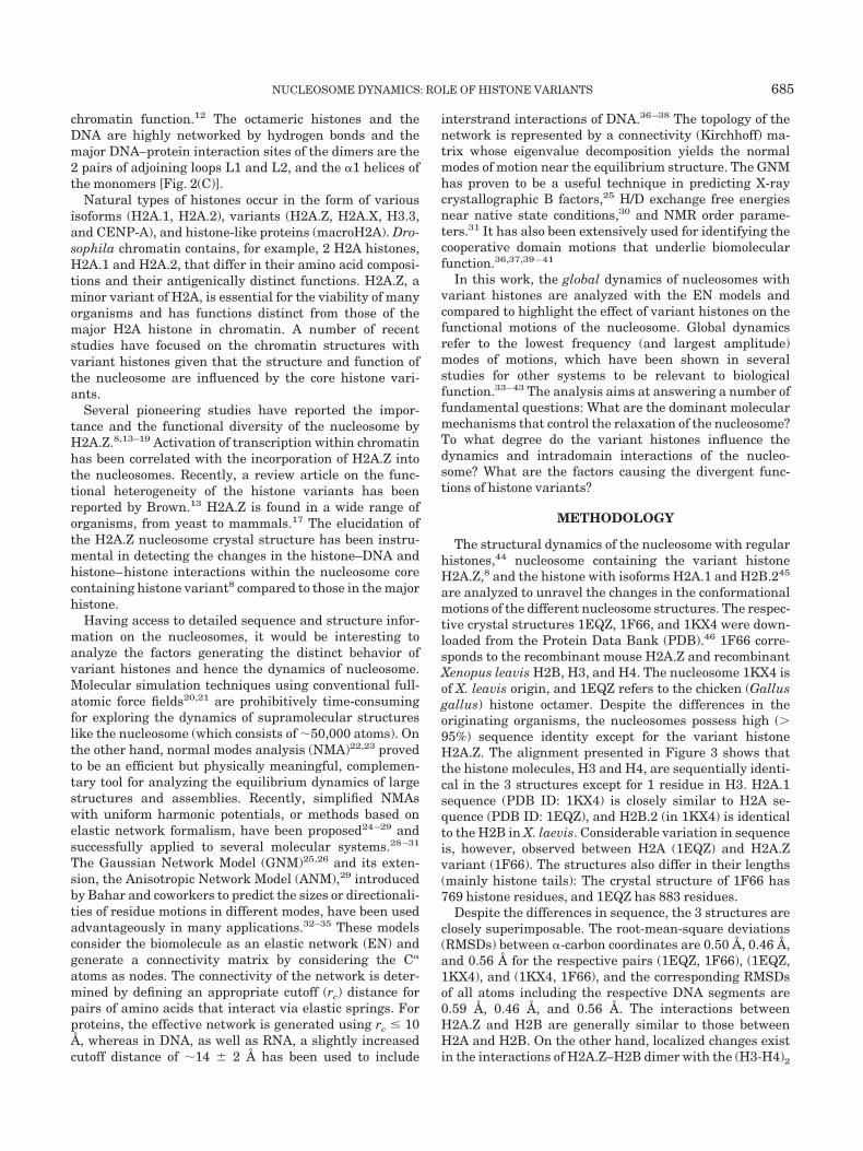
are analyzed to unravel the changes in the conformationalmotions of the different nucleosome structures. The respec-tive crystal structures 1EQZ, 1F66, and 1KX4 were down-loaded from the Protein Data Bank (PDB).46 1F66 corre-sponds to the recombinant mouse H2A.Z and recombinantXenopus leavis H2B, H3, and H4. The nucleosome 1KX4 isof X. leavis origin, and 1EQZ refers to the chicken (Gallusgallus) histone octamer. Despite the differences in theoriginating organisms, the nucleosomes possess high (�95%) sequence identity except for the variant histoneH2A.Z. The alignment presented in Figure 3 shows thatthe histone molecules, H3 and H4, are sequentially identi-cal in the 3 structures except for 1 residue in H3. H2A.1sequence (PDB ID: 1KX4) is closely similar to H2A se-quence (PDB ID: 1EQZ), and H2B.2 (in 1KX4) is identicalto the H2B in X. laevis. Considerable variation in sequenceis, however, observed between H2A (1EQZ) and H2A.Zvariant (1F66). The structures also differ in their lengths(mainly histone tails): The crystal structure of 1F66 has769 histone residues, and 1EQZ has 883 residues.
Despite the differences in sequence, the 3 structures areclosely superimposable. The root-mean-square deviations(RMSDs) between �-carbon coordinates are 0.50 Å, 0.46 Å,and 0.56 Å for the respective pairs (1EQZ, 1F66), (1EQZ,1KX4), and (1KX4, 1F66), and the corresponding RMSDsof all atoms including the respective DNA segments are0.59 Å, 0.46 Å, and 0.56 Å. The interactions betweenH2A.Z and H2B are generally similar to those betweenH2A and H2B. On the other hand, localized changes existin the interactions of H2A.Z–H2B dimer with the (H3-H4)2
NUCLEOSOME DYNAMICS: ROLE OF HISTONE VARIANTS 685
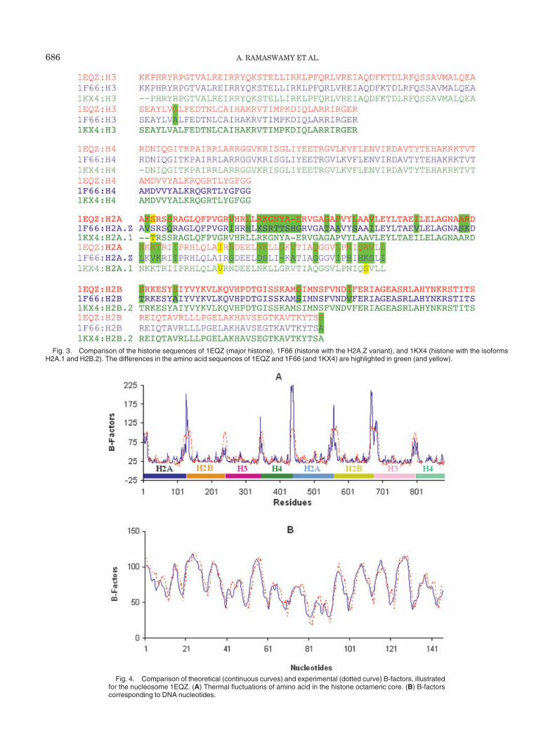
Fig. 3. Comparison of the histone sequences of 1EQZ (major histone), 1F66 (histone with the H2A.Z variant), and 1KX4 (histone with the isoformsH2A.1 and H2B.2). The differences in the amino acid sequences of 1EQZ and 1F66 (and 1KX4) are highlighted in green (and yellow).
Fig. 4. Comparison of theoretical (continuous curves) and experimental (dotted curve) B-factors, illustratedfor the nucleosome 1EQZ. (A) Thermal fluctuations of amino acid in the histone octameric core. (B) B-factorscorresponding to DNA nucleotides.
686 A. RAMASWAMY ET AL.
tetramer and those between the 2 H2A.Z–H2B dimers,which induce local perturbations in the structure near theinterfaces between the dimers and central tetramer.
The NMA of the global dynamics of nucleosome has beenperformed using the EN models. Fluctuation amplitudesare predicted using either the GNM or ANM, while thedetermination of fluctuation vectors requires the use of theANM.29 The GNM has the advantage of being one order ofmagnitude faster, and is resorted to unless the directionali-ties of the motions are explored. The structures 1F66 and1EQZ differ in their lengths (see above). The dynamics ofcommon residues have been compared. The Kirchhoffmatrix of inter-residue contacts is constructed using theC� atoms for representing the amino acids, and the P andO4* atoms for representing the DNA nucleotides. Cutoffdistances of 10 Å, 15 Å, and 18 Å have been adopted forprotein–protein, protein–DNA, and DNA–DNA interac-tions, respectively. Molecular graphics images were pro-duced using the UCSF Chimera package from the UCSFComputer Graphics Laboratory.47
RESULTS AND DISCUSSION
For a better understanding of the nucleosome dynamics,a 2-step analysis has been performed. First, we examinethe overall dynamics of the nucleosome. Two essentialquantities, the mean-square fluctuations of residues andtheir cross-correlations, are analyzed and compared withexperimental data. Second, we proceed to a more detailedanalysis by dissecting the overall dynamics into the contri-butions of individual modes of motions, and focusing on theslowest (or global) modes that dominate the observedbehavior. The global mode shapes of each histone mono-mer in the context of the octameric, DNA-bound structureare analyzed to identify the rigid and mobile parts of thestructure, as well as the dominant mechanisms of motionand the type of couplings between the cooperative motionsof different structural elements. Major differences in thecollective dynamics of the nucleosome with ordinary his-tones and the nucleosome with histone variants are eluci-dated.
Thermal Fluctuations of the Nucleosome
Figure 4 compares the experimental (from X-ray crystal-lographic studies; dotted curves) and presently computed(from EN analysis; continuous curves) B-factors. Figure4(A) displays the B-factors corresponding to the �-carbonsof octameric histones as a function of residues index. Thecurves also reflect the distribution of the mean-square (ms)fluctuations of individual residues in the folded state, asthe B-factors (Bi) scale with the ms fluctuations, �(�Ri)
2�,in the equilibrium positions, as Bi � (82/3) �( �Ri)
2� forresidue i.
Figure 4(B) describes the B-factors of the P atoms of oneof the 2 DNA strands. The periodicity of the curves reflectsthe different mobilities of the solvent- and protein-exposedsegments of the helical turns, with solvent-exposed re-gions enjoying higher mobility. The agreement betweentheory and experiment is excellent and supports the use ofthe present approach for further analysis of nucleosomedynamics.
Cooperative Inter- and Intradomain Motions of theHandshake Dimers
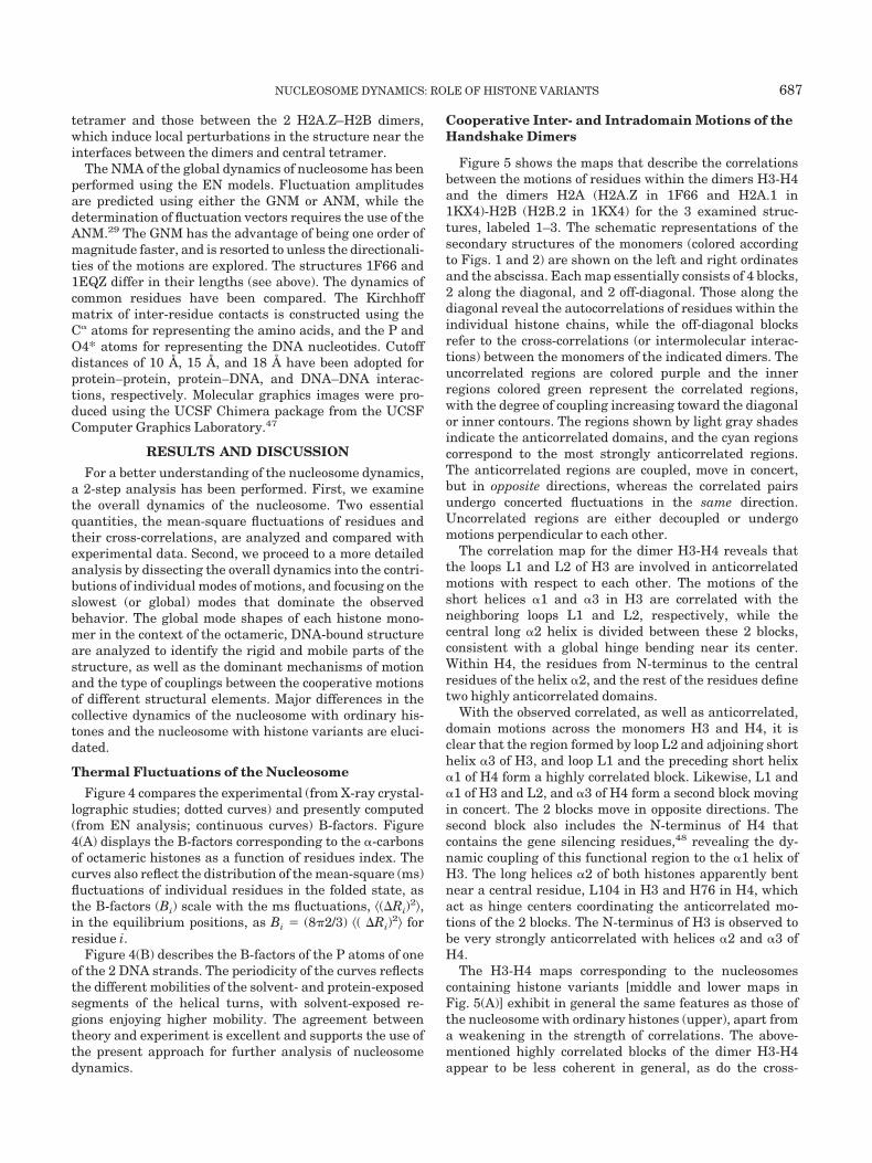
Figure 5 shows the maps that describe the correlationsbetween the motions of residues within the dimers H3-H4and the dimers H2A (H2A.Z in 1F66 and H2A.1 in1KX4)-H2B (H2B.2 in 1KX4) for the 3 examined struc-tures, labeled 1–3. The schematic representations of thesecondary structures of the monomers (colored accordingto Figs. 1 and 2) are shown on the left and right ordinatesand the abscissa. Each map essentially consists of 4 blocks,2 along the diagonal, and 2 off-diagonal. Those along thediagonal reveal the autocorrelations of residues within theindividual histone chains, while the off-diagonal blocksrefer to the cross-correlations (or intermolecular interac-tions) between the monomers of the indicated dimers. Theuncorrelated regions are colored purple and the innerregions colored green represent the correlated regions,with the degree of coupling increasing toward the diagonalor inner contours. The regions shown by light gray shadesindicate the anticorrelated domains, and the cyan regionscorrespond to the most strongly anticorrelated regions.The anticorrelated regions are coupled, move in concert,but in opposite directions, whereas the correlated pairsundergo concerted fluctuations in the same direction.Uncorrelated regions are either decoupled or undergomotions perpendicular to each other.
The correlation map for the dimer H3-H4 reveals thatthe loops L1 and L2 of H3 are involved in anticorrelatedmotions with respect to each other. The motions of theshort helices �1 and �3 in H3 are correlated with theneighboring loops L1 and L2, respectively, while thecentral long �2 helix is divided between these 2 blocks,consistent with a global hinge bending near its center.Within H4, the residues from N-terminus to the centralresidues of the helix �2, and the rest of the residues definetwo highly anticorrelated domains.
With the observed correlated, as well as anticorrelated,domain motions across the monomers H3 and H4, it isclear that the region formed by loop L2 and adjoining shorthelix �3 of H3, and loop L1 and the preceding short helix�1 of H4 form a highly correlated block. Likewise, L1 and�1 of H3 and L2, and �3 of H4 form a second block movingin concert. The 2 blocks move in opposite directions. Thesecond block also includes the N-terminus of H4 thatcontains the gene silencing residues,48 revealing the dy-namic coupling of this functional region to the �1 helix ofH3. The long helices �2 of both histones apparently bentnear a central residue, L104 in H3 and H76 in H4, whichact as hinge centers coordinating the anticorrelated mo-tions of the 2 blocks. The N-terminus of H3 is observed tobe very strongly anticorrelated with helices �2 and �3 ofH4.
The H3-H4 maps corresponding to the nucleosomescontaining histone variants [middle and lower maps inFig. 5(A)] exhibit in general the same features as those ofthe nucleosome with ordinary histones (upper), apart froma weakening in the strength of correlations. The above-mentioned highly correlated blocks of the dimer H3-H4appear to be less coherent in general, as do the cross-
NUCLEOSOME DYNAMICS: ROLE OF HISTONE VARIANTS 687
correlations across the monomers. We note in particularthe disappearance of the anticorrelations between the H3N-terminus and the H4 �2 and �3. The introduction ofH2A and H2B variants thus affects the global dynamics ofthe entire nucleosome.
The upper right map in Figure 5(B) reveals that thedimer H2A-H2B of ordinary nucleosome exists as a highlyinter- as well as intracoupled dimer. Almost the entiremonomers H2A and H2B are engaged in correlated mo-tions, except for the C-terminal segment of H2A and theN-terminal helix N� of H2B. We note that the C-terminusof H2A inserts into the H3-H4 dimer, which may explainits decoupling from the rest of the H2A-H2B dimer.Likewise, the N-terminus of H2B was invisible in the1EQZ X-ray structure, in accord with its decoupling fromthe collective dynamics of the dimer. In the H2A.Z variant,the coherent domain motions that exist within the H2Ahistone are highly disrupted, and the residues that areuncorrelated in H2A become anticorrelated.
The comparison of the maps 1 and 2 in Figure 5(B)indicates that significant differences in intra- and intermo-lecular correlations exist between the major H2A and thevariant H2A.Z. In particular, the H2A.Z residues Arg81-Lys119 located at the interface between the (H3-H4)2
tetramer and the (H2A-H2B) dimer exhibit substantialdecreases in their couplings to the helix–loop �1L1 on thesame monomer (H2A.Z), and to the loop–helix L2�3 on theneighboring (H2B) monomer (see the portions of the mapenclosed in the orange boxes). The loss of these long-rangecorrelations implies an inefficient propagation of motion,or communication, between the nucleosome core regionsnear the central tetramer, and those adjoining the wrappedDNA. This loss in communication, or cooperativity, is inaccord with the experimentally observed chromatin-destabilizing role of H2A.Z.15 The “destabilization” of thechromatin function is thus attributed, according to thisanalysis, to the disruption of the correlated, or concerted,changes in nucleosome conformation. The histone H2Balso exhibits inter- and intracorrelated domain motions,the cooperative nature of which is highly dependent onH2A mobility, with the cooperativity of the motions decreas-ing with enhanced mobility of the H2A.Z.
In general, the correlations between the motions of thechains H3 and H4 are quite similar in the 3 structures,whereas in H2A.Z-H2B, H2A.1-H2B.2, and H2A-H2Bdimers, different patterns of domain interactions areobserved. In H2A-H2B dimer, both monomers are involvedin highly concerted/cooperative intramolecular motions, as
Fig. 5. Cross-correlations between the motions of residues of dimers H3-H4 and H2A-H2B in 1EQZ (1),1F66 (2), and 1KX4 (3) crystal structures. The uncorrelated residues (colored purple) separate the correlated(where amplitude increases from light green, dark green, and dark gray) and anticorrelated domain (coloredgray) regions.
688 A. RAMASWAMY ET AL.
well as intermolecular interactions with their counterpartmonomer. That may be one reason for observing largerconserved domains in H2A-H2B dimer. On the other hand,in the dimers H2A.Z-H2B and H2A.1-H2B.2, the inter-and intramolecular correlations are weakened. The weakercouplings between the monomers are manifested by thehigher amplitude motions (see Fig. 6) in the variantscompared to their counterparts in the ordinary nucleo-some. Such changes in inter- as well as intramoleculardomain correlations might shed light into the distinctivetranscriptional activity of the nucleosome with varianthistone monomers.
Global Mode Shapes of the Handshake Motifs
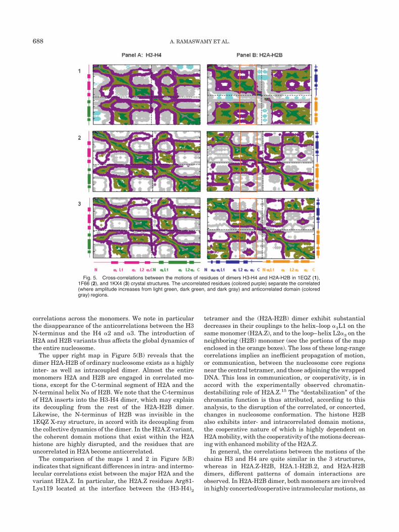
The behavior illustrated in Figures 4 and 5 reflects theresult from an ensemble of normal modes. Next, weproceed to a closer examination of the 2 lowest frequencymodes, shortly referred to as modes 1 and 2. The slowestmodes usually involve the entire structure and are therebyreferred to as global modes. They contribute to the ob-served spectrum of motions scales with their inversefrequencies (or corresponding eigenvalue of the Kirchhoffmatrix). A small subset of slow modes usually dominatesthe overall dynamics, and the slowest 1 to 2 among themhave been shown in numerous studies to drive motionsrelevant to biological function.33–43
Figure 6 illustrates the global mode dynamics of themonomeric histones, computed for 1EQZ (blue curves),1F66 (red), and 1KX4 (green). The results are displayed forone set of monomers (labeled as H3, H4, H2A, and H2B),with the global dynamics of the corresponding secondmonomers (copies) in the octameric core being almostidentical. The ordinate represents the distributions of thesquare displacements of individual residues induced bythe first (solid curves) and second (dotted curves) modes.The secondary structures of the monomers (colored accord-ing to Figs. 1 and 2) are shown along the abscissa. Thehistone–DNA interacting sites, L1, L2, and �1, are indi-cated, along with a few other interacting sites of interest(e.g., minima serving as global hinge sites). It is interest-ing to observe that (1) the nucleosomes with the varianthistones (red and green curves) generally exhibit largeramplitude of motions compared to the nucleosome withordinary histone monomers, and (2) the residues that aredynamic in one mode behave as rigid domains in the othermode, and vice versa.
In the first mode, the residues Arg24-Ile30 of H4 (i.e.,residues 1–7 in Fig. 6), which are involved in gene silenc-ing, exhibit relatively high mobility, which is consistentwith their active participation in functional dynamics.48 Inthe H2A monomer, the peak observed in the first modecorresponds to its dynamic L2 loop (Leu77), which inter-acts with the dynamic loop L1 of H2B. We note inparticular that the residues Lys79 in loop L2 of H2A andSer53 in loop L1 of H2B that interact with the minorgroove of DNA are highly dynamic. The regions L2 ofH2A.Z, L1 of H2B, and �1 of H3 of each monomer exhibithigher amplitude motions in the variant nucleosome. Ithas been determined that in the ordinary nucleosome,
these regions form hydrogen bonds with the neighboringDNA nucleotides, which are likely to be perturbed, if notbroken, in the variant. However, it should be noted thatour model pertains to the changes in �-carbon, and P- andO4*-atoms coordinates, and only the changes in hydrogen
Fig. 6. Comparison of the global mode shapes of monomeric histonesH3, H4, H2A, and H2B computed with the GNM for the crystal structuresof the ordinary nucleosome 1EQZ (blue), and 2 nucleosomes with histonevariants, 1F66 (red) and 1KX4 (green). The curves scale with theamplitude of motions undergone by different structural elements in mode1 (solid curves) and mode 2 (dotted curves). Arrows indicate thehydrogen-bonding sites between the amino acid residues and the nucleo-tides located at the L1, L2, �1, and �2 sites along with other interactingsites of interest (e.g., minima serving as global hinge sites).
NUCLEOSOME DYNAMICS: ROLE OF HISTONE VARIANTS 689
bonds that affect these backbone coordinates are takeninto consideration in the ANM.
In mode 2, we observe the docking domain of H2A (resi-dues from Ile82 to Ile120, i.e., residues 63–101 in the Fig. 6)to be highly stable (minimal fluctuations) except for theC-terminal region. This region is highly conserved accordingto the experimental results, and the equivalent region ofDrosophila H2A.Z is essential in fly development.18 Thehinge domain corresponding to �C helix of the docking do-main is indicated in Figure 6.
The complementary shapes of the 2 modes correspond-ing to the monomers H2A and H2B are noteworthy. Theseare essentially sinusoidal shapes with a 2-fold symmetricorigin at Gly47 (in H2A) and Ser39 (in H2B), the secondmode being almost the mirror image of the first. Ingeneral, it is observed that the hinge regions observed inone mode behave as highly mobile dynamic regions in theother mode, and vice versa. Overall, a strong cooperativitybetween the global dynamics of the H2A and H2B mono-mers is indicated.
Fig. 7. Color-coded representation of the dynamics of 1EQZ (1), 1F66 (2) and 1KX4 (3) in the first (A) andsecond (B) collective mode.
690 A. RAMASWAMY ET AL.
Global Dynamics of Nucleosome
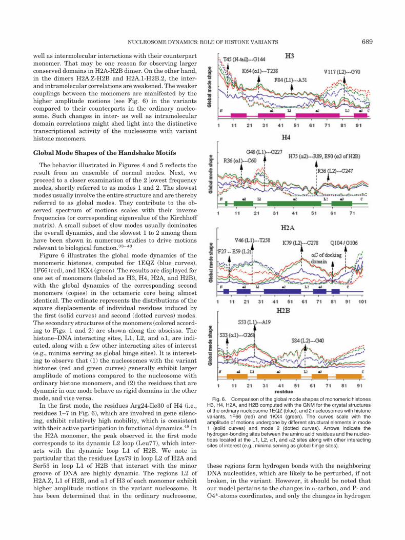
Figure 7 shows the dynamics of nucleosomes in acolor-coded fashion [from black (rigid) to red (most flexible)for the first (A) and second (B) slowest modes of structures[1–3]. The histone tails of the 3 nucleosomes vary inlength, so the calculations have been performed with allthe residues of histone tails, as well as with the commonhistone tail residues. Both observations of nucleosomedynamics (with full tail domains, as well as with commontail domains) revealed similar pattern of nucleosome dy-namics, reported here.
A major observation is the highly symmetrical dynamicsof the overall nucleosome with respect to the dyad axis(vertical axis in the present view), consistent with thecomparable mobilities of the copies of each histone pointedout above.
In the first mode, the rigid domains fall along the dyadaxis of nucleosome. The spatially conserved domains areidentified at (1) the H2B residues from L2 through the Cterminus, (2) the H2A residues around the loop L1, (3) theresidues from helix �3 to the C-tail of histone H3, and (4)the close neighborhood of His75 on H4 �2 helix. TheN-termini of histones have been experimentally proven tomediate most of the protein–DNA interactions, and theirmobilities are essential in the regulation of eukaryotictranscription.49 In our study, the N-termini are shown tobe highly dynamic in both modes. In particular the highlydynamic N-tail of H3 is engaged in a highly cooperativemotion with the neighboring DNA segments. As a result,the wrapped DNA also exhibits a symmetric dynamicswith respect to the dyad.
In the second slowest mode [Fig. 7(B)], the dynamics hasbeen identified as conjugate to the first slowest mode ofmotion; that is, the central region of nucleosome perpen-dicular to the dyad axis is highly constrained (rigid), whileseveral domains, which were severely almost rigid in mode1, show significant mobilities. All N-termini of histonesexcept for H4 in the nucleosome with ordinary histonesshow high mobilities, consistent with the disordered struc-tures of the N-termini of histones.50 In the nucleosome, thetetramer (H3-H4)2 is positioned on both sides of the dyadaxis and interacts with one of the DNA strands, and so thedynamics of (H3-H4)2 affect the dynamics of the particularDNA strand interacting with (H3-H4)2. Loop L1 andC-terminus in H2A, helix �2, and C-terminus in H2B,Loop L2 and N-tail domain in H3, and the H4 L1 loop showthe highest mobilities. The most constrained regions thatalso constrain and control the DNA motions on both sidesof the dyad axis are composed of H3 L1, H4 N-tail, �2, L2and �3, H2A �3, �C, and adjoining segments (includingQ104), and H2B L1 and L2.
The comparison of the dynamics of the ordinary nucleo-some (1) with that containing the variants (2 and 3) showsthat the direction of the loci of rigid (black) regionschanges from a diagonal orientation in (1) to a horizontalone in (2) and (3). Given that the first mode symmetry axisis along the perpendicular axis, the joint contribution ofthe first 2 modes would then be expected to induce anoverall higher and more evenly spread mobility in all
chains of the variants, compared to that in the ordinarynucleosome. Experimental report on the transcriptionallyactive conformation of nucleosome with the variant H2A.Z,8
suggests the perturbed dynamics of (H2A.Z-H2B) dimersand the N-terminus of H3 in physiological conditions.50
The second slowest mode could explain the different globaldynamics of transcriptionally active nucleosomes, whilethe first mode is invariably preserved in the 3 structures.
Directions of Global Mode Dynamics of Nucleosome
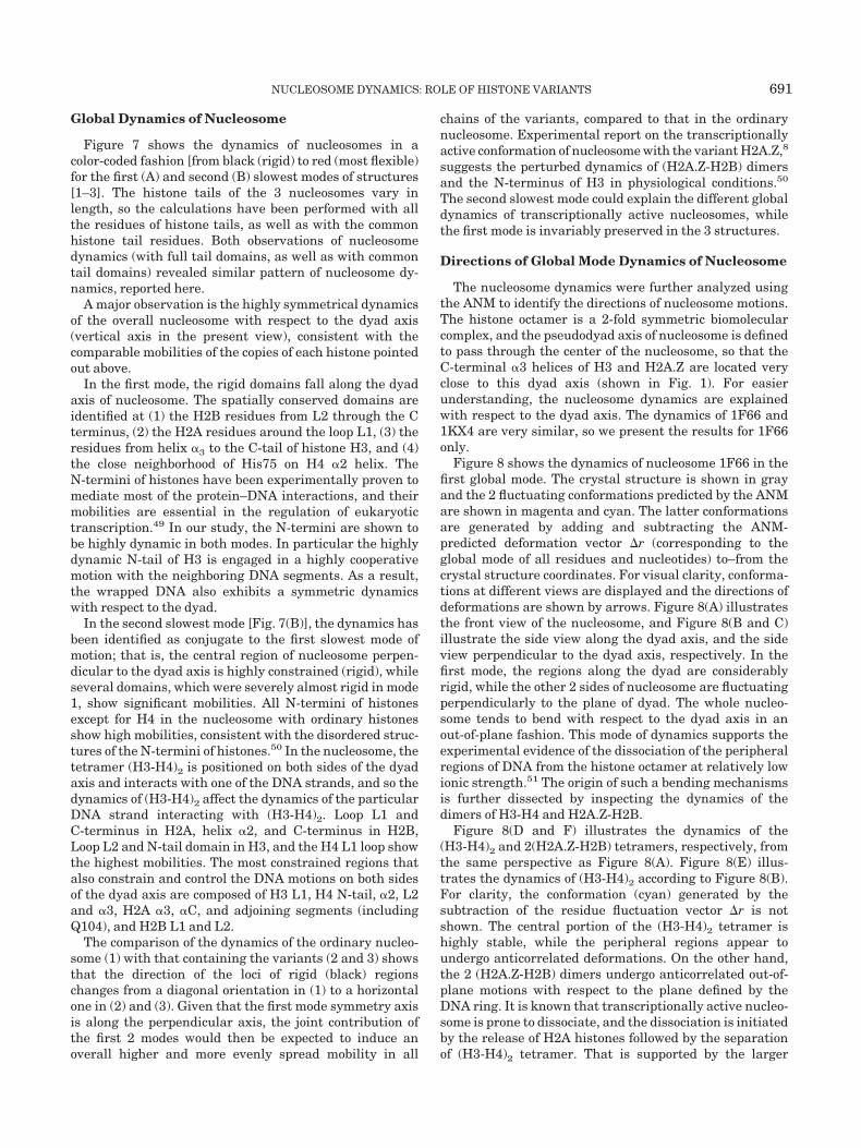
The nucleosome dynamics were further analyzed usingthe ANM to identify the directions of nucleosome motions.The histone octamer is a 2-fold symmetric biomolecularcomplex, and the pseudodyad axis of nucleosome is definedto pass through the center of the nucleosome, so that theC-terminal �3 helices of H3 and H2A.Z are located veryclose to this dyad axis (shown in Fig. 1). For easierunderstanding, the nucleosome dynamics are explainedwith respect to the dyad axis. The dynamics of 1F66 and1KX4 are very similar, so we present the results for 1F66only.
Figure 8 shows the dynamics of nucleosome 1F66 in thefirst global mode. The crystal structure is shown in grayand the 2 fluctuating conformations predicted by the ANMare shown in magenta and cyan. The latter conformationsare generated by adding and subtracting the ANM-predicted deformation vector �r (corresponding to theglobal mode of all residues and nucleotides) to–from thecrystal structure coordinates. For visual clarity, conforma-tions at different views are displayed and the directions ofdeformations are shown by arrows. Figure 8(A) illustratesthe front view of the nucleosome, and Figure 8(B and C)illustrate the side view along the dyad axis, and the sideview perpendicular to the dyad axis, respectively. In thefirst mode, the regions along the dyad are considerablyrigid, while the other 2 sides of nucleosome are fluctuatingperpendicularly to the plane of dyad. The whole nucleo-some tends to bend with respect to the dyad axis in anout-of-plane fashion. This mode of dynamics supports theexperimental evidence of the dissociation of the peripheralregions of DNA from the histone octamer at relatively lowionic strength.51 The origin of such a bending mechanismsis further dissected by inspecting the dynamics of thedimers of H3-H4 and H2A.Z-H2B.
Figure 8(D and F) illustrates the dynamics of the(H3-H4)2 and 2(H2A.Z-H2B) tetramers, respectively, fromthe same perspective as Figure 8(A). Figure 8(E) illus-trates the dynamics of (H3-H4)2 according to Figure 8(B).For clarity, the conformation (cyan) generated by thesubtraction of the residue fluctuation vector �r is notshown. The central portion of the (H3-H4)2 tetramer ishighly stable, while the peripheral regions appear toundergo anticorrelated deformations. On the other hand,the 2 (H2A.Z-H2B) dimers undergo anticorrelated out-of-plane motions with respect to the plane defined by theDNA ring. It is known that transcriptionally active nucleo-some is prone to dissociate, and the dissociation is initiatedby the release of H2A histones followed by the separationof (H3-H4)2 tetramer. That is supported by the larger
NUCLEOSOME DYNAMICS: ROLE OF HISTONE VARIANTS 691
amplitude of the out-of-plane distortions of the (H2A.Z-H2B) dimer compared to the more restricted in-planemotions of the (H3-H4)2 tetramer core.
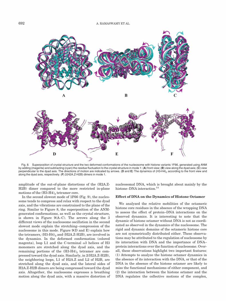
In the second slowest mode of 1F66 (Fig. 9), the nucleo-some tends to compress and relax with respect to the dyadaxis, and the vibrations are constrained to the plane of thering. Similar to Figure 8, the superposition of the ANM-generated conformations, as well as the crystal structure,is shown in Figure 9(A–C). The arrows along the 3different views of the nucleosome oscillation in the secondslowest mode explain the stretching–compression of thenucleosome in this mode. Figure 9(D and E) explain howthe tetramers, (H3-H4)2 and (H2A.Z-H2B), are involved inthe dynamics. In the deformed conformation (coloredmagenta), loop L1 and the C-terminal �3 helices of H3monomers are stretched along the dyad axis, and theremaining portions of the (H3-H4)2 tetramer are com-pressed toward the dyad axis. Similarly, in 2(H2A.Z-H2B),the neighboring loops, L1 of H2A.Z and L2 of H2B, arestretched along the dyad axis, and the lateral sides ofH2A.Z-H2B dimers are being compressed toward the dyadaxis. Altogether, the nucleosome expresses a breathingmotion along the dyad axis, with a massive distortion of
nucleosomal DNA, which is brought about mainly by thehistone–DNA interaction.51
Effect of DNA on the Dynamics of Histone Octamer
We analyzed the relative mobilities of the octamerichistone core residues in the absence of the wrapping DNAto assess the effect of protein–DNA interactions on theobserved dynamics. It is interesting to note that thedynamic of histone octamer without DNA is not as coordi-nated as observed in the dynamics of the nucleosome. Therigid and dynamic domains of the octameric histone coreare not symmetrically distributed either. These observa-tions may be attributed to the regulation of nucleosome byits interaction with DNA and the importance of DNA–protein interactions over the function of nucleosome. Over-all, these observations highlight two important features:(1) Attempts to analyze the histone octamer dynamics inthe absence of its interaction with the DNA, or that of theDNA in the absence of the histone octamer are likely tomiss the functional mechanisms of either component, and(2) the interaction between the histone octamer and theDNA regulates the collective motions of the complex,
Fig. 8. Superposition of crystal structure and the two deformed conformations of the nucleosome with histone variants 1F66, generated using ANMby adding (magenta) and subtracting (cyan) the residue fluctuation to the crystal structure in mode 1: (A) front view; (B) view along the dyad axis; (C) viewperpendicular to the dyad axis. The directions of motion are indicated by arrows. (D and E) The dynamics of (H3-H4)2 according to the front view andalong the dyad axis, respectively. (F) 2(H2A.Z-H2B) dimers in mode 1.
692 A. RAMASWAMY ET AL.
which in the absence of intermolecular interactions aresignificantly more disordered.
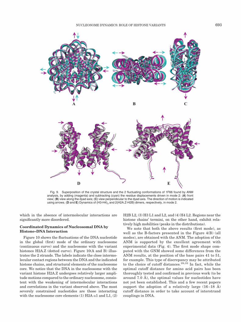
Coordinated Dynamics of Nucleosomal DNA byHistone–DNA Interaction
Figure 10 shows the fluctuations of the DNA nucleotidein the global (first) mode of the ordinary nucleosome(continuous curve) and the nucleosome with the varianthistones H2A.Z (dotted curve). Figure 10(A and B) illus-trates the 2 strands. The labels indicate the close intermo-lecular contact regions between the DNA and the indicatedhistone chains, and structural elements of the nucleosomecore. We notice that the DNA in the nucleosome with thevariant histone H2A.Z undergoes relatively larger ampli-tude motions compared to the ordinary nucleosome, consis-tent with the weakening of intermolecular interactionsand correlations in the variant observed above. The mostseverely constrained nucleotides are those interactingwith the nucleosome core elements (1) H2A �1 and L1, (2)
H2B L2, (3) H3 L1 and L2, and (4) H4 L2. Regions near thehistone chains’ termini, on the other hand, exhibit rela-tively high mobilities (peaks in the distributions).
We note that both the above results (first mode), aswell as the B-factors presented in the Figure 4(B) (allmodes), are obtained with the ANM. The adoption of theANM is supported by the excellent agreement withexperimental data (Fig. 4). The first mode shape com-puted with the GNM showed some differences from theANM results, at the position of the base pairs 41 to 51,for example. This type of discrepancy may be attributedto the choice of cutoff distances.29,32 In fact, while theoptimal cutoff distance for amino acid pairs has beenthoroughly tested and confirmed in previous work (to bearound 7.0 Å), the optimal values for nucleotides havenot yet been established. This and a few recent paperssupport the adoption of a relatively large (16 –18 Å)cutoff distance in order to take account of interstrandcouplings in DNA.
Fig. 9. Superposition of the crystal structure and the 2 fluctuating conformations of 1F66 found by ANManalysis, by adding (magenta) and subtracting (cyan) the residue displacements driven in mode 2: (A) frontview; (B) view along the dyad axis; (C) view perpendicular to the dyad axis. The direction of motion is indicatedusing arrows. (D and E) Dynamics of (H3-H4)2 and 2(H2A.Z-H2B) dimers, respectively, in mode 2.
NUCLEOSOME DYNAMICS: ROLE OF HISTONE VARIANTS 693
Changes in the Volume Over the Normal ModeDynamics
Table I lists the molecular dimensions along the 3principal axes (X, Y, and Z) of the structure, and thedeformations in these molecular dimensions induced bythe first two slowest modes. The Y axis coincides with the
dyadic axis (shown by the vertical arrow in Fig. 1), the Xaxis is the in-plane perpendicular axis, and the Z axis isthe out-of-plane axis that completes the right-handedreference frame. The dimensions of the nucleosome alongthese principal axes have been calculated using the In-sight II package. The changes in the dimensions have been
Fig. 10. Comparison of the fluctuations predicted by the ANM for the nucleosome DNA nucleotides. (A andB) Results for the two different strands in 1EQZ (continuous curve) and 1F66 (dotted curve), respectively. Thelabels indicate the histone chains and secondary structure elements that make intermolecular contacts with theDNA.
TABLE I. Changes in Molecular Dimensions Induced by Dominant Modes
Structures
Dimensions along the Cartesian Axes (Å)
X axis Y axis Z axis
Distance Difference Distance Difference Distance Difference
1EQZ
Crystal 103.0 106.2 68.1Mode 1 103.3 0.3 106.2 0.0 68.3 0.2Mode 1� 104.3 1.3 106.1 �0.1 67.9 �0.2Mode 2 103.0 0.0 106.4 0.2 67.9 �0.2Mode 2� 103.1 0.1 105.9 �0.3 68.3 0.2
1F66
Crystal 103.1 106.7 67.6Mode 1 103.8 0.7 107.9 1.2 68.6 2.0Mode 1� 107.4 4.3 105.4 �1.3 66.5 �1.1Mode 2 104.3 1.2 108.4 1.7 67.2 �0.4Mode 2� 104.3 1.2 105.0 �1.7 68.0 0.4
694 A. RAMASWAMY ET AL.
estimated by adding and subtracting the fluctuationspredicted by the two slowest ANM modes to the crystalstructure coordinates (used as a reference). The differ-ences in the molecular dimensions along the 3 axes providea measure of the voluminous changes induced by theglobal normal modes. Modes 1 and 1� refer to thedeformed conformations associated with mode 1, andmodes 2 and 2� refer to mode 2.
We note that the deformations observed in the varianthistones are generally higher than those occurring in thenucleosome of ordinary histones, consistent with the highermobility of the variants. Notably, an expansion of 4.3 Å isinduced by mode 1 perpendicular to the dyad axis, accom-panied by compressions of 1.3 Å along the X axis, followedby an out-of-plane expansion of 2.0 Å that is accommo-dated by 0.7 Å and 1.2 Å stretchings along the 2 radialdirections. Overall, the result is a bending (or flipping)motion around the dyadic axis accompanied by an expan-sion along the X axis, as illustrated in Figure 8. Mode 2, onthe other hand, is essentially manifested by in-planestretching–contraction (of 1.7Å) along the dyad axis, accom-panied by small (0.4 Å) compression–expansion along theZ direction.
CONCLUSIONS
Global dynamics of nucleosomes with native and varianthistones revealed the relaxation of nucleosome with sev-eral conformations during the dynamics. The most prob-able mechanisms of motions predicted by the presentanalysis are (1) the flip (or bending) of both the sides of thenucleosome in an out-of-plane fashion with respect to thedyad axis, accompanied by an expansion along the in-plane (X) axis perpendicular to the dyad axis (mode 1; Fig.8), and (2) the in-plane stretching–compression of nucleo-some core resulting in the deformation of the circularlysupercoiled DNA (mode 2; Fig. 9).
In the flipping motion of nucleosome, the N-termini ofthe H3 and H4 histones are highly mobile, consistent withthe functional role of these N-tails experimentally ob-served under physiological conditions.50 Other regionsdistinguished by their high mobility and closely interact-ing with the wrapped DNA superhelix are the H2A L2(peak at K79), H2B L1 (peak at S53; Fig. 6). The mostseverely constrained regions are, on the other hand, thecentral long helices in H3 and H4, and the respective loopsL1 and L2 of H2A and H2B monomers. These latter sitesplay a critical role in coordinating the collective mode 1.
In the stretching–compression mode, the nucleosomeexhibits a breathing motion along the dyad axis, with amassive distortion of nucleosomal DNA, which is broughtabout mainly by the histone-DNA interaction.51 Elementsexhibiting the highest mobilities in this mode are theN-terminal tail and loop L2 of H3, H4 L1, H2A L1, H2B L1,and the C-tails of H2A and H2B, while the most con-strained regions include H3 L1, H4 L2, H2A L2, and H2BL1.
The nucleosomes containing histone variants are foundto be significantly more mobile than the ordinary nucleo-some. They exhibit weaker intramolecular and intermolecu-
lar couplings/correlations resulting in more disordered orless coherent motions consistent with their higher flexibil-ity. A region exhibiting distinctive dynamics in the histonevariants is the helix �C of H2A, known to be the dockingregion of H2A.Z. While this region is severely constrainedand participating in the global hinge center in mode 1 ofthe ordinary nucleosome, it becomes significantly moreflexible in the nucleosome containing histone variants,consistent with the possible weakening of intra- andintermolecular couplings in the variant. The predictedhigher mobility/disorder of the variants may facilitate thedisruption of the nucleosome. A recent article by Lugerand coworkers52 described the variant H2A.Z histone asessential for the nucleosome stability at higher ionicstrength, which is in disagreement with the earlier studiesof Abbott et al.15 They attributed these discrepancies tothe differences in experimental conditions/preparations.However, the present study focuses on the structuraldynamics of nucleosome irrespective of ionic strengthdependence.
The coupling between the histone octamer and thesurrounding DNA superhelix is instrumental in regulat-ing the dynamics of the octamer, and conversely, theclosely interacting proteins significantly alter the dynam-ics of the DNA, signaling the important role of the complex-ation with histones in regulating the transcriptional activ-ity.
REFERENCES
1. van Holde KE. Chromatin. New York: Springer; 1989. 497 p.2. Wolffe A. Chromatin: structure and function. San Diego: Academic
Press; 1998. 447 p.3. Ramakrishnan V. Histone structure and the organization of
nucleosome. Ann Rev Biophys Biomol Struct 1997;26:83–112.4. Workman JL, Kingston RE. Alteration of nucleosome structure as
a mechanism of transcriptional regulation. Ann Rev BiophysBiomol Struct 1998;67:545–579.
5. Luger K, Mader AW, Richmond RK, Sargent DF, Richmond TJ.Crystal structure of the nucleosome core particle at 2.8 Å resolu-tion. Nature 1997;389:251–260.
6. Richmond TJ, Finch JT, Rushton B, Rhodes D, Klug A. Structureof the nucleosome core particle at 7Å resolution. Nature 1984;311:532–537.
7. Arents G, Burlingame RW, Wang BC, Love WE, MoudrianakisEN. The nucleosomal core histone octamer at 3.1 Å resolution: atripartite protein assembly and a left-handed super helix. ProcNatl Acad Sci USA 1991;88:10148–10152.
8. Suto RK, Clarkson MJ, Tremethick DJ, Luger K. Crystal struc-ture of a nucleosome core particle containing the variant histoneH2A.Z. Nat Struct Biol 2000;7:1121–1124.
9. Eickbush TH, Moudrianakis EN. The histone core complex: Anoctamer assembled by two sets of protein–protein interactions.Biochem 1978;17:4955–4964.
10. Hayes JJ, Clark DJ, Wolffe AP. Histone contributions to thestructure of DNA in the nucleosome. Proc Natl Acad Sci USA1991;88:6829–6833.
11. Hayes JJ, Tullius TD, Wolffe AP. The structure of DNA in anucleosome. Proc Natl Acad Sci USA 1990;87:7405–7409.
12. Davie JR. Covalent modifications of histones: expression fromchromatin templates. Curr Opin Genet Dev 1998;8:173–178.
13. Brown DT. Histone variants: are they functionally heteroge-neous? Genome Biol 2001;2: reviews 0006.1–reviews 0006.6.
14. Adam M, Robert F, Larochelle M, Gaudreau L. H2A.Z is requiredfor global chromatin integrity and for recruitment of RNA polymer-ase II under specific conditions. Mol Cell Biol 2001;21:6270–6279.
15. Abbott DW, Ivanova VS, Wang X, Bonner WM, Ausio J. Character-ization of the stability and folding of H2A.Z chromatin particles:
NUCLEOSOME DYNAMICS: ROLE OF HISTONE VARIANTS 695
implications for transcriptional activation. J Biol Chem 2001;276:41945–41949.
16. Fan JY, Gordon F, Luger K, Hansen JC, Tremethick DJ. Theessential histone variant H2A.Z regulates the equilibrium be-tween different chromatin conformational states. Nat Struct Biol2002;9:172–176.
17. Jackson JD, Gorovsky MA. Histone H2A.Z has a conservedfunction that is distinct from that of the major H2A sequencevariants. Nucleic Acid Res 2000;28:3811–3816.
18. Clarkson MJ, Wells JRE, Gibson F, Saint R, Tremethick DJ.Regions of variant histone His2AvD required for Drosophiladevelopment. Nature 1999;399:694–697.
19. Santisteban MS, Kalashnikova T, Smith MM. Histone H2A.Zregulates transcription and is partially redundant with nucleo-some remodeling complexes. Cell 2000;103:411–422.
20. Cornell WD, Cieplak P, Bayly CI, Gould IR, Merz KMJr., Fergu-son DM, Spellmeyer DC, Fox T, Caldwell JW, Kollman PA. Asecond generation force field for the simulation of proteins, nucleicacids and organic molecules. J Am Chem Soc 1995;117:5179–5197.
21. Dauber-Osguthorpe P, Roberts VA, Osguthorpe DJ, Wolff J,Genest M, Hagler AT. Structure and energetics of ligand bindingto proteins: E. coli dihydrofolate reductase-trimethoprim, a drug-receptor system. Proteins 1988;4:31–47.
22. Levitt M, Sander C, Stern PS. Protein normal-mode dynamics:trypsin inhibitor, crambin, ribonuclease and lysozyme. J Mol Biol1985;181:423–447.
23. Gibrat JF, Go N. Normal mode analysis of human lysozyme: studyof the relative motion of the two domains and characterization ofthe harmonic motion. Proteins 1990;8:258–279.
24. Tirion MM. Large amplitude elastic motions in proteins from asingle-parameter, atomic analysis. Phys Rev Lett 1996;77:1905–1908.
25. Bahar I, Atilgan AR, Erman B. Direct evaluation of thermalfluctuations in proteins using a single-parameter harmonic poten-tial. Fold Des 1997;2:173–181.
26. Haliloglu T, Bahar I, Erman B. Gaussian dynamics of foldedproteins. Phys Rev Lett 1997;79:3090–3093.
27. Hinsen K. Analysis of domain motions by approximate normalmode calculations. Proteins 1998;33:417–429.
28. Tama F, Sanejouand Y.-H. Conformational change of proteinsarising from normal mode calculations. Protein Eng 2001;14:1–6.
29. Atilgan AR, Durrell SR, Jernigan RL, Demirel MC, Keskin O,Bahar I. Anisotropy of fluctuation dynamics of proteins with anelastic network model. Biophys J 2001;80:505–515.
30. Bahar I, Wallqvist A, Covell DG, Jernigan RL. Correlationbetween native-state hydrogen exchange and cooperative residuefluctuations from a simple model. Biochemistry 1998;37:1067–1075.
31. Haliloglu T, Bahar I. Structure-based analysis of protein dynam-ics: comparison of theoretical results for hen lysozyme with X-raydiffraction and NMR relaxation data. Proteins 1999;37:654–667.
32. Kundu S, Melton JS, Sorensen DC, Phillips GN Jr. Dynamics ofproteins in crystals: comparison of experiment with simple mod-els. Biophys J 2002;83:723–732.
33. Ming D, Kong Y, Lambert MA, Huang Z, Ma J. How to describeprotein motion without amino acid sequence and atomic coordi-nates. Proc Natl Acad Sci USA 2002;99:8620–8625.
34. Tama F, Wriggers W, Brooks CL III. Exploring global distortionsof biological macromolecules and assemblies from low-resolution
structural information and continuum elastic network theory. JMol Biol 2002;321:297–305.
35. Tama F, Valle M, Frank J, Brooks CL III. Dynamic reorganizationof the functionally active ribosome explored by normal modeanalysis and cryo-electron microscopy. Proc Natl Acad Sci USA2003;100:9319–9323.
36. Bahar I, Jernigan RL. Vibrational dynamics of transfer RNAs:comparison of the free and synthetase-bound forms. J Mol Biol1998;281:871–884.
37. Bahar I, Erman B, Jernigan RL, Atilgan AR, Covell DG. Collectivemotions in HIV-1 reverse transcriptase: examination of flexibilityand enzyme function. J Mol Biol 1999;285:1023–1037.
38. Temiz AN, Bahar I. Inhibitor binding alters the directions ofdomain motions in HIV-1 reverse transcriptase. Proteins 2002;49:61–70.
39. Bahar I, Atilgan AR, Demirel MC, Erman B. Vibrational dynamicsof folded proteins: significance of slow and fast motions in relationto function and stability. Phys Rev Lett 1998;80:2733–2736.
40. Bahar I, Jernigan RL. Cooperative fluctuations and subunitcommunication in tryptophan synthase. Biochemistry 1999;38:3478–3490.
41. Demirel MC, Atilgan AR, Jernigan RL, Erman B, Bahar I.Identification of kinetically hot residues in proteins. Protein Sci1998;7:2522–2532.
42. Keskin O, Jernigan RL, Bahar I. Proteins with similar architec-ture exhibit similar large-scale dynamic behavior. Biophys J2000;78:2093–2106.
43. Tama F, Wriggers W, Brooks III CL. Exploring global distortionsof biological macromolecules and assemblies from low-resolutionstructural information and elastic network theory. J Mol Biol2002;321:297–305.
44. Harp JM, Hanson BL, Timm DE, Bunick GJ. Asymmetries in thenucleosome core particle at 2.5Å resolution. Acta Crystallogr DBiol Crystallogr 2000;56:1513–1534.
45. Davey CA, Sargent DF, Luger K, Maeder AW, Richmond TJ.Solvent mediated interactions in the structure of the nucleosomecore particle at 1.9 Å resolution. J Mol Biol 2002;319:1097–1113.
46. Berman HM, Westbrook J, Feng Z, Gilliland G, Bhat TN, WeissigH, Shindyalov IN, Bourne PE. The Protein Data Bank. NucleicAcid Res 2000;28:235–242.
47. Huang CC, Couch GS, Petterse EF, Ferrin TE. Chimera: Anextensible molecular modeling application constructed using stan-dard components. Pacific Symposium on Biocomputing 1996;1:724. http://www.cgl.ucsf.edu/chimera
48. Johnson LM, Fisher Adams G, Grunstein M. Identification of anon-basic domain in the histone N-terminus required for repres-sion of the yeast silent mating loci. EMBO J 1992;11:2201–2209.
49. Ebralidse KK, Grachev SA, Mirzabekov AD. A highly basichistone H4 domain bound to the sharply bent region of nucleoso-mal DNA. Nature 1988;331:365–367.
50. Smith RM, Rill RL. Mobile histone tails in nucleosomes: assign-ments of mobile segments and investigations of their role inchromatin folding. J Biol Chem 1989;264:10574–10581.
51. Muthurajan UM, Park YJ, Edayathumangalam RS, Suto RK,Chakravarthy S, Dyer PN, Luger K. Structure and dynamics ofnucleosomal DNA. Biopolymers 2003;68:547–556.
52. Park YJ, Dyer PN, Tremethick DJ, Luger K. A new fluorescenceresonance energy transfer approach demonstrates that the his-tone variant H2AZ stabilizes the histone octamer within thenucleosome. J Biol Chem 2004;279:24274–24282.
696 A. RAMASWAMY ET AL.