histologic analysis of venous invasion...
TRANSCRIPT

Acta Medica et BiologicaVol. 33, No. 1, 19-34, 1985
HISTOLOGIC ANALYSIS OF VENOUS INVASION IN COLORECTAL CARCINOMAS IN RELATION
TO HEMATOGENOUS METASTASES
MUTSUO YAMAMOTO
Department of Surgery Niigata University
(Director: Professor Terukazu Muto)
(Received December 24, 1984)
INTRODUCTION
Hematogenous metastases have long been one of the most difficult problems in the
treatment of colorectal carcinomas; it was reported that 18% of these carcinomas had
liver metastases at the time of initial operation (Oxley et a1., 1976)1) and that 14.5% had
metachronous pulmonary metastases (Schulten et a1., 1976).2) In a series of investigation
conducted in Japan, 12.5% (735/5,890) of colorectal carcinomas had liver metastases at
the time of initial operation and 4.2% (100/2,353) had pulmonary metastases according to
the report by the Japanese Research Society for Cancer of Colon and Rectum (1982).3)
Welch et al. (1978)4) reported that only 76.2% of 1,566 colorectal carcinomas had been
curatively resected. Fortner et a1. (1984)5) reported that 50% of 120,000 colorectal
carcinomas had generated recurrence and over 50% of such recurrence had been liver
metastases. Tsuchiya (1982)6) expressed that 80% of colorectal carcinomas with meta
chronous hepatic metastases already had microscopic hepatic metastases at the time of
initial operation. At the present time, having achieved almost full development of
surgical treatments, early diagnosis of primary lesions and prevention of recurrence are
required most for the improvement of the prognosis of patients with colorectal car
cinoma. The fact that the potential of hematogenous metastases was well correlated
with venous invasion at primary sites has been accepted by many researchers. Venous
invasion is only a factor in everything that seems to affect the mechanism of hemato
genous metastases, and is demonstrated in specimens of colorectal carcinomas as
"intravasation" and "intravenous embolization" reported by Nishi et a1. (1962).7) In the
work of Brown and Warren (1938),8) the incidence of venous invasion in 165 rectal
carcinomas was 61%. Since this report, a considerable number of reports on venous

- 20 M. YAMAMOTO
invasion have been published. The incidence of venous invasion has ranged between 20%(Seefeld et al., 1943) and (Talbot et al., 1981). 13)
The fluctuation of this rate might be caused by the different methods for pathologic
examination. In our previous studies, the incidence of venous invasion examined in three
longitudinal sections with elastic tissue stain was This procedure was recom
mended by Konishi et al. (1982)14) as a reliable method, but not adequate to predict
hematogenous metastases because the incidence of venous invasion thus demonstrated is
too high to evaluate. The purpose of this study is to evaluate the mechanism of
hematogenous metastases on the basis of detailed analysis of venous invasion at the
primary sites of colorectal carcinomas. It is expected that active adjuvant chemother
apy or other methods used on the high-risk patients will be effective in preventing
hematogenous metastases.
MATERIALS AND METHODS
One hundred and fifty-six patients with primary colorectal carcinoma (76 with rectal
carcinoma and 80 with colonic carcinoma) with complete histologic examination and
clinical follow-up were selected for the present analysis. These patients consisted of 87
(55.8%) males and 69 (44.2%) females, with age ranging from 25 to 83 years, 59.9 years in
avarage (Table 1), and were divided into 3 groups: 102 patients (group A : metastasis-free
group) living over 3 years after surgery without recurrence; 26 (group B) with meta
chronous hematogenous metastases; and 28 (group C) with synchronous hematogenous
metastases. Patients with more than two primary lesions and those with indistinct or
other principal patterns of recurrence were excluded from this study. For histologic
analysis on specimens resected from 156 patients, 3 longitudinal sections from each lesion
were examined by elastic tissue stain (Figs. 1 and Each section of the lesions was
divided into four subsections of proximal outer (PO), proximal inner (PI), distal inner (DI)
and distal outer (DO) regions as indicated in Fig. 3. Each subsection was further divided
into three layers (submucosa, muscular layer and subserosa). Location and size (the
maximum length of the short axis of an elastic tissue ring being indicative of venous wall)
of invaded veins were individually recorded (Fig. 2). Uncertain involvement of veins was
judged as "negative".
Table l. Sex and Average Age
Group Total No. Cases Male (%) Female (%) Average Age
A 102 53(52.0) 49(48.0) 59.9B 26 19(73.1) 7(26.9) 59.3C 28 15(53.6) 13(46.4) 60.7
Total 156 87(55.8) 69(44.2) 59.9

distal f-
VENOUS INVASION IN COLORECTAL CA.RCINOMAS
I
II
-1! 2 em 7! em
I I
- 21
Fig. 1. Three longitudinal sections are taken from primary lesion, each including peripheral intactportions over 2 cm of length.
Fig. Venous invasion is well indicated by elastic tissue stain (Van Gieson); the venuleinvolved is otherwise not demonstrated. The arrow shows the maximum length of short axisof the venule as representing the size of invaded vein. x 400.

22 -
distal ~
...., 2 em f-
M. YAMAMOTO
--.c:qz.~"tZZ."tZZ.~rm222ZZZZ2-;) proximal
2 em f-
DO Dr PI PO
Fig. 3. Each longitudinal section is divided into four subsections (PO, PI, DI and DO), outer subsectionsincluding peripheral intact portions over 2 cm of length.
RESULTS
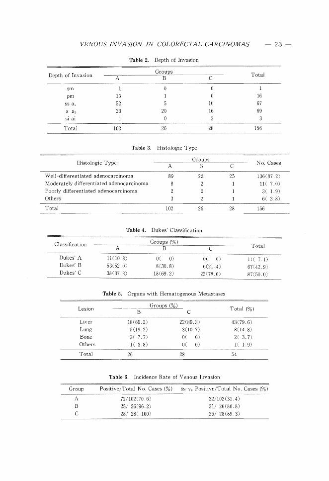
1. Basic Observation (Tables 2, 3, 4 and 5):
The depth of carcinomatous invasion shown in Table 2 was progressive with develop
ment of hematogenous metastases and no case with intramural invasion was seen in
group C. There was little difference among the distribution of histologic types in the
three groups and well-differentiated adenocarcinoma dominated in each group (Table 3).
Each lesion was classified by Dukes' Classification and advanced carcinomas were in
creased parallel with the development of hematogenous metastases. The incidence of
lesions in the Dukes' C Class was as follows; 37.3% in group A, 69.2% in Band 78.6% in
C (Table
Table 5 shows principal organs with hematogenous metastases in groups Band C.Liver metastases had a prominent incidence in both groups, 69.2% and 89.3%, re
spectively.
2. Incidence of Venous Invasion (Table 6):
Venous invasion was found in 72 patients (70.6%) in group A, 25 (96.2%) in Band 28
(100%) in C, totalling 125 (80.1%) out of 156. Only one (1.9%) of 54 patients with
hematogenous metastases had no recognizable venous invasion. Subserosal invaded
veins (ss-vs) were found in 32 patients (31.4%) in group A, as compared with a high
incidence of 21 (80.8%) in Band 25 (89.3%) in C.
3. Number and Rate of Invaded Veins in Each Layer (Table 7):
In this study, 950 invaded veins were analysed, 330 in group A, 177 in Band 443 in C.Table 7 shows the number of invaded veins in the submucosa, muscular layer andsubserosa in each group. In group A, 207 (62.7%) invaded veins were found in the
submucosa (sm-vs : submucosal invaded veins) and 102 (30.9%) in the subserosa. The
rate of sm-vs was twice as much as that of sS-Vs in group A. In group C, 151 (31.4%)
invaded veins were found in the submucosa and 273 (61.6%) in the subserosa. Inversely,
VENOUS INVASION IN COLORECTAL CARCINOMAS
Table 2. Depth of Invasion
Depth of Invasion
sm 1 0 0pm 15 1 0
ss al 52 5 10s a 2 33 20 16si ai 1 0 2
Total 102 26 28
Table 3. Histologic Type
Total
1
1667693
156
- 23-
Histologic Type
Well-differentiated adenocarcinoma
Moderately differentiated adenocarcinoma
Poorly differentiated adenocarcinoma
Others
Total
898
23
102
222o2
26
No. Cases
25 136(87.2)1 ll( 7.0)1 3( 1.9)1 6( 3.8)
28 156
Table 4. Dukes' Classification
Classification
Dukes' B
Dukes' C
.8)
53(52.0)38(37.3)
O( 0)6(21.4)
22(78.6)
Total
II( 7.1)67(42.9)
Table 5. Organs with Hematogenous Metastases
LesionGroups (%)
Total (%)B C
Liver 18(69.2) 22(89.3) 43(79.6)Lung 509.2) 300.7) 804.8)Bone 2( 7.7) O( 0) 2( 3.7)Others I( 3.8) O( 0) I( 1. 9)
Total 26 28 54
Table 6. Incidence Rate of Venous Invasion
Group
A
BC
Positive/Total No. Cases (%)
72/102(70.6)25/ 26(96.2)28/ 28( 100)
ss-vs Positive/Total No. Cases (%)
32/102 (31. 4)21/ 26(80.8)25/ 28(89.3)

24 M. YAMAMOTO
the rate of sS-Vs was twice as much as that of sm-vs in group C. In group B, sm-vs and
sS-Vs were exactly same in number (84) and rate Only 21 (6.4%) invaded veins
were found in the muscular layer in group and the rate was almost identical to that
in the other groups.
4. Average Number of Invaded Veins
Table 8 shows the average number of invaded veins in each layer. The overall
average number of invaded veins was 3.24 in group A, 6.81 in Band 15.82 in C. The
increase in the average number of sS-Vs was particularly remarkable with an increased
incidence of hematogenous metastases. The average number of sS-Vs was as follows;
1.00 in group A, 3.23 in Band 9.75 in C.5. Location of Venous Invasion (Tables 9 and 10):
In group A, 86 invaded veins were demonstrated in DO, 80 in DI, 76
(23.0%) in PI and 88 (26.7%) in PO (Table 9). T'he rates of venous invasion for the four
subsections were almost equal in all the layers. This equivalence was also seen in groups
Band C. The distribution of sm-vs and sS-Vs is indicated in Table 10. In group A, sm
-Vs were frequently seen in the outer part (DO PO) of the tumor (70.1 %), and sS-Vs in the
inner part (DI PI) (79.4%). The distribution of sm-vs was almost identical in groups B
and C, but the rate of sm-vs in the outer part was slightly greater in group B (73.8%) and
C (75.5%). Although high in incidence, the rates of sS-Vs in the inner part were obviously
lower in group B (66.7%) and in C the rates of sS-Vs in the outer
part were higher in group B (33.3%) and C as compared with A (20.6%). In each
group, sm-vs were slightly predominant in the distal half (DO DI) of the tumor, and ss
-Vs in the proximal half (PO +PI).
6. Average Number and Rate of Increase of Venous Invasions (Fig. 4 and Table 11):
The average numbers of invaded veins in each subsection are shown in Fig. 4. The
inner part of the subserosa and the outer part of the submucosa showed high averages.
The rates of increase of venous invasions are shown in Table 11, and are divided into two
groups: one in the outer part (OP) of the tumor and the other in the inner part (IP). The
outer part of the subserosa had the highest rate of increase of venous invasions (5.14 in
group Band 19.24 in C), and the inner part of the subserosa, which had the highest
average, showed the second highest rate of increase of venous invasions.
7. Size of Invaded Veins (Table 12):
The sizes of 950 invaded veins were analysed, and are shown in Table 12. The
average size of invaded veins for the three groups combined was 356 Ji, 342 Ji in group A,
314 Ji in Band 383 Ji in C. In each group, over 70% of all invaded veins showed a size
of under 400 Ji. In groups with hematogenous metastases, large invaded veins of over
400 Ji were slightly increased in incidence. The distribution of size of invaded veins was
almost identical among the three groups, and of invaded veins in group A were
unexpectedly over 1,000 Ji.

VEiVOUS INVASION IN COLORECTAL CARCINOMAS 25
Table 7. Invaded Veins in Each Layer
Group Totalsm pm ss
A 207(62.7) 21C 6.4) 102(30.9) 330B 84(47. 9( 5.1) 84(47.5) 177C 151(31.4) 19( 4.3) 273(61. 6) 443
Total 442 49 459 950
Table 8. Average Number of Venous Invasion
Group sm pm ss Total
A 2.03 0.21 1. 00 3.24B 3.23 0.35 3.23 6.81C 5.39 0.68 9.75 15.82
Table 9. Location of Venous Invasion
Group
A 86(26.1) 80(24.2)B 41(23.2) 45(25.4)C 130(29.3) 110(24.8)
DO : distal outer region PO : proximal outer regionDI : distal inner region PI: proximal inner region
76(23.0)39(22.0)97(21.9)
88(26.7)52(29.4)
106(23.9)
Table 10. Location of sm-vs and sS-Vs
Group DO(%)
A 32.7B 39.3C 45.7
Group DO(%)
A 7.8B 8.3C 19.8
Dr (%)
16.416.717.9
ss-vs
DI (%)
38.233.329.3
PI (%) PO(%)
13.5 32.99.5 34.56.6 29.8
PI (%) PO (%)
41.2 12.733.3 25.029.3 21.6
8. Size of sm-vs and sS-Vs (Table 13):
Table 13 shows the numbers and rates of sm-vs and sS-Vs in each size range. In each
group, over 80% of sm-vs were found in the range under 400 J.l. The rate of large
invaded veins of over 400 J.l was 13.0% in group A, 19.0% in Band 19.9% in C, a rate
slightly higher in groups Band C than in A. In group A, sS--Vs with a size of over 400 J1

- 26- M. YAMAMOTO
had the highest rate (41.2%) in comparison with the other two ranges. In group B, sS-Vs
of under 200 j..l were most frequent (48.8%), while in group C, there was no difference in
the rate of sS-Vs among the three size ranges.
Table 11. Rate of Increase of Venous Invasions in Each Part
Group
AB
C
1. 00
1.39
2.16
sm
1. 00
1.68
2.87
1. 00
2.727.23
1. 00
5.1419.24
IP : inner part of tumor (PI +DI)
OP : outer part of tumor (PO +DO)
group C
'"~ group B
~------------.-
DO -- DI -- PI -POgroup A
2.0
group C
Zca
group Bu~ 1.0
group A
1.0
3.0
Z 2.0cau~
ss-vs
Fig. 4. Average numbers of invaded veins in each subsection in thesubmucosa and the subserosa. 0 = group A; • = group B; = group C.

VENOUS INVASION IN COLORECTAL CARCINOMAS
Table 12. Size of Invaded Veins
- 27-
Size (JL)
-199
200-399
400-599
600-999
1000-
group A
131(39.7)
129(39.1)
30( 9.1)23( 7.0)
17( 5.2)
No. Invaded Veins (%)group B
82(46.3)
60(33.9)14( 7.9)
13( 7.3)
8( 4.5)
group C
178(40.2)
141C31.8)
4700.6)
4600.4)31C 7.0)
Size (JL)
-199
200-399
400-
Table 13. Size of sm-vs
No. Invaded Veins (%)group A group B group C
87(42.0) 40(47.6) 61C40.4)93(44.9) 28(33.3) 60(39.7)2703.0) 1609.0) 3009.9)
Size of sS-Vs
Size (JL)group
-199 30(29.4) 41(48.8) 99(36.3)200-399 30(29.4) 26(31. 0) 81(29.7)400- 42(41. 2) 17(20.2) 93(34.1)
Table 14. Location and Size of sm-vs
sm-vs under 400 JL
Group DO(%) DI (%) PI (%) PO(%)
A 37.2 17.8 13.9 31.1B 38.2 16.2 11.8 33.8C 47.1 17.4 7.4 28.1
sm-vs over 400 J.L
Group DO(%) DI (%) PI (%) PO(%)
A 37.0 7.4 11.1 44.4B 43.8 18.6 0 37.5C 40.0 20.0 3.3 36.7
9. Location and Size of sm-vs and sS-Vs (Table 14 and 15):
All sm-vs in each group were divided into two categories; one with sizes of under 400
11 and the other with sizes of over 400 11 (Table 14). Each group showed little difference
between the distribution of all sm-vs and sm-vs of under 400 11. The rate of sm-vs of
over 400 11 was slightly increased in the outer part of the tumor in each group as

28 YAMAMOTOM.
AB
C
Table 15.
5.0
9.011.7
. d Size of sS-VsLocatIOn an
. under 400 J1ss-vs
46.7
35.836.1
A
B
C
33.323.516.1
sm-vs
O--OOP
e---e IP
Fig. 5.
Whole-vs
Average numbers . h size range.sm-v, m eac
400p i
- 'nner part (IP).= outer part (OP); .-1

VENOUS INVASION IN COLORECTAL CARCINOMAS - 29
cOInp.arE~d with the rate of all the sm-vs . All sS-Vs were distributed (Table 15),
as each group exhibited no significant difference in the distribution between all ss-v5 and
sS-Vs of under 400 Ji. In the outer part of the tumor, the rate of sS-Vs of over 400 Ji was
19.0% in group A, in B and in C. This rate increase was remarkable.
especially in group C, as sS-Vs of over 400 Ji in the outer part comprised the majority of
incidences as opposed to the inner part. The number of invaded veins of over 400 Ji was
not large enough to make significant influence upon the distribution of all sm-vs and ss
10. Location and Average Number of sm-vs and sS-Vs in Size Range (Figs. 5 and 6):
The average number of sm-vs and sS-Vs in each size range is shown in Figs. 5 and 6.
The distribution of all sm-vs was closely corresponded with that of sm-vs of under 400
2.0
> 3.0<~CD
CfQ(])
Zca0"
4.0~
5.0
6.0
Whole-vs
ABC400,u L
ABC
sS-Vs
400ft iB C
0--<:> OP
-IP
Fig. 6. Average numbers of sS'-Vs in each size range.outer part (OP);. inner part (IP).

30 M. YAMAMOTO
j1. Similarly, the distribution of all sS-Vs closely correlated to that of sS-Vs of under 400
j1, but the increased number of sS-Vs of over 400 j1 in the outer part was remarkable, and
the average number of sS-Vs of over 400 j1 in the outer part exceeded that of the sS-Vs of
over 400 J.l in the inner part in group C.11. Rate of Increase in Number of sm-vs and sS-Vs in Size Range (Table 16):
Table 16. Rate of Increase in Each Size Range
sm-vs
Group
A 1. 00 1. 00 1. 00 1. 00 1. 00 1.00
B 1. 39 1. 68 1. 30 1. 55 2.40 2.09
C 2.16 2.87 1. 91 2.69 5.00 3.73
sS-Vs
Groupover 400 J1
IP OP
A 1. 00 1. 00 1. 00 1. 00 1. 00 1. 00
B 2.72 5.14 3.76 6.54 1. 27 2.88
C 7.23 19.24 9.78 14.85 3.67 26.38
IP : inner part of tumorOP : outer part of tumor
~ . .. ~ .. .. . ... ...... ... .
SS-Vs(lP)
Cancer cells in blood
Fig. 7. Schematic diagram showing three routes of hematogenous metastases.

VENOUS INVASION IN COLORECTAL CARCINOMAS 31 -
Table 16 shows rates of increase in number of invaded veins in each size range. The
rate of increase in the number of all sS-Vs in the outer part was 5.14 in group Band 19.42
in C, indicating relatively high rates as compared with those in the inner part. Such high
rates of increase were caused by the increase of sS-Vs of under 400 J.1. in group B and the
increase of sS-Vs of over 400 J.1. in group C.
DISCUSSION
In this study, venous invasion at primary sites was analysed in relation to its location
and size. With the development of hematogenous metastases, both sm-vs (submucosal
invaded veins) and sS-Vs (subserosal invaded veins) increased in number as compared with
the metastasis-free group (group A). The increase in the number of sS-Vs was especially
notable, and subserosal venous invasion might be one of the reliable indicators forpredicting hematogenous metastases. 15
,16J The incidence of intramuscular venous in
vasion was almost constant (approximately 5%) in all groups. The majority of intra
muscular invaded veins were found in the ulcer base of tumors, these veins being
collapsed by dense cancer tissue and their metastatic potentials doubtful. The increase
in the number of sm-vs in the outer part of tumors and sS-Vs in the inner part was
remarkable. Few sS-Vs were seen in the outer part in the metastasis-free group and the
increase in the number of invaded veins in this part was not so notable although this part
indicated the highest rate of increase in the number of invaded veins among the four
parts. 17J
A variety of estimations for the metastatic potential of sm-vs were described inprevious reports. 16
,lSl Even at present, no difinite estimation has been settled upon. It is,
however, a fact that venous invasion first occurs in the submucosa. It is thought that the
high incidence of sm-vs in the outer part is influenced by the following two factors: The
active growth and invasion in the peripheral region of tumors in the submucosa; and the
disappearance of the submucosal layer in the central region with the presence of ulcera
tion of the tumor. In spite of a high incidence of sm-vs in the outer part, few invaded
veins were found in the outer part of the muscular layer and the subserosa in the
metastasis-free group, and this fact suggests certain defensive mechanism to hemato
genous metastases in the muscular layer.
The route of direct metastases from sm-vs can not be ignored because 13.0% of the
colorectal carcinomas with hematogenous metastases had no sS-Vs . On the other hand,
the number of sS-Vs remarkably increased in the inner part, and it is conjectured that the
subserosal cancer tissue may result in the greater part of the direct venous involvements,
because few sS-Vs were demonstrated in the primary lesions with intramuscular carci
nomatous invasion.
The result of this study suggests three routes of hematogenous metastases (Fig. 7).
The first is the route of direct metastases from sm-vs in the outer part. This route is
regarded as a minor route because of the defensive mechanism of the muscular layer.

- 32 M. YAMAMOTO
The second is the route from sS-Vs in the inner part and is regarded as a main route
because it has the higest frequency of venous invasion among the four parts. The last
route is from sS-Vs in the outer part. It is thought that the invaded veins found in this
part originated in sm-vs in the outer part, sS-Vs in the inner part and the cancer tissue
itself in this part. The sS-Vs in this part may be considered to be caused largely by the
inflow from sS-Vs in the inner part. Such speculation could be made on the basis of the
defensive mechanism of the muscular layer and the remarkable increase in the number
of sS-Vs in the outer part.
The following three factors predicting hematogenous metastases may be pointed out
based on the present analysis:
1) Presence of more than six invaded veins in all layers.
2) Presence of more than three invaded veins in the extramuscular regions.
3) Presence of invaded veins in the peripheral part of the extramuscular regions.
The last factor is especially characteristic of groups with hematogenous metastases
and may have a close relation to the metastatic potential of colorectal carcinomas.
Lesions with one of these characteristics should be treated at least by active adjuvant
chemotherapy or other methods in an early postoperative period, and be obsearved by a
close long-term follow-up, In recent reports,5,19) resection of metastatic lesions from
colorectal carcinomas had fairly satisfactory results and it is considered that this should
be actively attempted, with the that these efforts, that include
chemotherapy, will improve the prognosis of with colorectal carcinomas.
Changes in the size of the invaded veins in accordance with the development of
hematogenous metastases were one of the interesting problems. Despite our prospection
that larger invaded veins might be demonstrated more frequently in groups with hemato
genous metastases B and C) than in a metastasis-free group (group A), there was
little difference in the distribution of sizes of invaded veins among the three groups.
Considerably large invaded veins were also found in the metastasis-free group. The
invaded veins were, therefore, divided into two groups; one with sizes of under 400 J1. in
the submucosa and subserosa (about 70% of all) and the other with sizes of over 400 J1..
With hematogenous metastases, sm-vs of over 400 J1. increased in the outer part. It is
thought that sm-vs were present in the submucosa for a considerably long period until the
occurrence of hematogenous metastases, and growing during all that time. On the other
hand, sS-Vs of under 400 J1. increased in both parts in the group with metachronous
hematogenous metastases (group B), the increase in the outer part being especially
remarkable. In the group with synchronous hematogenous metastases (group C), both ss
-Vs of under 400 J1. and sS-Vs of over 400 J1. increased remarkably in the outer part.
These results suggest the process of venous invasion occurring at the primary sites
with subsequent hematogenous metastases (the increase in the number of small sS-Vs in
the inner part-inflow to the outer part with migration of cancer cells or masses into the
initial venous flow-growth of sS-Vs in the outer part).

VENOUS INVASION IN COLORECTAL CARCINOMAS 33
It is emphasized that detection of sS-Vs in the outer part may indicate the
possibility of early hematogenous metastases.
CONCLUSION AND SUMMARY
1. The purpose of this study is to evaluate the mechanism of hematogenous
metastases on the basis of detailed analysis of venous invasion at primary sites of
colorectal carcinomas.
2. One hundred and fifty-six patients with primary colorectal carcinomas his
tologically scrutinized with rectal carcinoma and 80 with colonic carcinoma) were
selected for the present investigation, and 950 invaded veins demonstrated at primary
sites of their lesions were analysed in relation to the location and size.
3. With regard to hematogenous metastases, the following features were pointed
out:
a. Venous invasion increased remarkably in number with metastatic potentials; the
average number of invaded veins was 3.24 in metastasis-free group (group A), but was 6.
81 in the group with metachronolls hematogenous metastases (group B) and 15.82 in the
group with synchronous hematogenous metastases (group C).
b. The increase in the number of sm-vs (submucosal invaded veins) was marked in
the outer part of the tumor as was the increase in the number of sS-Vs (subserosal invaded
veins) in the inner part.
c. The rate of increase in venous invasion in the outer part of the subserosal region
was the highest of those in the four
d. The incidence of intramuscular venous invasion was almost constantly observed
at the incidence of approximately suggesting a defensive mechanism of the muscular
layer.
e. The distribution of the size of invaded veins showed no significant difference, but
sm-vs of over 400 fJ. increased in number in the outer part of groups Band C, sS-Vs of
under 400 fJ. increased in both the outer and inner parts in group B, and both ss-vs of under
400 fJ. and sS-Vs of over 400 fJ. increased in the outer part in group C.4. The following three routes of hematogenous metastases were suggested:
a. Direct metastasis from sm-vs .
b. From sS-Vs in the inner part.
c. From sS-Vs in the outer part.
5. The following three factors predicting hematogenous metastases were pointed
out:
a. Presence of more than six invaded veins in all layers.
b. Presence of more than three invaded veins in the extramuscular region.
c. Presence of invaded veins in the peripheral part of the extramuscular region.
It is particularly emphasized that the presence of sS-Vs of over 400 fJ. in this region
predict the possibility of early hematogenous metastases.

34 - M. YAMAMOTO
ACKNOWLEDGEMENT
The author is grateful to Dr. Jun Soga, Professor of Surgery, College of Biomedical
Technology, Niigata University, for having kindly supplied him with helpful suggestion
and advice during the investigation of the present work and the preparation of this
manuscript. Thanks are also due to Dr. Shin Koyama, Associate Professor, Department
of Surgery, Niigata University School of Medicine for his constant encouragement
throughout this investigation, and to Mr. Takashi Hatano for his technical assistance in
preparation of the histologic materials.
REFERENCES
1) Oxley, E. M. and Ellis, H.: Prognosis of carcinoma of the large bowel in the presence of livermetastases. Brit. j. Surg. 56: 149-152, 1969.
2) Schulten, M. F., Heiskell, C. A. and Shields, T. W.: The incidence of solitary pulmonary metastasisfrom carcinoma of the large intestine. Surg. Gynecol. Obstet. 143: 727-729, 1976.
3) Yasutomi, M., Matsuda, T. and Izumimoto, G.: Policy of treatment for colorectal carcinomas withmetastases. Surg. Diag. Treatm. 24: 149-156, 1982 (In ]pn).
4) Welch,]' P., and Donaldson, G. A.: Detection and treatment of recurrent cancer of the colon andrectum. Amer. j. Surg. 135: 505-511, 1978.
5) Fortner,]. G. et al.: Multivariate analysis of a personal series of 247 consecutive patients withliver metastases from colorectal cancer. Ann. Surg. 199: 306-324, 1984.
6) Tsuchiya, S.: Recent advance in diagnosis and treatment for carcinomas of the large intestine.-Factors influencing prognosis-. Herusu Publ. Co., Tokyo, pp. 119-128, 1982 (In ]pn).
7) Nishi, M., Tamura, T. and Takatsuki, H.: Clinico-pathological study of gastric cancer with livermetastases. lpn. j. Cance clinics 8: 759-767, 1962 (In]pn).
8) Brown, C. E. and Warren, S.: Visceral metastasis from rectal carcinoma. Surg. Gynecol. Obstet.66: 611-621, 1938.
9) Seefeld, P. H. and Bargen, ]. A.: The spread of carcinoma of the rectum. Invasion of lymphatics,veins and nerves. Ann. Surg. 118: 76-90, 1943.
10) Sunderland, D. A.: The significance of vein invasion by cancer of the rectum and sigmoid. Amicroscopic study of 210 cases. Cancer 2: 429-437, 1949.
11) Knudsen,]. B. et al.: Venous and nerve invasion as prognostic factors in postoperative survival ofpatients with resectable cancer of the rectum. Dis. Col. & Rect. 26: 613-617, 1983.
12) Grinnell, R. S.: The lymphatic and venous spread of carcinoma of the rectum. Ann. Surg. 116:200-216, 1942.
13) Talbot, I, C. et al.: The clinical significance of invasion of veins by rectal cancer. Brit. j. Surg.67: 439-442, 1980.
14) Konishi, F. et al.: On the methods of histologic analyses for venous invasion of carcinomas of thelarge intestine (Abst.). ]. lPn. Soc. Colo-Proctol. 33: 69, 1980 (In ]pn).
15) Shida, H., Kubo, T., Sakamoto, M. and Oya, G.: Extramural venous invasion as a prognosticfactor of postoperative liver metastasis in colorectal cancer. j. lpn. Surg. Soc. 82: 277-283, 1981.
16) Talbot, 1. C. et al.: Spread of rectal cancer within veins. Amer.]. Surg. 141: 15-17, 1981.17) Kato, H., Sugano, H., Nakamura, K. and Takahashi, T.: The evaluation on venous invasion (v
(+)) in resected specimens of carcinomas of the large intestine-with special emphasis on subserosaIvenous invasion- (Abst.). j. lpn. Soc. Colo-Proctol. 32: 73, 1979 (In ]pn).
18) Kam-Kei Lui, Enjoji, M. and Inokuchi, K.: Venous permeation of colorectal carcinoma. lpn.].Surg. 10: 284-289, 1980.
19) Adson, M. A. and Van Heerden, ]. A.: Major hepatic resections for metastatic colorectal cancer.Ann. Surg. 191: 576-583, 1980.