hippocampus-dependent learning is associated with adult...
TRANSCRIPT

Hippocampus-Dependent Learning Is Associated With AdultNeurogenesis in MRL/MpJ Mice
Sandrine Thuret,1,2 Nicolas Toni,1 Stefan Aigner,1 Gene W. Yeo,1 and Fred H. Gage1*
ABSTRACT: The hippocampus is involved in declarative memory andproduces new neurons throughout adulthood. Numerous experimentshave been aimed at testing the possibility that adult neurogenesis isrequired for learning and memory. However, progress has been encum-bered by the fact that abating adult neurogenesis usually affects otherbiological processes, confounding the interpretation of such experi-ments. In an effort to circumvent this problem, we used a reverseapproach to test the role of neurogenesis in hippocampus-dependentlearning, exploiting the low levels of adult neurogenesis in the MRL/MpJstrain of mice compared with other mouse strains. We observed thatadult MRL/MpJ mice produce 75% fewer new neurons in the dentategyrus than age-matched C57BL/6 mice. Learning-induced synapticremodeling, spatial learning, and visual recognition learning werereduced in MRL/MpJ mice compared with C57BL/6 mice. When MRL/MpJ mice were allowed unlimited access to running wheels, neurogene-sis along with spatial learning and visual recognition learning wereincreased to levels comparable to those in running C57BL/6 mice. To-gether, these results suggest that adult neurogenesis is correlated withspatial learning and visual recognition learning, possibly by modulatingmorphological plasticity in the dentate gyrus. VVC 2009 Wiley-Liss, Inc.
KEY WORDS: neural stem cells; synaptic plasticity; dendritic spines;voluntary exercise; ultrastructure
INTRODUCTION
Adult neurogenesis results in the addition of new neurons in twoareas of the adult mammalian adult brain, the subgranular zone of thehippocampus and the subventricular zone (Altman and Das, 1965;Kaplan and Bell, 1984; Eriksson et al., 1998; Gould et al., 1999).Although much effort has been devoted to understanding the function ofadult neurogenesis in the hippocampus, the rarity of appropriate mousemodels with decreased hippocampal neurogenesis has hindered conclusive
experiments. Indeed, knockout and transgenic miceshowing a decrease in neurogenesis are usually unheal-thy or die prematurely, because the targeted genes haveadditional functions in vital organs (Ferri et al., 2004;Shi et al., 2004; Filipkowski et al., 2005). Treatmentsthat reduce neurogenesis, such as gamma irradiation ofthe head (Snyder et al., 2005) or injection of the anti-mitotic agent methylazoxymethanol (Shors et al., 2001;Bruel-Jungerman et al., 2005), are accompanied byinflammatory reactions and immunosuppression thatare difficult to control and alter physiological and be-havioral responses.
In this study, we compared the MRL/MpJ strain ofmice with C57BL/6 mice. The MRL/MpJ mouse is ahealthy, inbred mouse derived from the interbreedingof four lines: LG/J, AKR/J, C3H/Hej, and C57BL/B6(Li et al., 2001). MRL/MpJ mice have a uniquecapacity for wound healing and tissue regeneration, asshown by the unusually rapid closure of ear holepunches (Clark et al., 1998) and the efficient regener-ation of injured heart tissue, which contrasts with thescar formation seen in C57BL/6 mice (Leferovichet al., 2001). Although the molecular bases for thesephenomena remain to be investigated, genetic linkageanalysis has revealed that wound healing is a complex,multigenic, and sexually dimorphic trait (McBreartyet al., 1998; Masinde et al., 2001, 2005; Blankenhornet al., 2003; Heber-Katz et al., 2004).
These observations prompted us to investigatewhether the exceptional regenerative properties ofthese mice might also be reflected in elevated baselineand inducible levels of adult neurogenesis. Usingimmunohistochemistry to assess adult neurogenesisand serial section transmission electron microscopy toanalyze synaptic morphology, we were surprised tofind that MRL/MpJ mice naturally show a 75%reduction in neurogenesis and an absence of learning-induced synaptic remodeling when compared withC57BL/6 mice. To analyze the learning abilities of theMRL/MpJ mice, we used two conventional types oflearning and memory tests: the Morris water maze forspatial learning (Morris, 1984) and the visual-pairedcomparison (VPC) test for object recognition memory(Ennaceur and Delacour, 1988). Both tests requirenormal hippocampal function. Our results suggestthat hippocampus-dependent learning depends on the
Sandrine Thuret and Nicolas Toni contributed equally to this work.
1 Laboratory of Genetics, The Salk Institute for Biological Studies, LaJolla, California; 2Centre for the Cellular Basis of Behaviour and MRCCentre for Neurodegeneration Research, The James Black Centre, Insti-tute of Psychiatry, King’s College London, United Kingdom
Grant sponsor: Damon Runyon Cancer Research Foundation; Grant num-ber: DRG-1859-05; Grant sponsors: Paralyzed Veterans of America Spi-nal Cord Research Foundation, CRF Research Consortium on Spinal CordInjury, Swiss National Fund for Scientific Research, Human FrontierScience Program Organization, NINDS, NIA, Lookout Fund.*Correspondence to: Fred H. Gage, Laboratory of Genetics, The SalkInstitute for Biological Studies, 10010 North Torrey Pines Road, La Jolla,CA 92037, USA. E-mail: [email protected] for publication 9 October 2008DOI 10.1002/hipo.20550Published online 12 January 2009 in Wiley InterScience (www.interscience.wiley.com).
HIPPOCAMPUS 19:658–669 (2009)
VVC 2009 WILEY-LISS, INC.

level of adult neurogenesis, possibly through a mechanisminvolving synaptic remodeling.
MATERIALS AND METHODS
Animals
Ten-week-old C57BL/6 and MRL/MpJ female mice wereobtained from The Jackson Laboratory. Animals were housedaccording to standard National Institutes of Health (NIH) reg-ulations in regular laboratory cages, five mice per cage. Run-ning mice were housed individually in cages containing a run-ning wheel. Eight mice per strain were used for each of the cellproliferation and survival studies. For each behavioral study, 10mice per strain were used; for serial section electron micros-copy, nine C57BL/6 (four learners and five nonlearners) andsix MRL/MpJ mice (three learners and three nonlearners) wereused. All animal experimentation reported in the article wasconducted in accordance with NIH guidelines and wasapproved by the Salk Institutional Animal Care and UseCommittee.
BrdU Injections
BrdU (5-bromo-2-deoxyuridine; Sigma) was dissolved at 10mg/ml in 0.9% NaCl and sterile-filtered at 0.22 lm. Animalsreceived i.p. injections of 50 lg/g body weight, one per day for6 days.
Microarray Analysis and QuantitativePCR Validation
Ten-week-old mice (n 5 4 for MRL/MpJ; n 5 6 for C57BL/6) were anesthetized [ketamine (100 mg/kg) and xylazine (10mg/kg)]. Hippocampi were dissected in the cold and immediatelydisrupted in Trizol RNA extraction reagent (1 ml per hippocam-pus pair; Invitrogen) by trituration with a fine gauge needle.RNA was prepared as per manufacturer’s instructions, dissolvedin water, and frozen at 2808C. The quality of RNA preparationswas verified by agarose gel electrophoresis and staining with ethi-dium bromide. RNA concentration was determined spectropho-tometrically. The synthesis of cDNA targets and hybridization toAffymetrix (Santa Clara, CA) Mouse 430 2.0 GeneChip arrayswere performed according to the manufacturer’s instructions.Affymetrix CEL files obtained from scanning of the GeneChiparrays were normalized using the apt-probeset-summarize func-tion (plier-mm-sketch) in the Affymetrix Power Tools. The gene-level normalized expression data were categorized into two sets,namely MRL and BL6. We computed t statistics for each probeset comparing MRL vs. BL6. For example, we defined the t statis-tic comparing MRL vs. BL6 as tMRL,BL6 5 (lMRL 2 lBL6)/sqrt(((nMRL 2 1)r2
MRL 1 (nBL6 2 1)r2BL6)(nMRL 1 nBL6))/
((nMRLnBL6) (nMRL 1 nBL6 2 2))), where nMRL and nBL6 are thenumber of replicates, lMRL and lBL6 are the means, and r2
MRL
and r2BL6 are the variances of the expression values for the two
datasets. Probe set mappings to Refseq genes were obtained viaEnsembl (www.ensembl.org). Gene descriptions for Refseq geneswere obtained via the University of California, Santa Cruz ge-nome browser (genome.ucsc.edu).
For quantitative reverse transcription PCR (qPCR), 10 lgRNA per sample was treated with RNase-free DNase (AmbionDNA-free; 4 U, 50 ll, 15 min at 378C). Two micrograms ofDNA-free RNA was reverse transcribed in the presence of ran-dom oligonucleotides (Multiscribe High Capacity cDNA ReverseTranscription Kit; Applied Biosystems). qPCR was carried out intriplicate using 20 ll reactions and Taqman predesigned geneexpression assays (Applied Biosystems). cDNA corresponding to10 ng input RNA was used for each qPCR reaction. Sample datawere fitted to standard curves generated from a four-logdilution series of pooled cDNAs. Taqman primer/probe setsfor candidate genes were as follows: FGF2, Mm00433287_m1;BDNF, Mm00432069_m1; IGF1, Mm00439561_m1;Sim1, Mm00441390_m1; Ang1, Mm00833184_s1; Galc,Mm00484646_m1; Homer1, Mm00516275_m1; Lhx9,Mm00495310_m1; Pik4ch, Mm00660064_m1; and Stk25,Mm00445502_m1. For each sample, relative expression levels ofthese genes were normalized to the average expression levels ofthe housekeeping gene, GAPDH.
Immunohistochemistry
Animals were anesthetized as described earlier and then per-fused transcardially with 4% paraformaldehyde in phosphatebuffer. The brains were removed, stored in fixative overnight,and transferred into 30% sucrose. Sections (40 lm thick) werecut coronally on a sliding microtome and stored at 2208C ina cryoprotectant containing 25% ethylene glycol and 25% glyc-erol in 0.05 M phosphate buffer. Primary and secondary anti-bodies were diluted in Tris-buffered saline containing 0.1%Triton X-100 and 3% donkey serum.
For BrdU immunohistochemistry, sections were incubated in50% formamide/50% 23 SSC buffer (23 SSC is 0.3 M NaCland 0.03 M sodium citrate, pH 7.0) at 658C for 2 h, rinsedtwice in SSC buffer, incubated in 2 M HCl for 30 min at 378C,and rinsed in 0.1 M borate buffer pH 8.5 for 10 min. Immuno-staining for BrdU and NeuN was done as described previously(Kempermann et al., 1997a). The following primary antibodieswere used: rat anti-BrdU ascites (Accurate, Harlan Sera-Lab,Loughborough, England) and mouse anti-NeuN (kindly pro-vided by R.J. Mullen, University of Utah, Salt Lake City, Utah).Secondary antibodies raised in donkey were as follows: anti-ratFITC, anti-mouse Cy3 (Jackson ImmunoResearch, West Grove,PA), and donkey anti-mouse biotinylated antibody (Vector Lab-oratories, Burlingame, CA). A 1-in-12 series of sections wasdouble-labeled as described earlier and analyzed by confocal mi-croscopy (Zeiss, Bio-Rad, Richmond, CA). One hundred BrdU-positive cells per animal were analyzed for coexpression of BrdUand NeuN to assess the neuronal phenotype, and ratios of cellscoexpressing BrdU and NeuN were determined.
LEARNING AND ADULT NEUROGENESIS IN MRL/MpJ MICE 659
Hippocampus

Stereology
BrdU-positive cells were counted in a 1-in-6 series of sec-tions (240 lm apart) through a 403 objective (Leitz) through-out the rostrocaudal extent of the granule cell layer. A 1-in-6series of adjacent sections between 21.06 and 23.04 mmfrom Bregma was stained with Hoechst 33342 (MolecularProbes, Eugene, Oregon; 0.5 mg/ml in Tris-buffered saline for15 min) and used to measure the granule cell layer volume.The granule cell area was traced using a semiautomatic stereol-ogy system (StereoInvestigator, MicroBrightfield) and a 103objective. The granule cell reference volume was determined bymultiplying the traced granule cell areas for each section by thedistance between sections sampled. The number of BrdU-labeled cells was then related to granule cell layer sectionalvolume and multiplied by the reference volume to estimate thetotal number of BrdU-positive cells.
Serial-Section Transmission Electron Microscopy
Ten to 12 h after the last water maze trial, mice were deeplyanesthetized as described earlier and perfused using a Ringer’ssolution followed by a solution containing 4% paraformalde-hyde and 0.5% glutaraldehyde. Brains were removed, postfixedin the same fixative for 24 h at 48C, and sliced at 100 lmthickness using a vibratome. Slices were postfixed again in 3%glutaraldehyde for 24 h at 48C, followed by osmium tetroxide2%, dehydration in an ascending series of ethanol concentra-tions, and finally impregnation in epoxy resin. Blocs weretrimmed to include both the dentate gyrus and the CA1. Seriesof 40–70 serial sections were cut at a thickness of 40 nm,counterstained using uranyl acetate and lead citrate solutions,and observed at a magnification of 19,0003 using a Jeol C10electron microscope. Synapses in the CA1 were imaged in themiddle third of the stratum radiatum, and synapses in the den-tate gyrus were imaged in the outer third of the stratum molec-ulare. Stereology was performed as previously described (Sterio,1984). Synapse density was determined by systematic randomsampling on 50 dissector pairs in each area, using the postsy-naptic density as counting unit.
To analyze the proportion of multiple synapse boutons(MSBs), 80–120 synapses per area were fully reconstructed inthree-dimensional space, and the proportion of MSBs over thetotal number of synapses was calculated. MSBs were defined asthe synaptic contact between an axon terminal and more thanone dendritic spine. Synapses in which the axon terminal couldnot be reconstructed in its entirety were omitted from theanalysis.
Spatial Learning
Mice were trained on a Morris water maze (Morris, 1984)with three daily blocks of four trials each, for either six blocks(for electron microscopic studies) or 13 blocks (for all otheranalyses). The platform was visible only during the first block.The starting points changed every trial and each trial lasteduntil the mouse found the platform, for a maximum of 60 s;
the time to reach the platform (escape latency) was recorded.At the end of each trial, the mice were allowed to rest on theplatform for 30 s and were returned to their cages for at least15 min until the following trial. Twenty-four hours after thelast block, we performed a probe trial: the platform wasremoved and mice were tested for 60 s. The proportion oftime spent in the quadrant that previously contained the plat-form was then calculated.
Visual-Paired Comparison Task
The VPC task has been adapted from previously describedprotocols (Clark et al., 2000; Korzus et al., 2004). The task isdivided into four phases: habituation, familiarization, delay,and test. (1) Mice were handled twice a day during the 3 dayspreceding the experiment. They were then placed in theirhome cage in the experimental room for 2 h before the experi-ment. (2) During familiarization, animals were individuallypresented with two identical objects in the testing chamber for30 min. (3) Animals were returned to their home cage for 30min or for 24 h until the test phase was started. (4) Duringthe test phase, animals were transferred to the testing chamberand, 2 min later, both the familiar object and a novel objectwere placed in the chamber. Exploration time was recorded fora 5-min test period. The animals’ movements were observed viaa camera located over the chamber and were recorded on vid-eotape and later analyzed on a large screen. Object exploration(animal’s head oriented toward the object and vibrissae moving)was scored for a total of 30 s of exploration time. Objects andchambers were cleaned with 85% ethanol and rinsed withwater in between each session and each animal.
Statistical Analysis
Values are expressed as mean 6 SEM. For water maze tasks,we determined the overall significance using repeated measuresANOVA test and post hoc Bonferroni test, using the Statviewsoftware. For all other data, we used the Student’s t-test.
RESULTS
Adult Neurogenesis
Neurogenesis is defined as the birth of new nerve cells andconsists of a series of distinct steps, three of which can beexamined separately: proliferation, survival, and differentiation.To compare the proliferation of adult neuronal stem cellsbetween MRL/MpJ and C57BL/6 strains, we gave 10-week-oldmice daily injections of the proliferation marker, BrdU (50 lg/g body weight), for 6 days, and we analyzed BrdU incorpora-tion 24 h after the last injection. The dentate gyrus of MRL/MpJ mice contained 60% fewer BrdU-labeled cells than that ofC57BL/6 mice (1,652 6 174 and 4,046 6 150 cells, respec-tively; P < 0.0001; Fig. 1). As in C57BL/6 mice (Lagace et al.,2007), these measures in MRL/MpJ were constant throughout
660 THURET ET AL.
Hippocampus
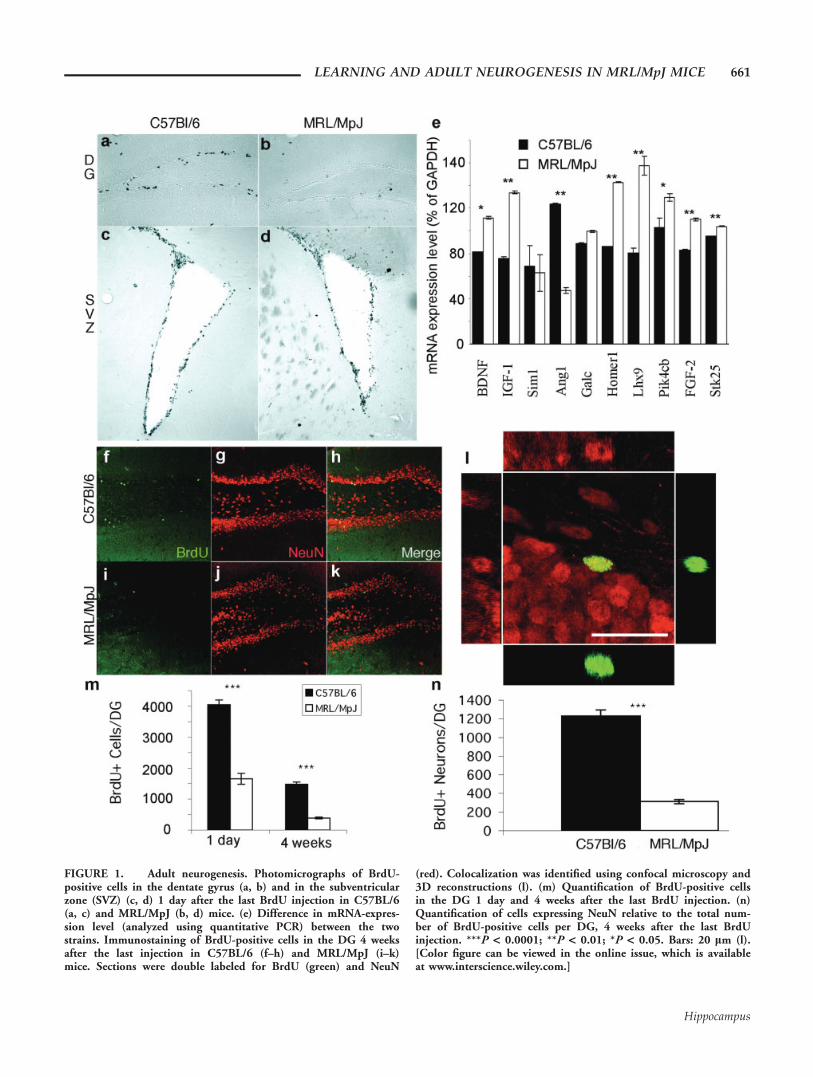
FIGURE 1. Adult neurogenesis. Photomicrographs of BrdU-positive cells in the dentate gyrus (a, b) and in the subventricularzone (SVZ) (c, d) 1 day after the last BrdU injection in C57BL/6(a, c) and MRL/MpJ (b, d) mice. (e) Difference in mRNA-expres-sion level (analyzed using quantitative PCR) between the twostrains. Immunostaining of BrdU-positive cells in the DG 4 weeksafter the last injection in C57BL/6 (f–h) and MRL/MpJ (i–k)mice. Sections were double labeled for BrdU (green) and NeuN
(red). Colocalization was identified using confocal microscopy and3D reconstructions (l). (m) Quantification of BrdU-positive cellsin the DG 1 day and 4 weeks after the last BrdU injection. (n)Quantification of cells expressing NeuN relative to the total num-ber of BrdU-positive cells per DG, 4 weeks after the last BrdUinjection. ***P < 0.0001; **P < 0.01; *P < 0.05. Bars: 20 lm (l).[Color figure can be viewed in the online issue, which is availableat www.interscience.wiley.com.]
LEARNING AND ADULT NEUROGENESIS IN MRL/MpJ MICE 661
Hippocampus

the estrous cycle (Proestus: 1,623 6 115 cells; Estrus: 1,560 6114 cells; Diestrus: 1,512 6 103 cells; ANOVA Single factor:F(2, 6) 5 0.25; P 5 0.8, data not shown). The observedreduction in the numbers of BrdU1 cells in the MRL/MpJhippocampus may be the result of a generally lower level ofBrdU bioavailability in this strain of mice, and not necessarilyreflect a hippocampus-specific reduction of cell proliferation.To test this possibility, we examined proliferation in the sub-ventricular zone. We did not detect any difference in the num-bers of BrdU1 cells in the subventricular zone between MRL/MpJ and C57BL/6 mice, indicating that the lower levels of cellproliferation in MRL/MpJ mice were restricted to the dentategyrus (Figs. 1c,d). Additionally, to test the possibility that lowerlevels of cell proliferation in MRL/MpJ mice were caused by asmaller hippocampus, we used unbiased stereology to measurethe volumes of the hippocampus and the granule cell layer andcounted the total number of granule cells in both strains. Forthese parameters, we did not detect any significant differencesbetween MRL/MpJ and C57BL/6 mice (hippocampal volume:5.59 6 0.13 mm3 vs. 5.41 6 0.31 mm3; granule cell layer vol-ume: 0.29 6 0.02 mm3 vs. 0.26 6 0.02 mm3; total numberof granule cells: 282,000 6 16,000 vs. 238,000 6 20,000,respectively; n 5 4 and P > 0.1 for all measures).
In an effort to begin identifying molecular mechanisms thatmay be involved in the interstrain proliferation differences, weperformed an mRNA microarray analysis on the hippocampi ofthe two strains of mice (see Methods section). At a P-value cut-off of 0.01 and correcting for multiple hypotheses testing, 1,024(2.3% of all probes) and 374 (0.8%) probes were differentiallyenriched in the MRL/MpJ and C57BL/6 strains, respectively.Gene ontology analysis revealed significant enrichment in trans-lational initiation (P < 3 3 1026), intracellular protein trans-port (P < 3.2 3 1025), and protein biosynthesis (P < 2 31024) in the MRL/MpJ mice. Therefore, we surmised that therewere no large-scale gene expression differences in neuron-relatedpathways. However, we did identify potential candidate geneswhose functions were related to neuronal proliferation, differen-tiation, or function that changed dramatically. The RNA-expres-sion level of these candidate genes was examined using qPCR(Fig. 1e). A Student’s t-test showed significant interstrain differ-ences for many genes related to neural stem cell proliferation,such as brain-derived neurotrophic factor (BDNF), insulin-likegrowth factor (IGF-1), and fibroblast growth factor (FGF2), butalso for kinases, such as the phosphatidylinositol-4-kinase, cata-lytic, beta (Pik4cb), and the serine-threonine kinase 25 (Stk25),for transcription factors such as the LIM homeobox protein 9(Lhx9), and for the ribonuclease angiogenin RNAse A family 5(Ang1) and the synaptic protein Homer1.
We next assessed the survival of newborn cells by comparingthe number of BrdU-positive cells 4 weeks after the last injec-tion with the number of labeled cells 24 h after the last injec-tion. The total number of BrdU-positive cells after 4 weeks inMRL/MpJ mice was 75% lower than in C57BL/6 mice (3896 28 and 1,473 6 75 cells, respectively; Student’s t-test, P <0.0001; Figs. 1f–m). When the number of labeled cells after 4weeks was expressed as a ratio of the number of labeled cells at
24 h after the last injection, we found that the survival rate was1 cell in 4.3 in MRL/MpJ mice and 1 in 2.7 in C57BL/6mice; therefore, survival was also reduced in MRL/MpJ mice.
Finally, we examined the differentiation of BrdU-positivecells into neurons 4 weeks after the last injection by colabelingfor BrdU and the neuron-specific marker, NeuN (Fig. 1l). InMRL/MpJ, neurons accounted for 80 6 0.8% of the survivingBrdU-positive cells when compared with 83.4 6 1.9% inC57BL/6 (Student’s t-test, P > 0.1; data not shown), indicat-ing that differentiation was similar in both strains.
Altogether, these data indicate that adult MRL/MpJ micegenerate 75% fewer neurons per dentate gyrus than C57BL/6mice (311 6 23 and 1,229 6 63 cells, respectively; Student’st-test, P < 0.0001; Fig. 1n). As in C57BL/6 mice (Lagaceet al., 2007), these measures in MRL/MpJ were constantthroughout the estrous cycle (Proestus: 289 6 37 cells; Estrus:378 6 11 cells; Diestrus: 409 6 41 cells; ANOVA Singlefactor: F(2, 6) 5 0.58; P 5 0.6, data not shown).
Learning Tests
We tested learning performances in MRL/MpJ mice usingspatial memory and object recognition memory tests. To testspatial learning, we used the Morris water-maze task. TenMRL/MpJ mice and 10 C57BL/6 mice were given four trialsper block, three blocks per day for a total of 13 blocks, andthe escape latency was recorded. To test the ability to find theplatform, the first block was performed using a visible plat-form, which was then hidden for the next 12 consecutiveblocks. Twenty-four hours after the last block, the platform wasremoved and all mice were subjected to a probe trial. Duringthe first five blocks, no interstrain differences were observed,indicating similar motivation and motor performances. Duringthe entire test, however, C57BL/6 mice reached the platformsignificantly faster than MRL/MpJ mice (one-way ANOVA,F(1,18) 5 10.62, P 5 0.0044; post hoc Bonferroni test P 50.006, Fig. 2a). This difference was not due to physical differ-ences, because swim speed was similar between both strains(MRL/MpJ: 18.2 6 0.7 cm/s; C57BL/6: 17.5 6 1.9 cm/s;Student’s t-test, P > 0.5, data not shown). Furthermore, thedifference was not due to a platform searching strategy, becauseduring the probe trial all mice stayed longer in the quadrantthat contained the platform than in any other quadrant. How-ever, MRL/MpJ mice stayed in the platform quadrant forsignificantly shorter periods of time than did C57BL/6 mice(37.7 6 1.6% vs. 50.4 6 2.8%, respectively, Student’s t-test,P < 0.001, data not illustrated). Thus, spatial learning isimpaired in MRL/MpJ mice.
To test recognition memory, we used the VPC task, whichexploits a subject’s natural preference for novel objects. TenC57BL/6 and 10 MRL/MpJ mice were first shown two identi-cal objects. We recorded the exploration times and observedthat the mice spent an identical amount of time observing bothobjects, indicating a lack of preference. Thirty minutes later,one of the familiar objects was replaced by a novel object.C57BL/6 and MRL/MpJ mice spent most of their time explor-
662 THURET ET AL.
Hippocampus

ing the new object (75.8 6 2.5% and 72.1 6 2%, respectively;Student’s t-test, P > 0.2; Fig. 2b), indicating that short-termrecognition memory was similar for both strains. After a 24-hdelay, a second novel object replaced the new object that waspresented for the 30-min test. C57BL/6 mice spent most oftheir time exploring the new object, whereas MRL/MpJ miceexplored the new object only for about half the time (74.2 63.9% and 50.7 6 1.9%, respectively; Student’s t-test, P <0.0001; Fig. 2b), indicating that memory of the familiar objectwas retained for less than 24 h in MRL/MpJ mice. Thus, long-term recognition memory is also impaired in MRL/MpJ mice.
Synaptic Remodeling
One of the mechanisms by which synaptic plasticity plays arole in the formation of new memories involves the learning-induced addition of new synapses on activated presynaptic ter-minals (Toni et al., 1999, 2001; Geinisman et al., 2001),resulting in the formation of MSB and the strengthening ofactivated synapses (Fig. 3b) (Sorra and Harris, 1993; Shepherdand Harris, 1998).
To analyze learning-induced synaptic remodeling in MRL/MpJ mice, we compared synapse ultrastructure between learnerand nonlearner animals using ssTEM. Three MRL/MpJ miceand five C57BL/6 mice were given three daily blocks of fourwater maze trials each, over two consecutive days. The first blockwas conducted using a visible platform. As control, three MRL/MpJ and four C57BL/6 nonlearner mice were housed in stand-ard conditions. Similar to the results shown in Figure 2a, we didnot detect any significant difference in the escape latencybetween groups for the five first blocks (One-way ANOVAF(1,4) 5 0.632, P 5 0.471, Fig. 3a), but the time to reach theplatform was significantly shorter for block 6 than for block 2for both strains (post hoc t-test P < 0.05), indicating that learn-ing had occurred in both C57BL/6 and MRL/MpJ mice. Ten to12 h after the last trial, all mice were perfused and processed forssTEM. In C57BL/6 mice, learning induced a significantincrease in the proportion of MSB in the dentate gyrus (36.2 61.9% in nonlearners, 51.1 6 4.9% in learners; Student’s t-test P< 0.01; Fig. 3c), but not in the CA1 hippocampal subfield. Incontrast, learning did not induce morphological modificationsin the dentate gyrus of MRL/MpJ mice (Fig. 3c). To testwhether this observed reduction of synaptic plasticity in MRL/MpJ mice might have resulted from differences in synapse den-sity, we measured synapse density in the CA1 area and the den-tate gyrus of both strains using stereology. Synapse density wassimilar in both strains in both the CA1 and the dentate gyrusand averaged 2.46 synapses/lm3 (Fig. 3d). Thus, synapse densitywas comparable between the two strains, but learning inducedmorphological modifications in the dentate gyrus of C57BL/6mice but not in MRL/MpJ mice.
Effect of Voluntary Running on Neurogenesis
Voluntary running increases neural stem cell proliferation,survival, and neuronal differentiation in rodents (van Praaget al., 1999b). To test whether neural stem cells of MRL/MpJmice retained the capacity to respond to such environmentalstimuli, eight MRL/MpJ and nine C57BL/6 mice were individ-ually housed in cages containing a running wheel for 4 weeksand were injected daily with BrdU for the first 6 days. Runningactivity was recorded on an hourly basis for the entire length ofthe housing (Fig. 4a). On an average, MRL/MpJ mice ran 13.96 1.0 km per 24 h and C57BL/6 mice ran 8.0 6 0.6 km per24 h (Student’s t-test, P < 0.001; data not illustrated). Fourweeks after the last injection, the number of BrdU-labeled cellswas counted. Running increased BrdU-labeled cells in bothMRL/MpJ and C57BL/6 mice. Surprisingly, the extent of thisincrease was such that the number of BrdU-labeled cells wasnow comparable between both strains (MRL/MpJ: 3096.1 655.8, and C57BL/6: 3,448 6 509.5 cells per dentate gyrus,Student’s t-test, P > 0.1; Figs. 4b–h). Therefore, runninginduced a 790% increase in BrdU incorporation in MRL/MpJmice, compared with a 230% increase in C57BL/6 mice.
We then stained for the neuronal marker, NeuN, andcounted the proportion of BrdU-labeled cells that colabeledwith NeuN. In MRL/MpJ mice, neurons accounted for
FIGURE 2. Water maze test and visual-paired comparison test.(a) Latency in seconds to reach the platform in the water maze testfor C57BL/6 (closed squares) and MRL/MpJ (open lozenges) mice.The platform was visible only during the first block. (b) Preferencein percentage for the novel object in the visual-paired comparisontest after 30 min and 24 h delay. C57BL/6, black; MRL/MpJ,white. **P < 0.0001; /P > 0.2.
LEARNING AND ADULT NEUROGENESIS IN MRL/MpJ MICE 663
Hippocampus

86.7 6 1.4% of the surviving BrdU-positive cells and for 88.36 0.8% in C57BL/6. Thus, voluntary running significantlyincreased neurogenesis in both strains (Student’s t-test, P <0.01), but there was no significant difference between the tworunning groups (Student’s t-test, P > 0.1; Fig. 4i). Altogether,MRL/MpJ runners generated on average a total of 2684.7 6168.8 BrdU-labeled neurons per dentate gyrus vs. 3044.5 6159 in C57BL/6 running mice during the 6 days of BrdUinjection (Student’s t-test, P > 0.1; Fig. 4j). Thus, voluntaryrunning completely rescued neurogenesis in MRL/MpJ mice.
Effect of Voluntary Running on Spatial Learningand Recognition Memory
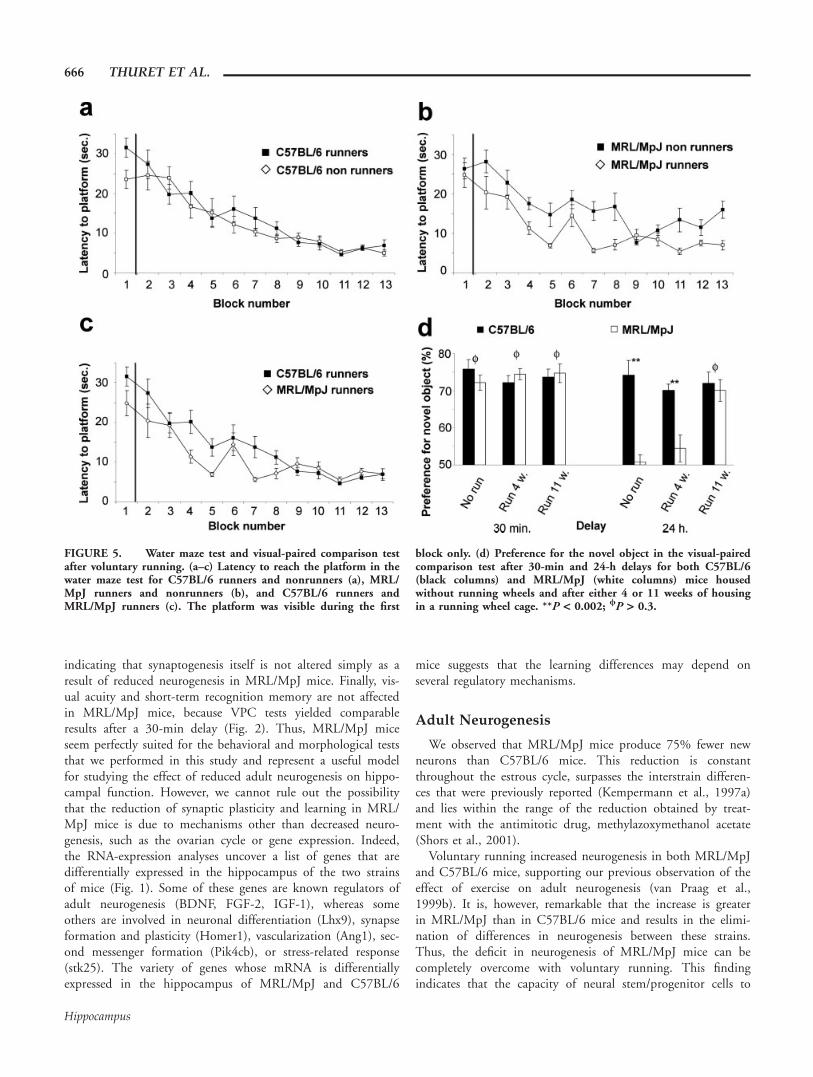
Nine MRL/MpJ mice and eight C57BL/6 mice were housedfor 4 weeks in individual cages with a running wheel and testedthereafter in the water maze using the same parameters as forFigure 2. When all four groups (runners, nonrunners, C57BL/6, MRL/MpJ) were compared using a two-way ANOVA testwith repeated measures, we found that exposure to runninghad a different effect on the strains (F(1,33) 5 11.7; P 50.02). Running significantly reduced the time to reach the plat-form for MRL/MpJ mice (One-way ANOVA, F(1,17) 5 21.7;P 5 0.0003; post hoc Bonferroni test, P 5 0.001, Fig. 5b) butnot for C57BL/6 mice (One-way ANOVA, F(1,16) 5 2.6;
P 5 0.1; Fig. 5a). Surprisingly, the time to reach the platformwas significantly shorter for MRL/MpJ mice than for C57BL/6mice (One-way ANOVA, F(1,15) 5 24.6; P 5 0.0002, posthoc Bonferroni test, P 5 0.045, Fig. 5c). Twenty-four hoursafter the last trial, the platform was removed and all runnermice were subjected to a probe trial. Both strains spent a simi-lar proportion of their time in the quadrant that previouslycontained the platform (MRL/MpJ: 32.9 6 1.3%, C57BL/6:37.2 6 2.1%, Student’s t-test, P > 0.05, data not illustrated).
Thus, after voluntary running, MRL/MpJ mice performed aswell or better than C57BL/6 mice on the water maze task.
The VPC test was performed following the same protocoldescribed in Figure 2, using the same set of objects in the sameconditions and environment. During the familiarization phase,the mice spent an equal amount of time exploring both identi-cal objects, indicating a lack of preference. After a delay of 30min, one object was changed and C57BL/6 and MRL/MpJspent 72.1 6 5.2% and 74.4 6 4.2%, respectively, of theirtime exploring the new object. Thus, there was no significantdifference in exploration time between the two strains (Stu-dent’s t-test, P > 0.3; Fig. 5d). After a delay of 24 h, the newobject was replaced again and the C57BL/6 mice spent most(70.1 6 4.7%) of their time exploring the new object, whereasMRL/MpJ mice explored the new object at chance (54.4 610.2%, Student’s t-test, P < 0.002; Fig. 5d). Thus, the deficit
FIGURE 3. Synapse ultrastructure and remodeling after spatialtraining. (a) Latency in seconds to reach the platform in the watermaze test for the mice used for ssTEM analyses. (b) Electronmicrograph of a multiple-synapse bouton (MSB, left) and a single-synapse bouton (SSB, right) showing axon terminals (A) makingsynaptic connections with dendritic spines (sp). Scale bar, 0.5 lm.
(c) Proportion of dendritic spines contacting MSB in the dentategyrus and in the CA1, under control conditions (nonlearners) andafter water maze training (Learners) for C57BL/6 and MRL/MpJmice. (d) Synapse density in the dentate gyrus and the CA1 areaof both C57BL/6 and MRL/MpJ mice. **P < 0.01; /P > 0.05.
664 THURET ET AL.
Hippocampus

of MRL/MpJ mice in long-term recognition memory was notrescued by 4 weeks of running. Hypothesizing that the dura-tion of voluntary running was too short to produce a detectableeffect on the VPC test, we returned all mice to their originalrunning wheel cages for 7 additional weeks, after which we per-formed another VPC test using a new set of objects. At thislater time point, after a 24-h delay, MRL/MpJ mice spent asmuch time as the C57BL/6 mice exploring the new object(70.1 6 2.9% and 72.0 6 2.9%, respectively, Student’s t-test,P > 0.3; Fig. 5d). Therefore, the deficit in spatial learning forMRL/MpJ mice was rescued after 4 weeks of voluntary run-ning, and the deficit in visual recognition memory was rescuedonly after 11 weeks of running.
DISCUSSION
In this study, we report that MRL/MpJ mice have a lowlevel of neurogenesis, which was associated with undetectablelearning-induced synaptic remodeling and poor performances
in hippocampus-dependent learning tasks when compared withC57BL/6 mice. We show that, after running, neurogenesis wasrestored in MRL/MpJ mice and their learning deficit wasrescued. These findings suggest that neurogenesis is correlatedwith hippocampus-dependent learning in MRL/MpJ mice andthat voluntary running can abolish interstrain differences inneurogenesis.
In this study, we test the function of adult neurogenesis byassessing a naturally occurring mutant strain that shows a dra-matic reduction in neurogenesis. These results support the pre-vious observation that adult neurogenesis plays a role in spatiallong-term memory (Snyder et al., 2005), and they extend theseobservations to another form of hippocampus-dependent mem-ory, the visual recognition task. The number of granule cells,the granule cell layer volume, the hippocampal volume, andstem cell proliferation in the subventricular zone are similarbetween the strains, indicating that MRL/MpJ mice do not suf-fer from general downregulation of stem cell/progenitor cellfunction in the brain but rather that neurogenesis is selectivelyreduced in the adult dentate gyrus. Furthermore, synapse den-sity is similar, both in the CA1 and the dentate gyrus (Fig. 3),
FIGURE 4. Adult neurogenesis in the dentate gyrus after vol-untary running. (a) Average running activity for both strains forthe first 10 days of housing in a cage containing a running wheel.Photomicrographs of BrdU-positive cells in the DG 4 weeks afterthe last injection in C57BL/6 (b–d) and MRL/MpJ (e–g) runningmice. Sections were double labeled for BrdU (green) and NeuN(red). (h) Quantification of BrdU-positive cells in the DG 4 weeks
after the last BrdU injection. (i, j) Quantification of BrdU-positivecells expressing NeuN in percentage of BrdU-positive cells per DG(i), and relative to the total number of BrdU-positive cells per DG,4 weeks after the last BrdU injection (j). ***P < 0.0001; *P <0.01; /P > 0.1. [Color figure can be viewed in the online issue,which is available at www.interscience.wiley.com.]
LEARNING AND ADULT NEUROGENESIS IN MRL/MpJ MICE 665
Hippocampus
indicating that synaptogenesis itself is not altered simply as aresult of reduced neurogenesis in MRL/MpJ mice. Finally, vis-ual acuity and short-term recognition memory are not affectedin MRL/MpJ mice, because VPC tests yielded comparableresults after a 30-min delay (Fig. 2). Thus, MRL/MpJ miceseem perfectly suited for the behavioral and morphological teststhat we performed in this study and represent a useful modelfor studying the effect of reduced adult neurogenesis on hippo-campal function. However, we cannot rule out the possibilitythat the reduction of synaptic plasticity and learning in MRL/MpJ mice is due to mechanisms other than decreased neuro-genesis, such as the ovarian cycle or gene expression. Indeed,the RNA-expression analyses uncover a list of genes that aredifferentially expressed in the hippocampus of the two strainsof mice (Fig. 1). Some of these genes are known regulators ofadult neurogenesis (BDNF, FGF-2, IGF-1), whereas someothers are involved in neuronal differentiation (Lhx9), synapseformation and plasticity (Homer1), vascularization (Ang1), sec-ond messenger formation (Pik4cb), or stress-related response(stk25). The variety of genes whose mRNA is differentiallyexpressed in the hippocampus of MRL/MpJ and C57BL/6
mice suggests that the learning differences may depend onseveral regulatory mechanisms.
Adult Neurogenesis
We observed that MRL/MpJ mice produce 75% fewer newneurons than C57BL/6 mice. This reduction is constantthroughout the estrous cycle, surpasses the interstrain differen-ces that were previously reported (Kempermann et al., 1997a)and lies within the range of the reduction obtained by treat-ment with the antimitotic drug, methylazoxymethanol acetate(Shors et al., 2001).
Voluntary running increased neurogenesis in both MRL/MpJand C57BL/6 mice, supporting our previous observation of theeffect of exercise on adult neurogenesis (van Praag et al.,1999b). It is, however, remarkable that the increase is greaterin MRL/MpJ than in C57BL/6 mice and results in the elimi-nation of differences in neurogenesis between these strains.Thus, the deficit in neurogenesis of MRL/MpJ mice can becompletely overcome with voluntary running. This findingindicates that the capacity of neural stem/progenitor cells to
FIGURE 5. Water maze test and visual-paired comparison testafter voluntary running. (a–c) Latency to reach the platform in thewater maze test for C57BL/6 runners and nonrunners (a), MRL/MpJ runners and nonrunners (b), and C57BL/6 runners andMRL/MpJ runners (c). The platform was visible during the first
block only. (d) Preference for the novel object in the visual-pairedcomparison test after 30-min and 24-h delays for both C57BL/6(black columns) and MRL/MpJ (white columns) mice housedwithout running wheels and after either 4 or 11 weeks of housingin a running wheel cage. **P < 0.002; /P > 0.3.
666 THURET ET AL.
Hippocampus
respond to environmental stimuli is retained in MRL/MpJmice. What accounts for the increased effect of running onMRL/MpJ mice?
One possibility is that inactive progenitors are more respon-sive to stimulation. This possibility is supported by the obser-vation of neurogenesis in the aged brain. Neurogenesisdecreases with aging, but environmental enrichment induces athreefold increase in neurogenesis in the aged brain and only atwofold increase in the younger brain (Kempermann et al.,1998, 2002; van Praag et al., 2005). This finding suggests thatextrinsic factors play an important role in the inhibition ofneurogenesis in the adult MRL/MpJ hippocampus.
Another possibility is greater distances run, because neuro-genesis was found to be linearly correlated with the amount ofrunning up to 6 km/day in outbred mice but not in mice bredfor increased running (Rhodes et al., 2003). In support of thispossibility, we found that MRL/MpJ mice ran, on average,60% greater distances than C57BL/6 mice, although their cir-cadian rhythm was similar. Combined with the behavioralexperiments of Figure 5, our results suggest that distance runalso influences learning performances. Indeed, spatial learningperformances were improved in good runner mice only (MRL/MpJ), and increasing exposure to running wheels also increasedthe learning performances on the visual learning task.
Synaptic Remodeling
MSBs result from the synaptogenesis induced by long-termpotentiation (LTP) (Toni et al., 1999) or associative learning(Geinisman et al., 2001). This form of synaptic plasticity isbelieved to play a role in the late phases of learning and tostrengthen synaptic transmission between activated neurons(Lamprecht and LeDoux, 2004).
Our observation that, in C57BL/6 mice, spatial learningincreased the proportion of MSBs in the dentate gyrus but notin the CA1 suggests that, under our experimental conditions,the dentate gyrus was more involved in spatial learning thanthe CA1 area. This finding, together with the absence of learn-ing-induced MSB formation in MRL/MpJ mice, indicates thatstructural plasticity is correlated with adult neurogenesis, sug-gesting that the function of new neurons may be mediated byan increase in synaptic remodeling. This possibility is sup-ported by the recent report that, in the dentate gyrus, imma-ture neurons integrate into the hippocampal circuitry (Toniet al., 2007) and express LTP more easily than mature neurons(Farmer et al., 2004; Schmidt-Hieber et al., 2004; Ge et al.,2007).
Spatial Learning
In standard housing, we observed that the performance ofMRL/MpJ mice in spatial learning was lower than that ofC57BL/6 mice, supporting our previous observation that theamount of neurogenesis predicts performance on the watermaze task in different mouse strains (Kempermann and Gage,2002).
We further observed that running MRL/MpJ mice had bet-ter performances than nonrunners in the water maze test,supporting previous reports that enriched environment andvoluntary exercise increase spatial learning in healthy mice(Kempermann et al., 1997b; van Praag et al., 1999a,b) and res-cue learning deficits associated with ethanol exposure (Crewset al., 2004; Christie et al., 2005), stroke (Luo et al., 2007), oraging (van Praag et al., 2005). We observed, however, that, af-ter 4 weeks of running, learning did not improve in C57BL/6mice. The discrepancy between our results and our previousreport (van Praag et al., 1999a) is very likely because of a dif-ference in the paradigm used for the water maze test. In thisstudy, mice were presented with 12 trials per day, whereas inthe previous study, mice were exposed to the water maze foronly four trials a day, which is probably more challenging.
The observation that running improved learning in MRL/MpJ but not in C57BL/6 mice suggests that spatial learningmay depend on the magnitude of the increase in neurogenesisrather than on the total number of new neurons (runningincreased neurogenesis by 2.3-fold for C57BL/6 and of 7.9-fold for MRL/MpJ mice), which may itself depend on distancerun by the mice (MRL/MpJ mice ran in average 170% the dis-tance covered by C57BL/6). Alternatively, MRL/MpJ mice mayuse compensatory mechanisms to cope with the reduction ofneurogenesis, such as a modified connectivity of granule neu-rons, as seen in the high proportion of MSBs (Fig. 3c), oroverexpression of genes regulating stem cell proliferation (Fig.1e). The running-induced neurogenesis may combine withthese mechanisms to produce the increased effect on learning.Although our study cannot provide a definite answer to thisquestion, it supports the idea that adult neurogenesis is associ-ated with spatial learning.
Object Recognition Memory
For the VPC task, the performance of MRL/MpJ andC57BL/6 mice at the 30-min delay was identical and bothmice recognized the familiar object. At 24-h delay, MRL/MpJmice exploration was at chance, whereas C57BL/6 mice stillremembered the familiar object. This finding indicates a deficitin long-term recognition memory in MRL/MpJ mice and sug-gests that neurogenesis may be involved in long-term recogni-tion memory.
Normal hippocampal function is required for performanceon the VPC task, and several reports indicate that hippocampallesions impair recognition memory (Wood et al., 1993; Clarket al., 2000; Zola et al., 2000). Therefore, in light of our watermaze results, we were expecting that 4 weeks of voluntary run-ning would also rescue the performance of MRL/MpJ mice inthe VPC test. The observation that 11 weeks of running arerequired to completely rescue VPC task performances, whereasjust 4 weeks of running rescue water maze performances, sug-gests that different mechanisms are involved in the completionof these tasks. One possibility is that the VPC task is moredemanding and requires the incorporation of more new neu-rons than the water maze task. Eleven weeks of running most
LEARNING AND ADULT NEUROGENESIS IN MRL/MpJ MICE 667
Hippocampus
likely results in an increase in neurogenesis that is greater thanafter 4 weeks. This difference may be sufficient to overcomethe visual learning deficit. In support of this possibility, wefound that 4 weeks of running resulted in a small (but not sig-nificant) increase in VPC learning in MRL/MpJ (Fig. 5d).Alternatively or maybe additionally, the VPC task may dependon the maturation stage of new neurons. The VPC taskdepends on CA1 area function, as indicated by the observationsof impaired performance on the VPC task in mice lacking theNMDAr1 subunit in the CA1 area (Rampon et al., 2000) orafter lesions of the CA1 area (Wood et al., 1993). Between 4and 11 weeks after cell division, new neurons may establishmore extended connections and recruit other hippocampal sub-fields. Indeed, our recent study shows that new neurons attaina mature connectivity only after 4–8 weeks of maturation, dur-ing which dendritic spines contact perforant path synapses andaxons are projected in the CA3 area (Zhao et al., 2006; Toniet al., 2007, 2008). The improved connectivity resulting fromprolonged maturation of new neurons may therefore result inthe involvement of the CA1 area that is necessary to performthe VPC task.
Acknowledgments
The authors thank Enhung Matthew Teng, John Jepsen, andHeather Lansford for technical help, Nathalie Kayadjanian andBoris Sakic for comments on the manuscript, and Mary LynnGage for editing the manuscript.
REFERENCES
Altman J, Das GD. 1965. Autoradiographic and histological evidenceof postnatal hippocampal neurogenesis in rats. J Comp Neurol124:319–335.
Blankenhorn EP, Troutman S, Clark LD, Zhang XM, Chen P, Heber-Katz E. 2003. Sexually dimorphic genes regulate healing and regen-eration in MRL mice. Mamm Genome 14:250–260.
Bruel-Jungerman E, Laroche S, Rampon C. 2005. New neurons in thedentate gyrus are involved in the expression of enhanced long-termmemory following environmental enrichment. Eur J Neurosci21:513–521.
Christie BR, Swann SE, Fox CJ, Froc D, Lieblich SE, Redila V, Web-ber A. 2005. Voluntary exercise rescues deficits in spatial memoryand long-term potentiation in prenatal ethanol-exposed male rats.Eur J Neurosci 21:1719–1726.
Clark LD, Clark RK, Heber-Katz E. 1998. A new murine model formammalian wound repair and regeneration. Clin Immunol Immu-nopathol 88:35–45.
Clark RE, Zola SM, Squire LR. 2000. Impaired recognition memoryin rats after damage to the hippocampus. J Neurosci 20:8853–8860.
Crews FT, Nixon K, Wilkie ME. 2004. Exercise reverses ethanol inhi-bition of neural stem cell proliferation. Alcohol 33:63–71.
Ennaceur A, Delacour J. 1988. A new one-trial test for neurobiologicalstudies of memory in rats. I. Behavioral data. Behav Brain Res31:47–59.
Eriksson PS, Perfilieva E, Bjork-Eriksson T, Alborn AM, Nordborg C,Peterson DA, Gage FH. 1998. Neurogenesis in the adult humanhippocampus. Nat Med 4:1313–1317.
Farmer J, Zhao X, van Praag H, Wodtke K, Gage FH, Christie BR.2004. Effects of voluntary exercise on synaptic plasticity and geneexpression in the dentate gyrus of adult male Sprague-Dawley ratsin vivo. Neuroscience 124:71–79.
Ferri AL, Cavallaro M, Braida D, Di Cristofano A, Canta A, VezzaniA, Ottolenghi S, Pandolfi PP, Sala M, DeBiasi S, Nicolis SK.2004. Sox2 deficiency causes neurodegeneration and impaired neu-rogenesis in the adult mouse brain. Development 131:3805–3819.
Filipkowski RK, Kiryk A, Kowalczyk A, Kaczmarek L. 2005. Geneticmodels to study adult neurogenesis. Acta Biochim Pol 52:359–372.
Ge S, Yang CH, Hsu KS, Ming GL, Song H. 2007. A critical periodfor enhanced synaptic plasticity in newly generated neurons of theadult brain. Neuron 54:559–566.
Geinisman Y, Berry RW, Disterhoft JF, Power JM, Van der Zee EA.2001. Associative learning elicits the formation of multiple-synapseboutons. J Neurosci 21:5568–5573.
Gould E, Reeves AJ, Fallah M, Tanapat P, Gross CG, Fuchs E. 1999.Hippocampal neurogenesis in adult Old World primates. Proc NatlAcad Sci USA 96:5263–5267.
Heber-Katz E, Chen P, Clark L, Zhang XM, Troutman S, Blanken-horn EP. 2004. Regeneration in MRL mice: Further genetic locicontrolling the ear hole closure trait using MRL and M.m. Casta-neus mice. Wound Repair Regen 12:384–392.
Kaplan MS, Bell DH. 1984. Mitotic neuroblasts in the 9-day-old and11-month-old rodent hippocampus. J Neurosci 4:1429–1441.
Kempermann G, Gage FH. 2002. Genetic determinants of adult hip-pocampal neurogenesis correlate with acquisition, but not probetrial performance, in the water maze task. Eur J Neurosci 16:129–136.
Kempermann G, Kuhn HG, Gage FH. 1997a. Genetic influence onneurogenesis in the dentate gyrus of adult mice. Proc Natl AcadSci USA 94:10409–10414.
Kempermann G, Kuhn HG, Gage FH. 1997b. More hippocampalneurons in adult mice living in an enriched environment. Nature386:493–495.
Kempermann G, Kuhn HG, Gage FH. 1998. Experience-induced neu-rogenesis in the senescent dentate gyrus. J Neurosci 18:3206–3212.
Kempermann G, Gast D, Gage FH. 2002. Neuroplasticity in old age:Sustained fivefold induction of hippocampal neurogenesis by long-term environmental enrichment. Ann Neurol 52:135–143.
Korzus E, Rosenfeld MG, Mayford M. 2004. CBP histone acetyltrans-ferase activity is a critical component of memory consolidation.Neuron 42:961–972.
Lagace DC, Fischer SJ, Eisch AJ. 2007. Gender and endogenous levelsof estradiol do not influence adult hippocampal neurogenesis inmice. Hippocampus 17:175–180.
Lamprecht R, LeDoux J. 2004. Structural plasticity and memory. NatRev Neurosci 5:45–54.
Leferovich JM, Bedelbaeva K, Samulewicz S, Zhang XM, Zwas D,Lankford EB, Heber-Katz E. 2001. Heart regeneration in adultMRL mice. Proc Natl Acad Sci USA 98:9830–9835.
Li X, Gu W, Masinde G, Hamilton-Ulland M, Xu S, Mohan S, Bay-link DJ. 2001. Genetic control of the rate of wound healing inmice. Heredity 86:668–674.
Luo CX, Jiang J, Zhou QG, Zhu XJ, Wang W, Zhang ZJ, Han X,Zhu DY. 2007. Voluntary exercise-induced neurogenesis in thepostischemic dentate gyrus is associated with spatial memory recov-ery from stroke. J Neurosci Res 85:1637–1646.
Masinde G, Li X, Baylink DJ, Nguyen B, Mohan S. 2005. Isolationof wound healing/regeneration genes using restrictive fragment dif-ferential display-PCR in MRL/MPJ and C57BL/6 mice. BiochemBiophys Res Commun 330:117–122.
Masinde GL, Li X, Gu W, Davidson H, Mohan S, Baylink DJ. 2001.Identification of wound healing/regeneration quantitative trait loci(QTL) at multiple time points that explain seventy percent of var-iance in (MRL/MpJ and SJL/J) mice F2 population. Genome Res11:2027–2033.
668 THURET ET AL.
Hippocampus
McBrearty BA, Clark LD, Zhang XM, Blankenhorn EP, Heber-KatzE. 1998. Genetic analysis of a mammalian wound-healing trait.Proc Natl Acad Sci USA 95:11792–11797.
Morris R. 1984. Developments of a water-maze procedure for study-ing spatial learning in the rat. J Neurosci Methods 11:47–60.
Rampon C, Tang YP, Goodhouse J, Shimizu E, Kyin M, Tsien JZ.2000. Enrichment induces structural changes and recovery fromnonspatial memory deficits in CA1 NMDAR1-knockout mice. NatNeurosci 3:238–244.
Rhodes JS, van Praag H, Jeffrey S, Girard I, Mitchell GS, Garland TJr, Gage FH. 2003. Exercise increases hippocampal neurogenesis tohigh levels but does not improve spatial learning in mice bred forincreased voluntary wheel running. Behav Neurosci 117:1006–1016.
Schmidt-Hieber C, Jonas P, Bischofberger J. 2004. Enhanced synapticplasticity in newly generated granule cells of the adult hippocam-pus. Nature 429:184–187.
Shepherd GM, Harris KM. 1998. Three-dimensional structure andcomposition of CA3–>CA1 axons in rat hippocampal slices:Implications for presynaptic connectivity and compartmentaliza-tion. J Neurosci 18:8300–8310.
Shi Y, Chichung Lie D, Taupin P, Nakashima K, Ray J, Yu RT, GageFH, Evans RM. 2004. Expression and function of orphan nuclearreceptor TLX in adult neural stem cells. Nature 427:78–83.
Shors TJ, Miesegaes G, Beylin A, Zhao M, Rydel T, Gould E. 2001.Neurogenesis in the adult is involved in the formation of tracememories. Nature 410:372–376.
Snyder JS, Hong NS, McDonald RJ, Wojtowicz JM. 2005. A role foradult neurogenesis in spatial long-term memory. Neuroscience130:843–852.
Sorra KE, Harris KM. 1993. Occurrence and three-dimensional struc-ture of multiple synapses between individual radiatum axons andtheir target pyramidal cells in hippocampal area CA1. J Neurosci13:3736–3748.
Sterio DC. 1984. The unbiased estimation of number and sizes ofarbitrary particles using the disector. J Microsc 134:127–136.
Toni N, Buchs PA, Nikonenko I, Bron CR, Muller D. 1999. LTPpromotes formation of multiple spine synapses between a singleaxon terminal and a dendrite. Nature 402:421–425.
Toni N, Buchs PA, Nikonenko I, Povilaitite P, Parisi L, Muller D.2001. Remodeling of synaptic membranes after induction of long-term potentiation. J Neurosci 21:6245–6251.
Toni N, Teng EM, Bushong EA, Aimone JB, Zhao C, Consiglio A,van Praag H, Martone ME, Ellisman MH, Gage FH. 2007. Syn-apse formation on neurons born in the adult hippocampus. NatNeurosci 10:727–734.
Toni N, Laplagne DA, Zhao C, Lombardi G, Ribak CE, Gage FH,Schinder AF. 2008. Neurons born in the adult dentate gyrus formfunctional synapses with target cells. Nat Neurosci 11:901–907.
van Praag H, Christie BR, Sejnowski TJ, Gage FH. 1999a. Runningenhances neurogenesis, learning, and long-term potentiation inmice. Proc Natl Acad Sci USA 96:13427–13431.
van Praag H, Kempermann G, Gage FH. 1999b. Running increasescell proliferation and neurogenesis in the adult mouse dentategyrus. Nat Neurosci 2:266–270.
van Praag H, Shubert T, Zhao C, Gage FH. 2005. Exercise enhanceslearning and hippocampal neurogenesis in aged mice. J Neurosci25:8680–8685.
Wood ER, Mumby DG, Pinel JP, Phillips AG. 1993. Impaired objectrecognition memory in rats following ischemia-induced damage tothe hippocampus. Behav Neurosci 107:51–62.
Zhao C, Teng EM, Summers RG Jr, Ming GL, Gage FH. 2006. Dis-tinct morphological stages of dentate granule neuron maturation inthe adult mouse hippocampus. J Neurosci 26:3–11.
Zola SM, Squire LR, Teng E, Stefanacci L, Buffalo EA, Clark RE.2000. Impaired recognition memory in monkeys after damagelimited to the hippocampal region. J Neurosci 20:451–463.
LEARNING AND ADULT NEUROGENESIS IN MRL/MpJ MICE 669
Hippocampus