high intra- and inter-tumoral heterogeneity of ras ... · high intra- and inter-tumoral...
TRANSCRIPT
International Journal of
Molecular Sciences
Article
High Intra- and Inter-Tumoral Heterogeneity of RASMutations in Colorectal CancerMarion Jeantet 1,2,3, David Tougeron 1,4,5, Gaelle Tachon 1,2, Ulrich Cortes 1,2,Céline Archambaut 1,2, Gaelle Fromont 3 and Lucie Karayan-Tapon 1,2,6,*
1 Faculté de Médecine Pharmacie, Université de Poitiers, 86021 Poitiers, France; [email protected] (M.J.);[email protected] (D.T.); [email protected] (G.T.);[email protected] (U.C.); [email protected] (C.A.)
2 Département de Cancérologie Biologique, Centre Hospitalo-Universitaire de Poitiers, 86021 Poitiers, France3 Département d’anatomopathologie, Centre Hospitalo-Universitaire de Poitiers, 86021 Poitiers, France;
[email protected] Département de Gastroentérologie, Centre Hospitalo-Universitaire de Poitiers, 86021 Poitiers, France5 Laboratoire Inflammation, Tissus Epithéliaux et Cytokines, EA 4331, Université de Poitiers,
86021 Poitiers, France6 INSERM1084, Laboratoire de Neurosciences Expérimentales et Cliniques, Université de Poitiers,
86021 Poitiers, France* Correspondence: [email protected]; Tel.: +33-5-4944-4988
Academic Editor: William Chi-shing ChoReceived: 5 September 2016; Accepted: 25 November 2016; Published: 1 December 2016
Abstract: Approximately 30% of patients with wild type RAS metastatic colorectal cancer arenon-responders to anti-epidermal growth factor receptor monoclonal antibodies (anti-EGFR mAbs),possibly due to undetected tumoral subclones harboring RAS mutations. The aim of this study was toanalyze the distribution of RAS mutations in different areas of the primary tumor, metastatic lymphnodes and distant metastasis. A retrospective cohort of 18 patients with a colorectal cancer (CRC)was included in the study. Multiregion analysis was performed in 60 spatially separated tumor areasaccording to the pathological tumor node metastasis (pTNM) staging and KRAS, NRAS and BRAFmutations were tested using pyrosequencing. In primary tumors, intra-tumoral heterogeneity for RASmutation was found in 33% of cases. Inter-tumoral heterogeneity for RAS mutation between primarytumors and metastatic lymph nodes or distant metastasis was found in 36% of cases. Moreover,28% of tumors had multiple RAS mutated subclones in the same tumor. A high proportion of CRCspresented intra- and/or inter-tumoral heterogeneity, which has relevant clinical implications foranti-EGFR mAbs prescription. These results suggest the need for multiple RAS testing in differentparts of the same tumor and/or more sensitive techniques.
Keywords: colorectal cancer; RAS mutation; intra-tumoral heterogeneity; inter-tumoral heterogeneity
1. Introduction
Colorectal cancer (CRC) is the third deadliest of all cancers [1]. Nearly one-third of the patientswill eventually die of the disease. Targeting the epidermal growth factor receptor (EGFR), an importantcomponent in CRC carcinogenesis, is one of the major therapeutic options in metastatic CRC (mCRC).Two anti-EGFR monoclonal antibodies (mAbs), cetuximab and panitumumab, are commonly usedin mCRC. Clinical trials have shown the benefit of anti-EGFR mAbs alone or in combination withchemotherapy in mCRC [2–4].
Several studies have demonstrated that KRAS mutation in exon 2 is a predictive marker ofresistance to anti-EGFR mAbs [5]. More recently, other activating RAS mutations (KRAS exons 3and 4 and NRAS exons 2, 3 and 4) were also shown to confer resistance to anti-EGFR mAbs [3,4].
Int. J. Mol. Sci. 2016, 17, 2015; doi:10.3390/ijms17122015 www.mdpi.com/journal/ijms
Int. J. Mol. Sci. 2016, 17, 2015 2 of 12
Approximately 50% of mCRC harbor mutations in exons 2, 3 or 4 of either KRAS or NRAS genes [6].The most frequent mutations are detected in exon 2 (codons 12 and 13) of KRAS (40%), and, to alesser extent, in exon 3 (codons 59 and 61) and exon 4 (codons 117 and 146) of KRAS (≈7% of cases).Activating mutations of NRAS occur only in a subset of mCRC (≈5% of cases), mostly at codons 12,13 and 61 [6]. The BRAFV600E mutation occurs in 10%–15% of mCRC [7,8]. BRAFV600E mutant mCRCis associated with poorer outcomes. However, whether this mutation is predictive of resistance toanti-EGFR mAbs is uncertain [7].
Only wild-type (WT) RAS mCRCs benefit from treatment with anti-EGFR mAbs. Nevertheless,nearly 35% of patients with WT RAS tumors do not respond to anti-EGFR treatment [3,4,6]. Severalmolecular mechanisms underlying the development of treatment resistance have been reported in theliterature [9]. One possible explanation lies in tumor heterogeneity with regard to RAS mutations [8,10].There is a general consensus that progression of cancer develops from a single mutated cell, followedby clonal expansion associated with genetic alterations. The acquisition of these alterations can resultin the emergence of new tumor subclones with different genotypes [11]. Intra-tumoral heterogeneityis defined by the presence of at least two different tumoral subclones within the same tumor mass.Inter-tumoral heterogeneity consists in the presence of at least two different tumor subclones atdifferent tumor sites in a single patient (i.e., primary tumor, metastatic lymph nodes or metastases) [12].Both intra- and inter-tumoral heterogeneity are important to identify since they could affect responseto targeted therapies.
Different levels of tumoral heterogeneity have already been observed in several tumortypes [13–15]. Nevertheless, there are few data concerning intra- and inter-tumoral heterogeneityin CRC. KRAS, NRAS and BRAF mutations are considered to be mutually exclusive in CRC [16].Inter-tumoral heterogeneity seems to be relatively low between primary and metastatic lesionsin mCRC since concordance of KRAS and BRAF status is over 95% [17–19]. Nevertheless, theseprevious works used sequencing methods with low sensitivity and did not study complete RASstatus. In addition, few data have been available concerning inter-tumoral heterogeneity of RAS andBRAF mutations between primary tumors and lymph node metastasis. Data concerning intra-tumoralheterogeneity of RAS and BRAF mutations between different areas of primary tumor data are lacking.In the present study, we investigated intra- and inter-tumoral heterogeneity of RAS and BRAFmutations in 60 tumor areas from 18 CRCs.
2. Results
2.1. Population
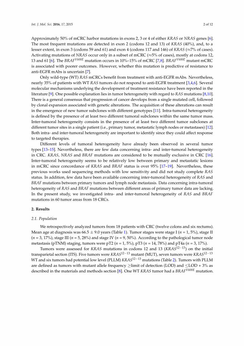
We retrospectively analyzed tumors from 18 patients with CRC (twelve colons and six rectums).Mean age at diagnosis was 66.5 ± 9.0 years (Table 1). Tumor stages were stage I (n = 1, 5%), stage II(n = 3, 17%), stage III (n = 5, 28%) and stage IV (n = 9, 50%). According to the pathological tumor nodemetastasis (pTNM) staging, tumors were pT2 (n = 1, 5%), pT3 (n = 14, 78%) and pT4a (n = 3, 17%).
Tumors were assessed for KRAS mutations in codons 12 and 13 (KRAS12−13) on the initialtransparietal section (ITS). Five tumors were KRAS12−13 mutant (MUT), seven tumors were KRAS12−13
WT and six tumors had potential low level (PLLM) KRAS12−13 mutations (Table 2). Tumors with PLLMare defined as tumors with mutant allele frequency ≥limit of detection (LOD) and ≤LOD + 3% asdescribed in the materials and methods section [8]. One WT KRAS tumor had a BRAFV600E mutation.
Int. J. Mol. Sci. 2016, 17, 2015 3 of 12
Table 1. Patients and tumor characteristics.
Patient Age Sex Tumor Site Stage pTNM 2009 Initial KRAS Initial BRAF Recurrence OS Status
2 77.9 F right colon III pT3N1bM0 WT WT yes 48.68 dead3 75.9 M left colon III pT4aN2bM0 WT WT no 2.50 alive4 79.2 F right colon II pT3N0M0 WT V600E no 59.14 alive6 55.9 M left colon I pT2N0M0 WT WT no 62.50 alive7 67.3 M left colon III pT3N1bM0 G12S WT yes 69.21 dead8 72.9 F left colon IV pT3N1bM1 G12V WT - 26.35 dead9 58.5 M left colon III pT4aN0M0 G12S WT yes 82.86 dead
12 77.8 F left colon IV pT4aN0M1 G12D WT - 22.60 dead14 56.4 F right colon IV pT3N1aM1 G12V WT - 48.39 dead15 77.7 M left colon IV pT3N2bM1 G12D WT - 14.41 dead16 74.7 F right colon II pT3N0M0 G12V WT no 37.70 alive13 66.0 M left colon II pT3N0M0 G12V WT - 49.00 alive1 61.9 M rectum IV pT3N2bM1 WT WT - 22.5 dead5 58.9 M rectum IV pT3N0M1 WT WT - 52.34 dead
10 57.1 F rectum IV pT3N2bM1 WT WT - 44.80 dead11 65.2 F rectum IV pT3N2bM1 G12D WT - 17.96 dead17 54.9 F rectum III pT3N2aM0 G12V WT yes 41.22 alive18 58.6 M rectum IV pT3N0M1 G12R WT - 43.82 dead
M: male; F: female; OS: overall survival; WT: wild-type; pTNM: pathological tumor node metastasis.

Int. J. Mol. Sci. 2016, 17, 2015 4 of 12
Table 2. RAS and BRAF mutations in the initial transparietal sections (n = 13) and spatially separatedtumor areas (n = 43) in 13 colorectal cancers.
Heterogeneity Patients pTNM %TC KRAS Other Genes
Intra-tumoralheterogeneity
4ITS 70 WT BRAF:V600E (PLLM)pT1 30 WT WTpT2 90 WT BRAF:V600E (MUT)
9
ITS 70 G12S (PLLM)/Q61R (PLLM) NRAS:Q61R (MUT)pT1 95 WT NRAS:Q61R (MUT)pT2 70 WT NRAS:Q61R (MUT)pT3 25 WT NRAS:Q61R (MUT)
18
ITS 80 G12R (MUT) WTpT1 30 G12R (MUT) NRAS : K117N (PLLM)pT2 35 G12R (MUT) WTpT3 20 G12R (MUT) WT
Intra-tumoral andInter-tumoralheterogeneity
2
ITS 80 WT NRAS: A59T (PLLM)pT2 15 WT WTpT3 20 G13D (MUT) WTN 5 WT WT
7
ITS 60 G12S (PLLM) FA (NRAS61 et KRAS146)pT1 70 G12D (MUT)/Q61L (PLLM) NRAS:Q61K (MUT)pT2 25 G12S (MUT) NRAS:Q61K (MUT)pT3 40 Q61L (PLLM) NRAS:Q61K (MUT)N 25 WT NRAS:Q61K (MUT)
12
ITS 40 G12D (PLLM)/A146T (MUT) WTpT2 15 A146T (MUT) WTpT3 20 Q61H(PLLM)/A146T (MUT) FA (BRAF)M 25 WT FA(NRAS59)
Inter-tumoralheterogeneity only 14
ITS 30 G12V (MUT) WTpT1 5 G12V (PLLM) WTpT2 40 G12V (MUT) WTpT3 5 G12V (PLLM) WTN 5 WT WT
Mutation withoutheterogeneity
8
ITS 30 G12V (MUT) WTpT1 60 G12V (MUT) WTpT2 75 G12V (MUT) WTpT3 60 G12V (MUT) WTN 80 G12V (MUT) WT
11
ITS 5 G12D (PLLM) WTpT2 5 G12D (MUT) WTpT3 5 G12D (PLLM) WTN 5 G12D (MUT) WT
13
ITS 70 G12V (MUT) WTpT1 70 G12V (MUT) WTpT2 10 G12V (MUT) WTpT3 15 G12V (MUT) WT
15
ITS 40 G12D (MUT) WTpT1 60 G12D (MUT) WTpT2 15 G12D (MUT) WTpT3 20 G12D (MUT) WTN 40 G12D (MUT) WT
16
ITS 40 G12V (MUT) WTpT1 80 G12V (MUT) WTpT2 40 G12V (MUT) WTpT3 10 G12V (MUT) WT
17
ITS 30 G12V (MUT) WTpT1 30 G12V (MUT) WTpT2 20 G12V (MUT) WTpT3 10 G12V (MUT) WTN 20 G12V (MUT) WT
%TC: percentage of tumoral cells; ITS: initial transparietal section; N: metastatic lymph node; M: metastasis;WT: wild-type; PLLM: potential low level mutation; MUT: mutant; FA: failed analysis.
2.2. Intra-Tumoral and Inter-Tumoral Heterogeneity of KRAS and NRAS Mutations
Thirty-nine percent of mCRC studied harbored intra- and/or inter-tumoral heterogeneity (Table 2).Among the six tumors with intra-tumoral heterogeneity, only two were KRAS12−13 WT in ITS
(cases 2 and 4). Three tumors (cases 2, 7, 12) presented KRAS12−13 PLLM or MUT in at least one of thetumor areas selected, but they also presented other KRAS or NRAS MUT or PLLM in other areas of
Int. J. Mol. Sci. 2016, 17, 2015 5 of 12
the tumor (case 2) or in the same area (case 7, 12). An additional NRAS117 PLLM in the submucosa(pT1) was observed for one tumor (case 18) (Table 2 and Figure S1). All in all, 33% of tumors (6/18)showed intra-tumoral heterogeneity for RAS mutation. The others presented either a WT pattern(cases 1, 3, 5, 6 and 10) or the same mutation with variation in allele frequency (cases 8, 11, 13–17) forall intra-tumoral areas (Table 2 and Table S1).
To evaluate the inter-tumoral heterogeneity, we tested the metastatic lymph nodes and distantmetastases for KRAS, NRAS and BRAF mutations when available. Among 11 cases analyzed, fourpresented inter-tumoral heterogeneity (36%). Three mutated primary tumors were WT in the metastaticlymph nodes (N+) or in the visceral metastases (M+) (cases 2, 12, 14) (Table 2).
It is worth noting that all patients with mCRC with RAS PLLM and/or RAS mutation in a limitedarea of the tumor (i.e., intra- or inter-tumoral heterogeneity) treated with anti-EGFR mAbs had diseaseprogression at 2–3 months (n = 5, cases 2, 7, 9, 11 and 12).
2.3. Mutational Intra-Tumoral Heterogeneity
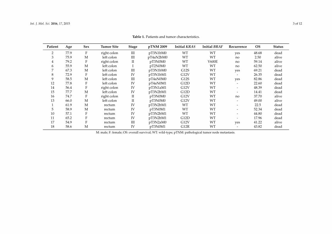
We focus on mutational intra-tumoral heterogeneity, defined as subclones with different mutantallele frequencies. Therefore, we calculated mutant allele frequency in neoplastic cells (MAFnc)and heterogeneity score (HS) for cases 7–9 and 13–18, which harbored the same mutation from thesubmucosa (pT1), the muscular layer (pT2) to the subserosa (pT3) (Figure 1). MAFnc is the mutantallele frequency normalized to 100% tumoral cells and HS corresponds to the fraction of neoplasticcells carrying a specific mutation and was calculated assuming that somatic mutations are usuallyheterozygous events [20]. Mean HS increased with T stage, 128.4 ± 46.1 standard deviation (SD) inpT1, 234.1 ± 186.8 SD in pT2 and 296.2 ± 156.9 SD in pT3 (p = 0.03) (Figure S2). We observed that HSwas higher in pT3 areas in 55% of cases (n = 5/9) regardless of the mutation. Mean total HS (sum ofHS for each mutation in one tumor) was 692.6 ± 262.6 SD. Total HS score was associated neither withtumor stage (p = 0.78), nor with tumor location (p = 0.90), nor with tumor recurrence (p = 0.56).
Int. J. Mol. Sci. 2016, 17, 2015 5 of 12
2.2. Intra-Tumoral and Inter-Tumoral Heterogeneity of KRAS and NRAS Mutations
Thirty-nine percent of mCRC studied harbored intra- and/or inter-tumoral heterogeneity (Table 2).
Among the six tumors with intra-tumoral heterogeneity, only two were KRAS12−13 WT in ITS (cases 2 and 4). Three tumors (cases 2, 7, 12) presented KRAS12−13 PLLM or MUT in at least one of the tumor areas selected, but they also presented other KRAS or NRAS MUT or PLLM in other areas of the tumor (case 2) or in the same area (case 7, 12). An additional NRAS117 PLLM in the submucosa (pT1) was observed for one tumor (case 18) (Table 2 and Figure S1). All in all, 33% of tumors (6/18) showed intra-tumoral heterogeneity for RAS mutation. The others presented either a WT pattern (cases 1, 3, 5, 6 and 10) or the same mutation with variation in allele frequency (cases 8, 11, 13–17) for all intra-tumoral areas (Table 2 and Table S1).
To evaluate the inter-tumoral heterogeneity, we tested the metastatic lymph nodes and distant metastases for KRAS, NRAS and BRAF mutations when available. Among 11 cases analyzed, four presented inter-tumoral heterogeneity (36%). Three mutated primary tumors were WT in the metastatic lymph nodes (N+) or in the visceral metastases (M+) (cases 2, 12, 14) (Table 2).
It is worth noting that all patients with mCRC with RAS PLLM and/or RAS mutation in a limited area of the tumor (i.e., intra- or inter-tumoral heterogeneity) treated with anti-EGFR mAbs had disease progression at 2–3 months (n = 5, cases 2, 7, 9, 11 and 12).
2.3. Mutational Intra-Tumoral Heterogeneity
We focus on mutational intra-tumoral heterogeneity, defined as subclones with different mutant allele frequencies. Therefore, we calculated mutant allele frequency in neoplastic cells (MAFnc) and heterogeneity score (HS) for cases 7–9 and 13–18, which harbored the same mutation from the submucosa (pT1), the muscular layer (pT2) to the subserosa (pT3) (Figure 1). MAFnc is the mutant allele frequency normalized to 100% tumoral cells and HS corresponds to the fraction of neoplastic cells carrying a specific mutation and was calculated assuming that somatic mutations are usually heterozygous events [20]. Mean HS increased with T stage, 128.4 ± 46.1 standard deviation (SD) in pT1, 234.1 ± 186.8 SD in pT2 and 296.2 ± 156.9 SD in pT3 (p = 0.03) (Figure S2). We observed that HS was higher in pT3 areas in 55% of cases (n = 5/9) regardless of the mutation. Mean total HS (sum of HS for each mutation in one tumor) was 692.6 ± 262.6 SD. Total HS score was associated neither with tumor stage (p = 0.78), nor with tumor location (p = 0.90), nor with tumor recurrence (p = 0.56).
Figure 1. Mutation allele frequency and heterogeneity score. Variability of mutation allele frequency and heterogeneity score between tumoral zone selections in cases which harbored the same mutation in pT1 to pT3. MAF: mutation allele frequency; HS: heterogeneity score.
Figure 1. Mutation allele frequency and heterogeneity score. Variability of mutation allele frequencyand heterogeneity score between tumoral zone selections in cases which harbored the same mutationin pT1 to pT3. MAF: mutation allele frequency; HS: heterogeneity score.
2.4. RAS Mutations Are Not Exclusive
RAS mutations are considered to be mutually exclusive. Only a few articles have reported casesof coexisting KRAS and NRAS mutations [21]. However, in our study, we observed that different KRASmutations as well as KRAS and NRAS mutations may coexist. Five tumors had multiple RAS mutatedsubclones in the same tumor (28%). Moreover, four presented, in one of their tumor selections, at least

Int. J. Mol. Sci. 2016, 17, 2015 6 of 12
two RAS mutated clones (22%) (cases 7, 9, 12, and 18) (Table 2). Indeed, we observed the coexistence ofdifferent KRAS mutations in three tumors (17%) (cases 7, 9, 12) as well as KRAS and NRAS mutationsin three tumors (17%) (cases 7, 9 and 18).
Tumors with two or more RAS mutations (cases 2, 7, 9, 12, and 18) as compared with tumorsshowing one RAS mutation (cases 8, 11, 13 to 17) were associated neither with tumor stage (p = 0.29)nor with tumor recurrence (p = 0.07).
3. Discussion
This study revealed a high proportion of intra- and inter-tumoral heterogeneity for RAS mutationsin metastatic colorectal cancer. Nearly 40% of the mCRC studied harbored intra- and/or inter-tumoralheterogeneity. We also demonstrated the coexistence of different RAS mutations within the same tumor.These results have relevant clinical implications for anti-EGFR monoclonal antibodies prescription inmCRC since RAS mutations confer resistance to this treatment. Hence, testing of KRAS and NRASmutations (codons 12, 13, 59, 61, 117 and 146) is a prerequisite for anti-EGFR mAbs used in mCRC.In daily practice, this testing usually relies on a single tumor sample with high tumor cell content.However, some patients with RAS WT tumor have primary resistance to anti-EGFR mAbs. Oneexplanation is the limited sensitivity of testing methods, leading to false negative results [6,8,10].Nevertheless, intra- and/or inter-tumoral heterogeneity of RAS mutations could be the most importantcause of therapeutic failure.
Indeed, recently, the “Big Bang” model emphasized that clonal alterations and subclonal eventsare an early event in CRC carcinogenesis with subclone mixing [22]. Several neoplastic subcloneswith co-existing mutations in different genes (as well as different molecular alterations) could bepresent in a single primary tumor with different mutant allele frequencies [19,20,23–25]. In our study,by testing KRAS and NRAS mutations in histologically relevant macrodissected zones accordingto pTNM, we observed the presence of (i) mutational intra-tumor heterogeneity with at least twoco-existing KRAS and/or NRAS mutations within the same tumor areas and (ii) spatial intra-tumoralheterogeneity with coexistence within the same tumor of KRAS and/or NRAS mutated zones andWT zones. This intra-tumoral heterogeneity was found in 33% of cases in our study. Kosmidouet al. reported similar results (44%) of intra-tumoral heterogeneity for KRAS mutations when theycompared tumor center and tumor periphery [23]. Recently, Kim et al. observed a substantial levelof intra-tumoral heterogeneity (46% to 80%) on multiregion biopsies from five mCRCs [26]. Up untilnow, our study is one of the largest study concerning intra-tumoral heterogeneity and the only oneconcerning an extended RAS status.
KRAS, NRAS and BRAF mutations are considered to be mutually exclusive in CRC [16].Nevertheless, in our study, we observed the coexistence of two different RAS mutations in 28% ofcases. We identified the coexistence of different KRAS mutations as well as KRAS and NRAS mutations.Coexistence of different KRAS12−13 mutations has been reported by others on small series [24–26].To our knowledge, the present study is one of the first reports concerning the coexistence of mutationsin codons 12–13, 61 and 146 of KRAS (17% of cases). In addition, we demonstrated the coexistence ofmutations in KRAS and NRAS, which has been reported only once in Vagaja, N.’s article [21].
Concordance of KRAS and BRAF status between primary and metastatic lesions in mCRC hasbeen considered to be over 95% [17–19]. To our knowledge, few studies have evaluated inter-tumoralheterogeneity of RAS mutations between primary tumors and lymph nodes or distant metastaticlesions. We observed 36% of inter-tumoral heterogeneity, with RAS mutated primary tumors beingWT in lymph nodes and/or distant metastatic lesions. In addition, metastatic lymph nodes may havedifferent RAS mutations as compared to primary tumors. Concerning metastatic lymph nodes, onlylimited data concerning KRAS mutations are available. In one study, heterogeneity in KRAS mutationsbetween primary tumors and lymph node metastases was found in approximately 30% of cases [27].To summarize, in some tumors, KRAS or NRAS mutations were “universal” as they were present inall macrodissected areas in primary lesions and in metastases. In other cases, we observed multiple
Int. J. Mol. Sci. 2016, 17, 2015 7 of 12
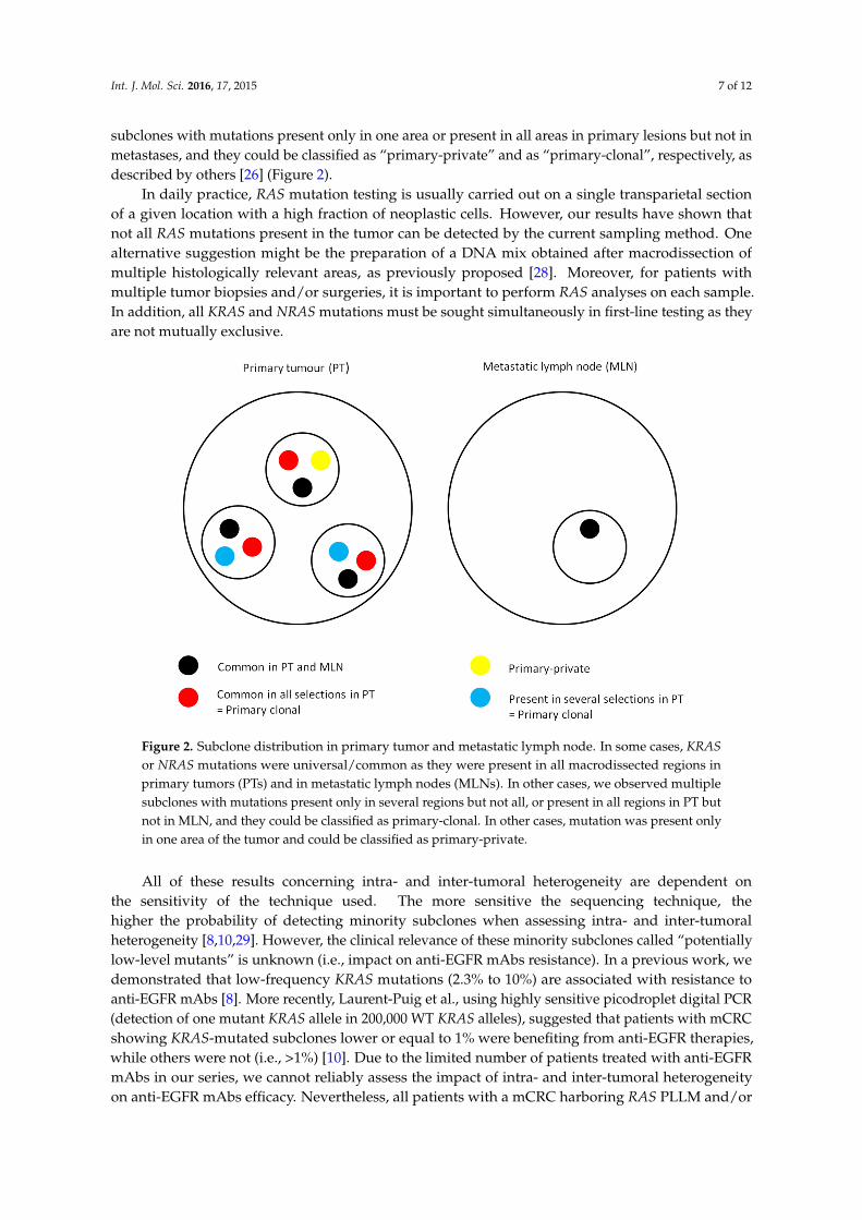
subclones with mutations present only in one area or present in all areas in primary lesions but not inmetastases, and they could be classified as “primary-private” and as “primary-clonal”, respectively, asdescribed by others [26] (Figure 2).
In daily practice, RAS mutation testing is usually carried out on a single transparietal sectionof a given location with a high fraction of neoplastic cells. However, our results have shown thatnot all RAS mutations present in the tumor can be detected by the current sampling method. Onealternative suggestion might be the preparation of a DNA mix obtained after macrodissection ofmultiple histologically relevant areas, as previously proposed [28]. Moreover, for patients withmultiple tumor biopsies and/or surgeries, it is important to perform RAS analyses on each sample.In addition, all KRAS and NRAS mutations must be sought simultaneously in first-line testing as theyare not mutually exclusive.
Int. J. Mol. Sci. 2016, 17, 2015 7 of 12
lymph nodes, only limited data concerning KRAS mutations are available. In one study, heterogeneity in KRAS mutations between primary tumors and lymph node metastases was found in approximately 30% of cases [27]. To summarize, in some tumors, KRAS or NRAS mutations were “universal” as they were present in all macrodissected areas in primary lesions and in metastases. In other cases, we observed multiple subclones with mutations present only in one area or present in all areas in primary lesions but not in metastases, and they could be classified as “primary-private” and as “primary-clonal”, respectively, as described by others [26] (Figure 2).
In daily practice, RAS mutation testing is usually carried out on a single transparietal section of a given location with a high fraction of neoplastic cells. However, our results have shown that not all RAS mutations present in the tumor can be detected by the current sampling method. One alternative suggestion might be the preparation of a DNA mix obtained after macrodissection of multiple histologically relevant areas, as previously proposed [28]. Moreover, for patients with multiple tumor biopsies and/or surgeries, it is important to perform RAS analyses on each sample. In addition, all KRAS and NRAS mutations must be sought simultaneously in first-line testing as they are not mutually exclusive.
Figure 2. Subclone distribution in primary tumor and metastatic lymph node. In some cases, KRAS or NRAS mutations were universal/common as they were present in all macrodissected regions in primary tumors (PTs) and in metastatic lymph nodes (MLNs). In other cases, we observed multiple subclones with mutations present only in several regions but not all, or present in all regions in PT but not in MLN, and they could be classified as primary-clonal. In other cases, mutation was present only in one area of the tumor and could be classified as primary-private.
All of these results concerning intra- and inter-tumoral heterogeneity are dependent on the sensitivity of the technique used. The more sensitive the sequencing technique, the higher the probability of detecting minority subclones when assessing intra- and inter-tumoral heterogeneity [8,10,29]. However, the clinical relevance of these minority subclones called “potentially low-level mutants” is unknown (i.e., impact on anti-EGFR mAbs resistance). In a previous work, we demonstrated that low-frequency KRAS mutations (2.3% to 10%) are associated with resistance to anti-EGFR mAbs [8]. More recently, Laurent-Puig et al., using highly sensitive picodroplet digital PCR (detection of one mutant KRAS allele in 200,000 WT KRAS alleles), suggested that patients with mCRC showing KRAS-mutated subclones lower or equal to 1% were benefiting from anti-EGFR therapies, while others were not (i.e., >1%) [10]. Due to the limited number of patients treated with
Figure 2. Subclone distribution in primary tumor and metastatic lymph node. In some cases, KRASor NRAS mutations were universal/common as they were present in all macrodissected regions inprimary tumors (PTs) and in metastatic lymph nodes (MLNs). In other cases, we observed multiplesubclones with mutations present only in several regions but not all, or present in all regions in PT butnot in MLN, and they could be classified as primary-clonal. In other cases, mutation was present onlyin one area of the tumor and could be classified as primary-private.
All of these results concerning intra- and inter-tumoral heterogeneity are dependent onthe sensitivity of the technique used. The more sensitive the sequencing technique, thehigher the probability of detecting minority subclones when assessing intra- and inter-tumoralheterogeneity [8,10,29]. However, the clinical relevance of these minority subclones called “potentiallylow-level mutants” is unknown (i.e., impact on anti-EGFR mAbs resistance). In a previous work, wedemonstrated that low-frequency KRAS mutations (2.3% to 10%) are associated with resistance toanti-EGFR mAbs [8]. More recently, Laurent-Puig et al., using highly sensitive picodroplet digital PCR(detection of one mutant KRAS allele in 200,000 WT KRAS alleles), suggested that patients with mCRCshowing KRAS-mutated subclones lower or equal to 1% were benefiting from anti-EGFR therapies,while others were not (i.e., >1%) [10]. Due to the limited number of patients treated with anti-EGFRmAbs in our series, we cannot reliably assess the impact of intra- and inter-tumoral heterogeneityon anti-EGFR mAbs efficacy. Nevertheless, all patients with a mCRC harboring RAS PLLM and/or
Int. J. Mol. Sci. 2016, 17, 2015 8 of 12
RAS mutations in a limited part of the tumor (i.e., intra- or inter-tumoral heterogeneity), and treatedwith anti-EGFR mAbs showed disease progression (n = 5). To conclude, RAS mutated subclones (>1%)partially explain primary anti-EGFR mAbs resistance. In contrast, secondary resistances to anti-EGFRmAbs are partially due to RAS and EGFR mutation [30–32].
One limitation of our study is the limited number of patients included, even though 78 samplesfrom 18 tumors were analyzed. Moreover, in some cases, PPLM was found only in ITS but not inselected macrodissected areas in primary tumors. Interestingly, these results involved high tumoralcellularity cases, thereby confirming that the mutation is present but remains undetectable in theselected macrodissected areas. The major strengths of this study are: (1) analysis of extended RASstatus; (2) multiple testing in primary tumors, metastatic lymph nodes and metastasis; and (3) use of ahighly sensitive method.
With regard to tumor infiltration, the mutations were more frequently found in subserosaselections (pT3) and heterogeneity score increased with wall invasion; thus, pT3 selection seemed tobe the best sampling zone for molecular analysis. In addition, we observed that HS was very highin several areas with a low fraction of neoplastic cells, thereby suggesting possible bias due to lowtumoral cellularity or genomic amplification of the mutant allele or loss of the wild-type allele as wellas mutation in both alleles [33].
4. Materials and Methods
4.1. Tumor Samples
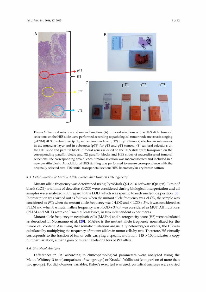
Tumoral zone selection was performed under HES (Haematoxylin-Erythrosin-Saffron) stainingby two experienced pathologists (Marion Jeantet and Gaelle Fromont) according to the 2009 pTNMclassification [34]. For each tumor, selections involved the submucosa (pT1), the muscular layer (pT2),the subserosa (pT3), the metastatic lymph nodes (N+) and/or the visceral metastases (M+) whenavailable. For instance, for a pT2 tumor, we performed pT1 and pT2 selections. For a pT3 or pT4tumor, we performed pT1, pT2 and pT3 selections (Figure 3). The corresponding area of each tumoralzone selection was macrodissected and included in a new paraffin block. Another HES staining wasperformed to verify correspondence with the originally selected area. The percentage of tumor cellswas assessed and selections with less than 5% of tumor cells were excluded.
A total of 60 tumoral macrodissections were analyzed. Among these samples, 15 were in pT1, 18 inpT2, 16 in pT3, 10 in N+ and 1 in M+. Four to six 10 µm thick sections were used for molecular analysis.
4.2. KRAS, NRAS and BRAF Mutation Testing
Genomic DNA from macrodissected sections was extracted with the QIAamp DNA FFPE Tissuekit (Qiagen, Hilden, Germany). The 18 initial samples and the 60 newly macrodissected samples weretested for KRAS and NRAS mutations in codons 12–13 (KRAS12−13 and NRAS12−13), codon 59 (KRAS59
and NRAS59), codon 61 (KRAS61 and NRAS61), codon 117 (KRAS117 and NRAS117) and codon 146(KRAS146 and NRAS146). BRAF was tested for mutation in codon 600 (BRAFV600E). Mutations weredetected by pyrosequencing using TheraScreen KRAS PyroKit CE-IVD kit (Qiagen) for KRAS12−13 andKRAS61 or the PyroMark PCR kit (Qiagen) with homemade primers using PyroMark Assay Design2.0 software (Qiagen, Hilden, Germany) for other RAS [35] and BRAF mutations (Table S2). PCRwas carried out using 50 ng of DNA in a total volume of 25 µL according to the Qiagen supplier’sinstructions. Each series included a known-mutated and a known wild-type sample, as positiveand negative controls. Pyrosequencing was performed using a Pyromark Q24 MDx according to themanufacturer’s instructions (Qiagen).
Int. J. Mol. Sci. 2016, 17, 2015 9 of 12Int. J. Mol. Sci. 2016, 17, 2015 9 of 12
Figure 3. Tumoral selection and macrodissection. (A) Tumoral selections on the HES slide: tumoral selections on the HES slide were performed according to pathological tumor node metastasis staging (pTNM) 2009 in submucosa (pT1), in the muscular layer (pT2) for pT2 tumors, selection in submucosa, in the muscular layer and in subserosa (pT3) for pT3 and pT4 tumors; (B) tumoral selections on the HES slide and paraffin block: tumoral zones selected on the HES slide were transposed on the corresponding paraffin block; and (C) paraffin blocks and HES slides of macrodissected tumoral selections: the corresponding area of each tumoral selection was macrodissected and included in a new paraffin block. An additional HES staining was performed to ensure correspondence with the originally selected area. ITS: initial transparietal section; HES: haematoxylin-erythrosin-saffron.
4.3. Determination of Mutant Allele Burden and Tumoral Heterogeneity
Mutant allele frequency was determined using PyroMark Q24 2.0.6 software (Qiagen). Limit of blank (LOB) and limit of detection (LOD) were considered during biological interpretation and all samples were analyzed with regard to the LOD, which was specific to each nucleotide position [35]. Interpretation was carried out as follows: when the mutant allele frequency was <LOD, the sample was considered as WT; when the mutant allele frequency was ≥LOD and ≤LOD + 3%, it was considered as PLLM and when the mutant allele frequency was >LOD + 3%, it was considered as MUT. All mutations (PLLM and MUT) were confirmed at least twice, in two independent experiments.
Mutant allele frequency in neoplastic cells (MAFnc) and heterogeneity score (HS) were calculated as described in Normanno et al. [20]. MAFnc is the mutant allele frequency normalized for the tumor cell content. Assuming that somatic mutations are usually heterozygous events, the HS was calculated by multiplying the frequency of mutant alleles in tumor cells by two. Therefore, HS virtually corresponds to the fraction of tumor cells carrying a specific mutation. HS > 100 indicates a copy number variation, either a gain of mutant allele or a loss of WT allele.
4.4. Statistical Analyses
Differences in HS according to clinicopathological parameters were analyzed using the Mann–Whitney U test (comparison of two groups) or Kruskal–Wallis test (comparison of more than
Figure 3. Tumoral selection and macrodissection. (A) Tumoral selections on the HES slide: tumoralselections on the HES slide were performed according to pathological tumor node metastasis staging(pTNM) 2009 in submucosa (pT1), in the muscular layer (pT2) for pT2 tumors, selection in submucosa,in the muscular layer and in subserosa (pT3) for pT3 and pT4 tumors; (B) tumoral selections onthe HES slide and paraffin block: tumoral zones selected on the HES slide were transposed on thecorresponding paraffin block; and (C) paraffin blocks and HES slides of macrodissected tumoralselections: the corresponding area of each tumoral selection was macrodissected and included in anew paraffin block. An additional HES staining was performed to ensure correspondence with theoriginally selected area. ITS: initial transparietal section; HES: haematoxylin-erythrosin-saffron.
4.3. Determination of Mutant Allele Burden and Tumoral Heterogeneity
Mutant allele frequency was determined using PyroMark Q24 2.0.6 software (Qiagen). Limit ofblank (LOB) and limit of detection (LOD) were considered during biological interpretation and allsamples were analyzed with regard to the LOD, which was specific to each nucleotide position [35].Interpretation was carried out as follows: when the mutant allele frequency was <LOD, the sample wasconsidered as WT; when the mutant allele frequency was ≥LOD and ≤LOD + 3%, it was considered asPLLM and when the mutant allele frequency was >LOD + 3%, it was considered as MUT. All mutations(PLLM and MUT) were confirmed at least twice, in two independent experiments.
Mutant allele frequency in neoplastic cells (MAFnc) and heterogeneity score (HS) were calculatedas described in Normanno et al. [20]. MAFnc is the mutant allele frequency normalized for thetumor cell content. Assuming that somatic mutations are usually heterozygous events, the HS wascalculated by multiplying the frequency of mutant alleles in tumor cells by two. Therefore, HS virtuallycorresponds to the fraction of tumor cells carrying a specific mutation. HS > 100 indicates a copynumber variation, either a gain of mutant allele or a loss of WT allele.
4.4. Statistical Analyses
Differences in HS according to clinicopathological parameters were analyzed using theMann–Whitney U test (comparison of two groups) or Kruskal–Wallis test (comparison of more thantwo groups). For dichotomous variables, Fisher's exact test was used. Statistical analyses were carried

Int. J. Mol. Sci. 2016, 17, 2015 10 of 12
out with a two-sided test with a significance value of 0.05. All analyses were performed using Statviewsoftware (Statview for Windows, SAS Institute, version 5.0, Cary, NC, USA).
5. Conclusions
Concordance of KRAS and BRAF status between primary and metastatic lesions in mCRC hasbeen considered to be over 95% [17–19]. To our knowledge, the present study is one of the first reportsconcerning inter-tumoral heterogeneity of RAS mutations between primary tumors and lymph nodesor distant metastatic lesions. In conclusion, we have demonstrated the tumoral heterogeneity of RASmutation and the co-existence of different RAS mutations in CRC, which may have major clinicalimplications. Our results question our daily practice and expose the limits of single transparietalsampling to ensure optimal molecular analysis. Macrodissection of multiple histologically relevantareas can be an interesting solution as proposed by Richman [28]. The use of new sensitive newgeneration sequencing methods can also be an alternative option [36]. However, the best solution mayarise from peripheral blood testing since circulating tumor cells and circulating tumor DNA are thereflection of the whole tumor [37,38]. These technologies provide another avenue to detect mutations,especially in patients with lymph nodes or distant metastasis.
Supplementary Materials: Supplementary materials can be found at www.mdpi.com/1422-0067/17/12/2015/s1.
Acknowledgments: Funding for this work was provided by the Ligue contre le Cancer of Vienne and ofDeux-Sèvres, Poitou-Charentes region, Institut National du Cancer and the “Sport et Collection” and “RotaryClub de Civray” foundations. The authors wish to thank Jeffrey Arsham, an American translator, for havingreviewed and revised the original English-language text. The authors thank Vanessa Le Berre, a research secretary,for her help in editing and formatting the manuscript.
Author Contributions: Lucie Karayan-Tapon, Gaelle Fromont, and David Tougeron conceived and designedthe experiments; Marion Jeantet and Céline Archambaut performed the experiments; Marion Jeantet,Gaelle Tachon, and Lucie Karayan-Tapon analyzed the data; Ulrich Cortes and Gaelle Tachon contributedreagents/materials/analysis tools; Marion Jeantet, David Tougeron, Gaelle Tachon, and Lucie Karayan-Taponwrote the paper.
Conflicts of Interest: The authors declare no conflict of interest. The founding sponsors had no role in the designof the study; in the collection, analyses, or interpretation of data; in the writing of the manuscript, and in thedecision to publish the results.
References
1. Siegel, R.L.; Miller, K.D.; Jemal, A. Cancer statistics, 2016. CA Cancer J. Clin. 2016, 66, 7–30. [CrossRef][PubMed]
2. Cunningham, D.; Humblet, Y.; Siena, S.; Khayat, D.; Bleiberg, H.; Santoro, A.; Bets, D.; Mueser, M.;Harstrick, A.; Verslype, C.; et al. Cetuximab monotherapy and cetuximab plus irinotecan inirinotecan-refractory metastatic colorectal cancer. N. Engl. J. Med. 2004, 351, 337–345. [CrossRef] [PubMed]
3. Douillard, J.Y.; Oliner, K.S.; Siena, S.; Tabernero, J.; Burkes, R.; Barugel, M.; Humblet, Y.; Bodoky, G.;Cunningham, D.; Jassem, J.; et al. Panitumumab-FOLFOX4 treatment and RAS mutations in colorectalcancer. N. Engl. J. Med. 2013, 369, 1023–1034. [CrossRef] [PubMed]
4. Van Cutsem, E.; Lenz, H.J.; Kohne, C.H.; Heinemann, V.; Tejpar, S.; Melezinek, I.; Beier, F.; Stroh, C.;Rougier, P.; van Krieken, J.H.; et al. Fluorouracil, leucovorin, and irinotecan plus cetuximab treatment andRAS mutations in colorectal cancer. J. Clin. Oncol. 2015, 33, 692–700. [CrossRef] [PubMed]
5. Lievre, A.; Bachet, J.B.; le Corre, D.; Boige, V.; Landi, B.; Emile, J.F.; Cote, J.F.; Tomasic, G.; Penna, C.;Ducreux, M.; et al. KRAS mutation status is predictive of response to cetuximab therapy in colorectal cancer.Cancer Res. 2006, 66, 3992–3995. [CrossRef] [PubMed]
6. Sorich, M.J.; Wiese, M.D.; Rowland, A.; Kichenadasse, G.; McKinnon, R.A.; Karapetis, C.S. ExtendedRAS mutations and anti-EGFR monoclonal antibody survival benefit in metastatic colorectal cancer:A meta-analysis of randomized, controlled trials. Ann. Oncol. 2015, 26, 13–21. [CrossRef] [PubMed]
Int. J. Mol. Sci. 2016, 17, 2015 11 of 12
7. Rowland, A.; Dias, M.M.; Wiese, M.D.; Kichenadasse, G.; McKinnon, R.A.; Karapetis, C.S.; Sorich, M.J.Meta-analysis of BRAF mutation as a predictive biomarker of benefit from anti-EGFR monoclonal antibodytherapy for RAS wild-type metastatic colorectal cancer. Br. J. Cancer 2015, 112, 1888–1894. [CrossRef][PubMed]
8. Tougeron, D.; Lecomte, T.; Pages, J.C.; Villalva, C.; Collin, C.; Ferru, A.; Tourani, J.M.; Silvain, C.; Levillain, P.;Karayan-Tapon, L. Effect of low-frequency KRAS mutations on the response to anti-EGFR therapy inmetastatic colorectal cancer. Ann. Oncol. 2013, 24, 1267–1273. [CrossRef] [PubMed]
9. Wheeler, D.L.; Dunn, E.F.; Harari, P.M. Understanding resistance to EGFR inhibitors-impact on futuretreatment strategies. Nat. Rev. Clin. Oncol. 2010, 7, 493–507. [CrossRef] [PubMed]
10. Laurent-Puig, P.; Pekin, D.; Normand, C.; Kotsopoulos, S.K.; Nizard, P.; Perez-Toralla, K.; Rowell, R.; Olson, J.;Srinivasan, P.; le Corre, D.; et al. Clinical relevance of KRAS-mutated subclones detected with picodropletdigital PCR in advanced colorectal cancer treated with anti-EGFR therapy. Clin. Cancer Res. 2015, 21,1087–1097. [CrossRef] [PubMed]
11. Greaves, M.; Maley, C.C. Clonal evolution in cancer. Nature 2012, 481, 306–313. [CrossRef] [PubMed]12. Blanco-Calvo, M.; Concha, A.; Figueroa, A.; Garrido, F.; Valladares-Ayerbes, M. Colorectal Cancer
Classification and Cell Heterogeneity: A Systems Oncology Approach. Int. J. Mol. Sci. 2015, 16, 13610–13632.[CrossRef] [PubMed]
13. Taniguchi, K.; Okami, J.; Kodama, K.; Higashiyama, M.; Kato, K. Intratumor heterogeneity of epidermalgrowth factor receptor mutations in lung cancer and its correlation to the response to gefitinib. Cancer Sci.2008, 99, 929–935. [CrossRef] [PubMed]
14. Yancovitz, M.; Litterman, A.; Yoon, J.; Ng, E.; Shapiro, R.L.; Berman, R.S.; Pavlick, A.C.; Darvishian, F.;Christos, P.; Mazumdar, M.; et al. Intra- and inter-tumor heterogeneity of BRAFV600E mutations in primaryand metastatic melanoma. PLoS ONE 2012, 7, e29336. [CrossRef] [PubMed]
15. Katona, T.M.; Jones, T.D.; Wang, M.; Eble, J.N.; Billings, S.D.; Cheng, L. Genetically heterogeneous andclonally unrelated metastases may arise in patients with cutaneous melanoma. Am. J. Surg. Pathol. 2007, 31,1029–1037. [CrossRef] [PubMed]
16. Rajagopalan, H.; Bardelli, A.; Lengauer, C.; Kinzler, K.W.; Vogelstein, B.; Velculescu, V.E. Tumorigenesis:RAF/RAS oncogenes and mismatch-repair status. Nature 2002, 418, 934. [CrossRef] [PubMed]
17. Brannon, A.R.; Vakiani, E.; Sylvester, B.E.; Scott, S.N.; McDermott, G.; Shah, R.H.; Kania, K.; Viale, A.;Oschwald, D.M.; Vacic, V.; et al. Comparative sequencing analysis reveals high genomic concordancebetween matched primary and metastatic colorectal cancer lesions. Genome Biol. 2014, 15, 454. [CrossRef][PubMed]
18. Santini, D.; Loupakis, F.; Vincenzi, B.; Floriani, I.; Stasi, I.; Canestrari, E.; Rulli, E.; Maltese, P.E.; Andreoni, F.;Masi, G.; et al. High concordance of KRAS status between primary colorectal tumors and related metastaticsites: Implications for clinical practice. Oncologist 2008, 13, 1270–1275. [CrossRef] [PubMed]
19. Baldus, S.E.; Schaefer, K.L.; Engers, R.; Hartleb, D.; Stoecklein, N.H.; Gabbert, H.E. Prevalence andheterogeneity of KRAS, BRAF, and PIK3CA mutations in primary colorectal adenocarcinomas and theircorresponding metastases. Clin. Cancer Res. 2010, 16, 790–799. [CrossRef] [PubMed]
20. Normanno, N.; Rachiglio, A.M.; Lambiase, M.; Martinelli, E.; Fenizia, F.; Esposito, C.; Roma, C.; Troiani, T.;Rizzi, D.; Tatangelo, F.; et al. Heterogeneity of KRAS, NRAS, BRAF and PIK3CA mutations in metastaticcolorectal cancer and potential effects on therapy in the CAPRI GOIM trial. Ann. Oncol. 2015, 26, 1710–1714.[CrossRef] [PubMed]
21. Vagaja, N.N.; Parry, J.; McCallum, D.; Thomas, M.A.; Bentel, J.M. Are all RAS mutations the same?Coexisting KRAS and NRAS mutations in a caecal adenocarcinoma and contiguous tubulovillous adenoma.J. Clin. Pathol. 2015, 68, 657–660. [CrossRef] [PubMed]
22. Sottoriva, A.; Kang, H.; Ma, Z.; Graham, T.A.; Salomon, M.P.; Zhao, J.; Marjoram, P.; Siegmund, K.; Press, M.F.;Shibata, D.; et al. A Big Bang model of human colorectal tumor growth. Nat. Genet. 2015, 47, 209–216.[CrossRef] [PubMed]
23. Kosmidou, V.; Oikonomou, E.; Vlassi, M.; Avlonitis, S.; Katseli, A.; Tsipras, I.; Mourtzoukou, D.;Kontogeorgos, G.; Zografos, G.; Pintzas, A. Tumor heterogeneity revealed by KRAS, BRAF, and PIK3CApyrosequencing: KRAS and PIK3CA intratumor mutation profile differences and their therapeuticimplications. Hum. Mutat. 2014, 35, 329–340. [CrossRef] [PubMed]
Int. J. Mol. Sci. 2016, 17, 2015 12 of 12
24. Improta, G.; Zupa, A.; Possidente, L.; Tartarone, A.; Pedicini, P.; Nappi, A.; Molinari, S.; Fraggetta, F.; Vita, G.Coexistence of two different mutations in codon 12 of the KRAS gene in colorectal cancer: Report of a casesupporting the concept of tumoral heterogeneity. Oncol. Lett. 2013, 5, 1741–1743. [CrossRef] [PubMed]
25. Baisse, B.; Bouzourene, H.; Saraga, E.P.; Bosman, F.T.; Benhattar, J. Intratumor genetic heterogeneity inadvanced human colorectal adenocarcinoma. Int. J. Cancer 2001, 93, 346–352. [CrossRef] [PubMed]
26. Kim, T.M.; Jung, S.H.; An, C.H.; Lee, S.H.; Baek, I.P.; Kim, M.S.; Park, S.W.; Rhee, J.K.; Lee, S.H.; Chung, Y.J.Subclonal Genomic Architectures of Primary and Metastatic Colorectal Cancer Based on IntratumoralGenetic Heterogeneity. Clin. Cancer Res. 2015, 21, 4461–4472. [CrossRef] [PubMed]
27. Oliveira, C.; Velho, S.; Moutinho, C.; Ferreira, A.; Preto, A.; Domingo, E.; Capelinha, A.F.; Duval, A.;Hamelin, R.; Machado, J.C.; et al. KRAS and BRAF oncogenic mutations in MSS colorectal carcinomaprogression. Oncogene 2007, 26, 158–163. [CrossRef] [PubMed]
28. Richman, S.D.; Chambers, P.; Seymour, M.T.; Daly, C.; Grant, S.; Hemmings, G.; Quirke, P. Intra-tumoralheterogeneity of KRAS and BRAF mutation status in patients with advanced colorectal cancer (aCRC) andcost-effectiveness of multiple sample testing. Anal. Cell. Pathol. (Amst.) 2011, 34, 61–66. [CrossRef] [PubMed]
29. Molinari, F.; Felicioni, L.; Buscarino, M.; de Dosso, S.; Buttitta, F.; Malatesta, S.; Movilia, A.; Luoni, M.;Boldorini, R.; Alabiso, O.; et al. Increased detection sensitivity for KRAS mutations enhances the predictionof anti-EGFR monoclonal antibody resistance in metastatic colorectal cancer. Clin. Cancer Res. 2011, 17,4901–4914. [CrossRef] [PubMed]
30. Misale, S.; Yaeger, R.; Hobor, S.; Scala, E.; Janakiraman, M.; Liska, D.; Valtorta, E.; Schiavo, R.; Buscarino, M.;Siravegna, G.; et al. Emergence of KRAS mutations and acquired resistance to anti-EGFR therapy in colorectalcancer. Nature 2012, 486, 532–536. [CrossRef] [PubMed]
31. Montagut, C.; Dalmases, A.; Bellosillo, B.; Crespo, M.; Pairet, S.; Iglesias, M.; Salido, M.; Gallen, M.;Marsters, S.; Tsai, S.P.; et al. Identification of a mutation in the extracellular domain of the Epidermal GrowthFactor Receptor conferring cetuximab resistance in colorectal cancer. Nat. Med. 2012, 18, 221–223. [CrossRef][PubMed]
32. Tougeron, D.; Cortes, U.; Ferru, A.; Villalva, C.; Silvain, C.; Tourani, J.M.; Levillain, P.; Karayan-Tapon, L.Epidermal growth factor receptor (EGFR) and KRAS mutations during chemotherapy plus anti-EGFRmonoclonal antibody treatment in metastatic colorectal cancer. Cancer Chemother. Pharmacol. 2013, 72,397–403. [CrossRef] [PubMed]
33. Paguirigan, A.L.; Smith, J.; Meshinchi, S.; Carroll, M.; Maley, C.; Radich, J.P. Single-cell genotypingdemonstrates complex clonal diversity in acute myeloid leukemia. Sci. Transl. Med. 2015, 7, 281re2.[CrossRef] [PubMed]
34. Gunderson, L.L.; Jessup, J.M.; Sargent, D.J.; Greene, F.L.; Stewart, A.K. Revised TN categorization for coloncancer based on national survival outcomes data. J. Clin. Oncol. 2010, 28, 264–271. [CrossRef] [PubMed]
35. Cortes, U.; Guilloteau, K.; Rouvreau, M.; Archaimbault, C.; Villalva, C.; Karayan-Tapon, L. Developmentof pyrosequencing methods for the rapid detection of RAS mutations in clinical samples. Exp. Mol. Pathol.2015, 99, 207–211. [CrossRef] [PubMed]
36. Haley, L.; Tseng, L.H.; Zheng, G.; Dudley, J.; Anderson, D.A.; Azad, N.S.; Gocke, C.D.; Eshleman, J.R.;Lin, M.T. Performance characteristics of next-generation sequencing in clinical mutation detection ofcolorectal cancers. Mod. Pathol. 2015, 28, 1390–1399. [CrossRef] [PubMed]
37. Mohamed Suhaimi, N.-A.; Foong, Y.M.; Lee, D.Y.S.; Phyo, W.M.; Cima, I.; Lee, E.X.W.; Goh, W.L.; Lim, W.-Y.;Chia, K.S.; Kong, S.L.; et al. Non-invasive sensitive detection of KRAS and BRAF mutation in circulatingtumor cells of colorectal cancer patients. Mol. Oncol. 2015, 9, 850–860. [CrossRef] [PubMed]
38. Basnet, S.; Zhang, Z.-Y.; Liao, W.-Q.; Li, S.-H.; Li, P.-S.; Ge, H.-Y. The Prognostic Value of Circulating Cell-FreeDNA in Colorectal Cancer: A Meta-Analysis. J. Cancer 2016, 7, 1105–1113. [CrossRef] [PubMed]
© 2016 by the authors; licensee MDPI, Basel, Switzerland. This article is an open accessarticle distributed under the terms and conditions of the Creative Commons Attribution(CC-BY) license (http://creativecommons.org/licenses/by/4.0/).