hhaappppyy nneeww yyeeaarr 22001100 - espcrhim to honorary membership. as for the personal data:...
TRANSCRIPT

ESP CRBULLETIN
Editorial Office: G. Ghanem (Editor), C. Meunier (Secretary), Laboratory of Oncology and Experimental Surgery (L.O.C.E.), Université Libre de Bruxelles, Institut J. Bordet, Rue Héger-Bordet 1, B – 1000 Brussels, Belgium. Phone: 32-2-541.32.96 E-Mail:[email protected]
E S
P C
R
B
U L
L E
T I
N
PUBL
ISH
ED B
Y TH
E EU
ROPE
AN S
OC
IETY
FO
R PI
GM
ENT
CEL
L RE
SEAR
CH
E
DIT
OR
:
G
. GH
ANEM
(Bru
ssel
s)
I
NTE
RN
ATI
ON
AL
F
. BEE
RMAN
N (L
ausa
nne)
, M. B
ÖH
M (M
ünst
er),
J. B
ORO
VAN
SKY
(Pra
gue)
, M. d
’ISC
HIA
(Nap
les)
, N. S
MIT
(Lei
den)
,
ED
ITO
RIA
L B
OA
RD
: JC
GAR
CIA
-BO
RRO
N (M
urci
a),
R. M
ORA
ND
INI (
Brus
sels
), A
. NAP
OLI
TAN
O (N
aple
s), M
. PIC
ARD
O (R
ome)
.
N° 6
5 D
ec 2
009
N° 65 Dec 2009
HHHAAAPPPPPPYYY NNNEEEWWW YYYEEEAAARRR 2 22000111000
CONTENT
Discussion, Letters to the editor, Reviews, Short communications, ...
- EuMelaNet, A new ESPCR Special Interest Group, by Pr Marco d’Ischia - Obituary Pr Jiri Duchon, by Pr Jan Borovansky - 15 ESPCR, Münster, Meeting Report
th
Review of the literature
1. Chemistry of Melanins and other pigments (Prof A. Napolitano)
2. Biology of pigment cells and pigmentary disorders (Dr M. Picardo)
3. MSH, MCH, other hormones (Prof M. Böhm) 4. Photobiology (Dr N. Smit) 5. Neuromelanins (Prof M. d'Ischia) 6. Genetics, molecular and developmental biology
(Dr F. Beermann) 7. Tyrosinase, TRPs, other enzymes
(Prof JC. Garcia-Borron) 8. Melanosomes (Prof J. Borovansky) 9. Melanoma experimental, cell culture (Dr R. Morandini)
Announcements and related activities Calendar of events.

LETTER TO THE EDITOR DISCUSSION, REVIEW, SHORT COMMUNICATION, ...
EuMelaNet
A new ESPCR Special Interest Group "Dear Colleagues and Friends, We are very pleased to inform you that, following preliminary and informal contacts dating
back to 2007, and after the formal steps made in 2009 during the XV ESPCR meeting in Muenster, the ESPCR Council has approved the establishment of a new Special Interest Group within our society. The group is named EuMelaNet, to emphasize the European origins, the focus on the black eumelanins and related metabolites as potential added value products, and its organization as a multidisciplinary research network involving different centres in Europe and other continents. For more information please visit the web site (http://www.espcr.org/eumelanet/).
The special interest group EuMelaNet is basically aimed at promoting and revitalizing melanin
research at all levels. The main goal is to attract the interests of industries and companies on melanins and melanogenesis as a valuable, yet so far little explored, source of new molecules, polymers and processes of potential practical interest in a range of fields, from health care to technology. The expected outcome of the EuMelaNet activities is to create new links of cooperation between different research centres, to offer new opportunities for research projects and grant applications, to promote exchange of knowledge and information about scientists, especially beyond the boundaries of the pigment cell community, and to enhance the scientific role and visibility of our Society. In view of these goals, all members who wish to apply for inclusion in EuMelaNet should make their best to involve companies as potential partners for research projects on melanins and to participate in the activities of our Society.
Interest in melanins is increasing all over the world and we strongly believe that the new
interest group may be a useful means of gathering together researchers interested in these intriguing biopolymers within the aims and scope of the ESPCR.
We hope you find this initiative of interest and look forward to your comments and suggestions
as to how to pursue the EuMelaNet goals and create a solid and efficient organization under the aegis of our Society." Marco d'Ischia
1938
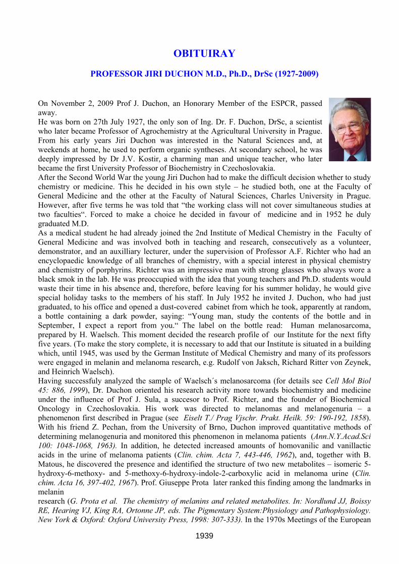
OBITUIRAY
PROFESSOR JIRI DUCHON M.D., Ph.D., DrSc (1927-2009)
On November 2, 2009 Prof J. Duchon, an Honorary Member of the ESPCR, passed away. He was born on 27th July 1927, the only son of Ing. Dr. F. Duchon, DrSc, a scientist who later became Professor of Agrochemistry at the Agricultural University in Prague. From his early years Jiri Duchon was interested in the Natural Sciences and, at weekends at home, he used to perform organic syntheses. At secondary school, he was deeply impressed by Dr J.V. Kostir, a charming man and unique teacher, who later became the first University Professor of Biochemistry in Czechoslovakia. After the Second World War the young Jiri Duchon had to make the difficult decision whether to study chemistry or medicine. This he decided in his own style – he studied both, one at the Faculty of General Medicine and the other at the Faculty of Natural Sciences, Charles University in Prague. However, after five terms he was told that “the working class will not cover simultaneous studies at two faculties“. Forced to make a choice he decided in favour of medicine and in 1952 he duly graduated M.D. As a medical student he had already joined the 2nd Institute of Medical Chemistry in the Faculty of General Medicine and was involved both in teaching and research, consecutively as a volunteer, demonstrator, and an auxilliary lecturer, under the supervision of Professor A.F. Richter who had an encyclopaedic knowledge of all branches of chemistry, with a special interest in physical chemistry and chemistry of porphyrins. Richter was an impressive man with strong glasses who always wore a black smok in the lab. He was preoccupied with the idea that young teachers and Ph.D. students would waste their time in his absence and, therefore, before leaving for his summer holiday, he would give special holiday tasks to the members of his staff. In July 1952 he invited J. Duchon, who had just graduated, to his office and opened a dust-covered cabinet from which he took, apparently at random, a bottle containing a dark powder, saying: “Young man, study the contents of the bottle and in September, I expect a report from you.“ The label on the bottle read: Human melanosarcoma, prepared by H. Waelsch. This moment decided the research profile of our Institute for the next fifty five years. (To make the story complete, it is necessary to add that our Institute is situated in a building which, until 1945, was used by the German Institute of Medical Chemistry and many of its professors were engaged in melanin and melanoma research, e.g. Rudolf von Jaksch, Richard Ritter von Zeynek, and Heinrich Waelsch). Having successfuly analyzed the sample of Waelsch´s melanosarcoma (for details see Cell Mol Biol 45: 886, 1999), Dr. Duchon oriented his research activity more towards biochemistry and medicine under the influence of Prof J. Sula, a succesor to Prof. Richter, and the founder of Biochemical Oncology in Czechoslovakia. His work was directed to melanomas and melanogenuria – a phenomenon first described in Prague (see Eiselt T./ Prag Vjschr. Prakt. Heilk. 59: 190-192, 1858). With his friend Z. Pechan, from the University of Brno, Duchon improved quantitative methods of determining melanogenuria and monitored this phenomenon in melanoma patients (Ann.N.Y.Acad.Sci 100: 1048-1068, 1963). In addition, he detected increased amounts of homovanilic and vanillactic acids in the urine of melanoma patients (Clin. chim. Acta 7, 443-446, 1962), and, together with B. Matous, he discovered the presence and identified the structure of two new metabolites – isomeric 5-hydroxy-6-methoxy- and 5-methoxy-6-hydroxy-indole-2-carboxylic acid in melanoma urine (Clin. chim. Acta 16, 397-402, 1967). Prof. Giuseppe Prota later ranked this finding among the landmarks in melanin research (G. Prota et al. The chemistry of melanins and related metabolites. In: Nordlund JJ, Boissy RE, Hearing VJ, King RA, Ortonne JP, eds. The Pigmentary System:Physiology and Pathophysiology. New York & Oxford: Oxford University Press, 1998: 307-333). In the 1970s Meetings of the European
1939

Pigment Cell Community witnessed friendly competition between J. Duchon, advocating the marker value of Thormählen-positive melanogens, and Prof Hans Rorsman, who preferred his 5-S-cysteinyldopa. This competition on the diagnostic and prognostic value of melanogens resulted in a draw when it was shown in large groups of melanoma patients (n=690 in Prague, n=570 in Lund) that the positivity of both types of markers was the same in about 35% patients (Eur. J. Cancer 16: 383-388, 1980). Many unanswered questions concerning the structure and analysis of melanogens were answered later by a former Ph.D.student of Jiri Duchon – Stan Pavel in the Netherlands. Thanks to his outstanding work in the field of melanogens, Dr. Duchon received a Eleanor Roosevelt Fellowship from UICC and WHO in 1967 and spent 15 months at the Dept. of Dermatology, Harvard Medical School, in the lab of the late Prof. T.B. Fitzpatrick. There he met the “father of melanosomes“ Prof. M. Seiji, and learned his method of melanosome isolation and started to study their composition. After return to Prague he taught us how to isolate and handle melanosomes and a series of articles followed. At first it was necessary to obtain pure samples of native melanosomes and, to achieve this, we reproduced all the methods of melanosome isolation developed since 1938 (when H. Waelsch had critically reviewed the previous techniques). At that time it was not known whether melanosomes represented simply an assembly of melanin with tyrosinase or whether they contained any other constituents of a protein or non-protein nature. Thanks to the leadership of Dr. Duchon, many blank spots were subsequently filled in for the first time in Prague where it was shown that: melanosomes are complex organelles containing many proteins (Cas.lek.ces. 111: 218-220, 1972, Neoplasma 22: 195-199, 1975), lipids (Sborník lék 79:335-339, 1977, Neoplasma 30:317-321, 1983), and metals, including zinc and copper (Hoppe-Seyler´s Z.Physiol. Chem. 354: 203-204, 1973). To summarize the research activities in the field of melanogensis and melanoma between 1976-1990 it is sufficient to say that 3 habilitation theses and 10 Ph.D. theses were defended under Jiri Duchon‘s supervision. In 1981 Prof Duchon was President of the 3rd European Workshop on Melanin Pigmentation held in Prague; and at the 8th Meeting of the ESPCR in Prague, which was organized by Dr. Matous and myself on the occasion of the 650th anniversary of the foundation of Charles University, the ESPCR Council elected him to Honorary Membership. As for the personal data: Jiri Duchon defended his Ph.D. Thesis “Urinary melanogens in melanoma disease“ on 18th May1962. His Habilitation Thesis “Study on melanins and on melanogenesis“ was defended on 29th June 1964, and he became Associate Professor on 1st July 1965. He defended his DrSc Thesis: “Contribution to the biochemistry of malignant melanoma“ on 4th May 1992 and, on 1st December 1993, President Vaclav Havel conferred on him the decree of Full Professor. Prof. Duchon retired on 1.4.1996. Prof. Duchon was not only a notable scientist but also a highly popular university teacher. His lectures were clear and his love for biochemistry radiated from him. He was the leading author of the standard Textbook of Biochemistry (successive editions appearing in 1985,1988, 1991, 1996) used at all medical faculties in the Czech and Slovak Republics. He was always sensitive to novel trends. With another excellent and successful pupil of his, Prof. Vachtenheim, he published “Molecular Biology for Medical Students and Physicians“ in 1992 - the first textbook of this kind in the Czech Republic. I had the privilege of seeing Prof. Duchon regularly for 48 years both at our Institute and also outside it. Attending scientific meetings before the Velvet Revolution often meant long train journeys, about which it would be possible to write a book of humourous stories. Jiri Duchon was Head of the Institute for 26 years. We were privileged to have Head who was wise and full of knowledge, a friend prepared to engage in scientific disputation, and a gentleman adhering to democratic principles and generating peaceful conditions for work. He was also an excellent companion with a deep knowledge of Latin, literature, and history and with two lifelong hobbies: mineralogy and yachting. We miss him enormously Jan Borovansky
1940

MEETING REPORT (15th ESPCR Meeting, Münster sep 2009)
Not all the session reports are available, many thanks for those who contributed
Missing contributions will appear in the next issue Symposium I: Recent advances in the treatment of pigmentary disorders of the skin Chairs: M. Picardo, A. Taïeb Symposium II: Perspectives in melanoma treatment Chairs: G. Ghanem, T.A. Luger Symposium II was chaired by T. Lüger (Münster) and G. Ghanem (Brussels). It had 4 invited speakers and 3 oral presentations. The first presentation, by Leon van Kempen (Nijmegen) discussed the relationship between events occurring within tumor microenvironment and tumor progression. He presented a 3D coculture model highlighting fibroblast role on angiogenesis and collagen type I turnover in cell proliferation and invasion. A. Eberle (Basle) reviewed targeted peptides to melanoma cells with a focus on melanocortin peptides agonists bound to various chelator groups able to efficiently bind radioisotopes mainly for Petscanning and therapy. He described validation studies in animal models with different linkers and chelators and concluded that MSH agonists may be promising radiopharmaceuticals. R. Dummer reviewed different molecular targeting approaches that are currently under clinical investigation. Interesting data came from clinical trials with drugs targeting BRAF, cKit, VEGFR and multikinase inhibitors. A special focus is dedicated to the MAPK pathway because of the frequent BRAF, NRAS mutations in melanomas. Some good results were effectively obtained kinase inhibitors such as Sorafenib, MEK and BRAFV600E inhibitors. However, a resistance phenomenon to the drugs was observed in some patients. A number of different trials are on going in many countries. D. Schadendorf discussed melanoma resistance to chemotherapy and possible solutions. One of the interesting observations he presented dealt with a trial using the antisense bcl-2 Oblimersen in combination with Dacarbazine that yeilded a significant effect on patient survival. The benefit is however modest: 1-2 months but shows the validity of such an approach. F. Journé (Brussels) showed a significant correlation between melanoma patient survival and a gene expression signature related to pigmentation found in metastases. The signature could be represented by one single gene, validated in an additional set of tumors also at the protein TYRP1, levels. J. van den Boorn (Amsterdam) an immunotherapy approach in B16-F10 melanoma-bearing C57BL/6 mice using a combination of CpG, imiquimod and a hydroquinone derivative. Thus combining TLR7 and 9 agonists to a bleaching agent. The authors evaluated a significant immune response in terms of melanoma specific CD8+ and IgG induction with a sustained NK cell expansion. The immune response resulted in an efficient B16 melanoma tumor growth inhibition. M.A.Zmijewski (Gdansk) examined vit.D analog derivatives as anti-melanoma agents. The authors used UVB photoconversion to generate these derivatives from synthetic precursors, identified them and evaluated their effect on the growth of a human melanoma cell line. They reported a specific inhibitory effect on melanoma cells and underlined their original approach. Session I: Developmental biology of pigment cells Chairs: F. Beermann, H. Arnheiter The Session I had 4 invited speakers and 1 selected presentation, which was chosen from the submitted abstracts.
Melanocytes have the remarkable capacity to be regenerated from stem cells during the normal hair cycle. It was appropriate, therefore, to start the session with a general lecture that covered recent findings on the induction of stem cells and that was given by the internationally recognized stem cell expert Hans R. Schöler (Münster, Germany). Prof. Schöler reported the exciting finding that a
1941

unipotent germ cell line can give rise to pluripotent cells simply as a result of manipulating culture conditions. Most protocols leading to induced pluripotent stem (iPS) cells, however, rely on the introduction of at least four factors, Oct4, Klf4, c-Myc, and Sox2. For generating iPS cells from neural stem cells, Schöler’s group eliminated these factors one by one and finally managed to obtain the desired cell type by using just one factor, Oct4 (Nature 2009, 461, 649-653). Although this approach required longer culture periods compared to those using multiple factors, it highlighted the fact that relatively simple manipulations can lead to cells that may eventually be used in cell-based therapies.
The following talks were more neural crest and pigment cell-centered and touched on the role of extracutaneous, “non-classical” melanocytes, different genetic approaches to identify novel genes involved in neural crest and melanocyte development and specific aspects of the role of genes in the Notch pathway for the development of cutaneous melanocytes. Lionel Larue (Orsay, France) reported on non-melanocytic cells and melanocytes located in the mouse heart. Apparently, presence of an activated form of ß-catenin in the heart (using the Tyrosinase::Cre mice and mice carrying a floxed allele of exon 3 of ßcatenin) led to premature death with mutant mice showing an increased left atrium of the heart. The basic defect was then localized to a structure called ductus arteriosus, containing amongst others two vagal neural crest derivatives - smooth muscle cells and melanocytes. Tyr::Cre- mediated recombination was found in both populations, however, further genetic studies involving mice deficient in only melanocytes indicated that the effect is only due to the population of neural crest-derived smooth muscle cells carrying an activated ß-catenin allele.
William Pavan (NIH, Bethesda, USA) presented new data emerging from the Sox10-sensitized mouse mutagenesis screen. For example, 5 modifier genes have been mapped which affect the Sox10 phenotype in heterozygous Sox10::lacZ knockin mice. Amongst these genes was a member of the Patched/sonic hedgehog pathway (Mos1: chastity, Gli3), as well as unknown proteins or a ribosomal protein (Mos4: RPS7). In addition to postnatal phenotypes acting on melanocytes, the use of the Sox10::lacZ reporter allows to screen embryos for deviation from the expected Sox10::lacZ expression pattern.
Tatjana Hochgreb (Pasadena, USA) reported on the mechanisms of formation of neural crest cells using a zebrafish model. A genetic approach based on the transposon-dependent cell trap and protein fusion screen called FlipTrap allowed her to identify a large number (~150) of novel genes affecting neural crest development, some of which were presented in more detail. The selected short presentation was given by Geneviève Aubin-Houzelstein (Maisons-Alfort and Paris, France), who reported on recent findings using melanocyte-specific activation or inactivation of the genes strawberry notch (Sbno2) and Notchless (Nle1). Transgenic expression of Dct::Sbno2 led to a gray hair phenotype indicating loss of melanocyte precursors within the bulge. Tyrosinase::Cre- mediated inactivation of Nle1 led to an essential white coat with Nle1-dependent effects starting from E12.5 during embryogenesis. From these observations, it might already be concluded that both Sbno2 and Nle1 are genes important in melanocyte homeostasis. Session II: Genetics of pigmentation Chairs: R. Spritz, E. Healy Session III: Melanins and melanogenesis Chairs: J. Borovansky, C. Jimenez-Cervantes (Contributed by C. Jimenez-Cervantes) Session III was chaired by Drs Jan Borovansky, from Charles University (Czech Republic) and Celia Jiménez-Cervantes from the University of Murcia (Spain). It was composed by three invited lectures and three short oral presentations. They were mainly related to the discussion of recent advances in the knowledge of the chemical structure of melanin, and the physical properties of the polymer. Structural information about proteins of the tyrosinase family was also reviewed and some new data on signalling pathways that regulate melanogenesis were presented.
1942

Dr Borovansky opened the session by presenting the first invited lecture delivered by Dr. Marco d’Ischia, from Naples. The Naples group is part of the history of the ESPCR and has been a leader in understanding the complexity of the melanin polymer structure. By means of physico-chemical approaches, and using synthetic eumelanins derived both from DHI or DHICA as simplest models, the group is unravelling the details of the process of monomer polymerization leading to the melanin polymer. Data on the positions in the monomers involved in polymerization steps were discussed. In addition, the preparation of a synthetic soluble DHI-based polymer allowed Dr d’Ischias’s group to dig into the structural reasons that explain the differences in the physical properties of the different types of eumelanin molecules.
The second invited lecture was presented by Dr. Hans Decker, from the University of Mainz (Germany). Dr. Decker is a very well known scientist in the field of protein modelling, with a huge experience in exploring the active site of catecholoxidases and hemocyanins from different species in comparison with the one of tyrosinase. By means of X-ray crystallographic data and computational modelling of these type 3 copper proteins, he proposed that the molecular basis underlying some pigment related disorders, such us albinism, resides in defects of proper tridimensional conformation of the tyrosinase active site.
The last invited lecture to the session was entitled “High temperature incandescence as a new optical signature of melanin: fundamental results and applications” and was delivered by Dr. Amblard. He described studies on heat accumulation and dissipation by a source of melanin that has been previously excited by laser. The resulting thermal spectra from natural melanin suspensions of hair or melanosomes in comparison with the ones from melanin suspensions point to the requirement of a solid state organization to generate visible incandescence. This physical property may be used to detect circulating melanoma cells in blood.
The short oral talks delivered in this session were presented by Drs. Bellei (from Rome), Ballotti (Nice) and Pezzella (Naples), respectively. The first one, entitled ”The role of p38 MAPK signalling pathway in melanogenesis in B16 mouse melanoma cells” dealt with several aspects of signalling by p38 in melanoma cells, whose relationship with the regulation of melanogenesis is currently under study in the laboratory of Dr Mauor Picardo, in Rome. Dr Bellei presented evidence that silencing of p38 in melanoma cells results in an increase of both basal and MSH-induced melanogenesis. Based on this and other data, it was proposed that there is an inverse correlation between p38 MAPK signalling and melanin synthesis in melanoma cells, probably related with a stimulation of tyrosinase ubiquitylation and degradation. On the other hand, the authors found that one type of widely used p38 inhibitor that effectively blocks melanogenesis could act independently of this MAPK. This is an interesting result that highlights the need of caution in interpreting pharmacological studies, owing to the lack of an absolute specificity of most, if not all, the kinase inhibitors synthesized thus far.
The second oral presentation explored the molecular mechanisms underlying the increase of melanosome pH observed after treatment of melanoma cells with either MSH or cAMP elevating agents. Dr Ballotti’s group has observed a regulation of the expression of vacuolar ATPases such as SLC45A2, SLC24A4 and SLC24A5 and the P protein. In addition, using pharmacological treatments, they conclude that the melanosome pH is regulated by the second messenger cAMP. The last short oral presentation came from the same lab as the first invited lecture of this session. Dr Pezzella presented detailed studies on a method designed to produce water soluble melanins, a tool to better study the intrinsic absorption properties of DHI-derived melanins. Working with a galactose-derived soluble brown pigment they can conclude that the dark colour of the DHI pigments is based not only in electronic delocalization occurring in this molecule but is also dependent on the redox state of the subunits. (Contributed by J. Borovansky) Session III (chaired by J. Borovanský and C. Jiménez –Cervantes) comprised 3 invited lectures, 3 oral presentation chosen from the abstracts and it was complemented with 10 posters.
1943

Invited lectures: M. d´Ischia summarized the recent results on integrated chemical, mass spectrometric and pulse-radiolytic bottom up approach to the mode and degree of polymerization of 5,6dihydroxyindole (DHI) and 5,6-dihydroyindole-2-carboxylic acid (DHICA). He pointed out that the first soluble DHI-based polymer yielded a novel insight into the origin of eumelanin „black chromophore“ and the underlying broad band UV-visible absorption. H. Decker reviewed the data on catecholoxidases, tyrosinases and hemocyanins obtained in his laboratory over the previous 11 years. It is interesting that hemocyanins can be converted into tyrosinases and/or catecholoxidase without any chemical modification. On behalf of his coworkers F. Amblard spoke about the high-temperature incadescence as a new optical signature of melanin with a potential for detecting melanized cells. His lecture was fascinating but required a higher level of biophysical knowledge. Oral presentations: A.Pezzela et al. supplemented the lecture of prof. d´Ischia et al. by details on the synthesis and chartacterization of the first water soluble 5,6-DHI polymer. B. Bellei et al showed that the down-regulation of p38 can in some circumstances contribute to melanogenesis stimulation. They also demonstrated that the widely used p38 MAP kinase specific inhibitors repressed the melanogenic pathway via an uknown target molecule different from p38 MAP kinase. Ballotti et al. underlined the key role of the melanosome pH in the regulation of melanin synthesis and brought a first evidence for the roles of cAMP and αMSH in the regulation of melanosome pH. Session IV: Signal transduction in melanocytes and melanoma cells Chairs: J. Garcia-Borrón, R. Halaban Session IV was chaired by Drs Ruth Halaban from Yale University and José Carlos García-Borrón from the University of Murcia, and consisted of 3 invited lectures and 3 oral presentations. During the session relatively new players in the game of regulation of melanocyte and melanoma cell biology were considered, in addition to classic stars such as MITF and p53. The session was opened by Ruth Halaban who presented the first lecturer, Dr Krutmann from Dusseldorf, Germany. His talk dealt with the role of the arylhydrocarbon receptor (AhR) pathway in human melanocytes. Interest in this novel potential regulatory pathway has been fostered by demonstration of AhR activation in UVB-irradiated keratinocytes as well as hyperpigmentation caused by the AhR ligand dioxin. Using cultured melanocytes and syngeneic AhR-/- mice, the German group has found: i) expression of functional AhR in cultured human and mouse melanocytes, ii) AhR-dependent induction of tyrosinase and related genes and iii) some evidence on the possible involvement of the AhR in the tanning response. Accordingly, the AhR signaling pathway may provide new targets to modulate pigmentation. The second invited lecture was delivered by Dr Behrmann, from the University of Luxembourg. Its title was “Signal transduction in melanoma via Jaks, STATS and SOCS”. The Janus kinase (Jak)/STAT signal transduction pathway is important in the cellular response to several cytokines. Cytokine signaling is often deregulated in melanoma as shown by the acquisition of resistance to cytokine-induced growth inhibition in melanoma cells. STAT1 and STAT3 transcription factors are involved in growth inhibition by interferon gamma and IL6, respectively. However, the actual role of STATs in the regulation of melanoma cell survival and growth remains unclear since inhibition of Jak-dependent STAT3 phosphorylation has no effect on density-dependent growth arrest. The lecture also highlighted the interest of cytokine-regulated microRNAs, a new field with broad potential implications. The last invited lecture, entitled “Activated kinases and signal transducers in melanomas”, was presented by Ruth Halaban, from the Yale Cancer Center (YCC). This is a comprehensive translational research center designated by the US National Cancer Institute. The final goal of the YCC is to provide a framework for the development of individual therapeutic protocols for cancer patients. A series of “omic” approaches to the study of signaling deregulation in melanoma were reviewed, aiming at the identification of druggable targets. Dr. Halaban focused on protein phosphorylation profiles in short-term cultures of melanoma cells, with emphasis on kinases and their downstream signaling
1944

intermediates. This type of studies is interesting not only for the knowledge of melanoma biology, but also for testing technologies amenable to an individualized analysis of melanoma cases. Together with high throughput techniques for the establishment of drug sensitivity profiles, they could open the door to a rational and individualized therapy of melanoma. The oral presentations in the session dealt with new aspects of well-known, classic players in the game of the regulation of melanocyte biology, namely MITF and p53. In spite of the large and ever-increasing number of studies devoted to their roles in melanocyte proliferation and differentiation, both transcription factors still hide surprising features and unsuspected interactions of bewildering complexity. R Ballotti, from Nice, presented new data suggesting that MITF controls the DNA damage response and a lineage-specific senescence program in melanomas. MITF silencing was reported to induce melanoma cell senescence by engaging a DNA damage response. Interestingly, this lineage-specific DNA damage response involved p53 upregulation, thus suggesting the MITF participates in the control of p53 levels and signaling in melanocytic cells. The next presentation, by A Schepsky, dealt with the site-specific acetylation of MITF as related with the regulation of binding affinity and transcriptional activity. The lecture emphasized the complexity of the functional regulation of MITF and lent further support to the MITF rheostat model by highlighting potential changes in MITF transcriptional activity as a result of regulated and site-specific post-translational covalent modifications. The closing presentation, by S Haferkamp, aimed at the assessment of the relative contributions of key tumour suppressors, namely p53, p21Waf1, pRb and p16INK4a in oncogene-induced senescence. The data presented suggested a major role pRb and p16INK4a/pRb pathway, at least when oncogenic N-RAS-induced senescence was considered. Oxidative Stress and Pigment Cells Chairs: K.U. Schallreuter, N. Smit Session VI: The Melanocyte under the sun Chairs: T. Schwarz, V.J. Hearing Alessandra Napolitano (University of Naples Frederico II) reported a study that used an improved procedure for the analysis of pheomelanic tissues, including notably red human hair. They showed that the pheomelanin pigment suffers extensive degradation affecting mainly the 5-S-cysteinyldopa-derived components during hair growth, whereas the 2-S-cysteinyldopa-derived units remain largely unaffected. A closely similar trend was also observed upon prolonged exposure of hair to sunlight. The noticeable persistence of the 2-S-cysteinyldopa component during the growth of red hair might reflect its intrinsic stability to reactive oxygen species or to UV-induced degradation. Marie Dominique Galibert (University of Rennes 1) discussed how the skin is the first body barrier that is exposed to various physical, chemical and biological hazards that can alter DNA structure. To maintain the integrity of the genome, cells are equipped with specific defense mechanisms, including the tanning response and the DNA-repair machinery. Having decrypted the molecular mechanism implicated in the UV-response of the tanning process, they hypothesized that in response to UV, the DNA-repair machinery is also regulated. Using a combination of in vivo assays and a genetic approach involving USF-1 knock-out mice, they show for the first time that HR23 members of the NER-DNA repair pathway, implicated in the stabilization of the XPC-protein, are differentially regulated in response to cumulative UV-irradiation. Expression of the gene encoding HR23A, but not HR23B, is up-regulated in response to UV, which leads to a 4-fold increase of HR23A protein. UV-regulated HR23A gene expression is dependent on the presence of conserved cis-regulatory elements (E-box motifs) and the USF-1 transcription factor. USF-1 is thus shown to mediate skin protection against UV by controlling two independent and complementary pathways: UV-induced pigmentation and DNA-repair processes. Vince Hearing (National Cancer Institute) reported a study characterizing gene expression patterns in human skin (skin types II-III) that had been repetitively exposed for 2 weeks to UVA and/or UVB compared with control unexposed skin. To test the hypothesis that different mechanisms are involved in the pigmentary responses of the skin to different types of UV, they used immunohistochemical and whole human genome microarray analyses to characterize human skin in situ to examine how
1945

melanocyte-specific proteins and paracrine melanogenic factors are regulated by repetitive exposure to different types of UV. Increased pigmentation was elicited with either or both types of UV, but the mechanisms involved were quite distinct. UVB dramatically up-regulated the expression of most of the genes involved in pigment production (as well as other cellular functions) in melanocytes and caused increased levels of melanin synthesis, but UVA had no such effect, thus the mechanism of the increased skin pigmentation was independent of increased melanin content. The gene expression patterns characterize the distinct responses of the skin to UVA or UVB, and identify several potential new melanogenic factors involved in the UV-induced pigmentation of human skin. Karl Gledhill (Bradford University) reported that epidermal melanocytes (EM) derived from individuals with skin phototypes 1 or 4 had very little intrinsic differences (in vitro) in terms of melanogenesis, i.e. expression/activity of tyrosinase, expression of dopachrome tautomerase, levels of melanin and dendricity. Interestingly, expression of tyrosinase related protein-1 appeared to be significantly greater in EM derived from skin phototype 1 compared to EM derived from skin phototype 4. Overall, this may suggest that the skin phototype is largely determined by the keratinocyte partner acting in a paracrine manner on the EM and not by intrinsic differences of the EM. However, although EM in skin phototype 1 do not actively contribute to melanin production post-UVR exposure they may instead contribute to the inflammation that occurs in this skin type. This was suggested as they were able to show that EM derived from skin phototype 1 responded to a single dose of UVB by increasing their production of the pro-inflammatory eicosanoid, prostaglandin E2 (PGE2), while EM derived from skin phototype 4 did not. Session VII: Melanocortin peptides, pigment cells and beyond Chairs: A. Eberle, M. Böhm This session was devoted to the most recent developments in melanocortin peptides, an area highly important melanocytes physiology, pathophysiology and also for translational research. In the first invited lecture, M. Böhm (Münster, Germany), described novel findings related to the protective activity of α-MSH. He highlighted the capacity of α-MSH to induce the transcription factor Nrf2, a master regulator of a panel of anti-oxidative enzymes (e. g. HO-1), in both normal human melanocytes and keratinocytes. Interestingly, this effect of α-MSH could also be reproduced with selected truncated MSH peptides and derivatives which do not bind to the MC1R. Of note, α-MSH, moreover, attenuated UVB-induced suppression of Nrf2 and Nrf-dependent enzymes thereby supporting the concept hat melanocortins function as endogenous protectors against UV-induced oxidative stress. In the second invited talk, Z. Abdel-Malek (Cincinatti, USA) further extended those findings by new data on the regulation of catalase by UV and α-MSH. In analogy to Nrf2 and Nrf-dependent enzymes, UV exposure decreased catalase protein expression and activity in normal human melanocytes, and this effect was counteracted by α-MSH. In accordance with these findings α-MSH reduced UV-mediated generation of hydrogen peroxide and reduced the amounts of 8-oxodG, a marker of oxidative DNA damage. In the third invited talk, J. C. Garcia-Borrón (Murcia, Spain), presented new findings on the crosstalk between the MC1R-mediated cAMP pathway and ERK signaling. Interestingly, the cAMP but not the ERK pathway was impaired in PC12 cells transfected with defined red hair fair skin MC1R plasmids, i. e. those with R151C, R160W and D294H mutations. Further experiments revealed that this maintained ERK signal transduction crosstalk occurs most likely via activation of c-kit, the receptor for mast cell growth expressed by melanocytes. In the final invited lecture of the session, K. Loser (Münster, Germany), reported on experiments employing α-MSH-treated regulatory T cells in a murine melanoma model. Importantly, α-MSH increased the activated state of these T cells by increasing the expression of distinct cytotoxic enzymes such as perforin. The latter findings were of special interest for potential future therapies using adoptive immunotherapy with α-MSH-treated T cells for melanoma patients. These invited lectures were followed by three short oral presentations. In the first one, M. L. Dell’Anna (Rome) presented data on functional differences in the phosphorylated state of fokal adhesion kinase p120FAK and of CREB between normal and vitiligo melanocytes after stimulation with α-MSH. In the second talk, E. Healy (Southampton) reported on the potential localization of
1946

MC1R within the melanosomes of melanocytes. In the last short talk, V. Maresca (Rome) introduced PPAR-γ as a new target for α-MSH in B16.F10 melanoma cells. Session VIII: Epidermal melanocytes and their cellular neighbours Chairs: Z. Abdel-Malek, R. Paus Session IX: Albinism and extracutanous melanin Chairs: L. Montoliu, B. McKay
This session started with a talk by Tadeusz Sarna (Department of Biophysics, Jagiellonian Univ., Krakow, Poland) in which he reported his research studies with purified bovine RPE melanosomes and their effects on peroxidation of lipids in a liposomal system, induced by intense visible light irradiation or by stains photosensitized reactions. Data presented showed that these melanosomes had a limited efficiency to scavenge or quench reactive oxygen species, which may have been generated by pigment granules appearing in photoaged melanosomes. Secondly, L. Zecca (Institute of Biomedical Technologies, INRC, Segrate-Milano, Italy) reviewed the presence and potential role of melanic pigments in the human brain. These melanic molecules are present at high concentrations in various major brain regions, such as cortexes, cerebellum, putamen, caudate nucleus and globus pallidus. Next, Brian McKay (Dep. Of Ophthalmology and Visual Science, University of Arizona, Tucson, AZ, USA), one of the co-chairs of this session, summarized his published work on L-DOPA as the suggested endogenous ligand for the OA1 receptor, a G-protein coupled receptor that is mutated in ocular albinism subjects. His data presented clearly demonstrated the role of L-DOPA as the OA1 endogenous ligand and the implications of the OA1 signaling pathway in retinal development. The next talk, by K.P. Hoffmann (Dep. General Zoology and Neurobiology, Ruhr University Bochum, Germany) discussed the role of cation-chloride co-transporters in ocular albinism, using albino and pigmented rats as experimental model. His data supported the idea that NKCC co-transporter may be the main cause of the observed elevated intracellular chloride level in albino visual cortex neurons. Next, M. Vittoria Schiaffino (San Raffaele Scientific Institute, Milan, Italy) reported her latest studies on the characterization of the OA1 phenotype, using the recently generated Oa1 knockout mice. Her data suggested a novel function for the OA1 protein in melanosomes motility, perhaps associated with the cytoskeletal proteins, as downstream effectors of this G-protein coupled receptor. She concluded that the observed OA1-deficiency phenotype might result from a different mechanism as previously recognized, involving a melanosome-autonomous signaling pathway implicated in the regulation of both membrane traffic and transport. The session concluded with two selected short oral presentations. The first one, by Lluis Montoliu (CNB-CSIC, Madrid, Spain), co-chair of the session, who presented his latest research on oculocutaneous type I albinism using mouse models. In a combined effort with Prof. S. Ito’s laboratory in Japan, they could demonstrate that L-DOPA contents in eyes and cochleas of albino mice were lower than those found in the corresponding pigmented or transgenic mice, expressing tyrosine hydroxylase under the control of tyrosinase regulatory elements, in agreement with the diverse sensory phenotypes (visual and hearing deficits associated to albinism in mice) observed in these animals. Finally, Heinz Arnheiter (Mammalian Development Section, NINDS, NIH, Bethesda, MD, USA) reviewed the fundamental role of Pax6 in the development of the retinal pigment epithelium (RPE). He concluded that this transcription factor assumes different roles in the RPE and the retina, despite the fact these two issues are both derived from the optic neuroepithelium. Session X: Pigment cell biology of the hair follicle Chairs: A. Slominski, S. Commo Session XI: Vitiligo and other pigmentary disorders Chairs: J. Lambert, J.P. van der Veen Session XII: Molecular biology of melanoma and nevi
1947

Chairs: D. Bennett, M. Schartl This session included quite a range of novel findings that continue the expansion of our molecular understanding of progression in melanoma. There were two talks on microRNAs and their growing importance for melanoma research, and potentially diagnosis and therapy. Anja Bosserhoff reported the results of global miRNA profiling of melanocytes and melanoma cell lines representing different stages of progression. New regulated miRNA candidates included miR-23b, which is upregulated in early melanoma development. In advanced melanoma, expression of miR-let-7a is lost. Interestingly, miR-let-7a is a negative regulator of integrin ß3 mRNA, and the loss of miR-let-7a results in increased invasive behavior of melanocytes. D. Müller from the same lab reported that miR-196 is significantly downregulated in melanoma cell lines compared to melanocytes. This RNA is encoded in three HOX gene clusters and can downregulate HOXC8, which in turn is overexpressed in melanoma, with target genes including osteopontin that may contribute to progression. miR-196 is also an interferon target. Moving to germline genetics, N. Gruis talked about the search for susceptibility genes for nevi and especially atypical nevi – both being potential melanoma precursors and known risk factors for melanoma. She commented that comparisons between studies are hampered by varying clinical definitions. A number of loci have shown association with nevus susceptibility, including a weak linkage to 7q21 near CDK6 (cyclin-dependent kinase 6). Stronger associations were shown in a recent genome-wide association study: MTAP (9p21), encoding an enzyme of polyamine metabolism, and PLA2G6 (22q13), a phospholipase needed for arachidonic acid synthesis. MTAP is adjacent to melanoma gene CDKN2A (encoding p16 and ARF), so a role for CDKN2A in this association may also be possible. Nevi are believed to be in a state of oncogene-induced senescence, and D. Peeper discussed the use of an RNAi library in human fibroblasts to screen for additional regulators of BRAF-induced senescence (besides p16). Early findings suggested roles for transcription factor TSC22 and the p16 paralog p15 (INK4B). Knockdown of p15 was not sufficient to block induced senescence in fibroblasts. Nor was knockdown of p16, but this combined with knockdown of PTEN did appear sufficient. C. Müller reported expression of NOTCH1 and 2 and their receptor Jagged1 in uncultured melanomas and nevi and a cultured melanoma line; she suggested interaction between this and the vitamin D signalling pathway. D. Widmer presented an update on the phenotype-switching model of his group (Hoek et al.), which hypothesizes that melanoma cells can switch back and forth between transcriptional programs leading to proliferative or invasive phenotypes, to drive metastatic progression. Hypoxic conditions led to upregulation of invasive phenotype-specific genes (and invasiveness), and downregulation of proliferative phenotype-specific genes. Thus hypoxia is a candidate microenvironmental condition for effecting this switch in vivo. Lastly, the grey gene in horses is strongly associated with a high incidence of melanoma; A. Golovko reported on this interesting model system. The team found that the underlying mutation affects the syntaxin (STX) 17 gene and leads to upregulation of STX17 and the neighboring NR4A3 gene in grey horse melanoma. In these tumors as a direct effect of both genes the RAS/RAF/MAPK pathway is activated, although mutations in NRAS and BRAF are absent. Interestingly STX17 transfection into human melanoma cells enhanced MAPK activation upon α-MSH stimulation. Workshop: Experimental approaches in melanoma research Chairs: A. Bosserhoff, L. Larue
The workshop on “Experimental Approaches in Melanoma Research" was the last session of the 15th Meeting of the European Society for Pigment Cell Research. Anja Bosserhoff and Lionel Larue were chair(wo)men of this session. This session was really appreciated by the attendees and it should certainly be repeated in the future. The speakers presented in a very didactic manner various technical and technological aspects classically used in the field of pigment cell biology. Dot Bennett from St George’s, University of London, presented «Melanocyte and melanoma cell culture – any chance of standardization?». Dot went through the establishment of melanocytes in culture in detail as it is highly important for the field to result in standards of cultivation.
1948

She presented the different methods in particular defined media, different gas mixtures, use serum or not, select from a variety of supplements, and perhaps use growth-inactivated feeder cells – keratinocytes or fibroblasts. She presented the importance of immortalization in culture of melanocytes as well as melanoma, which required different growth conditions. Keith Hoek from the University Hospital of Zurich presented «Expression analysis in melanoma using chip technology: problems, solutions and future directions». Keith presented, in a very pleasant way, the major keys that scientists have to pay attention when they analyze their microarrays. For instance, he reminds the audience that the misinterpretation of the analysis of microarray is classical and that correlation is not a proof. He went back on the famous BRAFV600E signature that people dreamt of. Such signature does not seem to exist. Unfortunately! Stefan Schneider from the Department of Dermatology, University of Heidelberg focussed on «In vitro models of melanoma vessel interaction”. Stefan presented an excited in vitro models mimicking nicely microvasculature of blood vessels. This system can be manipulated in a nice and efficient way. It will continue to bring some exciting results. For instance, Stefan and his colleagues could show that melanoma cells are able to activate endothelial cells via melanoma cell derived MMP1 or IL-1 that targets endothelial cell receptors (PAR1 and IL-1 receptor respectively). Finally, Thomas Tüting from the Department of Dermatology and Allergology, University Hospital Bonn presented « Choosing experimental mouse models for melanoma research: a tumor immunologist’s point of view » Thomas commented on the advantages and inconvenience of mouse models to be pertinent for humans. Especially, he presented the Hgf-Cdk4R24C mouse model which seems to be a good model system to understand and modulate some of the processes leading to melanoma development and progression.
1949

CURRENT LITERATURE ___________________________________
111... CCChhheeemmmiiissstttrrryyy ooofff MMMeeelllaaannniiinnnsss aaannnddd ooottthhheeerrr PPPiiigggmmmeeennntttsss (Pr A. Napolitano)
Many interesting reports on melanin structure and properties have appeared in this term. Using nonlinear transient absorption spectroscopy a group at Duke university (Piletic et al, J Chem Phys) provided a quantification of the molar absorptivities for eumelanin and pheomelanin from which the average molecular weight of the pigments could be estimated concluding that eumelanin contains approximately 46, and pheomelanin 28 monomer units. Meredith’s group at Brisbane (Watt et al Soft Matter) used low voltage-high resolution transmission electron microscopy (LVHRTEM) to investigate natural and synthetic eumelanin and observed that sheets of protomolecules stack to form onion-like nanostructures. The inter-sheet spacings within these structures are between 3.7 and 4.0 Angstrom consistent with non-covalent �� stacking in heteroaromatic systems and not far from previous estimation. Melanin-superoxide radical pairs were observed to form by TR-EPR spectra on UV irradiated air-equilibrated synthetic pigments and both the electron dipolar interaction D and the exchange interaction J between the two radicals could be observed (Toffoletti et al, Chem Commun). This further supports the interpretation on the triplet excited states of photoexcited natural RPE melanin published very recently by Norris and Sarna (Wang et al J Phys Chem B. 2009)The current status of knowledge on the structure of melanins with special emphasis on the organization of the pigment within melanosomes is presented by the excellent joint review by Ito’s and Simon’s group (Simon et al PCMR) which also discusses the important issues that must be addressed in future research efforts. A positive correlation between tobacco use, dependence, nicotine exposure and melanin pigmentation was found among African American smokers, highlighting a peculiar property of melanin so far little investigated (King et al, Pharmacol, Biochem and Behavior). Difficult to choose among the so many reports of melanin pigmentation controlling agents including single molecules and plant extracts. Of interest are the tyrosinase inhibitory effects of serotonin derivatives such as N-caffeoylserotonin and N-protocatechuoylserotonin (Yamazaki et al Bioorg Med Chem Letts) Even simple sulphur compounds 1-propylmercaptan, di-Me disulfide, diallyl disulfide, Pr disulfide, and 2,5-dimethylthiophene proved efficient pigmentation controlling agents both in vivo and in vitro. The hyperpigmentation of patients undergoing hemodialysis was correlated to a rise in the levels of serum cysteinyldopa resulting ultimately in pheomelanin production (Murakami et al, Blood Purification). Structure, Reactivity and Properties - King, G; Yerger, VB; Whembolua, G-L; Bendel, RB; Kittles, R; Moolchan, ET.
Link between facultative melanin and tobacco use among African Americans. Pharmacol, Biochem and Behavior (2009), 92(4), 589-596.
- Neifar, A; Ben Rebah, F.; Gargouri, A.; Abdelmouleh, A.
Physicochemical characterization of Sepia officinalis ink and the effects of storage conditions on the coagulation process. J Marine Biol Ass United Kingdom (2009), 89(4), 803-807.
- Piletic, IR; Matthews, TE; Warren, WS.
Estimation of molar absorptivities and pigment sizes for eumelanin and pheomelanin using femtosecond transient absorption spectroscopy. J Chem Phys (2009), 131(18), 181106/1-181106/4.
- Ren, G; Miao, Z; Liu, H; Jiang, L; Limpa-Amara, N; Mahmood, A; Gambhir, SS; Cheng, Z
Melanin -targeted preclinical PET imaging of melanoma metastasis. J Nuclear Med (2009), 50(10), 1692-1699.
1950

- Simon, JD; Peles, D; Wakamatsu, K; Ito, S. Current challenges in understanding melanogenesis: bridging chemistry, biological control, morphology, and function. Pigment Cell & Melanoma Research (2009), 22(5), 563-579.
- Toffoletti, A; Conti, F; Sandron, T; Napolitano, A; Panzella, L; D'Ischia, M
Time-resolved EPR observation of synthetic eumelanin-superoxide radical pairs. Chem Commun (Cambridge, United Kingdom) (2009), (33), 4977-4979.
- Tu, Y; Sun, Y; Tian, Y; Yang, M; Xie, M.
The antioxidant activity of melanin from taihe black-bone silky fowl in vitro. Shipin Yu Shengwu Jishu Xuebao (2009), 28(2), 145-149
- Watt, AAR; Bothma, JP; Meredith, P.
The supramolecular structure of melanin. Soft Matter (2009), 5(19), 3754-3760. Melanogenesis and its Modulation - Akihisa, T; Noto, T; Takahashi, A; Fujita, Y; Banno, N; Tokuda, H; Koike, K; Suzuki, T; Yasukawa, K;
Kimura, Y. Melanogenesis inhibitory, anti-inflammatory, and chemopreventive effects of limonoids from the seeds of Azadirachta indicia A. Juss. (neem). J Oleo Sci (2009), 58(11), 581-594.
- Arung, ET; Kusuma, IW; Christy, EO; Shimizu, K; Kondo, R.
Evaluation of medicinal plants from Central Kalimantan for antimelanogenesis. J Nat Medicines (2009), 63(4), 473-480.
- Chen, L-G; Chang, W-L; Lee, C-J; Lee, L-T; Shih, C-M; Wang, C-C.
Melanogenesis inhibition by gallotannins from Chinese galls in B16 mouse melanoma cells. Biol Pharmac Bull (2009), 32(8), 1447-1452.
- Chu, H-L; Wang, B-S; Duh, P-D.
Effects of Selected Organo-sulfur Compounds on Melanin. Journal of Agricultural and Food Chemistry (2009), 57(15), 7072-7077.
- Iida, Y; Satoh, I; Maezumi, N.
Evaluation of the properties of the tyrosinase inhibitor with use of immobilized tyrosinase. Chemical Sensors (2009), 25(Suppl. B), 22-24.
- Jeon, S; Kim, N-H; Koo, B-S; Kim, J-Y; Lee, A-Y.
Lotus (Nelumbo nuficera) flower essential oil increased melanogenesis in normal human melanocytes. Exp Molecular Med (2009), 41(7), 517-524.
- Kim, J; Kim, J-H; Jeung, E-S; Park, I-S; Choe, C-H; Kwon, T-H; Yu, K-Y; Jeong, S-I.
Inhibitory effects of stilbene glucoside isolated from the root of Polygonum multiflorum on tyrosinase activity and melanin biosynthesis. J Korean Soc Appl Biol Chem (2009), 52(4), 342-345.
- Lee, S-J; Park, SG; Chung, H-M; Choi, J-S; Kim, D-D; Sung, J-H.
Antioxidant and Anti-Melanogenic Effect of the Novel Synthetic Hexapeptide (SFKLRY-NH2). Intl J Peptide Res Therap (2009), 15(4), 281-286.
- Le Mellay-Hamon, V; Criton, M.
Phenylethylamide and phenylmethylamide derivatives as new tyrosinase inhibitors. Biol & Pharmaceutical Bulletin (2009), 32(2), 301-303
- Maruyama, S; Yamazaki, Y; Kono, Y; Yamanaka, A.
1951

Melanin generation inhibitors containing caffeoyl tryptamine or feruloyl tryptamine, and use thereof. Jpn. Kokai Tokkyo Koho (2009), 17pp. CODEN: JKXXAF JP 2009263282 A 20091112
- Matsuyama, K; Villareal, MO; El Omri, A; Han, J; Kchouk, ME; Isoda, H.
Effect of Tunisian Capparis spinosa L. extract on melanogenesis in B16 murine melanoma cells. J Nat Medicines (2009), 63(4), 468-472.
- Sato, K; Toriyama, M.
Depigmenting effect of catechins. Molecules (2009), 14(11), 4425-4432. - Yamazaki, Y; Kawano, Y; Yamanaka, A; Maruyama, S.
N-[(Dihydroxyphenyl)acyl]serotonins as potent inhibitors of tyrosinase from mouse and human melanoma cells. Bioorg Med Chem Letts (2009), 19(15), 4178-4182.
- Zhang, X; Hu, X; Hou, A; Wang, H.
Inhibitory effect of 2,4,2',4'-tetrahydroxy-3-(3-methyl-2-butenyl)-chalcone on tyrosinase activity and melanin biosynthesis. Biol & Pharm Bull (2009), 32(1), 86-90.
Melanin and related metabolites analysis - Lee, E; Tanaka, H; Wakamatsu, K; Sugita, S.
Melanin -based iridescent feather color in the jungle crow. Journal of Veterinary Medical Science (2009), 71(9), 1261-1263.
- Murakami, K; Nakanishi, Y; Wakamatsu, K; Yamamoto, K; Kohriyama, N; Hasegawa, M; Tomita, M;
Nabeshima, K; Hiki, Y; Asano, S; Kawashima, S; Ito, Y; Fujita, Y; Asada, H; Nakai, S; Sugiyama, S; Ito, S Serum Levels of 5-S-Cysteinyldopa Are Correlated with Skin Colors in Hemodialysis Patients but Not in Peritoneal Dialysis Patients. Blood Purification (2009), 28(3), 209-215.
Melanin application - Hamada, M; Nagai, A.
Melanin -containing epoxy resin molding materials and electronic devices sealed with them. Jpn. Kokai Tokkyo Koho (2009), 20pp. CODEN: JKXXAF JP 2009249392 A 20091029
- Schweitzer, AD; Howell, RC; Jiang, Z; Bryan, RA; Gerfen, G; Chen, C-C; Mah, D; Cahill, S; Casadevall,
A; Dadachova, E. Physico-chemical evaluation of rationally designed melanins as novel nature-inspired radioprotectors. PLoS One (2009), 4(9).
Other pigments - Pihet, M; Vandeputte, P; Tronchin, G; Renier, G; Saulnier, P; Georgeault, S; Mallet, R; Chabasse, D;
Symoens, F; Bouchara, J-P. Melanin is an essential component for the integrity of the cell wall of Aspergillus fumigatus conidia. BMC Microbiology (2009), 9.
- Saparrat, MCN; Fermoselle, GE.; Stenglein, SA; Aulicino, MB; Balatti, PA.
Pseudocercospora griseola causing angular leaf spot on Phaseolus vulgaris produces 1,8-Dihydroxynaphthalene- melanin. Mycopathologia (2009), 168(1), 41-47.
1952

222... BBBiiiooolllooogggyyy ooofff pppiiigggmmmeeennnttt ccceeellllllsss aaannnddd pppiiigggmmmeeennntttaaarrryyy dddiiisssooorrrdddeeerrrsss (Dr M. Picardo)
This critical overview of the current literature on the “biology of pigment cells” topic will include some recent papers on vitiligo. Van Raamsdonk approached the study of the intracellular pathways accounting for the hair and epidermis pigmentation through a mouse model. She used three mutant mice and dissected the G protein-coupled pathways dependent on EDNRb and MC1R receptors, respectively. By evaluating both the number of melanocytes and the amount of pigment, she underlined the different processes occurring in hair and epidermis, respectively. In the skin, Gaq signaling regulates pigment cell number in the dermis, and Gas induces eumelanin production in the epidermis; in the hair follicles, Gaq promotes eumelanogenesis, and Gas negatively regulates pheomelanogenesis. These findings are in agreement with previous data indicating that melanocyte survival is not conditioned by the endothelin/Gaq signaling. This study underlined the crucial relevance of the selective activation of different intracellular pathways in melanocyte biology, as widely demonstrated by the intense and productive research of the Garcia-Borron team. In the last papers published by this group, Herraiz and Sanchez-Laorden provided further evidence for the biological significance of the MC1R-dependent signaling. Herraiz used three MC1R mutants and dissected the corresponding intracellular pathway. The new and relevant result is that cAMP production and ERK activation events are not always connected. The time- and dose-related kinetic of the two phenomena is indeed some time independent. The functional studies suggested to the authors that the activation of ERK involves effectors with higher affinity for MC1R with respect to that suitable for cAMP release. In the other work of the same team, Sanchez-Laorden demonstrated that different MC1R mutants follow specific different post-ER retention, accounting for their loss of function and defective membrane arrangement. The study, complete and of high-impact, is based on multidisciplinary molecular and functional approach. Kokot’s study shed light on the relationship between aMSH and Nrf2 pathways, suggesting new interesting role of a-MSH in the cellular redox network in normal human melanocytes and keratinocytes. Currently, the biological research focused on the stem cells even in pigmentation world. The clinical impact of this topic is high, considering the possibility to in vitro study the molecular mechanisms leading to several different diseases. Accordingly, Utikal tested the in vitro generation of induced pluripotent cells starting from adult mouse and human melanocytes. The standard cocktail of reprogramming factors (Sox2, Oct4, Kfl4, c-Myc) used for fibroblasts reprogramming can be modified for melanocytes. Utikal indeed demonstrated that melanocytes and melanoma cells did not require Sox2, at least when used at low passages, indicating that melanocytes expressing one of reprogramming factors are more easily reprogrammed than fibroblasts. Raymond Boissy and Caroline Le Poole’s research group approached the cellular mechanisms of action of two know chemical depigmenting agents, MBEH and tBP. The two compounds act through different pathways, by removing the epidermal melanocytes and by modulating TRP1/MITF respectively. Both the tested compounds are specifically toxic for the melanocytes; yet, the consequent death occurs by apoptosis and necrosis for tBP and MBEH, respectively. The authors provided flow cytometric, western blot and microscopic supporting data. According to the widely multidisciplinary approached here used, the authors suggest an inflammatory mechanism of action for the extensively used MBEH. The histological study performed by Anbar’s team proposes a punctual evaluation of the morphological modifications of the epidermal cells after PUVA therapy, indicating the regression of vacuolization, as well as the occurrence of melanocytes containing melanosomes at all the stages. The clinical relevance is evident even if the study should be enlarged. Bam and Bagchi proposed in two separate and similar papers that the absence, detected after RNA extraction and RT-PCR analysis, of transcripts for TRP2 and Mart1 in PBMC from vitiligo patients may account for the loss of tolerance against these antigens at cutaneous level. Mulekar tested the effectiveness and the pertinence of the non-cultured cell suspension graft in childood vitiligo. Based on his own experience, he suggested this treatment even for large lesion of localized vitiligo; the effectiveness appears to be indeed independent on the area of the lesion. Cho, while studying the potential whiting agent cardamonin, demonstrated that this compound inhibits melanogenesis by the inhibition of Wnt/ß-catenin signaling. This study interestingly strengthened the role of ß-catenin in human normal melanocytes. Interestingly, authors suggested that the degradation of ß-catenin observed in presence of cardamonin is independent from GSK3-ß activity.
1953
- Anbar TS, El-Sawy AE, Attia SK, El-Tonsy MM. Pattern of repigmentation in two cases of hypopigmented type of vitiligo. Photoderm Photoimmunol Photomed 25: 156-158, 2009.
- Bam M and Bagchi T.
Absence of tyrosinase-related protein-2/dopachrome tautomerase transcripts in PBMCs from vitiligo patients. Scand J Immunol 69: 366-373, 2009.
- Bam M and Bagchi T.
MART-1 transcript is absent in PBMCs from vitiligo patients. Centr Eur J Biol 4:528-535, 2009. - Cho M, Ryu M, Jeong Y, Chung YH, Kim DE, Cho HS, Kang S, et al.
Cardamonin suppresses melanogenesis by inhibition of Wnt/β-catenin signaling. Biocem Biophys Res Commun 390: 500-505, 2009.
- Hariharan V, Klarquist J, Reust MJ, Koshoffer A, McKee MD, Boissy RE, Le Poole IC.
Monobenzyl ether of hydroquinone and 4-tertiary butyl pieno activate markedly different physiological responses in melanocytes: relevance to skin depigmentation. J Invest Dermatol DOI 10.1038/jid.2009.214.
- Herraiz C, Jimenez-Cervantes C, Zanna P, Garcia-Borron JC.
Melanocortin 1 receptor mutations impact differentially on signalling to the cAMP and ERK mitogen-activated protein kinase pathways. FEBS Lett 583: 3269-3274, 2009.
- Kokot A, Metze D, Mouchet N, Galibert MD, Schiller M, Luger TA, Bohm M.
a-melanocyte-stimulating hormone counteracts the suppressive effect of UVB on Nrf2 and Nrf-dependent gene expression in human skin. Endocrinology 150: 3197-3206, 2009.
- Mulekar SV, Al Eisa A, Delvi MBD, Al Issa A, Al Saeed AH.
Childood vitiligo: a long-term study of localized vitiligo treated by noncultured cellular grafting. Ped Dermatol 1-5, 2009.
- Van Raamsdonk CD, Barsh GS, Wakamatsu K, Ito S.
independent regulation of hair and skin color by two G protein-coupled pathways. Pigment Cell Mel Res 22: 819-826, 2009.
- Sanchez-Laorden BL, Herraiz C, Valencia JC, Hearing VJ, Jimenez-Cervantes C, Garcia-Borron JC.
Aberrant trafficking of human melanocortin 1 receptor variants associated with red hair and skin cancer: steady-state retention of mutant forms in the proximal golgi. J Cell Physiol 220: 640-654, 2009.
- Utikal J, Maherali N, Kulalert W, Hochedlinger K.
Sox2 is dispensable for the reprogramming of melanocytes and melanoma cells into induced pluripotent stem cells. J Cell Sci 122: 3502-3510, 2009.
1954

333... MMMSSSHHH,,, MMMCCCHHH,,, ooottthhheeerrr hhhooorrrmmmooonnneeesss,,, dddiiiffffffeeerrreeennntttiiiaaatttiiiooonnn (Pr M. Böhm) UV, oxidative stress and defense responses by α-MSH There is accumulating evidence that melanocortins such as α-MSH not only protect epidermal cells from UV-mediate stress by turning on melanogenesis and by reducing UVB-induced DNA damage, but also by modulating the anti-oxidative defense machinery of certain target cells. In a recent paper by Song et al. (Song et al., Pigment Cell Melanoma Res. 2009; 22: 809-818) the authors demonstrate that UVA/B - as expected - increases the amounts of hydrogen peroxide in cultures of normal human melanocytes. α-MSH reduced the amount of oxidative DNA damage as determined by comet assays and by immunofluorescence analysis of 8-oxodG. This effect of α-MSH was mediated by the MC1R. Most interestingly, α-MSH also attenuated the UV-induced decrease of catalase protein expression and enzyme activity. The latter effect is clearly supporting and extending previous data from Böhm and his coworkers who recently described other key enzymes of the anti-oxidative defence system like superoxide dismutase (SOD2) or heme oxygenase 1 (HO1) to be upregulated by α-MSH (Kokot et al., Endocrinology 2009; 150: 3197-206; Kokot et al. Arthritis Rheum. 2009; 60: 592-603). HaCaT cells – not an ideal tool for studying immunomodulatory effects of α-MSH unless transduced with MC1R Although HaCaT cells (“keratinocytes”) were previously shown to respond to melanocortin peptide including α-MSH and to express MC1R immunoreactivity, a number of reports have challenged these data. Of note, HaCaT cells are often used as a surrogate for normal human epidermal keratinocytes. However, it is well established that these cells have mutated p53, a finding not ideal for studying p53-mediated gene responses (e. g. p53-mediated POMC expression). In a recent paper by Garcin et al. (Photochemistry and Photobiology 2009; 85: 1440-1450) specific NDP-MSH binding sites in fact could not be detected in HaCaT cells. The authors thus generated HaCaT cells that stably express wild-type MC1R as well as the Arg151Cys non-functional MC1R mutant. The transfectants displayed increased basal cAMP levels confirming earlier results on a high constitutive activity of MC1R in absence of the α-MSH (e. g. work from Garcia-Borrón´s group). Of note, the cells had reduced basal NF-κB promoter activity and TNF-α transcription. However, whereas TNF-α-induced activation remained unaffected by α-MSH UVB-induced TNF-α-production was strongly suppressed by the peptide in HaCaT-MC1R cells. These data highlight again an intrinsic potential of functioning MC1R as an endogenous modulator of UVB-induced inflammatory responses within the epidermis. α-MSH tripeptide analogues – novel therapeutic future tools for the prevention of UV-induced DNA damage? There is ongoing need for the development of new preventive strategies against UV-induced skin cancer including melanoma. Therapeutic application of α-MSH has been indeed a dream for many years, e. g. for the treatment of vitiligo as anecdotally tested by Aaron B. Lerner more than 50 years ago. Exploitation of truncated MSH peptides and tripeptide derivatives for the treatment of inflammatory diseases including those of the skin is furthermore a hot topic at present (Brzoska & Böhm, Endocrine Rev. 2008; 29; 561-602). In a recent paper by Abdel-Malek et al. (Pigment Cell Melanoma Res. 2009; 22: 635-644) a panel of tripeptide analogues consisting of a modified α-MSH core, His -D-Phe -Arg containing different N-capping groups, C-terminal modification, or Arg mimics, were tested for their cAMP-inducing capacity in normal human melanocytes as well as on cells with non-functional MC1R. The overall idea behind was to create artificial peptides with more lipophilic properties amenable for transdermal delivery. Three of these peptides proved capable of not only inducing cAMP but also of reducing UVB-induced hydrogen peroxide generation and reduction in the amount of UVB-induced cyclopyrimidine dimers. It will be quite interesting to test the transdermal penetration potential and the possible metabolism of these tripeptides in human skin.
6 7 8
1955

444... PPPhhhoootttooobbbiiiooolllooogggyyy (Dr N. Smit)
Melanoma and ultraviolet. In the recent literature on melanoma and ultraviolet radiation different studies appeared where education about photoprotection is the main topic. Aulbert describes training of staff members and parents in a child day care center in order to improve knowledge on sun protection. Autier describes the sunscreen abuse that results in increased duration of sun exposure without decreasing sunburn occurrence. Also the papers by Bakija-Konsuo and Bolanca stress the importance of better education to influence people’s behaviour towards suntanning and photoprotection. In Medical Hypothesis Godar et al discus the increase in melanoma since 1940 and they propose a role for the increased exposure to UVA. Gruber et al, MacKie et al, Tucker MA and Young C all mention the worldwide increase in melanoma incidence with UV exposure as one of the risk factors that may be responsible. The paper by Roberts et al describes an increase in melanoma incidence of 21.9 in 1994 to 31.3 in 2004 per 100.000 people for combined invasive and pre-invasive melanoma among Kentucky residents. The authors make a comparison with increased use of tanning beds and UV exposure. Triay et al studied the incidence of conjunctival melanoma in Sweden and found an increase during the period 1960 to 2005 and tumors developed more frequently from parts of the conjunctiva exposed to UV radiation. Two papers point out the risk of diagnostic drift being responsible for the observed increase in melanoma incidence. Levell et al conclude that the increased ratio is likely due to the classification of benign lesions as stage 1 melanoma. Shuster also describes this diagnostic drift but gives an additional explanation for the increase in melanoma diagnosis showing little or no change in mortality. The error in diagnosis of melanoma by patient screening with routine histopathology may become amplified when the equality of positive and negative misdiagnosis is lost and there is no longer self-correction. The paper by Aalborg et al compares nevus counts among very light skinned children. This group of children was split in two for their ability to tan or not. Interestingly, the tanning group developed more nevi than the non tanning group suggesting that the capacity to produce a tan and possibly the process of melanin production is somehow involved in the nevus development. Yarak et al studied the prevalence of nevus development in a heterogeneous population of Brazilian schoolchildren. In this population also the light skin type group was associated with high numbers of acquired melanocytic nevi. Downs and Parisi described that school children in Southern Queensland obtained excessive erythemal UV exposure (up to 50 SED) during a school swimming carnival. Pettijohn et al studied nevus development for children after vacations spend at waterside locations or not. Nevus development was stronger after waterside vacation >1 year after the holidays. The authors suggest that a threshold UV dose is received more easily during waterside vacations, which seems to be in agreement with the work of Downs and Parisi.
Melanin and skin whitening. Plant lice (aphids) may cause the formation of galls, an abnormal swelling of plant tissues. The lice can live inside the galls that provide protection to them. The galls are rich in tannins and in China the “Galla Chinensis or Chinese galls” are used in medicine for treatment of various diseases. Chen LG et al have tested different herbal medicines and found tyrosinase inhibitory activity for Chinese galls. In B16 melanoma’s where pigmentation was induced either by ultraviolet A or α-MSH the melanin synthesis was inhibited by the Chinese galls. Three gallotannins were isolated and their tyrosinase inhibition characteristics were further evaluated. Di Domenico et al have tested the ethyl ester derivative of ferulic acid (FAEE). FA has been shown earlier to protect against photoinduced damage. The FAEE was found to reduce ROS in human melanocytes exposed to UVB. FAEE induced HSP70 and heme oxygenase, reduced PARP activation and prevented apoptosis. Nicols and Katiyar reviewed the literature on polyphenols of various natural sources and their photoprotective effects and they suggest that such polyphenols may be useful for supplementing sunscreen protection.
Various. Interesting papers appeared on cell signaling in relation to skin cancer about AKT and mTOR in keratinocytes (Cao et al) and PTEN which is connected with the same pathway (Ming et al). Platz et al review NRAS and BRAF mutations in melanoma and Chernoff et al describe GAB2 amplification as critical for a subset of melanomas arising from sun protected sites. The GAB2 amplifications occurred independently of mutations in BRAF, NRAS or C-Kit. Kannengieser report on the differential gene expression associated with BRAF mutations. An interesting discussion appeared in the J Invest Dermatol on p53 dependent apoptosis in the commentary by Noonan and de Fabo and in the paper by Waster and Ollinger showing different effects of UVB and UVA on the translocation of p53 and thus on the p53 induced apoptosis.
1956

- Aalborg J, Morelli JG, Mokrohisky ST, Asdigian NL, Byers TE, Dellavalle RP, Box NF, Crane LA. Tanning and increased nevus development in very-light-skinned children without red hair. Arch.Dermatol. 2009, 145:989-996.
- An SM, Koh JS, Boo YC. Inhibition of melanogenesis by tyrosinase siRNA in human melanocytes. BMB.Rep. 2009, 42:178-183.
- Aspinwall LG, Leaf SL, Kohlmann W, Dola ER, Leachman SA.
Patterns of photoprotection following CDKN2A/p16 genetic test reporting and counseling. J.Am.Acad.Dermatol. 2009, 60:745-757.
- Aulbert W, Parpart C, Schulz-Hornbostel R, Hinrichs B, Kruger-Corcoran D, Stockfleth E.
Certification of sun protection practices in a German child day-care centre improves children's sun protection--the 'SunPass' pilot study. Br.J.Dermatol. 2009, 161 Suppl 3:5-12.
- Autier P. Sunscreen abuse for intentional sun exposure. Br.J.Dermatol. 2009, 161 Suppl 3:40-45.
- Bakija-Konsuo A, Mulic R. Educating people about importance of photoprotection results of campaign on the islands in Dubrovnik area. Coll.Antropol. 2008, 32 Suppl 2:189-193.
- Bolanca Z, Bolanca I, Buljan M, Blajic I, Penavic ZJ, Situm M. Trends, habits and attitudes towards suntanning. Coll.Antropol. 2008, 32 Suppl 2:143-146.
- Cao C, Wan Y. Parameters of protection against ultraviolet radiation-induced skin cell damage. J.Cell Physiol 2009, 220:277-284.
- Chen LG, Chang WL, Lee CJ, Lee LT, Shih CM, Wang CC.
Melanogenesis inhibition by gallotannins from Chinese galls in B16 mouse melanoma cells. Biol.Pharm.Bull. 2009, 32:1447-1452.
- Chen N, Hu Y, Li WH, Eisinger M, Seiberg M, Lin CB.
The role of keratinocyte growth factor in melanogenesis: a possible mechanism for the initiation of solar lentigines. Exp.Dermatol. 2009.
- Chernoff KA, Bordone L, Horst B, Simon K, Twadell W, Lee K, Cohen JA, Wang S, Silvers DN, Brunner
G, Celebi JT. GAB2 amplifications refine molecular classification of melanoma. Clin.Cancer Res. 2009, 15:4288-4291. CONCLUSIONS: GAB2 amplification is critical for melanomas arising from sun-protected sites. Genetic alterations in GAB2 will help refine the molecular classification of melanomas.
- Coelho SG, Choi W, Brenner M, Miyamura Y, Yamaguchi Y, Wolber R, Smuda C, Batzer J, Kolbe L, Ito S, Wakamatsu K, Zmudzka BZ, Beer JZ, Miller SA, Hearing VJ. Short- and long-term effects of UV radiation on the pigmentation of human skin. J.Investig.Dermatol.Symp.Proc. 2009, 14:32-35.
- Council ML, Gardner JM, Helms C, Liu Y, Cornelius LA, Bowcock AM: Contribution of genetic factors for melanoma susceptibility in sporadic US melanoma patients. Exp.Dermatol. 2009, 18:485-487.
- Cust AE, Schmid H, Maskiell JA, Jetann J, Ferguson M, Holland EA, gha-Hamilton C, Jenkins MA, Kelly
J, Kefford RF, Giles GG, Armstrong BK, Aitken JF, Hopper JL, Mann GJ. Population-based, Case-Control-Family Design to Investigate Genetic and Environmental Influences on Melanoma Risk: Australian Melanoma Family Study. Am.J.Epidemiol. 2009.
1957

- De Giorgi V, Sestini S, Grazzini M, Janowska A, Boddi V, Lotti T. Prevalence and distribution of melanocytic naevi on the scalp: a prospective study. Br.J.Dermatol. 2009. Compared with subjects without alopecia, whose hair shields the scalp from UV rays, subjects with androgenetic alopecia showed no significant increase in number of scalp naevi.
- de la Fuente H., Lamana A, Mittelbrunn M, Perez-Gala S, Gonzalez S, Garcia-Diez A, Vega M, Sanchez-Madrid F. Identification of genes responsive to solar simulated UV radiation in human monocyte-derived dendritic cells. PLoS.One. 2009, 4:e6735.
- Di Domenico F., Perluigi M, Foppoli C, Blarzino C, Coccia R, De MF, Butterfield DA, Cini C. Protective effect of ferulic acid ethyl ester against oxidative stress mediated by UVB irradiation in human epidermal melanocytes. Free Radic.Res. 2009, 43:365-375.
- Downs NJ, Parisi AV. Ultraviolet exposures in different playground settings: a cohort study of measurements performed in a school population. Photodermatol. Photoimmunol. Photomed. 2009, 25:196-201. The UV exposures increased significantly for school activities scheduled primarily outdoors, reaching a maximum of 50 standard erythema doses (SED) recorded to a vertex site measured during a school swimming carnival. CONCLUSION: The excessive erythemal UV exposures measured in this research have the potential to significantly contribute to the later development of melanoma and non melanoma skin cancers.
- Downs NJ, Schouten PW, Parisi AV, Turner J: Measurements of the upper body ultraviolet exposure to
golfers: non-melanoma skin cancer risk, and the potential benefits of exposure to sunlight. Photodermatol.Photoimmunol.Photomed. 2009, 25:317-324.
- Elgoweini M, Nour El DN.
Response of vitiligo to narrowband ultraviolet B and oral antioxidants. J. Clin. Pharmacol. 2009, 49:852-855. In conclusion, oral vitamin E may represent a valuable adjuvant therapy, preventing lipid peroxidation in the cellular membrane of melanocytes and increasing the effectiveness of NB-UVB.
- Godar DE, Landry RJ, Lucas AD. Increased UVA exposures and decreased cutaneous Vitamin D(3) levels may be responsible for the increasing incidence of melanoma. Med.Hypotheses 2009, 72:434-443. We measured the outdoor and indoor solar irradiances and found indoor solar UVA irradiances represent about 25% (or 5-10 W/m(2)) of the outdoor irradiances ……
- Gruber F, Kastelan M, Brajac I, Saftic M, Peharda V, Cabrijan L, Stanic ZZ, Simonic E. Molecular and genetic mechanisms in melanoma. Coll.Antropol. 2008, 32 Suppl 2:147-152.
- Hacker E, Muller HK, Hayward N, Fahey P, Walker G. Enhancement of DNA repair using topical T4 endonuclease V does not inhibit melanoma formation in Cdk4/Tyr-Nras mice following neonatal UVR. Pigment Cell Melanoma Res. 2009. Our study indicates that neonatal UVR-initiated melanomas may be driven by mechanisms other than solely that of a large CPD load and/or their inefficient repair. This is further suggestive of different mechanisms by which UVR may enhance the transformation of keratinocytes and melanocytes.
- Hammer GP, Blettner M, Zeeb H. Epidemiological studies of cancer in aircrew. Radiat.Prot.Dosimetry. 2009, 136:232-239.
- Hu ZM, Zhou Q, Lei TC, Ding SF, Xu SZ. Effects of hydroquinone and its glucoside derivatives on melanogenesis and antioxidation: Biosafety as skin whitening agents. J.Dermatol.Sci. 2009, 55:179-184.
1958

- Izykowska I, Gebarowska E, Cegielski M, Podhorska-Okolow M, Piotrowska A, Zabel M, Dziegiel P. Effect of melatonin on melanoma cells subjected to UVA and UVB radiation in In vitro studies. In Vivo 2009, 23:733-738.
- Kannengiesser C, Spatz A, Michiels S, Eychene A, Dessen P, Lazar V, Winnepenninckx V, Lesueur F, Druillennec S, Robert C, van den Oord JJ, Sarasin A, Bressac-de PB. Gene expression signature associated with BRAF mutations in human primary cutaneous melanomas. Mol.Oncol. 2008, 1:425-430. The genes whose expression is associated with BRAF mutations are not simply restricted to the MAPK/ERK signaling but also converge to enhanced immune responsiveness, cell motility and melanosomes processing involved in the adaptative UV response.
- Levell NJ, Beattie CC, Shuster S, Greenberg DC.
Melanoma epidemic: a midsummer night's dream? Br.J.Dermatol. 2009, 161:630-634.
- Li J, Wang Y, Wong RP, Li G. The role of ING tumor suppressors in UV stress response and melanoma progression. Curr.Drug Targets. 2009, 10:455-464.
- MacKie RM, Hauschild A, Eggermont AM. Epidemiology of invasive cutaneous melanoma. Ann.Oncol. 2009, 20 Suppl 6:vi1-vi7.
- Mahe E, Qattini S, Beauchet A, Saiag P.
Web-based resources for sun protection information--a French-language evaluation. Eur.J.Cancer 2009, 45:2160-2167. CONCLUSION: We showed the limits of web-based resources on the French-speaking web regarding sun protection advice. The poor quality of most of the websites and commercial conflict of interest with sunscreen manufacturers may explain these limits.
- Maresca V, Flori E, Fabbri C, Briganti S, Mariani G, Catricala C, Picardo M. Acidic catalase in human skin in vivo: a new marker of permanent damage. Melanoma Res. 2009.
- Marin YE, Seiberg M, Lin CB. Aldo-keto reductase 1C subfamily genes in skin are UV-inducible: possible role in keratinocytes survival. Exp.Dermatol. 2009, 18:611-618.
- McGee HM, Dharmadasa T, Woods GM.
Solar simulated ultraviolet radiation damages murine neonatal skin and alters Langerhans cell development, but does not induce inflammation. Photochem. Photobiol.Sci. 2009, 8:881-886.
- Ming M, He YY. PTEN: new insights into its regulation and function in skin cancer. J.Invest Dermatol. 2009, 129:2109-2112.
- Nichols JA, Katiyar SK.
Skin photoprotection by natural polyphenols: anti-inflammatory, antioxidant and DNA repair mechanisms. Arch.Dermatol.Res. 2009.
- Noonan FP, De Fabo EC. UVB and UVA initiate different pathways to p53-dependent apoptosis in melanocytes. J.Invest Dermatol. 2009, 129:1608-1610.
- Pettijohn KJ, Asdigian NL, Aalborg J, Morelli JG, Mokrohisky ST, Dellavalle RP, Crane LA. Vacations to waterside locations result in nevus development in Colorado children. Cancer Epidemiol.Biomarkers Prev. 2009, 18:454-463.
- Philips N, Conte J, Chen YJ, Natrajan P, Taw M, Keller T, Givant J, Tuason M, Dulaj L, Leonardi D, Gonzalez S.
1959

Beneficial regulation of matrixmetalloproteinases and their inhibitors, fibrillar collagens and transforming growth factor-beta by Polypodium leucotomos, directly or in dermal fibroblasts, ultraviolet radiated fibroblasts, and melanoma cells. Arch.Dermatol.Res. 2009, 301:487-495.
- Platz A, Egyhazi S, Ringborg U, Hansson J. Human cutaneous melanoma; a review of NRAS and BRAF mutation frequencies in relation to histogenetic subclass and body site. Mol.Oncol. 2008, 1:395-405. The purpose of this paper is to summarize the literature on NRAS and BRAF activating mutations in melanoma tumors with respect to available data on histogenetic classification as well as body site and presumed UV-exposure.
- Rao TN, Bhagyalaxmi A, Ahmed K, Mohana Rao TS, Venkatachalam K. A case of melanoma in xeroderma pigmentosum. Indian J.Pathol.Microbiol. 2009, 52:524-526.
- Rigano D, Cardile V, Formisano C, Maldini MT, Piacente S, Bevelacqua Y, Russo A, Senatore F. Genista sessilifolia DC. and Genista tinctoria L. inhibit UV light and nitric oxide-induced DNA damage and human melanoma cell growth. Chem.Biol.Interact. 2009, 180:211-219.
- Roberts DJ, Hornung CA, Polk HC, Jr. Another duel in the sun: weighing the balances between sun protection, tanning beds, and malignant melanoma. Clin.Pediatr.(Phila) 2009, 48:614-622.
- Shuster S. Malignant melanoma: how error amplification by screening creates spurious disease. Br.J.Dermatol. 2009, 161:977-979.
- Song X, Mosby N, Yang J, Xu A, Abdel-Malek Z, Kadekaro AL. alpha-MSH activates immediate defense responses to UV-induced oxidative stress in human melanocytes. Pigment Cell Melanoma Res. 2009, 22:809-818.
- Thingnes J, Oyehaug L, Hovig E, Omholt SW. The mathematics of tanning. BMC.Syst.Biol. 2009, 3:60. CONCLUSION: Despite the paucity of experimental validation data the model is constrained enough to serve as a foundation for the establishment of a theoretical-experimental research programme aimed at elucidating the more fine-grained regulatory anatomy underlying the tanning response.
- Tomihari M, Hwang SH, Chung JS, Cruz PD, Jr., Ariizumi K. Gpnmb is a melanosome-associated glycoprotein that contributes to melanocyte/keratinocyte adhesion in a RGD-dependent fashion. Exp. Dermatol. 2009, 18:586-595.
- Triay E, Bergman L, Nilsson B, ll-Ericsson C, Seregard S. Time trends in the incidence of conjunctival melanoma in Sweden. Br. J. Ophthalmol. 2009, 93:1524-1528.
- Tucker MA. Melanoma epidemiology. Hematol.Oncol.Clin.North Am. 2009, 23:383-95, vii.
- Vie K, Fitoussi R, Mathieu E, Benetti LD, Gooris E, Hemmerle J.
Ultrastructural assessments of the melanosome distribution patterns and pigmentation features in human epidermal cells after UV irradiation and kojic acid treatment. Int.J.Cosmet.Sci. 2009, 31:461-473.
- Wang Y, Digiovanna JJ, Stern JB, Hornyak TJ, Raffeld M, Khan SG, Oh KS, Hollander MC, Dennis PA,
Kraemer KH. Evidence of ultraviolet type mutations in xeroderma pigmentosum melanomas. Proc. Natl. Acad. Sci. U.S.A 2009, 106:6279-6284.
- Waster PK, Ollinger KM.
1960

Redox-dependent translocation of p53 to mitochondria or nucleus in human melanocytes after UVA- and UVB-induced apoptosis. J.Invest Dermatol. 2009, 129:1769-1781.
- Welsh MM, Applebaum KM, Spencer SK, Perry AE, Karagas MR, Nelson HH. CTLA4 variants, UV-induced tolerance, and risk of non-melanoma skin cancer. Cancer Res. 2009, 69:6158-6163.
- Xiao L, Kaneyasu K, Saitoh Y, Terashima Y, Kowata Y, Miwa N. Cytoprotective effects of the lipoidic-liquiform pro-vitamin C tetra-isopalmitoyl-ascorbate (VC-IP) against ultraviolet-A ray-induced injuries in human skin cells together with collagen retention, MMP inhibition and p53 gene repression. J.Cell Biochem. 2009, 106:589-598.
- Yarak S, Ogawa MM, Hirata S, de Almeida FA. Prevalence of acquired melanocytic naevi in Brazilian schoolchildren. Clin. Exp. Dermatol. 2009.
- Young C. Solar ultraviolet radiation and skin cancer. Occup.Med.(Lond) 2009, 59:82-88.
1961

555... NNNeeeuuurrrooommmeeelllaaannniiinnnsss (Pr M. d'Ischia) The involvement of neuromelanin in the pathogenesis of Parkinson’s Disease is a focus of continuing interest. Following a previous report showing that neuromelanin can induce mitochondria-mediated apoptosis in human dopaminergic SH-SY5Y cells with a prominent participation of the protein component, Naoi et al. (2009) extended the study to address the role of the pigment component by exposing SH-SY5Y cells to synthetic melanin samples obtained from dopamine and cysteinyldopamine. The results showed complex and somehow contrasting effects of the synthetic pigments and natural neuromelanin on the redox state of cellular glutathione and sulphydryl groups and drew attention on the regulation of apoptotic processes by sulphydryl groups in relation to neuronal degeneration. A caveat is thus raised about the use of synthetic melanins from dopamine to model nigrostriatal neuromelanin: the lack in the former of important molecular constituents like proteins and lipids must be kept in due account when extrapolating data from in vitro studies to the in vivo situation. In a most relevant study Tribl et al. (2009) provided direct demonstration of the presence in neuromelanin granules of ferritin as an iron-storing protein. This finding, which must be discussed in the light of the apparent lack of ferritin expression in dopaminergic neurones, not only supports a regulatory role of ferritin in neuromelanin synthesis and accumulation but offers also new insights into the mechanisms regulating iron homeostasis in dopaminergic neurones of human substantia nigra. In another paper, Paris et al. (2009) propose that aminochrome formation by oxidation of dopamine is implicated in the death of dopaminergic neurons in Parkinson’s disease and that DT-diaphorase plays an important protective role in preventing neurotoxic reactions associated to aminochrome formation and accumulation.
- Naoi Makoto, Yi Hong, Maruyama Wakako, Inaba Keiko, Shamoto-Nagai Masayo, Akao Yukihiro, Gerlach Manfred, Riederer Peter. Glutathione redox status in mitochondria and cytoplasm differentially and sequentially activates apoptosis cascade in dopamine-melanin-treated SH-SY5Y cells. Neuroscience Letters 465(2):118-122, 2009. Abstract: Neuromelanin (NM)-contg. dopaminergic neurons in the substantia nigra are selectively vulnerable in Parkinson's disease (PD), suggesting the involvement of NM in the pathogenesis. NM is composed of protein, lipid, trace metals and melanin component, a mixt. of eumelanin produced from dopamine (DA)-quinone and pheomelanin contg. 5-S-cyteinyl-DA-quinone. We reported that NM induces mitochondria-mediated apoptosis in human dopaminergic SH-SY5Y cells, which was suppressed completely by Protease K-treatment, suggesting the essential requirement for the protein component. In this paper, the role of the melanin component in NM-dependent apoptosis was studied using SH-SY5Y cells and synthesized DA-melanin (DAM) and L-cysteinyl-DAM (Cys-DAM). DAM oxidatively decreased glutathione (GSH) and sulfhydryl (SH) content in mitochondria, whereas NM increased GSH by de-S-glutathionylation of complex I. DAM induced mitochondrial permeability transition (mPT), leading to membrane potential collapse and cytochrome c release, whereas Cys-DAM did not. However, the cytotoxicity of DAM itself was rather mild and thiol-targeting reducing reagents, including GSH, dithiothreitol and N-acetyl-cysteine, increased apoptosis significantly. The reducing SH reagents activated caspase 3 and induced apoptosis, but did not affect mPT. On the other hand, NM itself activated mitochondria-initiated apoptotic cascade, which GSH suppressed completely. The results indicate that DAM induces apoptosis through the sequential activation by oxidn. of SH status in mitochondria and redn. in cytoplasm, in contrast to the case with NM. The regulation of apoptotic processing by SH redox state is discussed in relation to degeneration of nigra-striatal DA neurons in aging and PD, where oxidative stress is increased with impaired antioxidant capacity.
- Paris Irmgard, Lozano Jorge, Perez-Pastene Carolina, Munoz Patricia, Segura-Aguilar Juan. Molecular and neurochemical mechanisms in PD pathogenesis. Neurotoxicity Research 16(3):271-279, 2009. Abstract: Oxidn. of dopamine to aminochrome seems to be a normal process leading to aminochrome polymn. to form neuromelanin, since normal individuals have this pigment in their dopaminergic neurons in the substantia nigra. The neurons lost in individuals with Parkinson's disease are dopaminergic neurons contg. neuromelanin. This raises two questions. First, why are those cells contg. neuromelanin lost in this disease. Second, what is the identity of the neurotoxin that induces this cell death. We propose that aminochrome is the agent responsible for the death of dopaminergic neurons contg. neuromelanin in individuals with Parkinson's disease. The normal oxidative pathway of dopamine, in which aminochrome polymerizes to form neuromelanin, can be neurotoxic if DT-diaphorase is inhibited under certain conditions. Inhibition of DT-
1962

diaphorase allows two neurotoxic reactions to proceed: (i) the formation of aminochrome adducts with alpha-synuclein, which induce and stabilize the formation of neurotoxic protofibrils; and (ii) the one electron redn. of aminochrome to the neurotoxic leukoaminochrome o-semiquinone radical. Therefore, we propose that DT-diaphorase is an important neuroprotective enzyme in dopaminergic neurons contg. neuromelanin.
- Tribl Florian, Asan Esther, Arzberger Thomas, Tatschner Thomas, Langenfeld Elmar, Meyer Helmut E., Bringmann Gerhard, Riederer Peter, Gerlach Manfred, Marcus Katrin. Identification of L-ferritin in neuromelanin granules of the human substantia nigra. Targeted proteomics approach. Molecular and Cellular Proteomics 8(8):1832-1838, 2009. Abstract: In the pigmented dopaminergic neurons of the human substantia nigra pars compacta the system relevant in iron storage is the polymer neuromelanin (NM). Although in most cells this function is mainly accomplished by ferritin, this protein complex appears not to be expressed in NM-contg. neurons. Nevertheless the conceivable presence of iron-storing proteins as part of the NM granules has recently been discussed on the basis of Mossbauer spectroscopy and synchrotron x-ray microspectroscopy. Intriguingly by combining subcellular fractionation of NM granules, peptide sequencing via tandem mass spectrometry, and the addnl. confirmation by multiple reaction monitoring and immunogold labeling for electron microscopy, L-ferritin could now be unambiguously identified and localized in NM granules for the first time. This finding not only supports direct evidence for a regulatory role of L-ferritin in neuroectodermal cell pigmentation but also integrates a new player within a complicated network governing iron homeostasis in the dopamine neurons of the human substantia nigra. Thus the authors' finding entails far reaching implications esp. when considering etiopathogenetic aspects of Parkinson disease.
1963

666... GGGeeennneeetttiiicccsss,,, mmmooollleeecccuuulllaaarrr aaannnddd dddeeevvveeelllooopppmmmeeennntttaaalll bbbiiiooolllooogggyyy (Dr F. Beermann) Selected highlights: 1. Melanocytes and the heart: Lewin et al. (Journal of Clinical Investigation) now identify cells in mouse and human hearts that resemble melanocytes and, most importantly, provide evidence that these cells are linked to cardial arrhythmia in mice, being connected to neighboring cardiomyocytes. 2. Melanocytes and Schwann cells: Adameyko et al. (Cell) provide evidence that Schwann cell precursors (originating from growing nerves) give rise to melanoblasts during mouse development (thus questioning the importance of the dorsolateral pathway), and can give rise to melanocytes in the adult under specific conditions. - Adameyko I, Lallemend F, Aquino JB, Pereira JA, Topilko P, Muller T, Fritz N, Beljajeva A, Mochii M,
Liste I, Usoskin D, Suter U, Birchmeier C, Ernfors P. Schwann cell precursors from nerve innervation are a cellular origin of melanocytes in skin. Cell 139: 366-379, 2009 Abstract: Current opinion holds that pigment cells, melanocytes, are derived from neural crest cells produced at the dorsal neural tube and that migrate under the epidermis to populate all parts of the skin. Here, we identify growing nerves projecting throughout the body as a stem/progenitor niche containing Schwann cell precursors (SCPs) from which large numbers of skin melanocytes originate. SCPs arise as a result of lack of neuronal specification by Hmx1 homeobox gene function in the neural crest ventral migratory pathway. Schwann cell and melanocyte development share signaling molecules with both the glial and melanocyte cell fates intimately linked to nerve contact and regulated in an opposing manner by Neuregulin and soluble signals including insulin-like growth factor and platelet-derived growth factor. These results reveal SCPs as a cellular origin of melanocytes, and have broad implications on the molecular mechanisms regulating skin pigmentation during development, in health and pigmentation disorders.
- Aydin IT, Beermann F
Melanocyte and RPE-specific expression in transgenic mice by mouse MART-1/Melan-A/mlana regulatory sequences. Pigment Cell Melanoma Res 22: 854-856, 2009 Comment: Expression of lacZ from a MART-1 BAC results in pigment cell-specific expression thus suggesting an alternative to promoters of the tyrosinase family.
- Balani K, Brito FC, Kos L, Agarwal A
Melanocyte pigmentation stiffens murine cardiac tricuspid valve leaflet. J R Soc Interface 6: 1097-1102, 2009
- Bauer GL, Praetorius C, Bergsteinsdottir K, Hallsson JH, Gisladottir BK, Schepsky A, Swing DA,
O'Sullivan TN, Arnheiter H, Bismuth K, Debbache J, Fletcher C, Warming S, Copeland NG, Jenkins NA, Steingrimsson E. The role of MITF phosphorylation sites during coat color and eye development in mice analyzed by bacterial artificial chromosome transgene rescue. Genetics 183: 581-594, 2009. Shortened abstract: Here, we use a BAC transgene rescue approach to introduce specific mutations in MITF to study the importance of specific phospho-acceptor sites and protein domains. We show that mice that carry a BAC transgene where single-amino-acid substitutions have been made in the Mitf gene rescue the phenotype of the loss-of-function mutations in Mitf. This may indicate that signaling from KIT to MITF affects other phospho-acceptor sites in MITF or that alternative sites can be phosphorylated when Ser73 and Ser409 have been mutated.
- Becker JC, Houben R, Schrama D, Voigt H, Ugurel S, Reisfeld RA
Mouse models for melanoma: a personal perspective. Exp Dermatol, 2009 (Epub ahead of print) - Bhawan J, Whren K, Panova I, Yaar M
Keratin 16 expression in epidermal melanocytes. Br J Dermatol, 2009 (Epub ahead of print)
1964

- Birlea SA, Gowan K, Fain PR, Spritz RA Genome-wide association study of generalized vitiligo in an isolated European founder population identifies SMOC2, in close proximity to IDDM8. J Invest Dermatol, 2009 (Epub ahead of print)
- Braasch I, Liedtke D, Volff JN, Schartl M
Pigmentary function and evolution of tyrp1 gene duplicates in fish. Pigment Cell Melanoma Res 22: 839-850, 2009
- Buac K, Xu M, Cronin J, Weeraratna AT, Hewitt SM, Pavan WJ
NRG1 / ERBB3 signaling in melanocyte development and melanoma: inhibition of differentiation and promotion of proliferation. Pigment Cell Melanoma Res 22: 773-784, 2009
- Chen D, Lin Q, Box N, Roop D, Ishii S, Matsuzaki K, Fan T, Hornyak TJ, Reed JA, Stavnezer E,
Timchenko NA, Medrano EE SKI knockdown inhibits human melanoma tumor growth in vivo. Pigment Cell Melanoma Res 22: 761-772, 2009
- Chen KG, Valencia JC, Gillet JP, Hearing VJ, Gottesman MM
Involvement of ABC transporters in melanogenesis and the development of multidrug resistance of melanoma. Pigment Cell Melanoma Res 22: 740-749, 2009
- Chen N, Hu Y, Li WH, Eisinger M, Seiberg M, Lin CB
The role of keratinocyte growth factor in melanogenesis: a possible mechanism for the initiation of solar lentigines. Exp Dermatol, 2009 (Epub ahead of print)
- Chen SY, Huang Y, Zhu Q, Fontanesi L, Yao YG, Liu YP
Sequence characterization of the MC1R gene in yak (Poephagus grunniens) breeds with different coat colors. J Biomed Biotechnol 2009: 861046, 2009
- Chimge NO, Bayarsaihan D
Generation of neural crest progenitors from human embryonic stem cells. J Exp Zoolog B Mol Dev Evol, 2009 (Epub ahead of print)
- Cho IH, Lu ZR, Yu JR, Park YD, Yang JM, Hahn MJ, Zou F
Towards profiling the gene expression of tyrosinase-induced melanogenesis in HEK293 cells: a functional DNA chip microarray and interactomics studies. J Biomol Struct Dyn 27: 331-346, 2009
- Cho M, Ryu M, Jeong Y, Chung YH, Kim DE, Cho HS, Kang S, Han JS, Chang MY, Lee CK, Jin M, Kim
HJ, Oh S Cardamonin suppresses melanogenesis by inhibition of Wnt/beta-catenin signaling. Biochem Biophys Res Commun, 2009 (Epub ahead of print)
- Choi TY, Park SY, Jo JY, Kang G, Park JB, Kim JG, Hong SG, Kim CD, Lee JH, Yoon TJ
Endogenous expression of TRPV1 channel in cultured human melanocytes. J Dermatol Sci 56: 128-130, 2009
- Degenhardt Y, Huang J, Greshock J, Horiates G, Nathanson K, Yang X, Herlyn M, Weber B
Distinct MHC gene expression patterns during progression of melanoma. Genes Chromosomes Cancer, 2009 (Epub ahead of print)
- Delevoye C, Hurbain I, Tenza D, Sibarita JB, Uzan-Gafsou S, Ohno H, Geerts WJ, Verkleij AJ, Salamero J,
Marks MS, Raposo G AP-1 and KIF13A coordinate endosomal sorting and positioning during melanosome biogenesis. J Cell Biol 187: 247-264, 2009
- Duffy DL, Zhao ZZ, Sturm RA, Hayward NK, Martin NG, Montgomery GW
1965

Multiple pigmentation gene polymorphisms account for a substantial proportion of risk of cutaneous malignant melanoma. J Invest Dermatol, 2009 (Epub ahead of print)
- Fontanesi L, Beretti F, Riggio V, Dall'Olio S, Gonzalez EG, Finocchiaro R, Davoli R, Russo V, Portolano B
Missense and nonsense mutations in melanocortin 1 receptor (MC1R) gene of different goat breeds: association with red and black coat colour phenotypes but with unexpected evidences. BMC Genet 10: 47, 2009
- Giordano F, Bonetti C, Surace EM, Marigo V, Raposo G
The ocular albinism type 1 (OA1) G-protein-coupled receptor functions with MART-1 at early stages of melanogenesis to control melanosome identity and composition. Hum Mol Genet 18: 4530-4545, 2009
- Gratten J, Pilkington JG, Brown EA, Beraldi D, Pemberton JM, Slate J
The genetic basis of recessive self-colour pattern in a wild sheep population. Heredity, 2009 (Epub ahead of print)
- Hacker E, Muller HK, Hayward N, Fahey P, Walker G
Enhancement of DNA repair using topical T4 endonuclease V does not inhibit melanoma formation in Cdk4/Tyr-Nras mice following neonatal UVR. Pigment Cell Melanoma Res, 2009 (Epub ahead of print)
- Herraiz C, Jimenez-Cervantes C, Zanna P, Garcia-Borron JC
Melanocortin 1 receptor mutations impact differentially on signalling to the cAMP and the ERK mitogen-activated protein kinase pathways. FEBS Lett 583: 3269-3274, 2009
- Hida T, Wakamatsu K, Sviderskaya EV, Donkin AJ, Montoliu L, Lynn Lamoreux M, Yu B, Millhauser GL,
Ito S, Barsh GS, Jimbow K, Bennett DC Agouti protein, mahogunin, and attractin in pheomelanogenesis and melanoblast-like alteration of melanocytes: a cAMP-independent pathway. Pigment Cell Melanoma Res 22: 623-634, 2009
- Hoashi T, Tamaki K, Hearing VJ
The secreted form of a melanocyte membrane-bound glycoprotein (Pmel17/gp100) is released by ectodomain shedding. Faseb J, 2009 (Epub ahead of print)
- Huizing M, Pederson B, Hess RA, Griffin A, Helip-Wooley A, Westbroek W, Dorward H, O'Brien KJ,
Golas G, Tsilou E, White JG, Gahl WA Clinical and cellular characterization of Hermansky-Pudlak Syndrome Type-6. J Med Genet, 2009 (Epub ahead of print)
- Hwang KC, Cho SK, Lee SH, Park JY, Kwon DN, Choi YJ, Park C, Kim JH, Park KK, Hwang S, Park SB,
Kim JH Depigmentation of skin and hair color in the somatic cell cloned pig. Dev Dyn 238: 1701-1708, 2009
- Kandavelou K, Ramalingam S, London V, Mani M, Wu J, Alexeev V, Civin CI, Chandrasegaran S
Targeted manipulation of mammalian genomes using designed zinc finger nucleases. Biochem Biophys Res Commun 388: 56-61, 2009 Comment: A technical paper on Zinc finger nucleases, where they also succeeded in correcting the tyrosinase mutation in albino melanocytes.
- Keenen B, Qi H, Saladi SV, Yeung M, de la Serna IL
Heterogeneous SWI/SNF chromatin remodeling complexes promote expression of microphthalmia-associated transcription factor target genes in melanoma. Oncogene, 2009 (Epub ahead of print)
- Kido K, Sumimoto H, Asada S, Okada SM, Yaguchi T, Kawamura N, Miyagishi M, Saida T, Kawakami Y
Simultaneous suppression of MITF and BRAF V600E enhanced inhibition of melanoma cell proliferation. Cancer Sci 100: 1863-1869, 2009
1966

- Lakkaraju A, Carvajal-Gonzalez JM, Rodriguez-Boulan E It takes two to tango to the melanosome. J Cell Biol 187: 161-163, 2009 Commentary on Delevoye et al., JCB 2009.
- Levin MD, Lu MM, Petrenko NB, Hawkins BJ, Gupta TH, Lang D, Buckley PT, Jochems J, Liu F, Spurney
CF, Yuan LJ, Jacobson JT, Brown CB, Huang L, Beermann F, Margulies KB, Madesh M, Eberwine JH, Epstein JA, Patel VV Melanocyte-like cells in the heart and pulmonary veins contribute to atrial arrhythmia triggers. J Clin Invest 119: 3420-3436, 2009 Abstract: Atrial fibrillation is the most common clinical cardiac arrhythmia. It is often initiated by ectopic beats arising from the pulmonary veins and atrium, but the source and mechanism of these beats remains unclear. The melanin synthesis enzyme dopachrome tautomerase (DCT) is involved in intracellular calcium and reactive species regulation in melanocytes. Given that dysregulation of intracellular calcium and reactive species has been described in patients with atrial fibrillation, we investigated the role of DCT in this process. Here, we characterize a unique DCT-expressing cell population within murine and human hearts that populated the pulmonary veins, atria, and atrioventricular canal. Expression profiling demonstrated that this population expressed adrenergic and muscarinic receptors and displayed transcriptional profiles distinct from dermal melanocytes. Adult mice lacking DCT displayed normal cardiac development but an increased susceptibility to atrial arrhythmias. Cultured primary cardiac melanocyte-like cells were excitable, and those lacking DCT displayed prolonged repolarization with early afterdepolarizations. Furthermore, mice with mutations in the tyrosine kinase receptor Kit lacked cardiac melanocyte-like cells and did not develop atrial arrhythmias in the absence of DCT. These data suggest that dysfunction of melanocyte-like cells in the atrium and pulmonary veins may contribute to atrial arrhythmias.
- Lin J, Takata M, Murata H, Goto Y, Kido K, Ferrone S, Saida T
Polyclonality of BRAF mutations in acquired melanocytic nevi. J Natl Cancer Inst 101: 1423-1427, 2009 Abstract: Melanocytic nevi are thought to be senescent clones of melanocytes that have acquired an oncogenic BRAF mutation. BRAF mutation is considered to be a crucial step in the initiation of melanocyte transformation. However, using immunomagnetic separation or laser-capture microdissection, we examined BRAF mutations in sets of approximately 50 single cells isolated from acquired melanocytic nevi from 13 patients and found a substantial number of nevus cells that contained wild-type BRAF mixed with nevus cells that contained BRAF(V600E). Furthermore, we simultaneously amplified BRAF exon 15 and a neighboring single nucleotide polymorphism (SNP), rs7801086, from nevus cell samples obtained from four patients who were heterozygous for this SNP. Subcloning and sequencing of the polymerase chain reaction products showed that both SNP alleles harbored the BRAF(V600E) mutation, indicating that the same BRAF(V600E) mutation originated from different cells. The polyclonality of BRAF mutations in acquired melanocytic nevi suggests that mutation of BRAF may not be an initial event in melanocyte transformation.
- Liu L, Harris B, Keehan M, Zhang Y
Genome scan for the degree of white spotting in dairy cattle. Anim Genet, 2009 (Epub ahead of print) - Liu Y, Ye F, Li Q, Tamiya S, Darling DS, Kaplan HJ, Dean DC
Zeb1 represses Mitf and regulates pigment synthesis, cell proliferation, and epithelial morphology. Invest Ophthalmol Vis Sci 50: 5080-5088, 2009
- Longbottom R, Fruttiger M, Douglas RH, Martinez-Barbera JP, Greenwood J, Moss SE
Genetic ablation of retinal pigment epithelial cells reveals the adaptive response of the epithelium and impact on photoreceptors. Proc Natl Acad Sci U S A 106: 18728-18733, 2009 Abstract: The retinal pigment epithelium (RPE) plays a critical role in the maintenance of the outer retina. RPE cell death or dysfunction drives the pathophysiology of many retinal diseases, but the physiological response of the retina to RPE cell loss is poorly understood, mainly because of the absence of suitable experimental models. Here, we generated a transgenic mouse in which an inducible Cre recombinase is expressed exclusively in the RPE under the control of the monocarboxylate transporter 3 gene promoter (RPE(CreER)). This was crossed with a transgenic mouse harboring a diphtheria toxin A (DTA) chain gene rendered transcriptionally silent by a floxed stop sequence. We show that activation of DTA in the double transgenic mouse (RPE(CreER)/DTA) led to 60-80% RPE cell death, with surviving cells maintaining the integrity of the monolayer by increasing their size. Despite the apparent morphological normality of the
1967

enlarged RPE cells in the RPE(CreER)/DTA mice, functional analysis revealed significant deficits on electroretinography, and retinal histopathology showed regions of photoreceptor rosetting and degeneration although with retention of a normal vascular network. Our study reveals that whilst the RPE monolayer has a remarkable intrinsic capacity to cope with cellular attrition, specific aspects of RPE multifunctionality essential for photoreceptor survival are compromised. The RPE(CreER)/DTA mouse offers advantages over models that employ chemical or mechanical strategies to kill RPE cells, and should be useful for the development and evaluation of RPE-based therapies, such as stem cell transplantation. Comment: See also the paper by Raymond and Jackson (Curr Biol. 1995 Nov 1;5(11):1286-95), entitled" The retinal pigmented epithelium is required for development and maintenance of the mouse neural retina" where a Tyrp1::DT-A transgene was used.
- Lucotte G, Mercier G, Dieterlen F, Yuasa I
A decreasing gradient of 374F allele frequencies in the skin pigmentation gene SLC45A2, from the North of West Europe to North Africa. Biochem Genet, 2009 (Epub ahead of print)
- Maloney SC, Marshall JC, Antecka E, Orellana ME, Fernandes BF, Martins C, Castiglione E, S DIC, Logan
P, Burnier MN, Jr. SPARC is expressed in human uveal melanoma and its abrogation reduces tumor cell proliferation. Anticancer Res 29: 3059-3064, 2009
- McRobie H, Thomas A, Kelly J
The genetic basis of melanism in the gray squirrel (Sciurus carolinensis). J Hered 100: 709-714, 2009 - Minvielle F, Cecchi T, Passamonti P, Gourichon D, Renieri C
Plumage colour mutations and melanins in the feathers of the Japanese quail: a first comparison. Anim Genet, 2009 (Epub ahead of print)
- Monsel G, Ortonne N, Bagot M, Bensussan A, Dumaz N
c-Kit mutants require hypoxia-inducible factor 1alpha to transform melanocytes. Oncogene, 2009 (Epub ahead of print)
- Montel V, Suzuki M, Galloy C, Mose ES, Tarin D
Expression of melanocyte-related genes in human breast cancer and its implications. Differentiation, 2009 (Epub ahead of print)
- Mullen LM, Vignieri SN, Gore JA, Hoekstra HE
Adaptive basis of geographic variation: genetic, phenotypic and environmental differences among beach mouse populations. Proc Biol Sci 276: 3809-3818, 2009
- Murillo-Cuesta S, Contreras J, Zurita E, Cediel R, Cantero M, Varela-Nieto I, Montoliu L
Melanin precursors prevent premature age-related and noise-induced hearing loss in albino mice. Pigment Cell Melanoma Res, 2009 (Epub ahead of print) Abstract: Strial melanocytes are required for normal development and correct functioning of the cochlea. Hearing deficits have been reported in albino individuals from different species, although melanin appears to be not essential for normal auditory function. We have analyzed the auditory brainstem responses (ABR) of two transgenic mice: YRT2, carrying the entire mouse tyrosinase (Tyr) gene expression-domain and undistinguishable from wild-type pigmented animals; and TyrTH, non-pigmented but ectopically expressing tyrosine hydroxylase (Th) in melanocytes, which generate the precursor metabolite, L-DOPA, but not melanin. We show that young albino mice present a higher prevalence of profound sensorineural deafness and a poorer recovery of auditory thresholds after noise-exposure than transgenic mice. Hearing loss was associated with absence of cochlear melanin or its precursor metabolites and latencies of the central auditory pathway were unaltered. In summary, albino mice show impaired hearing responses during ageing and after noise damage when compared to YRT2 and TyrTH transgenic mice, which do not show the albino-associated ABR alterations. These results demonstrate that melanin precursors, such as L-DOPA, have a protective role in the mammalian cochlea in age-related and noise-induced hearing loss.
- Ng A, Uribe RA, Yieh L, Nuckels R, Gross JM
1968

Zebrafish mutations in gart and paics identify crucial roles for de novo purine synthesis in vertebrate pigmentation and ocular development. Development 136: 2601-2611, 2009
- Orgaz JL, Ladhani O, Hoek KS, Fernandez-Barral A, Mihic D, Aguilera O, Seftor EA, Bernad A,
Rodriguez-Peralto JL, Hendrix MJ, Volpert OV, Jimenez B 'Loss of pigment epithelium-derived factor enables migration, invasion and metastatic spread of human melanoma'. Oncogene, 2009 (Epub ahead of print)
- Pennisi E
Evolutionary biology. How beach life favors blond mice. Science 325: 1330-1333, 2009 - Philips MA, Abramov U, Lillevali K, Luuk H, Kurrikoff K, Raud S, Plaas M, Innos J, Puussaar T, Koks S,
Vasar E Myg1-deficient mice display alterations in stress-induced responses and reduction of sex-dependent behavioural differences. Behav Brain Res, 2009 (Epub ahead of print) Comment: Myg1 = melanocyte-proliferating gene 1. No pigment cell abnormalities are reported in knockout mice.
- Rutledge LY, Wilson PJ, Kyle CJ, Wheeldon TJ, Patterson BR, White BN
How the gray wolf got its color. Science 325: 33-34; author reply 34, 2009 - Scott TL, Wakamatsu K, Ito S, D'Orazio JA
Purification and growth of melanocortin 1 receptor (Mc1r)-defective primary murine melanocytes is dependent on stem cell factor (SFC) from keratinocyte-conditioned media. In Vitro Cell Dev Biol Anim, 2009 (Epub ahead of print)
- Shimada T, Sato JJ, Aplin KP, Suzuki H
Comparative analysis of evolutionary modes in Mc1r coat color gene in wild mice and mustelids. Genes Genet Syst 84: 225-231, 2009
- Shin SS, Wall BA, Goydos JS, Chen S
AKT2 is a downstream target of metabotropic glutamate receptor 1 (Grm1). Pigment Cell Melanoma Res, 2009 (Epub ahead of print)
- Simon JD, Peles D, Wakamatsu K, Ito S
Current challenges in understanding melanogenesis: bridging chemistry, biological control, morphology, and function. Pigment Cell Melanoma Res 22: 563-579, 2009
- Stanescu H, Wolfsberg TG, Moreland RT, Ayub MH, Erickson E, Westbroek W, Huizing M, Gahl WA,
Helip-Wooley A Identifying putative promoter regions of Hermansky-Pudlak syndrome genes by means of phylogenetic footprinting. Ann Hum Genet 73: 422-428, 2009
- Truschel ST, Simoes S, Setty SR, Harper DC, Tenza D, Thomas PC, Herman KE, Sackett SD, Cowan DC,
Theos AC, Raposo G, Marks MS ESCRT-I function is required for Tyrp1 transport from early endosomes to the melanosome limiting membrane. Traffic 10: 1318-1336, 2009
- Uong A, Zon LI
Melanocytes in development and cancer. J Cell Physiol 222: 38-41, 2010 - Usovetskii IA, Burunova VV, Kovtun NE, Suzdal'tseva YG, Krasil'nikova YB, Korotkii NG, Yarygin KN
Preparation of mixed keratinocyte and melanocyte cultures from biopsy specimens of pigmented skin sites of patients with vitiligo. Bull Exp Biol Med 148: 103-108, 2009
- Utikal J, Maherali N, Kulalert W, Hochedlinger K
1969

Sox2 is dispensable for the reprogramming of melanocytes and melanoma cells into induced pluripotent stem cells. J Cell Sci 122: 3502-3510, 2009 Abstract: Induced pluripotent stem cells (iPSCs) have been derived at low frequencies from different cell types through ectopic expression of the transcription factors Oct4 and Sox2, combined with either Klf4 and c-Myc or Lin28 and Nanog. In order to generate iPSCs more effectively, it will be crucial to identify somatic cells that are easily accessible and possibly require fewer factors for conversion into iPSCs. Here, we show that both human and mouse melanocytes give rise to iPSCs at higher efficiencies than fibroblasts. Moreover, we demonstrate that a mouse malignant melanoma cell line, which has previously been reprogrammed into embryonic stem cells by nuclear transfer, remains equally amenable to reprogramming into iPSCs by these transcription factors. In contrast to skin fibroblasts, melanocytes and melanoma cells did not require ectopic Sox2 expression for conversion into iPSCs. iPSC lines from melanocytic cells expressed pluripotency markers, formed teratomas and contributed to viable chimeric mice with germ line transmission. Our results identify skin melanocytes as an alternative source for deriving patient-specific iPSCs at increased efficiency and with fewer genetic elements. In addition, our results suggest that cancer cells remain susceptible to transcription factor-mediated reprogramming, which should facilitate the study of epigenetic changes in human cancer.
- Van Raamsdonk CD, Barsh GS, Wakamatsu K, Ito S
Independent regulation of hair and skin color by two G protein-coupled pathways. Pigment Cell Melanoma Res 22: 819-826, 2009
- Vanover JC, Spry ML, Hamilton L, Wakamatsu K, Ito S, D'Orazio JA
Stem cell factor rescues tyrosinase expression and pigmentation in discrete anatomic locations in albino mice. Pigment Cell Melanoma Res 22: 827-838, 2009 Comment: This paper shows that the Tyrc2J albino mutation can result in a functional tyrosinase activity upon c-kit signaling (provided by K14::SCF transgenic expression).
- Watt B, van Niel G, Fowler DM, Hurbain I, Luk KC, Stayrook SE, Lemmon MA, Raposo G, Shorter J,
Kelly JW, Marks MS N-terminal domains elicit formation of functional Pmel17 amyloid fibrils. J Biol Chem, 2009 (Epub ahead of print)
- Wei A, Wang Y, Long Y, Wang Y, Guo X, Zhou Z, Zhu W, Liu J, Bian X, Lian S, Li W
A comprehensive analysis reveals mutational spectra and common alleles in Chinese patients with oculocutaneous albinism. J Invest Dermatol, 2009 (Epub ahead of print)
- Yamaguchi Y, Hearing VJ
Physiological factors that regulate skin pigmentation. Biofactors 35: 193-199, 2009 - Yamaguchi Y, Morita A, Maeda A, Hearing VJ
Regulation of skin pigmentation and thickness by Dickkopf 1 (DKK1). J Investig Dermatol Symp Proc 14: 73-75, 2009
- Yang G, Thieu K, Tsai KY, Piris A, Udayakumar D, Njauw CN, Ramoni MF, Tsao H
Dynamic gene expression analysis links melanocyte growth arrest with nevogenesis. Cancer Res, 2009 (Epub ahead of print)
- Yokoyama S, Salma N, Fisher DE
MITF pathway mutations in melanoma. Pigment Cell Melanoma Res 22: 376-377, 2009 - Yu N, Zhang S, Zuo F, Kang K, Guan M, Xiang L
Cultured human melanocytes express functional Toll-like receptors 2-4, 7 and 9. J Dermatol Sci 56: 113-120, 2009
- Zabierowski SE, Herlyn M
Embryonic stem cells as a model for studying melanocyte development. Methods Mol Biol 584: 301-316, 2009
1970

- Zhu S, Wurdak H, Wang Y, Galkin A, Tao H, Li J, Lyssiotis CA, Yan F, Tu BP, Miraglia L, Walker J, Sun
F, Orth A, Schultz PG, Wu X A genomic screen identifies TYRO3 as a MITF regulator in melanoma. Proc Natl Acad Sci U S A 106: 17025-17030, 2009 Abstract: Malignant melanoma is the most aggressive form of cutaneous carcinoma, accounting for 75% of all deaths caused by skin cancers. Microphthalmia-associated transcription factor (MITF) is a master gene regulating melanocyte development and functions as a "lineage addiction" oncogene in malignant melanoma. We have identified the receptor protein tyrosine kinase TYRO3 as an upstream regulator of MITF expression by a genome-wide gain-of-function cDNA screen and show that TYRO3 induces MITF-M expression in a SOX10-dependent manner in melanoma cells. Expression of TYRO3 is significantly elevated in human primary melanoma tissue samples and melanoma cell lines and correlates with MITF-M mRNA levels. TYRO3 overexpression bypasses BRAF(V600E)-induced senescence in primary melanocytes, inducing transformation of non-tumorigenic cell lines. Furthermore, TYRO3 knockdown represses cellular proliferation and colony formation in melanoma cells, and sensitizes them to chemotherapeutic agent-induced apoptosis; TYRO3 knockdown in melanoma cells also inhibits tumorigenesis in vivo. Taken together, these data indicate that TYRO3 may serve as a target for the development of therapeutic agents for melanoma.
1971

777... TTTyyyrrrooosssiiinnnaaassseee,,, TTTRRRPPPsss,,, ooottthhheeerrr eeennnzzzyyymmmeeesss (Pr. J.C. Garcia-Borron) - Abdel-Malek ZA, Ruwe A, Kavanagh-Starner R, Kadekaro AL, Swope V, Haskell-Luevano C, Koikov L,
Knittel JJ. alpha-MSH tripeptide analogs activate the melanocortin 1 receptor and reduce UV-induced DNA damage in human melanocytes. Pigment Cell Melanoma Res. 22(5):635-44, 2009. One skin cancer prevention strategy that we are developing is based on synthesizing and testing melanocortin analogs that reduce and repair DNA damage resulting from exposure to solar ultraviolet (UV) radiation, in addition to stimulating pigmentation. Previously, we reported the effects of tetrapeptide analogs of alpha-melanocortin (alpha-MSH) that were more potent and stable than the physiological alpha-MSH, and mimicked its photoprotective effects against UV-induced DNA damage in human melanocytes. Here, we report on a panel of tripeptide analogs consisting of a modified alpha-MSH core His(6)-d-Phe(7)-Arg(8), which contained different N-capping groups, C-terminal modifications, or arginine mimics. The most potent tripeptides in activating cAMP formation and tyrosinase of human melanocytes were three analogs with C-terminal modifications. The most effective C-terminal tripeptide mimicked alpha-MSH in reducing hydrogen peroxide generation and enhancing nucleotide excision repair following UV irradiation. The effects of these three analogs required functional MC1R, as they were absent in human melanocytes that expressed non-functional receptor. These results demonstrate activation of the MC1R by tripeptide melanocortin analogs. Designing small analogs for topical delivery should prove practical and efficacious for skin cancer prevention.
- Akyilmaz E, Yorganci E, Asav E.
Do copper ions activate tyrosinase enzyme? A biosensor model for the solution. Bioelectrochemistry. 2009 Sep 24. [Epub ahead of print] Some metal ions play a cofactor role for the activity of tyrosinase enzyme and one of them is copper ion. In this study an amperometric biosensor was developed in order to investigate the effect of the copper ions on the activity of tyrosinase enzyme. In the construction of the biosensor tyrosinase enzyme was immobilized on a Clark-type dissolved oxygen probe which was covered with a oxygen sensitive teflon membrane, by using a chemical covalent immobilization method based on gelatine and bifunctional reagent, glutaraldehyde. The principle of the measurement was based on the determination of the differentiation of dissolved oxygen level in the enzymatic reaction catalyzed by tyrosinase in the absence and the presence of copper ions. Differences between the dissolved oxygen concentrations were related to copper ion concentration which was added in to the reaction medium. The biosensor response depends linearly on copper ion concentration between 2.5-20.0muM with a response time 1min. The detection limit of the biosensor is 0.95muM. In the optimization studies of the biosensor, the most suitable amounts of tyrosinase, gelatin and glutaraldehyde ratio were determined to be 69.0U/cm(-2), 4.21mg/cm(-2), and 2.5%, respectively. In the optimization studies of the biosensor, phosphate buffer (pH 7.0 ,50mM) and 30 degrees C were detected to be working conditions. For the characterization of the biosensor some parameters such as reproducibility, thermal and pH stability were carried out.
- Braasch I, Liedtke D, Volff JN, Schartl M.
Pigmentary function and evolution of tyrp1 gene duplicates in fish. Pigment Cell Melanoma Res. 22(6):839-50, 2009. The function of the tyrosinase-related protein 1 (Tyrp1) has not yet been investigated in vertebrates basal to tetrapods. Teleost fishes have two duplicates of the tyrp1 gene. Here, we show that the teleost tyrp1 duplicates have distributed the ancestral gene expression in the retinal pigment epithelium (RPE) and melanophores in a species-specific manner. In medaka embryos, tyrp1a expression is found in the RPE and in melanophores while tyrp1b is only expressed in melanophores. In zebrafish embryos, expression of tyrp1 paralogs overlaps in the RPE and in melanophores. Knockdown of each zebrafish tyrp1 duplicate alone does not show pigmentary defects, but simultaneous knockdown of both tyrp1 genes results in the formation of brown instead of black eumelanin accompanied by severe melanosome defects. Our study suggests that the brown melanosome color in Tyrp1-deficient vertebrates is an effect of altered eumelanin synthesis. Black eumelanin formation essentially relies on the presence of Tyrp1 and some of its function is most likely conserved from the common ancestor of bony vertebrates.
1972

- Chen KG, Leapman RD, Zhang G, Lai B, Valencia JC, Cardarelli CO, Vieira WD, Hearing VJ, Gottesman MM. Influence of melanosome dynamics on melanoma drug sensitivity.J Natl Cancer Inst. 101(18):1259-71, 2009. BACKGROUND: Malignant melanomas are intrinsically resistant to many conventional treatments, such as radiation and chemotherapy, for reasons that are poorly understood. Here we propose and test a model that explains drug resistance or sensitivity in terms of melanosome dynamics. METHODS: The growth and sensitivity to cisplatin of MNT-1 cells, which are melanotic and enriched with mature stage III and IV melanosomes, and SK-MEL-28 cells, which have only immature stage I and II melanosomes, were compared using clonogenic assays. Differences in pigmentation, melanosome stages, melanosome number, and cellular structures in different cell lines in response to various treatments were examined by electron microscopy. The relative numbers of melanosomes of different stages were compared after treatment with 1-phenyl-2-thiourea. The relationship between drug transporter function and endogenous melanogenic toxicity was assessed by treating cells with the cyclosporin analog PSC-833 and by assessing vacuole formation and cell growth inhibition. All statistical tests were two-sided. RESULTS: Endogenous melanogenic cytotoxicity, produced by damaged melanosomes, resulted in pronounced cell growth inhibition in MNT-1 cells compared with amelanotic SK-MEL-28 cells. The sensitivity to CDDP of MNT-1 cells was 3.8-fold higher than that of SK-MEL-28 cells (mean IC(50) for SK-MEL-28 and MNT-1 = 2.13 microM and 0.56 microM, respectively; difference = 1.57 microM, 95% confidence interval = 1.45 to 1.69; P = .0017). After treatment with 6.7 microM CDDP for 72 hours, the number of stage II-III melanosomes in surviving MNT-1 cells was 6.8-fold that of untreated cells. Modulation of MNT-1 cells to earlier-stage (II, II-III, III) melanosomes by treatment with the tyrosinase inhibitor 1-phenyl-2-thiourea dramatically increased CDDP resistance. Furthermore, PSC-833 principally suppressed MNT-1 melanotic cell growth via an elevation of autophagosome-like vacuolar structures, possibly by inhibiting melanosome membrane transporters. CONCLUSIONS: Melanosome dynamics (including their biogenesis, density, status, and structural integrity) regulate the drug resistance of melanoma cells. Manipulation of melanosome functions may be an effective way to enhance the therapeutic activity of anticancer drugs against melanoma.
- Cho M, Ryu M, Jeong Y, Chung YH, Kim DE, Cho HS, Kang S, Han JS, Chang MY, Lee CK, Jin M, Kim
HJ, Oh S. Cardamonin suppresses melanogenesis by inhibition of Wnt/beta-catenin signaling. Biochem Biophys Res Commun. 2009 Oct 1. [Epub ahead of print] Wnt/beta-catenin signaling plays important roles in many developmental processes, including neural crest-derived melanocyte development. Here we show that cardamonin, a calchone from Aplinia katsumadai Hayata, inhibited pigmentation in melanocytes through suppression of Wnt/beta-catenin signaling pathway. Cardamonin significantly suppressed the expression of microphthalmia-associated transcription factor (MITF) and tyrosinase, which are melanocyte differentiation-associated markers, in human normal melanocytes, thereby decreasing intracellular melanin production. In addition, cardamonin promoted the degradation of intracellular beta-catenin that was accumulated by Wnt3a-conditioned medium (Wnt3a CM) or bromoindirubin-3'-oxime (BIO), a glycogen synthase kinase-3beta (GSK-3beta) inhibitor, in HEK293 reporter cells and human normal melanocytes. Our findings indicate that cardamonin may be a potential whitening agent for use in cosmetics and in the medical treatment of hyperpigmentation disorders.
- Choi TY, Sohn KC, Kim JH, Kim SM, Kim CH, Hwang JS, Lee JH, Kim CD, Yoon TJ. Impact of NAD(P)H:Quinone Oxidoreductase-1 on Pigmentation. J Invest Dermatol. 2009 Sep 17. [Epub ahead of print] We obtained metastasized melanoma tissue from a primary acral lentiginous melanoma (ALM) patient and established a melanoma cell line named primary culture of melanoma cell derived from lymph node (PML)-1. PML-1 cells had a light brown color and decreased the expression of melanogenesis markers, including tyrosinase (TYR), microphthalmia-associated transcription factor, and tyrosinase-related protein-1. To identify genes differentially regulated in PML-1 melanoma cells, we performed DNA microarray and two-dimensional matrix-assisted laser desorption ionization-time of flight mass spectrometry analyses. Among the candidate genes identified, we chose NAD(P)H:quinone oxidoreductase-1 (NQO1) for further study. Reverse transcription-PCR and western blot analyses showed that NQO1 was markedly decreased in PML-1 cells and in several amelanotic melanoma cell lines. To investigate whether NQO1 affects the melanogenesis, we treated the cultured normal human melanocytes (NHMC) and zebrafish with NQO1 inhibitors, ES936 and dicoumarol. Interestingly, melanogenesis was significantly decreased by the addition
1973

of NQO1 inhibitors in both NHMC and zebrafish models. In contrast, overexpression of NQO1 using a recombinant adenovirus clearly induced melanogenesis, concomitantly with an increase of TYR protein level. These results suggest that NQO1 is a positive regulator of the pigmentation process.
- Delevoye C, Hurbain I, Tenza D, Sibarita JB, Uzan-Gafsou S, Ohno H, Geerts WJ, Verkleij AJ, Salamero J,
Marks MS, Raposo G. AP-1 and KIF13A coordinate endosomal sorting and positioning during melanosome biogenesis. J Cell Biol. 187(2):247-64, 2009. Specialized cell types exploit endosomal trafficking to deliver protein cargoes to cell type-specific lysosome-related organelles (LROs), but how endosomes are specified for this function is not known. In this study, we show that the clathrin adaptor AP-1 and the kinesin motor KIF13A together create peripheral recycling endosomal subdomains in melanocytes required for cargo delivery to maturing melanosomes. In cells depleted of AP-1 or KIF13A, a subpopulation of recycling endosomes redistributes to pericentriolar clusters, resulting in sequestration of melanosomal enzymes like Tyrp1 in vacuolar endosomes and consequent inhibition of melanin synthesis and melanosome maturation. Immunocytochemistry, live cell imaging, and electron tomography reveal AP-1- and KIF13A-dependent dynamic close appositions and continuities between peripheral endosomal tubules and melanosomes. Our results reveal that LRO protein sorting is coupled to cell type-specific positioning of endosomes that facilitate endosome-LRO contacts and are required for organelle maturation.
- Devi S, Kedlaya R, Maddodi N, Bhat KM, Weber CS, Valdivia H, Setaluri V.
Calcium homeostasis in human melanocytes: role of transient receptor potential melastatin 1 (TRPM1) and its regulation by ultraviolet light. Am J Physiol Cell Physiol. 297(3):C679-87, 2009. Transient receptor potential melastatin (TRPM) is a subfamily of ion channels that are involved in sensing taste, ambient temperature, low pH, osmolarity, and chemical ligands. Melastatin 1/TRPM1, the founding member, was originally identified as melanoma metastasis suppressor based on its expression in normal pigment cells in the skin and the eye but not in aggressive, metastasis-competent melanomas. The role of TRPM1 and its regulation in normal melanocytes and in melanoma progression is not understood. Here, we studied the relationship of TRPM1 expression to growth and differentiation of human epidermal melanocytes. TRPM1 expression and intracellular Ca(2+) levels are significantly lower in rapidly proliferating melanocytes compared to the slow growing, differentiated melanocytes. We show that lentiviral short hairpin RNA (shRNA)-mediated knockdown of TRPM1 results in reduced intracellular Ca(2+) and decreased Ca(2+) uptake suggesting a role for TRPM1 in Ca(2+) homeostasis in melanocytes. TRPM1 knockdown also resulted in a decrease in tyrosinase activity and intracellular melanin pigment. Expression of the tumor suppressor p53 by transfection or induction of endogenous p53 by ultraviolet B radiation caused repression of TRPM1 expression accompanied by decrease in mobilization of intracellular Ca(2+) and uptake of extracellular Ca(2+). These data suggest a role for TRPM1-mediated Ca(2+) homeostasis, which is also regulated by ultraviolet B, in melanogenesis.
- Ebanks JP, Wickett RR, Boissy RE.
Mechanisms regulating skin pigmentation: the rise and fall of complexion coloration. Int J Mol Sci. 10(9):4066-87, 2009. Skin pigmentary abnormalities are seen as aesthetically unfavorable and have led to the development of cosmetic and therapeutic treatment modalities of varying efficacy. Hence, several putative depigmenting agents aimed at modulating skin pigmentation are currently being researched or sold in commercially available products. In this review we will discuss the regulation of processes that control skin complexion coloration. This includes direct inhibition of tyrosinase and related melanogenic enzymes, regulation of melanocyte homeostasis, alteration of constitutive and facultative pigmentation and down-regulation of melanosome transfer to the keratinocytes. These various processes, in the complex mechanism of skin pigmentation, can be regulated individually or concomitantly to alter complexion coloration and thus ameliorate skin complexion diseases.
- Elfakir A, Ezzedine K, Latreille J, Ambroisine L, Jdid R, Galan P, Hercberg S, Gruber F, Malvy D,
Tschachler E, Guinot C. Functional MC1R-Gene Variants Are Associated with Increased Risk for Severe Photoaging of Facial Skin. J Invest Dermatol. 2009 Nov 19. [Epub ahead of print]
1974

The objective of this study was to assess the association between melanocortin-1 receptor (MC1R) variants and the severity of facial skin photoaging. The study population comprised 530 middle-aged French women. A trained dermatologist graded the severity of facial skin photoaging from photographs using a global scale. Logistic regressions were performed to assess the influence of MC1R polymorphisms on severe photoaging with adjustment for possible confounders (demographic and phenotypic data and sun exposure intensity). Among the fifteen MC1R variants identified, the nine most common were V60L, V92M, R151C, R160W, R163Q, R142H, D294H, D84E, and I155T. One hundred and eighty-five individuals (35%) were WT homozygotes, 261 (49%) had one common variant, 78 (15%) had two common variants, and six (1%) had at least one rare variant. After adjustment for possible confounders, the presence of two common variants was already a risk factor for severe photoaging (AOR (95% confidence interval): 2.33 (1.17-4.63)). This risk reached 5.61 (1.43-21.96) when two major diminished-function variants were present. Surprisingly, the minor variant, V92M, was associated with increased risk of photoaging (2.57 (1.23-5.35)). Our results suggest that genetic variations of MC1R are important determinants for severe photoaging.
- Fagutao FF, Koyama T, Kaizu A, Saito-Taki T, Kondo H, Aoki T, Hirono I.
Increased bacterial load in shrimp hemolymph in the absence of prophenoloxidase. FEBS J. 276(18):5298-306, 2009. Invertebrates rely on their innate immune responses to protect themselves from pathogens, one of which is melanization of bacteria mediated by the activation of phenoloxidase (PO). Furthermore, invertebrate hemolymph, even that of healthy individuals, has been shown to contain bacterial species. The mechanisms that prevent these bacteria from proliferating and becoming deleterious to the host are, however, poorly understood. Here, we show that knocking down the activity of the inactive precursor of PO [prophenoloxidase (proPO)] by RNA interference resulted in a significant increase in the bacterial load of kuruma shrimp, Marsupenaeus japonicus, even in the absence of a bacterial or viral challenge. Silencing of proPO also led to a sharp increase in shrimp mortality. In addition, the hemolymph of proPO-depleted shrimp had significantly lower hemocyte counts and PO activity than control samples. Microarray analysis after proPO silencing also showed a decrease in the expression of a few antimicrobial peptides, but no effect on the expression of the genes involved in the clotting system. Treatment with antibiotics prior to and after proPO dsRNA injection, to counteract the loss of proPO, resulted in a significant increase in shrimp survival. Our results therefore show that the absence of proPO renders the shrimp incapable of controlling bacteria present in the hemolymph, and that proPO is therefore essential for its survival.
- García-Molina F, Munoz-Munoz JL, Acosta JR, García-Ruiz PA, Tudela J, García-Cánovas F, Rodríguez-
López JN. Melanogenesis inhibition by tetrahydropterines. Biochim Biophys Acta. 1794(12):1766-74, 2009. There is controversy in the literature concerning the action of tetrahydropterines on the enzyme tyrosinase and on melanogenesis in general. In this study, we demonstrate that tetrahydropterines can inhibit melanogenesis in several ways: i) by non-enzymatic inhibition involving purely chemical reactions reducing o-dopaquinone to L-dopa, ii) by acting as substrates which compete with L-tyr and L-dopa, since they are substrates of tyrosinase; and iii) by irreversibly inhibiting the enzymatic forms met-tyrosinase and deoxy-tyrosinase in anaerobic conditions. Three tetrahydropterines have been kinetically characterised as tyrosinase substrates: 6-R-L-erythro-5,6,7,8-tetrahydrobiopterin, 6-methyl-5,6,7,8-tetrahydropterine and 6,7-(R,S)-dimethyl-5,6,7,8-tetrahydropterine. A kinetic reaction mechanism is proposed to explain the oxidation of these compounds by tyrosinase.
- Gasparetti C, Faccio G, Arvas M, Buchert J, Saloheimo M, Kruus K.
Discovery of a new tyrosinase-like enzyme family lacking a C-terminally processed domain: production and characterization of an Aspergillus oryzae catechol oxidase. Appl Microbiol Biotechnol. 2009 Oct 2. [Epub ahead of print] A homology search against public fungal genome sequences was performed to discover novel secreted tyrosinases. The analyzed proteins could be divided in two groups with different lengths (350-400 and 400-600 residues), suggesting the presence of a new class of secreted enzymes lacking the C-terminal domain. Among them, a sequence from Aspergillus oryzae (408 aa, AoCO4) was selected for production and characterization. AoCO4 was expressed in Trichoderma reesei under the strong cbh1 promoter. Expression of AoCO4 in T. reesei resulted in high yields of extracellular enzyme, corresponding to 1.5 g L(-1) production of the enzyme. AoCO4 was purified with a two-step purification procedure, consisting of cation and anion exchange chromatography. The N-terminal analysis of the protein revealed N-terminal processing
1975

taking place in the Kex2/furin-type protease cleavage site and removing the first 51 amino acids from the putative N-terminus. AoCO4 activity was tested on various substrates, and the highest activity was found on 4-tert-butylcatechol. Because no activity was detected on L-tyrosine and on L: -dopa, AoCO4 was classified as a catechol oxidase. AoCO4 showed the highest activity within an acidic and neutral pH range, having an optimum at pH 5.6. AoCO4 showed good pH stability within a neutral and alkaline pH range and good thermostability up to 60 degrees C. The UV-visible and circular dichroism spectroscopic analysis suggested that the folding of the protein was correct.
- Herraiz C, Jiménez-Cervantes C, Zanna P, García-Borrón JC.
Melanocortin 1 receptor mutations impact differentially on signalling to the cAMP and the ERK mitogen-activated protein kinase pathways. FEBS Lett. 583(19):3269-74, 2009. Melanocortin 1 receptor (MC1R), a Gs protein-coupled receptor expressed in melanocytes, is a major determinant of skin pigmentation, phototype and cancer risk. MC1R activates cAMP and mitogen-activated protein kinase ERK1/ERK2 signalling. When expressed in rat pheochromocytoma cell line cells, the R151C, R160W and D294H MC1R variants associated with melanoma and impaired cAMP signalling mediated ERK activation and ERK-dependent, agonist-induced neurite outgrowth comparable with wild-type. Dose-response curves for ERK activation and cAMP production indicated higher sensitivity of the ERK response. Thus, the melanoma-associated MC1R mutations impact differently on cAMP and ERK signalling, suggesting that cAMP is not responsible for functional coupling of MC1R
- Jawaid S, Khan TH, Osborn HM, Williams NA.
Tyrosinase activated melanoma prodrugs. Anticancer Agents Med Chem. 2009 Sep;9(7):717-27, 2009. Metastatic malignant melanoma remains a highly aggressive form of skin cancer for which no reliable methods for treatment exist. Given the increasing incidence of this cancer, considerable attention has focused on the development of new and improved methods for tackling this disease. Within this article, methods for treating melanoma are reviewed and discussed with particular attention focusing on prodrugs that are activated by the tyrosinase enzyme. This enzyme is up-regulated and is of elevated activity within malignant melanomas compared with healthy melanocytes, providing an ideal in-situ tool for the activation of melanoma prodrugs. By way of background to the prodrug strategies discussed within this review, the causes of melanoma, the enzymology of tyrosinase, and the chemistry of the biosynthetic pathways associated with melanogenesis are presented. Aspects of the design, mode of action, and biological profiles of key prodrugs that are activated by tyrosinase, and that show potential for the treatment of melanoma, are then presented and compared.
- Manini P, Napolitano A, Westerhof W, Riley PA, d'Ischia M.
A Reactive ortho-Quinone Generated by Tyrosinase-Catalyzed Oxidation of the Skin Depigmenting Agent Monobenzone: Self-Coupling and Thiol-Conjugation Reactions and Possible Implications for Melanocyte Toxicity. Chem Res Toxicol. 2009 Jul 17. [Epub ahead of print] Monobenzone (hydroquinone monobenzylether, 1) is a potent skin depigmenting agent that causes irreversible loss of epidermal melanocytes by way of a tyrosinase-dependent mechanism so far little understood. Herein, we show that 1 can be oxidized by mushroom tyrosinase to an unstable o-quinone (1-quinone) that has been characterized by comparison of its properties with those of a synthetic sample obtained by o-iodoxybenzoic acid-mediated oxidation of 1. Preparative scale oxidation of 1 with tyrosinase and catalytic l-DOPA, followed by reductive workup and acetylation, led to the isolation of two main products that were identified as the acetylated catechol derivative 4 and an unusual biphenyl-type dimer of 4, acetylated 5, arising evidently by coupling of 4 with 1-quinone. In the presence of l-cysteine or N-acetyl-l-cysteine, formation of 4 and 5 was inhibited, and the reaction led instead to monoadducts (6 or 9) and diadducts (7 and 8). A similar behavior was observed when the tyrosinase-promoted oxidation of 1 was carried out in the presence of sulfhydryl-containing peptides, such as reduced glutathione, or proteins, such as bovine serum albumin (BSA), as inferred by detection of adduct 9 by high pressure liquid chromatography-electrochemical detection (HPLC-ED) after acid hydrolysis. The generation and reaction chemistry of 1-quinone described in this article may bear relevance to the etiopathogenetic mechanisms of monobenzone-induced leukoderma as well as to the recently proposed haptenation hypothesis of vitiligo, a disabling pigmentary disorder characterized by irreversible melanocyte loss.
- Murillo-Cuesta S, Contreras J, Zurita E, Cediel R, Cantero M, Varela-Nieto I, Montoliu L.
1976

Melanin precursors prevent premature age-related and noise-induced hearing loss in albino mice. Pigment Cell Melanoma Res. 2009 Oct 19. [Epub ahead of print] Summary Strial melanocytes are required for normal development and correct functioning of the cochlea. Hearing deficits have been reported in albino individuals from different species, although melanin appears to be not essential for normal auditory function. We have analyzed the auditory brainstem responses (ABR) of two transgenic mice: YRT2, carrying the entire mouse tyrosinase (Tyr) gene expression-domain and undistinguishable from wild-type pigmented animals; and TyrTH, non-pigmented but ectopically expressing tyrosine hydroxylase (Th) in melanocytes, which generate the precursor metabolite, L-DOPA, but not melanin. We show that young albino mice present a higher prevalence of profound sensorineural deafness and a poorer recovery of auditory thresholds after noise-exposure than transgenic mice. Hearing loss was associated with absence of cochlear melanin or its precursor metabolites and latencies of the central auditory pathway were unaltered. In summary, albino mice show impaired hearing responses during ageing and after noise damage when compared to YRT2 and TyrTH transgenic mice, which do not show the albino-associated ABR alterations. These results demonstrate that melanin precursors, such as L-DOPA, have a protective role in the mammalian cochlea in age-related and noise-induced hearing loss.
- Neeley E, Fritch G, Fuller A, Wolfe J, Wright J, Flurkey W.
Variations in IC(50) Values with Purity of Mushroom Tyrosinase. Int J Mol Sci. 10(9):3811-23, 2009. The effects of various inhibitors on crude, commercial and partially purified commercial mushroom tyrosinase were examined by comparing IC(50) values. Kojic acid, salicylhydroxamic acid, tropolone, methimazole, and ammonium tetrathiomolybdate had relatively similar IC(50) values for the crude, commercial and partially purified enzyme. 4-Hexylresorcinol seemed to have a somewhat higher IC(50) value using crude extracts, compared to commercial or purified tyrosinase. Some inhibitors (NaCl, esculetin, biphenol, phloridzin) showed variations in IC(50) values between the enzyme samples. In contrast, hydroquinone, lysozyme, Zn(2+), and anisaldehyde showed little or no inhibition in concentration ranges reported to be effective inhibitors. Organic solvents (DMSO and ethanol) had IC(50) values that were similar for some of the tyrosinase samples. Depending of the source of tyrosinase and choice of inhibitor, variations in IC(50) values were observed.
- Olivares C, Solano F.
New insights into the active site structure and catalytic mechanism of tyrosinase and its related proteins. Pigment Cell Melanoma Res. 22(6):750-60, 2009. Tyrosinases are widely distributed in nature. They are copper-containing oxidases belonging to the type 3 copper protein family, together with catechol oxidases and haemocyanins. Tyrosinases are essential enzymes in melanin biosynthesis and therefore responsible for pigmentation of skin and hair in mammals, where two more enzymes, the tyrosinase-related proteins (Tyrps), participate in the pathway. The structure and catalytic mechanism of mammalian tyrosinases have been extensively studied but they are not completely understood because of the lack of information on the tertiary structure. The availability of crystallographic data of one plant catechol oxidase and one bacterial tyrosinase has improved the model of the three-dimensional structure of the active site of the enzyme. Furthermore, sequence comparison of tyrosinase and the Tyrps reveals that the three orthologue proteins share many key structural features, because of their common origin from an ancestral gene, although the specific residues responsible for their different catalytic capabilities have not been identified yet. This review summarizes our current knowledge of tyrosinase and Tyrps structure and function and describes the catalytic mechanism of tyrosinase and Dct/Tyrp2, which are better characterized.
- Pérez-Oliva AB, Olivares C, Jiménez-Cervantes C, García-Borrón JC.
Mahogunin ring finger-1 (MGRN1) E3 ubiquitin ligase inhibits signaling from melanocortin receptor by competition with Galphas. J Biol Chem. 284(46):31714-25, 2009. Mahogunin ring finger-1 (MGRN1) is a RING domain-containing ubiquitin ligase mutated in mahoganoid, a mouse mutation causing coat color darkening, congenital heart defects, high embryonic lethality, and spongiform neurodegeneration. The melanocortin hormones regulate pigmentation, cortisol production, food intake, and body weight by signaling through five G protein-coupled receptors positively coupled to the cAMP pathway (MC1R-MC5R). Genetic analysis has shown that mouse Mgrn1 is an accessory protein for melanocortin signaling that may inhibit MC1R and MC4R by unknown mechanisms. These melanocortin receptors (MCRs) regulate pigmentation and body weight, respectively. We show that human melanoma cells express 4 MGRN1 isoforms differing in the C-terminal exon 17 and in usage of exon 12. This exon
1977

contains nuclear localization signals. MGRN1 isoforms decreased MC1R and MC4R signaling to cAMP, without effect on beta(2)-adrenergic receptor. Inhibition was independent on receptor plasma membrane expression, ubiquitylation, internalization, or stability and occurred upstream of Galpha(s) binding to/activation of adenylyl cyclase. MGRN1 co-immunoprecipitated with MCRs, suggesting a physical interaction of the proteins. Significantly, overexpression of Galpha(s) abolished the inhibitory effect of MGRN1 and decreased co-immunoprecipitation with MCRs, suggesting competition between MGRN1 and Galpha(s) for binding to MCRs. Although all MGRN1s were located in the cytosol in the absence of MCRs, exon 12-containing isoforms accumulated in the nuclei upon co-expression with the receptors. Therefore, MGRN1 inhibits MCR signaling by a new mechanism involving displacement of Galpha(s), thus accounting for key features of the mahoganoid phenotype. Moreover, MGRN1 might provide a novel pathway for melanocortin signaling from the cell surface to the nucleus.
- Peyroux E, Ghattas W, Hardré R, Giorgi M, Faure B, Simaan AJ, Belle C, Réglier M.
Binding of 2-Hydroxypyridine-N-oxide on Dicopper(II) Centers: Insights into Tyrosinase Inhibition Mechanism by Transition-State Analogs. Inorg Chem. 2009 Nov 2. [Epub ahead of print] 2-Hydroxypyridine-N-oxide (HOPNO) is described as a new and efficient transition-state analog (TS-analog) inhibitor for the mushroom tyrosinase with an IC(50) = 1.16 muM and a K(I) = 1.8 muM. Using the binuclear copper(II) complex [Cu(2)(BPMP)(mu-OH)](ClO(4))(2) (2) known as a functional model for the tyrosinase catecholase activity, we isolated and fully characterized a 1:1 (2)/OPNO adduct in which the HOPNO is deprotonated and chelates only one Cu-atom of the binuclear site in a bidentate mode. On the basis of these results, a structural model for the tyrosinase inhibition by HOPNO is proposed.
- Ramsden CA, Stratford MR, Riley PA.
The influence of catechol structure on the suicide-inactivation of tyrosinase. Org Biomol Chem. 7(17):3388-90, 2009. 3,6-Difluorocatechol, which cannot act as a monooxygenase tyrosinase substrate, is an oxidase substrate, and, in contrast to other catechols, oxidation does not lead to suicide-inactivation, providing experimental evidence for an inactivation mechanism involving reductive elimination of Cu(0) from the active site.
- Schallreuter KU, Hasse S, Rokos H, Chavan B, Shalbaf M, Spencer JD, Wood JM.
Cholesterol regulates melanogenesis in human epidermal melanocytes and melanoma cells. Exp Dermatol. 18(8):680-8, 2009. Cholesterol is important for membrane stability and is the key substrate for the synthesis of steroid hormones and vitamin D. Furthermore, it is a major component of the lipid barrier in the stratum corneum of the human epidermis. Considering that steroid hormone synthesis is taking place in epidermal melanocytes, we tested whether downstream oestrogen receptor/cAMP signalling via MITF/tyrosine hydroxylase/ tyrosinase/ pigmentation could be possibly modulated by cholesterol. For this purpose, we utilized human primary melanocyte cell cultures and human melanoma cells with different pigmentation capacity applying immunofluorescence, RT-PCR, Western blotting and determination of melanin content. Our in situ and in vitro results demonstrated that melanocytes can synthesize cholesterol via HMG-CoA reductase and transport cholesterol via LDL/Apo-B100/LDLR. Moreover, we show that cholesterol increases melanogenesis in these cells and in human melanoma cells of intermediate pigmentation (FM55) in a time- and dose-dependent manner. Cellular cholesterol levels in melanoma cells with different pigmentation patterns, epidermal melanocytes and keratinocytes do not differ except in the amelanotic (FM3) melanoma cell line. This result is in agreement with decreasing cholesterol content versus increasing pigmentation in melanosomes. Cholesterol induces cAMP in a biphasic manner i.e. after 30 min and later after 6 and 24 h, meanwhile protein expression of oestrogen receptor beta, CREB, MITF, tyrosine hydroxylase and tyrosinase is induced after 72 h. Taken together, we show that human epidermal melanocytes have the capacity of cholesterol signalling via LDL/Apo-B100/LDL receptor and that cholesterol under in vitro conditions increases melanogenesis.
- Shuster V, Fishman A.
Isolation, cloning and characterization of a tyrosinase with improved activity in organic solvents from Bacillus megaterium. J Mol Microbiol Biotechnol. 17(4):188-200, 2009. A tyrosinase-expressing bacterium was isolated from soil, and extracellular enzymatic activity was induced by the presence of tyrosine and CuSO(4). Amplification of the 16S rDNA genes revealed a high similarity with Bacillus megaterium. The enzyme was over-expressed in Escherichia coli BL21 and purified using an
1978

affinity column. The tyrosinase was composed of 297 amino acids and was determined to be a monomer with a relative molecular mass of 31 kDa according to gel filtration. The K(m) values for 3,4-dihydroxy-L-phenylalanine (L-DOPA) and L-tyrosine were 0.35 and 0.075 mM, respectively, and the K(cat)/K(m) values were 28.9 x 10(3) and 32.9 x 10(3) (s(-1) x M(-1)). The maximum activity for both monophenolase and diphenolase was observed at 50 degrees C and pH 7.0. Enzymatic activity was enhanced in the presence of 10-50% water-miscible organic solvents, which included ethanol, methanol, 2-propanol and dimethyl sulfoxide (DMSO). The activity in 30% DMSO was 170% of the activity in water and the enantioselectivity towards L-DOPA decreased by 40%. The residual activity following an incubation period of 17 h in 0-70% methanol was constant. This newly isolated and characterized tyrosinase may have potential applications in organic synthesis due to its high activity and stability at typically denaturing conditions. Copyright 2009 S. Karger AG, Basel.
- Song HS, Sim SS.
Acetoside inhibits alpha-MSH-induced melanin production in B16 melanoma cells by inactivation of adenyl cyclase. J Pharm Pharmacol. 61(10):1347-51, 2009. OBJECTIVES: The aim of the study was to determine the mechanism of the whitening effect of acteoside. METHODS: We used tyrosinase activity and melanin production stimulated in B16 melanoma cells by alpha-melanocyte stimulating hormone (alpha-MSH) or forskolin to measure the whitening effect of acteoside. KEY FINDINGS: Acteoside did not directly inhibit mushroom tyrosinase activity, but dose-dependently inhibited tyrosinase activity and melanin production in B16 melanoma cells stimulated by 1 micromol/l alpha-MSH. Acteoside also reduced cyclic AMP levels in cells stimulated by 1 micromol/l alpha-MSH, suggesting direct inhibition of adenyl cyclase. Acteoside also inhibited production of both melanin and cyclic AMP in cells stimulated by 1 micromol/l forskolin, an adenyl cyclase activator. Acteoside showed antioxidant activity in a cell-free DPPH (1-diphenyl-2-picrylhydroazyl) assay and inhibited generation of intracellular reactive oxygen species. CONCLUSIONS: These results suggest that the whitening activity of acteoside results from inhibition of adenyl cyclase and alpha-MSH signalling.
- Song X, Mosby N, Yang J, Xu A, Abdel-Malek Z, Kadekaro AL.
alpha-MSH activates immediate defense responses to UV-induced oxidative stress in human melanocytes. Pigment Cell Melanoma Res. 22(6):809-18, 2009. Exposure of cultured human melanocytes to ultraviolet radiation (UV) results in DNA damage. In melanoma, UV-signature mutations resulting from unrepaired photoproducts are rare, suggesting the possible involvement of oxidative DNA damage in melanocyte malignant transformation. Here we present data demonstrating immediate dose-dependent generation of hydrogen peroxide in UV-irradiated melanocytes, which correlated directly with a decrease in catalase activity. Pretreatment of melanocytes with alpha-melanocortin (alpha-MSH) reduced the UV-induced generation of 7,8-dihydro-8-oxyguanine (8-oxodG), a major form of oxidative DNA damage. Pretreatment with alpha-MSH also increased the protein levels of catalase and ferritin. The effect of alpha-MSH on 8-oxodG induction was mediated by activation of the melanocortin 1 receptor (MC1R), as it was absent in melanocytes expressing loss-of-function MC1R, and blocked by concomitant treatment with an analog of agouti signaling protein (ASIP), ASIP-YY. This study provides unequivocal evidence for induction of oxidative DNA damage by UV in human melanocytes and reduction of this damage by alpha-MSH. Our data unravel some mechanisms by which alpha-MSH protects melanocytes from oxidative DNA damage, which partially explain the strong association of loss-of-function MC1R with melanoma.
- Truschel ST, Simoes S, Setty SR, Harper DC, Tenza D, Thomas PC, Herman KE, Sackett SD, Cowan DC,
Theos AC, Raposo G, Marks MS. ESCRT-I function is required for Tyrp1 transport from early endosomes to the melanosome limiting membrane. Traffic. 10(9):1318-36, 2009. Melanosomes are lysosome-related organelles that coexist with lysosomes within melanocytes. The pathways by which melanosomal proteins are diverted from endocytic organelles toward melanosomes are incompletely defined. In melanocytes from mouse models of Hermansky-Pudlak syndrome that lack BLOC-1, melanosomal proteins such as tyrosinase-related protein 1 (Tyrp1) accumulate in early endosomes. Whether this accumulation represents an anomalous pathway or an arrested normal intermediate in melanosome protein trafficking is not clear. Here, we show that early endosomes are requisite intermediates in the trafficking of Tyrp1 from the Golgi to late stage melanosomes in normal melanocytic cells. Kinetic analyses show that very little newly synthesized Tyrp1 traverses the cell surface and that internalized Tyrp1
1979

is inefficiently sorted to melanosomes. Nevertheless, nearly all Tyrp1 traverse early endosomes since it becomes trapped within enlarged, modified endosomes upon overexpression of Hrs. Although Tyrp1 localization is not affected by Hrs depletion, depletion of the ESCRT-I component, Tsg101, or inhibition of ESCRT function by dominant-negative approaches results in a dramatic redistribution of Tyrp1 to aberrant endosomal membranes that are largely distinct from those harboring traditional ESCRT-dependent, ubiquitylated cargoes such as MART-1. The lysosomal protein content of some of these membranes and the lack of Tyrp1 recycling to the plasma membrane in Tsg101-depleted cells suggests that ESCRT-I functions downstream of BLOC-1. Our data delineate a novel pathway for Tyrp1 trafficking and illustrate a requirement for ESCRT-I function in controlling protein sorting from vacuolar endosomes to the limiting membrane of a lysosome-related organelle.
- Vanover JC, Spry ML, Hamilton L, Wakamatsu K, Ito S, D'Orazio JA.
Stem cell factor rescues tyrosinase expression and pigmentation in discreet anatomic locations in albino mice. Pigment Cell Melanoma Res. 22(6):827-38, 2009. The K14-SCF transgenic murine model of variant pigmentation is based on epidermal expression of stem cell factor (SCF) on the C57BL/6J background. In this system, constitutive expression of SCF by epidermal keratinocytes results in retention of melanocytes in the interfollicular basal layer and pigmentation of the epidermis itself. Here, we extend this animal model by developing a compound mutant transgenic amelanotic animal defective at both the melanocortin 1 receptor (Mc1r) and tyrosinase (Tyr) loci. In the presence of K14-Scf, tyrosinase-mutant animals (previously thought incapable of synthesizing melanin) exhibited progressive robust epidermal pigmentation with age in the ears and tails. Furthermore, K14-SCF Tyr(c2j/c2j) animals demonstrated tyrosinase expression and enzymatic activity, suggesting that the c2j Tyr defect can be rescued in part by SCF in the ears and tail. Lastly, UV sensitivity of K14-Scf congenic animals depended mainly on the amount of eumelanin present in the skin. These findings suggest that c-kit signaling can overcome the c2j Tyr mutation in the ears and tails of aging animals and that UV resistance depends on accumulation of epidermal eumelanin.
- Van Raamsdonk CD, Barsh GS, Wakamatsu K, Ito S.
Independent regulation of hair and skin color by two G protein-coupled pathways. Pigment Cell Melanoma Res. 22(6):819-26, 2009. Hair color and skin color are frequently coordinated in mammalian species. To explore this, we have studied mutations in two different G protein coupled pathways, each of which affects the darkness of both hair and skin color. In each mouse mutant (Gnaq(Dsk1), Gna11(Dsk7), and Mc1r(e)), we analyzed the melanocyte density and the concentrations of eumelanin (black pigment) and pheomelanin (yellow pigment) in the hair or skin to determine the mechanisms regulating pigmentation. Surprisingly, we discovered that each mutation affects hair and skin color differently. Furthermore, we have found that in the epidermis, the melanocortin signaling pathway does not couple the synthesis of eumelanin with pheomelanin, as it does in hair follicles. Even by shared signaling pathways, hair and skin melanocytes are regulated quite independently.
- Villareal MO, Han J, Yamada P, Shigemori H, Isoda H.
Hirseins inhibit melanogenesis by regulating the gene expressions of Mitf and melanogenesis enzymes. Exp Dermatol. 2009 Sep 17. [Epub ahead of print] Please cite this paper as: Hirseins inhibit melanogenesis by regulating the gene expressions of Mitf and melanogenesis enzymes. Experimental Dermatology 2009. Abstract: Previously, we reported that Thymelaea hirsuta extract has antimelanogenesis effect on B16 murine melanoma cells. The extract was subjected to fractionation, and hirsein A (HA) and hirsein B (HB) were discovered and tested for their ability to regulate melanogenesis in B16 cells. Western blot (WB) analysis was carried out to determine the expression of tyrosinase. Moreover, to elucidate the possible mechanism behind melanogenesis regulation, real-time PCR using primers for Mitf, Tyr, Trp1 and Dct genes, and protein kinase C (PKC) activity assay were carried out. Results clearly show that 0.1 mum HA and HB significantly reduced the melanin content. This reduction in melanin content was accompanied by reduced tyrosinase expression as detected by WB analysis. There was also a significant decrease in the expression level of Mitf gene in HA- and HB-treated cells. HA down-regulated the expressions of Tyr, Trp1 and Dct, whereas HB down-regulated only those of Trp1 and Dct. Interestingly, HB-treated cells had lower kinase activity than HA-treated cells indicating a possible difference in the activities of the compounds but with the same mechanism of melanogenesis regulation. We report for the first time that HA and HB can
1980

down-regulate melanogenesis by down-regulating Mitf gene expression, leading to reduced expressions of Tyr, Trp1 and Dct. The hirseins were also able to reduce the kinase activity, suggesting the possible involvement of PKC in the overall ability of the hirseins to down-regulate melanogenesis.
- Wakamatsu K, Ohtara K, Ito S.
Chemical analysis of late stages of pheomelanogenesis: conversion of dihydrobenzothiazine to a benzothiazole structure. Pigment Cell Melanoma Res. 22(4):474-86, 2009. Pheomelanogenesis is a complex pathway that starts with the oxidation of tyrosine (or DOPA, 3,4-dihydroxyphenylalanine) by tyrosinase in the presence of cysteine, which results in the production of 5-S-cysteinyldopa and its isomers. Beyond that step, relatively little has been clarified except for a possible intermediate produced, dihydro-1,4-benzothiazine-3-carboxylic acid (DHBTCA). We therefore carried out a detailed study on the course of pheomelanogenesis using DOPA and cysteine and the physiological enzyme tyrosinase. To elucidate the later stages of pheomelanogenesis, chemical degradative methods of reductive hydrolysis with hydroiodic acid and alkaline peroxide oxidation were applied. The results show that: (1) DHBTCA accumulates after the disappearance of the cysteinyldopa isomers, (2) DHBTCA is then oxidized by a redox exchange with dopaquinone to form ortho-quinonimine, which leads to the production of pheomelanin with a benzothiazine moiety, and (3) the benzothiazine moiety gradually degrades to form a benzothiazole moiety. This latter process is consistent with the much higher ratio of benzothiazole-derived units in human red hair than in mouse yellow hair. These findings may be relevant to the (photo)toxic effects of pheomelanin.
- Watt B, van Niel G, Fowler DM, Hurbain I, Luk KC, Stayrook SE, Lemmon MA, Raposo G, Shorter J,
Kelly JW, Marks MS. N-terminal domains elicit formation of functional Pmel17 amyloid fibrils. J Biol Chem. 2009 Oct 19. [Epub ahead of print] Pmel17 is a transmembrane protein that mediates the early steps in the formation of melanosomes, the subcellular organelles of melanocytes in which melanin pigments are synthesized and stored. In melanosome precursor organelles, proteolytic fragments of Pmel17 form insoluble, amyloid-like fibrils upon which melanins are deposited during melanosome maturation. The mechanism(s) by which Pmel17 becomes competent to form amyloid are not fully understood. To better understand how amyloid formation is regulated, we have defined the domains within Pmel17 that promote fibril formation in vitro. Using purified recombinant fragments of Pmel17, we show that two regions, an N-terminal domain of unknown structure and a downstream domain with homology to a polycystic kidney disease-1 (PKD) repeat, efficiently form amyloid in vitro. Analyses of fibrils formed in melanocytes confirm that the PKD domain forms at least part of the physiological amyloid core. Interestingly, this same domain is also required for the intracellular trafficking of Pmel17 to multivesicular compartments within which fibrils begin to form. Although a domain of imperfect repeats (RPT) is required for fibril formation in vivo and is a component of fibrils in melanosomes, RPT is not necessary for fibril formation in vitro and in isolation is unable to adopt an amyloid fold in a physiologically relevant time frame. These data define the structural core of Pmel17 amyloid, imply that the RPT domain plays a regulatory role in timing amyloid conversion, and suggest that fibril formation might be physically linked with multivesicular body sorting.
- Yang HY, Chen CW.
Extracellular and intracellular polyphenol oxidases cause opposite effects on sensitivity of streptomyces to phenolics: a case of double-edged sword. PLoS One. 4(10):e7462, 2009. Many but not all species of Streptomyces species harbour a bicistronic melC operon, in which melC2 encodes an extracellular tyrosinase (a polyphenol oxidase) and melC1 encodes a helper protein. On the other hand, a melC-homologous operon (melD) is present in all sequenced Streptomyces chromosomes and could be isolated by PCR from six other species tested. Bioinformatic analysis showed that melC and melD have divergently evolved toward different functions. MelD2, unlike tyrosinase (MelC2), is not secreted, and has a narrower substrate spectrum. Deletion of melD caused an increased sensitivity to several phenolics that are substrates of MelD2. Intracellularly, MelD2 presumably oxidizes the phenolics, thus bypassing spontaneous copper-dependent oxidation that generates DNA-damaging reactive oxygen species. Surprisingly, melC(+) strains were more sensitive rather than less sensitive to phenolics than melC(-) strains. This appeared to be due to conversion of the phenolics by MelC2 to more hydrophobic and membrane-permeable quinones. We propose that the conserved melD operon is involved in defense against phenolics produced by plants, and the sporadically present melC operon probably plays an aggressive role in converting the phenolics to the more
1981

permeable quinones, thus fending off less tolerant competing microbes (lacking melD) in the phenolic-rich rhizosphere.
1982

888... MMMeeelllaaannnooosssooommmeeesss (Pr J. Borovansky)
Reviews. The study by Simon et al. is, in my view, an essential reading for all the scientists interested in melanogenesis and in the melanin and melanosome research because it not only reviews the current state of art but it also discusses important issues that should be addressed in future research effort to get a closer understanding of the molecular and functional properties of melanins. Fistarol Itin summarized disorders of pigmentation – both the hypo- and hyperpigmentation in a clinically oriented paper. It includes diseases caused by abnormal migration of melanoblasts, diseases due to abnormal melanogenesis, diseases caused by abnormal melanosome formation, diseases caused by abnormaal melanosome transfer and other. The review by Ebanks et al is devoted mainly to depigmenting agents and to the regulation of processes that control skin pigmentation. Lakkaraju et al having been impressed by the report of Delevoye et al (see below) summarized the recent progress in our understanding of the melanosome biogenesis: A set of data support a scenario in which the adaptor and the motor interact, like in tango, to position the donor recycling endosomes near the nascent melanosomes at the cell periphery and to generate tubulovesicular intermediates that deliver the newly synthesized pigmenting enzymes to the melanosomes. A nice colour scheme on the role of clathrin adaptor proteins in the melanosome biogenesis dominates the paper. Melanosome transport/transfer. RPE cells cultured in vitro attach to the substrate with the apical projections extending radially from the central cell body. According to King-Smith melanosome migration in the RPE apical projections renders useful the model for the investigations of the actin-dependent mobility. Brunstein et al followed the motion of melanosomes in melanophores treated to depolymerize microtubules during aggregation and dispersion. They proposed a transport-diffusion model, in which melanosomes may detach from actin tracks and reattach to nearby filaments to resume the active motion after a given time of diffusion. This model predicts that organelles spend approximately 70% and 10% of the total time in active transport during dispersion and aggregation, respectively. A simple specific quantitative assay to assess the melanosome transfer from epidermal melanocytes to keratinocytes was described by Ma et al. Singh et al developed a new in vitro assay that exploits the specificity of Silv/gp100/Pmel17 expression for the melanosome/melanin granule to analyze the melanosome transfer from melanocytes to keratinocytes. Melanosome biogenesis. Delevoye et a showed that a close interaction between the clathrin adaptor AP-1 and a kinesin motor KIF13A is essential for the delivering of melanogenic enzymes from recycling endosomes to nascent melanosomes and for the organelle biogenesis. To determine the functional importance of the transmembrane domain in an organized fibril assembly Kuliawat and Santambrogio investigated the membrane trafficking and multimerization of the Silver/Pmel17/DWhite proteins. They showed that a DWhite mutation of the Pmel 17 changed lipid lipid interactions and disulfide bond-mediated associations of the lumenal domains. Giordano et al investigated the role of both the OA1 protein and MART-1 in melanosome biogenesis using the siRNA inactivation approach. They demonstrated that the melanosome biogenesis and composition are regulated at the early stages by OA1 cooperating with MART-1 and that MART-1 likely acts as an escort protein for OA1. Hoashi et al demonstrate that a secreted form of the Pmel17 protein is released by ectodomain shedding at the juxtamembrane and that this is independent of the cleavage by a furin-like proprotein convertase. The shedding was inhibited at low temperatures but not by metalloproteinase inhibitors. Watt et al performed experiments to define domains within Pmel17 that promote fibril formation. They suggest that the PKD domain (domain with homology to a polycystic kidney disease protein, polycystin 1), perhaps in conjunction with the NTR (N-terminal region) domain, participates directly as the core of the Pmel17 amyloid fibril and that both domains have dual functions, contributing to both Pmel17 sorting and fibril formation. Melanosomes in other contexts. Studying a case of pancreatic PEComa Hirabayshi et al demonstrated, by means of immunoelectron microscopy, the presence of typical premelanosomes and melanosomes and aberrant melanosomes that were positive for HMB45. In order to examine the role of melanosomes in the sensitivity of malignant melanoma to chemotherapeutic agents Chen et al. compared pigmentation and melanosome stage and number in melanoma cell lines in response to treatment with chemotherapeutic agents. They concluded that melanosome dynamics (including their biogenesis, density, status, and structural integrity) regulate the drug resistance of melanoma cells. Vie et al investigated spatial distribution and pigmentation features of the melanosomes within melanocytes and keratinocytes. They introduced new
1983

original parameters - aggregation and pigmentation indexes to characterize the pigmentation state of the skin. The first evidence of the preservation of a colour-producing nanostructure in a fossil feather was presented by Vinther et al. Feathers were preserved as arrays of fossilized melanosomes whereas the surrounding keratin had been degraded. (cf Vinther et al/Biol Lett 4:522-528, 2008). Although the substrate, the enzyme (not tyrosinase but laccase) and the localization of pigment formation are different from metazoan organisms, some human pathogenic fungi (e.g. Cryptococcus neoformans) are able to assemble melanin into relatively uniform spherical pigment particles stacked in the cell wall. In addition, extracellular vesicles produced by some fungi melanized when incubated in an L-DOPA solution as reported by Eisenman et al. (See also Gómez Nosanchuk – Curr Opinion Infect Dis 16:91-96, 2003).
- Brunstein M, Bruno L, Desposito M, Levi V.
Anomalous dynamics of melanosomes driven by myosin-V in Xenopus laevis melanophores. Biophys J 97(6): 1548-1557, 2009.
- Chen KG, Leapman RD, Zhang G, Lai B, Valencia JC, Cardarelli CO, Vieira WD, Hearing VJ, Gottesman MM. Influence of melanosome dynamics on melanoma drug sensitivity. J Natl Cancer Inst 101(18): 1259-1271, 2009.
- Delevoye CI, Hurbain I, Tenza D, Sibarita JB, Uzan-Gafsou S, Ohno H, Geerts WJC, Verkleij AJ, Salamero J, Marks MS, Raposo G. AP-1 and KIF13A coordinate endosomal sorting and positioning during melanosome biogenesis. J Cell Biol 187(2): 247-264, 2009.
- Ebanks JP, Randall Wickett R, Boissy RE. Mechanisms regulating skin pigmentation: The rise and fall of complexion coloration. Int J Mol Sci 10(9):4066-4087, 2009.
- Eisenman HC, Frases S, Nicola AM, Rodrigues ML, Casadevall A. Vesicle-associated melanization in Cryptococcus neoformans. Microbiology 2009 – [Epub ahead of print]
- Fistarol SK, Itin PH Disorders of pigmentation. J Dtsch Dermatol Ges 7: 2009 [E-pub ahead of print]
- Giordano F, Bonetti C, Surace EM, Mariga V, Raposo G. The ocular albinism type 1 (OA1) G-protein-coupled receptor functions with MARTZ-1 at early stages of melanogenesis to control melanosome identity and composition. Human Mol Genetics 18(23): 4530-4545, 2009.
- Hirabayshi K, Nakamura N, Kajiwara H, Hori S, Kawaguchi Y, Hamashita T, Dowaki S, Imaizumi T, Osamura RY. Perivascular epitheloid tumor (PEComa) of the pancreas: Immunoelectron microscopy and review of the literature. Pathology Int 59(9): 650-655, 2009.
- Hoashi T, Tamaki K, Hearing VJ. The secreted form of a melanocyte membrane-bound glycoprotein (Pmel17/gp100) is released by ectodomain shedding. FASEB J - doi 10.1096/fj.09-140921.
- King-Smith C Melanosome Motility in Fish Retinal Pigment Epithelial Cells. In: Gavin RH (ed) Cytoskeleton Methods and Protocols, Methods in Mol. Biol. 586: 275-282, Humana Press, 2009.
- Kuliawat R, Santambrogio L. A mutation within the transmembrane domain of melanosomal protein Silver(Pmel17) changes lumenal fragment interactions. Eur J Cell Biol. 88(11): 653-667, 2009.
- Lakkaraju A, Carvajal-Gonzales, Rodriguez-Boulan E.
1984

It takes two to tango to the melanosome. J Cell Biol 187(7): 161-163, 2009.
- Ma HJ, Zhao G, Zi SX, Li DG, Yang QQ. Efficacy of quantifying melanosome transfer with flow cytometry in a human melanocyte-HaCaT kerationocyte co-culture system in vitro. Exp Dermatol 2009 – [Epub ahead of print]
- Simon JD, Peles D, Wakamatsu K, Ito S. Current challenges in understanding melanogenesis: bridging chemistry, biological control, morphology, and function. Pigment Cell Mel Res. 22(5): 563-579, 2009.
- Singh SK, Nizard C, Kurfurst R, Bonte F, Schnebert S, Tobin DJ. The silver locus product (Silv/gp100, Pmel17) as a new tool for the analysis of melanosome transfer in human melanocyte-keratinocyte co-culture. Exp Dermatol 17:418-426, 2009.
- Vie K, Fitoussi R, Mathieu É, Bennetti LD, Gooris É, Hemmerlé J. Ultrastructural assessment of the melanosome distribution patterns and pigmentation features in human epidermal cells after UV irradiation and kojic acid treatment. Int J Cosmetic Sci 31: 461-473, 2009.
- Vinther J, Briggs DE, Clarke J, Mayr G, Prum RO. Structural coloration in a fossil feather. Biol Lett. 2009 – [Epub ahead of print]
- Watt B, van Niel G, Fowler DM, Hurbain I, Luk KC, Stayrook SE, Lemmon MA, Raposo G, Shorter J, Kelly N-terminal domains elicit formation of functional Pmel17 amyloid fibrils. J Biol Chem 2009; [E-pub ahead of print]
1985

999... MMMeeelllaaannnooommmaaa eeexxxpppeeerrriiimmmeeennntttaaalll,,, ccceeellllll cccuuullltttuuurrreee (Dr R. Morandini) "Dendritic cells (DCs) are the professional antigen-presenting cells of the immune system, with the potential to either stimulate or inhibit immune responses. Because dendritic cells (DC) are central to the induction of antigen-specific T cell responses, their use for the active immunotherapy of malignancies has been of considerable interest. Since clinical trials with DC-based vaccines have been initiated, a number of important developmental issues have become apparent. These include the ideal source and type of DC. DC are cultured in vitro with animal serum supplementation but this approach can lead to artefact: anti-FCS responses are difficult to separate from truly anti-tumour responses. Bouwer (2009) has recently developed the use of a defined serum-free medium for monitoring anti-melanoma responses induced by dendritic cell immunotherapy. The medium used is RPMI-1640 supplemented with bovine serum albumin, bovine insulin, bovine transferrin, sodium selenite, triiodothyrinine (T3) and hydrocortisone. Three-dimensional (3D) cell culture techniques are frequently used to model alterations in tissue architecture critically important for tumour development. 3D culture system may better reflect some in vivo aspects. There are some limitations to the 2D model that are apparent when compared to cells grown in a 3D matrix. For example, some proteins that are not expressed in a 2D model are found up-regulated in the 3D matrix. The aim of a 3D tissue model is to collect new information about cancer development and develop new potential treatment regimens that can be translated to in vivo models. Marrero has developed a vitro 3D model utilizing an original synthetic microgravity environment to facilitate studying the cell interactions. The system is based on keratinocytes spheroid that served as scaffolding for the growth of mouse melanoma. This model excludes the use of matrigel. Various technique used (immunohistochemistry, apoptose assay) support cell proliferation and synthesis of extracellular matrix. A. Signal transduction and cell culture - Danielczyk K, Dziegiel P.
T1 melatonin receptors and their role in the oncostatic action of melatonin. Postepy Hig Med Dosw (Online). 2009 Sep 15;63:425-34
- Du N, Pei X, Zhou J, Zhao H, Li X, Fu Y, Hao Y.
Transcriptional control of Flt3 ligand targeted by fluorouracil-induced Egr-1 promoter in hematopoietic damage. J Biomed Sci. 2009 Sep 21;16:85.
- Felicetti F, Parolini I, Bottero L, Fecchi K, Errico MC, Raggi C, Biffoni M, Spadaro F, Lisanti MP,
Sargiacomo M, Carè A. Caveolin-1 tumor-promoting role in human melanoma. Int J Cancer. 2009 Oct 1;125(7):1514-22.
- Franzini M, Corti A, Fornaciari I, Balderi M, Torracca F, Lorenzini E, Baggiani A, Pompella A, Emdin M,
Paolicchi A. Cultured human cells release soluble gamma-glutamyltransferase complexes corresponding to the plasma b-GGT. Biomarkers. 2009 Nov;14(7)
- Hachiya A, Sriwiriyanont P, Kobayashi T, Nagasawa A, Yoshida H, Ohuchi A, Kitahara T, Visscher MO,
Takema Y, Tsuboi R, Boissy RE. Stem cell factor-KIT signalling plays a pivotal role in regulating pigmentation in mammalian hair. J Pathol. 2009 May;218(1):30-9.
- Izykowska I, Gebarowska E, Cegielski M, Podhorska-Okolow M, Piotrowska A, Zabel M, Dziegiel P.
Effect of melatonin on melanoma cells subjected to UVA and UVB radiation in In vitro studies. In Vivo. 2009 Sep-Oct;23(5):733-8.
- Ivanov VN, Zhou H, Partridge MA, Hei TK.
Inhibition of ataxia telangiectasia mutated kinase activity enhances TRAIL-mediated apoptosis in human melanoma cells.Cancer Res. 2009 Apr 15;69(8):3510-9
1986

- Kokot A, Metze D, Mouchet N, Galibert MD, Schiller M, Luger TA, Böhm M. Alpha-melanocyte-stimulating hormone counteracts the suppressive effect of UVB on Nrf2 and Nrf-dependent gene expression in human skin. Endocrinology. 2009 Jul;150(7):3197-206.
- De Milito A, Canese R, Marino ML, Borghi M, Iero M, Villa A, Venturi G, Lozupone F, Iessi E, Logozzi
M, Mina PD, Santinami M, Rodolfo M, Podo F, Rivoltini L, Fais S. pH-dependent antitumor activity of proton pump inhibitors against human melanoma is mediated by inhibition of tumor acidity. Int J Cancer. 2009 Oct 28.
- Sasaki K, Pardee AD, Qu Y, Zhao X, Ueda R, Kohanbash G, Bailey LM, Okada H, Muthuswamy R,
Kalinski P, Basse PH, Falo LD, Storkus WJ. IL-4 Suppresses Very Late Antigen-4 Expression Which is Required for Therapeutic Th1 T-cell Trafficking Into Tumors. J Immunother. 2009 Sep 11.
- Scott TL, Wakamatsu K, Ito S, D'Orazio JA.
Purification and growth of melanocortin 1 receptor (Mc1r)-defective primary murine melanocytes is dependent on stem cell factor (SFC) from keratinocyte-conditioned media. In Vitro Cell Dev Biol Anim. 2009 Jul 25.
- Wang G, Cui Y, Zhang G, Garen A, Song X.
Regulation of proto-oncogene transcription, cell proliferation, and tumorigenesis in mice by PSF protein and a VL30 noncoding RNA. Proc Natl Acad Sci U S A. 2009 Sep 29;106(39):16794-8.
- Zhang Y, Wu X, He Y, Kastin AJ, Hsuchou H, Rosenblum CI, Pan W.
Melanocortin potentiates leptin-induced STAT3 signaling via MAPK pathway. J Neurochem. 2009 Jul;110(1):390-9.
B. Melanin and cell culture - Chen LG, Chang WL, Lee CJ, Lee LT, Shih CM, Wang CC.
Melanogenesis inhibition by gallotannins from Chinese galls in B16 mouse melanoma cells. Biol Pharm Bull. 2009 Aug;32(8):1447-52.
- Choi TY, Sohn KC, Kim JH, Kim SM, Kim CH, Hwang JS, Lee JH, Kim CD, Yoon TJ.
Impact of NAD(P)H:Quinone Oxidoreductase-1 on Pigmentation. J Invest Dermatol. 2009 Sep 17. - Choo SJ, Yun BS, Ryoo IJ, Kim YH, Bae KH, Yoo ID.
Aspochalasin I, a melanogenesis inhibitor from Aspergillus sp. J Microbiol Biotechnol. 2009 Apr;19(4):368-71.
- Fujii T, Saito M.
Inhibitory effect of quercetin isolated from rose hip (Rosa canina L.) against melanogenesis by mouse melanoma cells. Biosci Biotechnol Biochem. 2009 Sep;73(9):1989-93.
- Gasowska-Bajger B, Frackowiak-Wojtasek B, Koj S, Cichoń T, Smolarczyk R, Szala S, Wojtasek H.
Oxidation of carbidopa by tyrosinase and its effect on murine melanoma. Bioorg Med Chem Lett. 2009 Jul 1;19(13):3507-10.
- Hu DN, Wakamatsu K, Ito S, McCormick SA.
Comparison of eumelanin and pheomelanin content between cultured uveal melanoma cells and normal uveal melanocytes. Melanoma Res. 2009 Apr;19(2):75-9.
- Lim YJ, Lee EH, Kang TH, Ha SK, Oh MS, Kim SM, Yoon TJ, Kang C, Park JH, Kim SY.
Inhibitory effects of arbutin on melanin biosynthesis of alpha-melanocyte stimulating hormone-induced hyperpigmentation in cultured brownish guinea pig skin tissues. Arch Pharm Res. 2009 Mar;32(3):367-73.
1987

- Ma HJ, Zhao G, Zi SX, Li DG, Liu W, Yang QQ.
Efficacy of quantifying melanosome transfer with flow cytometry in a human melanocyte-HaCaT keratinocyte co-culture system in vitro. Exp Dermatol. 2009 Sep 16.
- Yamazaki Y, Kawano Y, Yamanaka A, Maruyama S.
N-[(Dihydroxyphenyl)acyl]serotonins as potent inhibitors of tyrosinase from mouse and human melanoma cells. Bioorg Med Chem Lett. 2009 Aug 1;19(15):4178-82.
C. 3D cell culture and/or skin reconstitution - Gil-Cardeza ML, Villaverde MS, Fiszman GL, Altamirano NA, Cwirenbaum RA, Glikin GC, Finocchiaro
LM. Suicide gene therapy on spontaneous canine melanoma: correlations between in vivo tumors and their derived multicell spheroids in vitro. Gene Ther. 2009 Sep 10.
- Li L, Dragulev B, Zigrino P, Mauch C, Fox JW.
The invasive potential of human melanoma cell lines correlates with their ability to alter fibroblast gene expression in vitro and the stromal microenvironment in vivo. Int J Cancer. 2009 Oct 15;125(8):1796-804.
- Marrero B, Messina JL, Heller R.
Generation of a tumor spheroid in a microgravity environment as a 3D model of melanoma. In Vitro Cell Dev Biol Anim. 2009 Oct;45(9):523-34.
- Usovetskii IA, Burunova VV, Kovtun NE, Suzdal'tseva YG, Krasil'nikova YB, Korotkii NG, Yarygin KN.
Preparation of mixed keratinocyte and melanocyte cultures from biopsy specimens of pigmented skin sites of patients with vitiligo. Bull Exp Biol Med. 2009 Jul;148(1):103-8.
- Seifert M, Tilgen W, Reichrath J.
Expression of 25-hydroxyvitamin D-1alpha-hydroxylase (1alphaOHase, CYP27B1) splice variants in HaCaT keratinocytes and other skin cells: modulation by culture conditions and UV-B treatment in vitro. Anticancer Res. 2009 Sep;29(9):3659-67.
- Tzukert K, Shimony N, Krasny L, Urieli-Shoval S, Gorodetsky R, Avrahami I, Nettelbeck DM, Haviv YS.
Human melanoma cells expressing the alphavbeta3 integrin are partially protected from necrotic cell death induced by dynamic matrix detachment. Cancer Lett. 2009 Oct 3.
D. Other tools and cell culture - Castro GA, Maria DA, Bouhallab S, Sgarbieri VC.
In vitro impact of a whey protein isolate (WPI) and collagen hydrolysates (CHs) on B16F10 melanoma cells proliferation. J Dermatol Sci. 2009 Oct;56(1):51-7.
- da Silva Correa ZM, Marshall JC, Souza Filho JP, Odashiro AN, Burnier MN Jr.
Fine needle aspiration biopsy to reestablish cell culture in an animal model of uveal melanoma. Arq Bras Oftalmol. 2009 Jul-Aug;72(4):515-8.
- Kornspan JD, Tarshis M, Rottem S.
Invasion of melanoma cells by Mycoplasma hyorhinis: Enhancement by protease treatment. Infect Immun. 2009 Nov 16.
- Motohashi T, Yamanaka K, Chiba K, Aoki H, Kunisada T.
Unexpected multipotency of melanoblasts isolated from murine skin. Stem Cells. 2009 Apr;27(4):888-97.
1988

- Ni CS, Zhao N, Sun T, Zhao XL, Wang XH, Gu Q, Sun BC.
Pilot study of differentiation of bone marrow derived mesenchymal stem cells into endothelial cells induced by B16 melanoma cells in vitro. Zhonghua Bing Li Xue Za Zhi. 2009 Jun;38(6):402-7.
- Sviderskaya EV, Easty DJ, Lawrence MA, Sánchez DP, Negulyaev YA, Patel RH, Anand P, Korchev YE,
Bennett DC. Functional neurons and melanocytes induced from immortal lines of postnatal neural crest-like stem cells. FASEB J. 2009 Sep;23(9):3179-92.
E. Melanoma Experimental - Bouwer AL, Netter P, Kemp RA, McLellan AD.
A defined serum-free medium useful for monitoring anti-melanoma responses induced by dendritic cell immunotherapy. J Immunol Methods. 2009 Nov 10.
- Chang JW, Hsieh JJ, Shen YC, Ho E, Chuang CK, Chen YR, Liao SK, Chen JS, Leong SP, Hou MM,
Chang NJ, Wang CH. Immunotherapy with dendritic cells pulsed by autologous dactinomycin-induced melanoma apoptotic bodies for patients with malignant melanoma. Melanoma Res. 2009 Oct;19(5):309-15.
- Dillman RO, Selvan SR, Schiltz PM, McClay EF, Barth NM, DePriest C, de Leon C, Mayorga C, Cornforth
AN, Allen K. Phase II trial of dendritic cells loaded with antigens from self-renewing, proliferating autologous tumor cells as patient-specific antitumor vaccines in patients with metastatic melanoma: final report. Cancer Biother Radiopharm. 2009 Jun;24(3):311-9.
- Do DP, Pai SB, Rizvi SA, D'Souza MJ.
Development of Sulforaphane-encapsulated Microspheres for Cancer Epigenetic Therapy. Int J Pharm. 2009 Nov 13.
- Gluck I, Simon AJ, Catane R, Pfeffer R, Schachter J, Rechavi G, Bar J.
Germline analysis of thymidine/guanidine polymorphism at position 309 of the Mdm2 promoter in malignant melanoma patients. Melanoma Res. 2009 Aug;19(4):199-202.
- Guimarães FS, Abud AP, Oliveira SM, Oliveira CC, César B, Andrade LF, Donatti L, Gabardo J, Trindade
ES, Buchi DF. Stimulation of lymphocyte anti-melanoma activity by co-cultured macrophages activated by complex homeopathic medication. BMC Cancer. 2009 Aug 22;9:293.
- Hasegawa S, Maruyama K, Takenaka H, Furukawa T, Saga T.
A medaka model of cancer allowing direct observation of transplanted tumor cells in vivo at a cellular-level resolution. Proc Natl Acad Sci U S A. 2009 Aug 18;106(33):13832-7.
- Le TT, Gardner J, Hoang-Le D, Schmidt CW, MacDonald KP, Lambley E, Schroder WA, Ogbourne SM,
Suhrbier A. Immunostimulatory cancer chemotherapy using local ingenol-3-angelate and synergy with immunotherapies. Vaccine. 2009 May 18;27(23):3053-62.
- Schartl M, Wilde B, Laisney JA, Taniguchi Y, Takeda S, Meierjohann S.
A Mutated EGFR Is Sufficient to Induce Malignant Melanoma with Genetic Background-Dependent Histopathologies. J Invest Dermatol. 2009 Jul 16.
- Yuan J, Ku GY, Gallardo HF, Orlandi F, Manukian G, Rasalan TS, Xu Y, Li H, Vyas S, Mu Z, Chapman
PB, Krown SE, Panageas K, Terzulli SL, Old LJ, Houghton AN, Wolchok JD.
1989

Safety and immunogenicity of a human and mouse gp100 DNA vaccine in a phase I trial of patients with melanoma. Cancer Immun. 2009 Jun 5;9:5.
1990

ANNOUNCEMENTS & RELATED ACTIVITIES
Calendar of events
2010 XVIth Meeting of the ESPCR 4-7 September, Hinxton-Cambridge, UK Contact: Dr Robert Kelsh 2010 40th Annual ESDR Meeting September 8-11, Helsinski, Finland 2010 16th Annual Meeting Pan American Society for Pigment Cell Research The Pigmentation and Melanoma Research Congress Sep 29-Nov 3, The BC Cancer Agency, Vancover, Canada
Contact: Dr. Youwen Zhou Web: www.paspcr2010.org
2011 41st Annual ESDR Meeting September 7-10 Barcelona, Spain
Contact: Website: www.esdr.org 2011 XXIth IPCC September, 21-24, Bordeaux, France Contact: Pr Alain Taïeb 2012 XVIIth Meeting of the ESPCR September, Geneva, Switzerland
Contact: Dr Bernhard Wehrle-Haller
1991