hepatobiliary trauma: current approach to...
TRANSCRIPT

Hepatobiliary Trauma:
Current Approach to
Management
William Schecter, MD
Professor of Clinical Surgery
University of California, San Francisco
Chief of Surgery
San Francisco General Hospital


OIS Liver Injury Grades I Haematoma Subcapsular, <10% surface area 2
Laceration Capsular tear, <1cm parenchymal depth 2
II Haematoma Subcapsular, 10-50% surface area 2
Intraparenchymal, <10cm diameter 2
Laceration 1-3cm parenchymal depth, <10cm length 2
III Haematoma Subcapsular, >50% surface area or expanding. Ruptured subcapsular or parenchymal haematoma 3
Intraparencymal haematoma >10cm or expanding 3
Laceration >3cm parenchymal depth 3
IV Laceration Parenchymal disruption involving 25-75% of hepatic lobe or 1-3 Coinaud's segments in a single lobe 4
V Laceration Parenchymal disruption involving >75% of hepatic lobe or >3 Coinaud's segments within a single lobe 5
Vascular Juxtahepatic venous injuries ie. retrohepatic vena cava/central major hepatic veins 5
VI Vascular Hepatic Avulsion 6
Advance one grade for multiple injuries to same organ up to Grade III.

BACKGROUND
• 1990: nonoperative ―therapy‖ for liver tx
• 2000: nonoperative approach = standard
• 85% blunt liver trauma: no surgery
• Most have Grade I-III injuries
• Hemodynamics uncompromised
Knudson/Lim: 1990 Pachter/Knudson:1996
Malholtra/Fabian: 2000

GRADE IV-V LIVER INJURIES
• Operative mortality: >50%
• Hemodynamically compromised

ADJUNCTIVE MEASURES
FOR LIVER INJURIES
• Angiography
• ERCP with stenting/sphincteroromy
• Percutaneous drainage: IAH

CASE PRESENTATION
• 20 year old man: restrained driver in MVA
• seat-belt mark; tense, tender abdomen
• BP-80 systolic: responsive to fluids
• FAST exam positive - transfusion initiated
• Abdominal CT scan performed

INITIAL CT SCAN: ACTIVE
EXTRAVASATION

ANGIOGRAM –ACTIVE
EXTRAVASATION

POST-EMBOLIZATION
ANGIOGRAM

ICU COURSE
• Bleeding controlled with embolization
• 9 unit transfusions: first 24 hours
• Progressive respiratory failure
• PT Day #4: FiO2-100%, PEEP of 14 cm
• PIP: 42
• IAP: 50
• OR for Decompression???

PELVIC COLLECTION -PRE
DECOMPRESSION

PELVIC DRAINS: POST US -
GUIDED DECOMPRESSION

HOSPITAL COURSE:
CONTINUING THE SAGA
• Abdominal decompression: 4Liters!
• IAP: 12
• Dramatic improvement in PIP/FiO2
• Continuous high-output bilious drainage
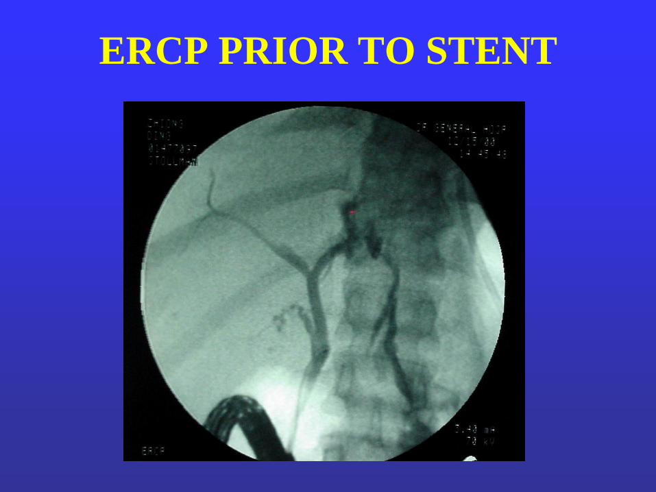
ERCP PRIOR TO STENT

SUMMARY: 3 PATIENTS
GRADE V LIVER INJURIES
PRE POST
• BLOOD 6-9 UNITS/first 24 hours
• IAP 35-50 12
• FIO2 50-100% 40-50%
• CREAT 0.9-1.5 0.6-0.8
• FLUID drained 3-5L
2/3 patients with bile leaks

REDUCTION IN LIVER
MORTALITY
• Grade IV-V injuries
• Mortality reduced from 40-80% to 8-22%
•Multi-modality therapy:
-early packing
-angioembolization
-ERCP/stents/drainage abscesses
Asensio et al J Trauma,2000

APPLICATION TO
PENETRATING TRAUMA
• Adjunctive techniques - complications of
penetrating liver trauma*
• Nonoperative management - selected cases
*Knudson/Lim:1994

PROPOSED ALGORITHM:
MAJOR LIVER TRAUMA
YES NO
OR
PACK
SPIRAL CT: EXTRAV.?
Bile leak – ERCP
IAH – U/S guided decompression ICU
STABLE / RESPONDS
TO RESUS.
NON-OP ANGIO &
CONTINUE
RESUSCITATION
UNSTABLE

SPIRAL CT CLASSIFICATION
• Type I: active extravasation-peritoneum
- unstable/required laparotomy
• Type II: intraparenchymal contrast +
hemoperioneum: 4/6 to OR
• Type III: only intraparenchymal contrast
- none required laparotomy
Feng et al, J Trauma, 2000

Autotransfusion

Perihepatic Packing • Damage control procedure
• Laparatomy pads compress areas of injury
• Avoid mobilization of the liver
– falciform and triangular ligaments
– diaphragmatic and retroperitoneal attachments
• Temporary abdominal wall closure
– Skin or ―Bogota‖ bag silo
• Return to OR for removal of lap pads in 24-48 hr


Fibrin Glue
Thrombin
Ca++ Fibrinogen Fibrin

Absorbable Mesh Packing

Pringle Maneuver
• First described in 1908*
• Can be tolerated for up to 60 minutes
– Causes ischemia reperfusion injury to liver
– Associated with massive bowel edema
• Controls hepatic parenchymal hemorrhage in 60-
80% of cases
– Helps diagnose hepatic vein/caval injuries
*J Pringle, Ann Surg 48:541, ‘08


Hepatic Artery Ligation
• Collateral flow through translobar and subcapsular
vessels
• Well tolerated if portal flow is preserved
– Portal vein supplies 80% of hepatic oxygen requirement
– Hepatic artery clamping increases portal vein oxygen
extraction

Hepatic Artery Extravasation
Successfully controlled by embolization

Vena Caval Blood Flow Percent
Superior Vena Cava 25
Inferior Vena Cava 75
Renal Veins 25
Portal Vein 40
Infrarenal IVC 10
J Malo, et.al., J Appl Physiol 56:1403, ‘84

Atrial-Caval Shunt
Pringle maneuver
suprahepatic &
subhepatic
snares
chest tube

Problems with Atrial Caval
Shunts
• Generally requires additional thoracotomy or
sternotomy
• Snaring the vena cava is technically challenging
• Insertion is associated with additional blood loss
• Potential for air embolism in a hypotensive patient

Total Vascular Occlusion
suprahepatic
& subhepatic
caval control aortic
control
Pringle maneuver


Indications for Total Vascular
Occlusion (TVO) • Penetrating injuries
– Major GSW with blast injury to parenchyma requiring
hepatotomy for control of hemorrhage
– Penetrating retrohepatic caval and hepatic vein injuries
• Blunt injuries
– Second-stage hepatic resections
– Liver avulsion
• Consider TVO when the Pringle maneuver and
packing together are insufficient

CVP After Total Vascular Occlusion 22 noncirrhotic patients
0
5
10
15
0 10 20 30 40 50
CVP
mm
Hg
minutes
+TVO –TVO
D Eyraud et.al. Anesth Analg 95:1173, ‘02

Hemodynamics of TVO 22 non-cirrhotic patients
0
20
40
60
80
100
120
140
0
20
40
60
80
100
120
140
0 10 20 30 40 50
SVRI
MAP
mm
Hg
IU
minutes
+TVO –TVO
D Eyraud et.al. Anesth Analg 95:1173, ‘02

Humoral Agents in TVO
22 non-cirrhotic patients
Hormone Baseline 5 minutes after
clamping
Arg vasopressin
(pg/ml) 8 ± 10 31 ± 26
Epinephrine
(pg/ml) 175 ± 128 347 ± 292
Norepinephrine
(pg/ml) 595 ± 366 1226 ± 1045
D Eyraud et.al. Anesth Analg 95:1173, ‘02

Extracorporeal Inferior Vena Caval
Bypass: study in 5 mongrel dogs
• Bypass all blood to suprahepatic vena cava
– Percutaneous femoral vein to internal jugular
vein
– Inferior mesenteric vein to internal jugular vein
– Heparin bonded shunts with extracorporeal
pump
• Less drop in MAP and CO
– Compared to Pringle maneuver + complete
caval interruption (TVO) or atrial-caval shunt
Howdieshell, et.al., Crit Care Med 24:631, ‘96

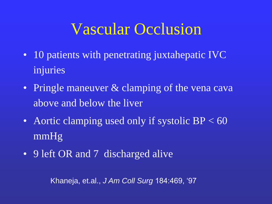
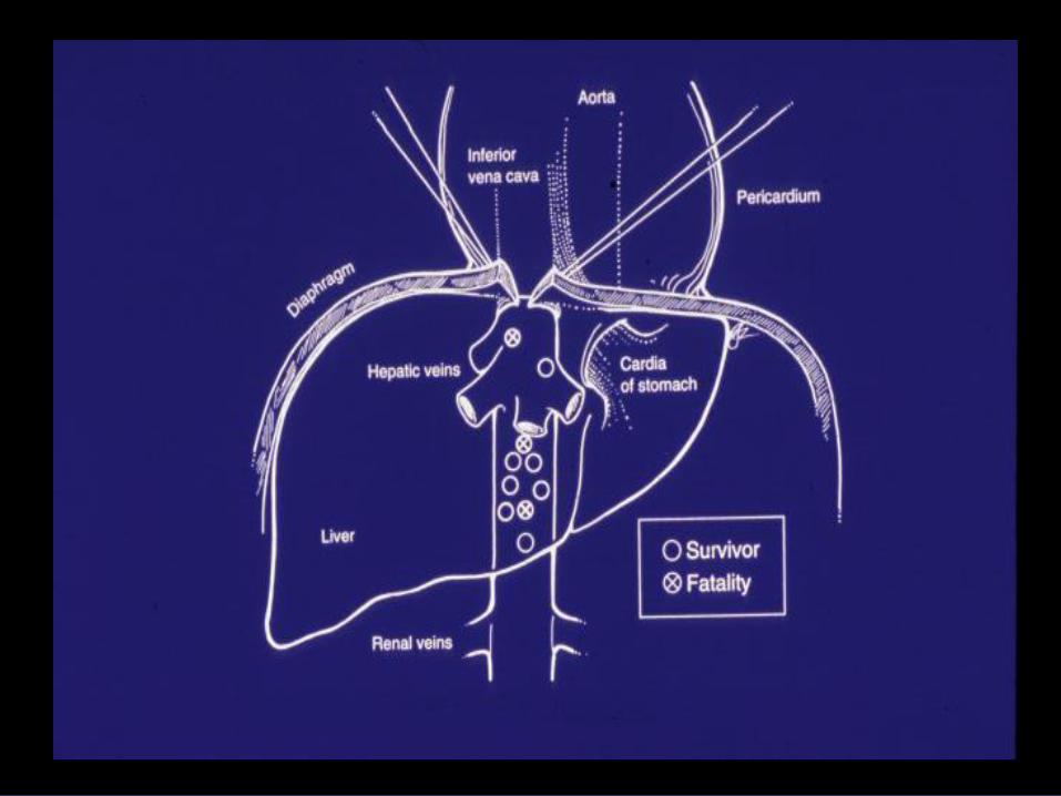
Vascular Occlusion
• 10 patients with penetrating juxtahepatic IVC
injuries
• Pringle maneuver & clamping of the vena cava
above and below the liver
• Aortic clamping used only if systolic BP < 60
mmHg
• 9 left OR and 7 discharged alive
Khaneja, et.al., J Am Coll Surg 184:469, ‘97

Selective Vascular Occlusion
• Pringle maneuver
• Dissection of the R side of the vena cava with isolation of the R hepatic vein trunk and middle/left hepatic vein confluence
– Be careful of an inferior R hepatic vein
• Application of bulldog clamps to the hepatic veins parallel to the vena cava
• Maintains flow in the IVC

Extrahepatic Biliary Injuries


Initial Therapy
• Splenectomy
• Closure of stomach wounds
• Repair of hepatic artery

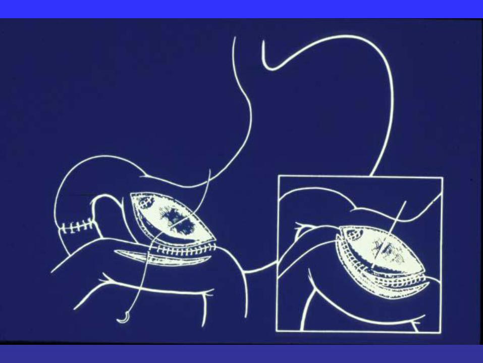


Technical Tips for
Hepaticojejunostomy
• Single layer absorbable suture
• Spatulate the duct
• Extend choledochotomy to left hepatic duct
• Place interrupted sutures in the anterior wall
of the duct prior to beginning the posterior
row of the anastomosis

Liver
Access loop
Hepaticojejunostomy

ERCP in Patients with Pancreatic
Trauma • 20 patients (ages 17-
54)
• 6 patients (30%) normal ERCP
• 13 patients with partial or complete PDD
• 1 patient with biliary injury (Rx biliary stent)
• 15 patients Rxed expectantly after ERCP
• 2 patients-distal pancreatectomy
• 7 patients sphincterotomy and/or pancreatic stent—none required surgery

Sphincterotomy Pancreatic stent

Normal ERCP

Blunt trauma
Pancreatic and peripancreatic
edema
Extravasation of contrast
From pancreatic duct
Rx- pancreatic sphincterotomy
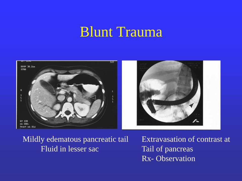
Blunt Trauma
Mildly edematous pancreatic tail
Fluid in lesser sac
Extravasation of contrast at
Tail of pancreas
Rx- Observation

2 cases of Blunt Trauma
Extravasation from
Pancreatic tail
Rx-Sphincterotomy
Extravasation from head of
Pancreas
Rx-IR perpancreatic drains

Blunt Trauma
Mild edema of body of
pancreas
Extensive extravasation
Rx- distal pancreatectomy

Distal Pancreatectomy
Distal Pancreatectomy with
Preservation of the Spleen
Lessons Learned
• Use ERCP to diagnose PDD after both blunt
and penetrating trauma
• Treat PDD in selected cases by pancreatic
sphincterotomy and/or pancreatic duct stent
• Early diagnosis of PDD can lead to prompt
minimally invasive or resection therapy and
minimize morbidity and mortality


Summary
• Cholecystectomy for gunshot wounds of the gallbladder in stable patients
• Cholecystorrhaphy vs cholecystectomy for small stab wounds of the gallbladder
• Tube cholecystostomy in unstable patients
• Choledochorrhapy for small stab wounds of the common bile duct
• Hepatojejunostomy for Common Duct Transections
• Drain the bile duct in the unstable patient